HAL Id: inserm-00381871
https://www.hal.inserm.fr/inserm-00381871
Submitted on 6 May 2009
HAL is a multi-disciplinary open access
archive for the deposit and dissemination of
sci-entific research documents, whether they are
pub-lished or not. The documents may come from
teaching and research institutions in France or
abroad, or from public or private research centers.
L’archive ouverte pluridisciplinaire HAL, est
destinée au dépôt et à la diffusion de documents
scientifiques de niveau recherche, publiés ou non,
émanant des établissements d’enseignement et de
recherche français ou étrangers, des laboratoires
publics ou privés.
Biological equivalent dose studies for dose escalation in
the stereotactic synchrotron radiation therapy clinical
trials.
Yolanda Prezado, G. Fois, M. Edouard, Christian Nemoz, Michel Renier,
Herwig Requardt, François Estève, Jean-François Adam, Hélène Elleaume,
Alberto Bravin
To cite this version:
Yolanda Prezado, G. Fois, M. Edouard, Christian Nemoz, Michel Renier, et al.. Biological
equiva-lent dose studies for dose escalation in the stereotactic synchrotron radiation therapy clinical trials..
Medical Physics, American Association of Physicists in Medicine, 2009, 36 (3), pp.725-33.
�inserm-00381871�
synchrotron radiation therapy clinical trials
Y. Prezadoa兲
ID17 Biomedical Beamline, European Synchrotron Radiation Facility (ESRF), 6 rue Jules Horowitz, BP 220, 38043 Grenoble Cedex, France
G. Fois
ID17 Biomedical Beamline, European Synchrotron Radiation Facility (ESRF), 6 rue Jules Horowitz, BP 220, 38043 Grenoble Cedex, France and Dipartimento di Fisica, Universitá degli Studi di Cagliari, S.P. Monserrato Sestu km 0.700, Monserrato, Cagliari 09042, Italy
M. Edouard
Equipe 6, Institut National de la Santé et de la Recherche Médicale U836, 6 rue Jules Horowitz, BP 220, 38043 Grenoble Cedex, France; Université Joseph Fourier, BP 53, 38041 Grenoble Cedex, France; and ID17 Biomedical Beamline, European Synchrotron Radiation Facility (ESRF), 6 rue Jules Horowitz, BP 220, 38043 Grenoble Cedex, France
C. Nemoz, M. Renier, and H. Requardt
ID17 Biomedical Beamline, European Synchrotron Radiation Facility (ESRF), 6 rue Jules Horowitz, BP 220, 38043 Grenoble Cedex, France
F. Estève
Equipe 6, Institut National de la Santé et de la Recherche Médicale U836, 6 rue Jules Horowitz, BP 220, 38043 Grenoble Cedex, France; Université Joseph Fourier, BP 53, 38041 Grenoble Cedex, France; ID17 Biomedical Beamline, European Synchrotron Radiation Facility (ESRF), 6 rue Jules Horowitz, BP 220, 38043 Grenoble Cedex, France; and Centre Hospitalier Universitaire, 38043 Grenoble, France
J. F. Adam
Equipe 6, Institut National de la Santé et de la Recherche Médicale U836, 6 rue Jules Horowitz, BP 220, 38043 Grenoble Cedex, France; Université Joseph Fourier, BP 53, 38041 Grenoble Cedex, France; and ID17 Biomedical Beamline, European Synchrotron Radiation Facility (ESRF), 6 rue Jules Horowitz, BP 220, 38043 Grenoble Cedex, France
H. Elleaume
Equipe 6, Institut National de la Santé et de la Recherche Médicale U836, 6 rue Jules Horowitz, BP 220, 38043 Grenoble Cedex, France; Université Joseph Fourier, BP 53, 38041 Grenoble Cedex, France; ID17 Biomedical Beamline, European Synchrotron Radiation Facility (ESRF), 6 rue Jules Horowitz, BP 220, 38043 Grenoble, France; and Centre Hospitalier Universitaire, 38043 Grenoble, France
A. Bravin
ID17 Biomedical Beamline, European Synchrotron Radiation Facility (ESRF), 6 rue Jules Horowitz, BP 220, 38043 Grenoble Cedex, France
共Received 27 July 2008; revised 3 December 2008; accepted for publication 16 December 2008; published 4 February 2009兲
Synchrotron radiation is an innovative tool for the treatment of brain tumors. In the stereotactic synchrotron radiation therapy共SSRT兲 technique a radiation dose enhancement specific to the tumor is obtained. The tumor is loaded with a high atomic number 共Z兲 element and it is irradiated in stereotactic conditions from several entrance angles. The aim of this work was to assess dosimetric properties of the SSRT for preparing clinical trials at the European Synchrotron Radiation Facility 共ESRF兲. To estimate the possible risks, the doses received by the tumor and healthy tissues in the future clinical conditions have been calculated by using Monte Carlo simulations共PENELOPEcode兲. The dose enhancement factors have been determined for different iodine concentrations in the tumor, several tumor positions, tumor sizes, and different beam sizes. A scheme for the dose escalation in the various phases of the clinical trials has been proposed. The biological equivalent doses and the normalized total doses received by the skull have been calculated in order to assure that the tolerance values are not reached. © 2009 American Association of Physicists in Medicine.
关DOI:10.1118/1.3070538兴
Key words: clinical trials, synchrotron radiation, Monte Carlo simulations, biological equivalent dose, normalized total dose
I. INTRODUCTION
Glioma is one of the most frequent primary brain tumors in adults, with an incidence of approximately 5–10 per 100 000 general population.1High-grade glioma is still of poor prog-nostic value despite the development of many innovative therapies. Stereotactic radiosurgery,2intensity-modulated ra-diation therapy,3 and boron neutron capture therapy4 are some examples. Radiotherapy of such tumors requires high doses, whereas the tolerance of the healthy brain tissues limits the maximum allowable dose because of the high risk of normal tissue morbidity.5The use of concomitant and ad-juvant temozolomide and radiotherapy has allowed signifi-cant prolongation of survival.6,7 However, the outcome re-mains still unsatisfactory. Therefore the management of glioblastomas is still mainly palliative. The key parameter is to increase the radiation dose delivered to the tumor relative to the one absorbed by the healthy tissues. A radiation dose enhancement, specific to the tumor, can be achieved if the volume to be treated is previously loaded with an iodinated contrast agent and exposed to kilovoltage x rays.
This concept was first proposed by Norman and co-workers8–10 by using a simple irradiation scheme and a conventional scanner x-ray source. The iodinated contrast media are injected to the patient and it accumulates preferentially in the tumor interstitium due to the increased permeability of the blood-brain barrier caused by invasive tumor growth. As already stressed by different authors,8,9,11 because of the local blood brain barrier disruption, tumor iodine concentrations of up to 4.0 and 5.0 mg/ml are rou-tinely obtained by intravenous administration. These concen-trations were observed using standard administration proce-dures for brain tumor imaging. With the specific aim of maximizing the iodine concentration, one may obtain tumor iodine concentrations of 10–20 mg/ml.8 Then, the iodine concentration achieved in the tumor undergoes a slow decay, whereas the iodine concentration in the surrounding normal tissue is negligible since no blood brain barrier leakage is observed in healthy brain tissue. At kilovoltage energies, the high photoelectric cross sections of iodine result in substan-tial photoelectric interactions 关the atomic cross section for photoelectric absorption exhibits a Z3 共Ref. 12兲兴. The high
linear energy transfer and short range of the photoelectric interaction products 共photoelectrons and Auger electrons兲 produce a localized dose enhancement. Therefore, the photon absorption is augmented specifically in the tumor volume and thus the absorbed dose is enhanced relatively to the sur-rounding tissues.
The dose enhancement yielded by this technique has been calculated previously by using Monte Carlo simulations.10,13 Its potential benefits and practical application have been demonstrated by the experimental treatment of tumor bearing rats,11,14 by the irradiation of human cells in vitro,9 and by the patient treatment of human brain metastasis by using a modified CT scanner without observing any bone toxicity or brain necrosis.10
The use of monochromatic x rays tuned at the optimal energy could significantly improve the dose distributions.15
Synchrotron sources, providing high-intensity, tunable monochromatic x rays, seem therefore ideal for this thera-peutic modality. At the European Synchrotron Radiation Fa-cility 共ESRF兲, one beamline has been dedicated to medical applications of synchrotron radiation.16–19At this beamline, SSRT preclinical trials were carried out in the past years in rats bearing high-grade F98 glioma. The survival of rats that received an intravenous injection of iodinated contrast agent before irradiation with monochromatic 50 keV x rays was significantly prolonged.20,21
Based on those encouraging results, ESRF has decided to plan the clinical trials and the first patients are expected in the near future. With this objective, the doses received by the tumor and healthy tissues have to be assessed to define the irradiation protocol. In the clinical trials the tumor will be loaded with different concentrations of iodine, the beam size will be adjusted to the tumor dimensions, and the tumor po-sitioned at the center of rotation. The irradiation will be per-formed over several incidences 共maximum of 10兲 with a monochromatic beam of 80 keV. The choice of 80 keV as the treatment energy among the technically possible ones at ESRF共from 20 to 100 keV兲 is based on the best compromise between the dose deposition in the tumor and the sparing of healthy tissues for a continuous arch irradiation.15 In previous works it has been shown that significant brain spar-ing is achieved in SSRT compared to conventional radio-therapy. In particular, comparisons have been performed with a stereotactic radiosurgery treatment 共using 10 MV beams兲9 and with a conventional treatment with a 6 MV linear accelerator.15 However, due to the high effective atomic number of bone 共approximately 12.3兲 relative to tissue 共ap-proximately 7.5兲, particular care has to be applied to avoid complications to the bone. The tolerance dose for necrosis in the skull is 60 Gy in a standard fractionation scheme 共2 Gy/session兲.22
In conventional radiotherapy, brain tumors are usually ir-radiated with 50 Gy in sessions of 2 Gy. In the SSRT clinical trials, it is foreseen that the patients undergo first the SSRT treatment 共“boost”兲 and that 1 week later they start a total cerebral irradiation with conventional radiotherapy at the hospital to deliver additional 40 Gy to the tumor. The clinical trials will be carried out in different phases with an escala-tion in doses. The SSRT irradiaescala-tion will be performed in one of the experimental stations of the Biomedical Beamline at the ESRF. During the days of the treatment, a complete priority will be given to the patient treatment and all other experimental activities in the beamline will be stopped. For that reason, single/hypofractionation schemes will be used. One of the goals of this work is to assess the possible dose prescriptions and fractionation scheme in order to optimize the number of sessions and the doses received by the skull in SSRT.
It is very important to take into account that the dose prescriptions will differ from the standard fractionation scheme of 2 Gy/session, and therefore the expected biologi-cal effects will be different. For that reason, the biologibiologi-cal equivalent doses共BEDs兲 have been assessed. To establish the equivalence with the standard fractionation scheme and
726 Prezado et al.: Biological equivalent dose studies for SSRT clinical trials 726
therefore to assess if the doses received by the skull are still within tolerances, the normalized total doses 共NTDs兲 have been studied.
II. MATERIALS AND METHODS II.A. Monte Carlo source code
To perform the dose calculations, a precise dosimetry sys-tem is required. However, none of the conventional treatment planning systems is able to compute dose distributions in the case of tumors loaded with high Z elements and irradiated with kilovoltage x rays. Monte Carlo simulations are well suited for calculating dose distributions because cumulative doses are computed using track histories of each incident photon. Monte Carlo is therefore the most accurate and sen-sitive method available for calculating the dose enhancement with high Z elements, as already shown in Refs.9 and15.
In this work thePENELOPEcode has been used.PENELOPE
is a general purpose Monte Carlo simulation package developed at the University of Barcelona.23 The developers of this code put special emphasis on the implementation of accurate low energy electron cross sections, which are of particular importance for this application. The simulation al-gorithm is based on a scattering model that combines nu-merical databases with analytical cross section models for the different interaction mechanisms and is applicable to en-ergies共kinetic energies in the case of electrons and positrons兲 from a few hundred eV to 1 GeV. It uses a mixed simulation scheme in which hard interactions are simulated collision by collision and small angular deflections and energy losses are treated in a grouped manner. That is to say, photon transport is simulated by means of the conventional detailed method. The simulation of electron and positron transport is per-formed by means of a mixed procedure. Hard interactions, with scattering angle or energy loss greater than preselected cutoff values, are simulated in detail. Soft interactions, with scattering angle or energy loss less than the corresponding cutoffs, are described by means of multiple scattering ap-proaches. In this work, due to the fact that the beam energy is low 共80 keV兲 and there are high Z elements present, the photoelectric interactions are of great relevance. PENELOPE
incorporates a detailed description of photoabsorption in K-and L-shells 共including the subsequent atomic relaxation兲. When the ionization occurs in an inner K- or L-shell, the residual atom, with a vacancy in the shell, subsequently re-laxes to its ground state by emitting x rays and Auger elec-trons. If the ionization occurs in an outer shell, it is assumed that the photoelectron leaves the target atom with kinetic energy equal to the energy deposited by the photon and the emission of subsidiary fluorescent radiation is disregarded. The photoelectric cross sections used in PENELOPE are ob-tained by interpolation in a numerical table that was ex-tracted from the LLNL Evaluated Photon Data Library 关EPDL 共Ref.24兲兴. ThePENELOPEdatabase for photoelectric absorption 共a subset of the EPDL兲 consists of tables of the total atomic cross section phi共E兲 and the cross sections for
the K- and L-shells, phi,i共E兲 共共i=K, L1, L2 and L3兲 for
the elements Z = 1 – 92, which span the energy range from 100 eV to 1000 GeV.
Compton scattering is as well a relevant photon interac-tion in low atomic number materials 共like the brain兲 of interest in radiation therapy, for the x-ray energies.25 PENE-LOPE considers bounding effects and Doppler broadening when simulating Compton interactions. The code has been widely used in medical-physics applications.26–30
In the simulations the heterogeneity is taken into account. The composition of a medium is specified by its stoichio-metric formula, i.e., atomic number Ziand number of atoms
per molecule niof all the elements present. In this work, the
material structure where radiation is transported consists of various regions with different compositions. In the code it is assumed that the interfaces between contiguous media are sharp 共i.e., there is no diffusion of chemical species across them兲. When a particle arrives at an interface, it is stopped there and the simulation is resumed with the interaction properties of the new medium. This is based on the Markovian property of the transport process, that means that the future values of a random variable共interaction event兲 are statistically determined by present events and depend only on the event immediately preceding. Owing to the Markovian character of the transport, the generation of a particle history can be stopped at an arbitrary state共any point of the track兲 and the simulation can be resumed from this state without introducing any bias in the results.
In this work the 2006 version of thePENELOPEcode was used. The number of primary photon stories is 109in all the
calculations. The standard deviation is around 1%. Some cross-checks have been performed with the code MNCPX
共Ref. 31兲 and an average difference of 2% has been found between both codes. To be conservative, the 2% average dif-ference has been taken as the error bar.
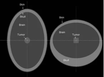
II.B. Phantom description and simulated irradiation An anthropomorphic head phantom has been constructed with the geometry package in PENELOPE. The phantom is inspired by the one described in the work of Harling et al.32 for neutron capture therapy. It consists of three nonconcen-tric ellipsoids representing the skin, the skull, and the brain. A sagital and a coronal view of the phantom is represented in Fig.1. In this model, the volume of the brain is 1470 cm3,
those of the skull bone and of the skin are 1021 and 184 cm3, respectively.
A cylindrical tumor of 2 cm height and 2 cm of diameter was placed in different positions in the brain and loaded with different concentrations of iodine. Nine equiangular-spaced beams with an energy of 80 keV and a size of 2⫻2 cm2
irradiating the phantom have been simulated. An odd number of beams is used to avoid hot spots in the skull due to the possible overlap of opposite beams.
II.C. Assessment of the dose enhancement factors The radiation dose enhancement is quantified by means of the dose enhancement factors共DEFs兲. The DEFs are defined as the ratio of the average radiation dose absorbed by the
tumor when it is loaded with contrast media共iodine兲 to the dose absorbed without contrast media. The DEFs have been calculated by using Monte Carlo simulations.
II.D. Assessment of the biological equivalent doses and normalized total doses
To determine the value of the BED, expression 共1兲 as derived by Fowler33 from the linear-quadratic model has been used,
BED = nd
冉
1 + d共␣/兲
冊
, 共1兲where n is the number of fractions, d is the dose per fraction, and␣ andare parameter characteristics of the population of cells. To relate the biological effect of a course of radia-tion to the total dose of radiaradia-tion administered with the stan-dard fractionation scheme 共2 Gy/fraction兲, the concept of normalized total dose 共NTD兲 has been used. Expression共2兲 is derived from the linear-quadratic model,34
NTD = BED
冒
冉
1 + d共␣/兲
冊
. 共2兲To establish the equivalence with the standard fractionation scheme 共2 Gy/fraction兲, in the Eq. 共2兲, d would take the value 2 Gy. The␣/ values are usually high for the tumors 共except melanoma, liposarcoma, and prostate cancers35兲 and
for early responding tissues 共rapidily renewing tissues兲; the values are usually in the range between 10 and 15 Gy.35–37 For the late responding tissues, such as the bone, the ␣/ values are small, around 2 Gy.35Therefore, the effect of the dose fractionation and the repair between consecutive frac-tions is especially important for the late responding tissues, but it is less important for the tumor response and the early responding tissues, where the total dose is more relevant. There is a few published information about the ␣/ values of the bone. Overgaad38estimated that the␣/ratios for the late bone damage are in the range from 1.8 to 2.8 Gy.
There-fore, to be conservative, the ␣/ value, which gives the highest BED in the bone, i.e., 1.8 Gy, has been used in the calculations. The value of ␣/ for the tumor was taken as 10, which is an average of the values found for tumors in Ref.39. This value has also been used in some other studies on gliomas.40,41
The linear-quadratic 共LQ兲 model, used in this work, is a useful tool to assess the dose radiation response and to inter-compare conventional fractionations since it closely fits the experimental cell survival curves in most of the cases of conventional fractionation schemes in radiotherapy. How-ever, it is worth to notice that, although it is one of the best available models, it is not always compatible with clono-genic cell survival studies at high dose per fraction, where the survival curves do not really follow the continuous bending predicted by the LQ model. The LQ formulation overestimates the magnitude of cell kill for doses greater than 6 Gy, and therefore, it underestimates the dose required to achieve a desired response for fractions of high dose.42In addition it has also been shown that the linear-quadratic model is not so well suited to describe the response at low doses共ⱕ1 Gy兲 either.43
III. RESULTS AND DISCUSSION
This section presents the calculated DEF in a human head phantom for different iodine concentrations, different tumor positions, tumor sizes, and beam sizes, as well as the calcu-lated doses received by the tumor and the skull.
III.A. Dose enhancement factors as a function of the iodine concentration
The variation in the DEF as a function of the iodine con-centration in the tumor has been studied. In the simulations, a cylindrical tumor, as described in Sec. II B, has been used. Table Ishows the DEF obtained for several iodine concen-trations in the range between 0 and 20 mg/ml. The variation in DEF versus iodine concentration follows a linear tendency 共r2⬎0.999兲 in that range, in agreement with Ref.44.
FIG. 1. Coronal共left兲 and sagital 共right兲 views of the anthropomorphic phan-tom. Three nonconcentric ellipsoids simulate the skin, the skull, and the brain. A cylindrical tumor is placed at the center of the brain.
TABLEI. Dose enhancement factors as a function of the iodine concentration in a cylindrical tumor of 2 cm of diameter and 2 cm height placed at the center of the brain.
关I兴 共mg/ml兲 DEF 1 1.10⫾0.01 5 1.47⫾0.01 8 1.74⫾0.02 9 1.84⫾0.02 10 1.93⫾0.02 15 2.37⫾0.02 20 2.80⫾0.03
728 Prezado et al.: Biological equivalent dose studies for SSRT clinical trials 728
III.B. Dose enhancement factors as a function of the tumor position
To study the variation in the DEF as a function of the tumor position, a cylindrical tumor共as defined in Sec. II B兲 loaded with 10 mg/ml of iodine was placed in four different positions: 共1兲 Central position 共from now on “central”兲, 共2兲 displaced by 5 cm in the frontal direction 共“frontal”兲, 共3兲 displaced by 1.5 cm in the left-right direction共“lateral”兲, and 共4兲 displaced by 3 cm in the left-right direction and by 4 cm in the frontal direction 共“eccentric”兲 共see Fig. 2兲. Table II shows that there is no variation in the DEF as a function of the tumor position within the error bars.
III.C. Dose enhancement factors as a function of the tumor size
The variation in the DEF as a function of the tumor size has also been investigated. One may expect a lower DEF with the increased tumor size due to strong attenuation in the tumor center. This would produce a depression at the center of the dose distribution in the tumor due to the high radiation absorption. This effect was found to be negligible for the concentrations expected to be used in the clinical trials, in particular, 10 mg/ml. On the other hand, the higher the inter-action volume, the higher will be the dose deposition in the tumor. DEF computations were carried out at increasing tu-mor diameters and heights with a constant 10 mg/ml iodine
concentration. A slight increase in the DEF with the tumor size was found and summarized in Table III. These results are not dependent on the tumor shape. Monte Carlo calcula-tions of the DEF values in spherical tumors have proven that the significant parameter is the tumor volume.
III.D. Dose enhancement factors as a function of the beam size
To study the possible influence of the safety margins around the tumor, the beam sizes were varied in the Monte Carlo simulations. A cylindrical tumor as described in Sec. II B loaded with 10 mg/ml of iodine was placed at the center of the brain and irradiated with increasing beam sizes up to 1 cm of margin around the tumor 共3.0⫻3.0 cm2兲. Table IV shows that there is no variation in the DEF within the error bars up to the maximum margins expected in clinical condi-tions.
III.E. Assessment of the doses received by the tumor and the skull
As it has already been indicated in Sec. I, the SSRT treat-ments will be used as a boost to enhance the doses received by the tumor while sparing the surrounding tissues. Due to the high atomic number of the bone, the doses received by the skull can be important. The clinical trials will be carried out in different phases. One of the goals of this work is to study the possible dose prescriptions and fractionation scheme taking into account that the patient will receive an additional dose of 40 Gy in conventional radiotherapy. One possible scheme for the escalation of the prescribed doses to the tumor for the groups of patients to be treated in each of the phases of the clinical trials could be the following.
FIG. 2. Different tumor positions. Upper row left: Central. Upper row right: Frontal. Lower row left: Lateral. Lower row right: Eccentric.
TABLEII. Dose enhancement factors as a function of the tumor position. The tumor is loaded with an iodine concentration of 10 mg/ml. No variation is observed within the error bars.
Tumor position DEF
Central共center of the brain兲 1.93⫾0.02 Frontal共5 cm displaced in the frontal direction兲 1.92⫾0.02 Lateral共1.5 cm displaced in the lateral direction兲 1.91⫾0.02 Eccentric共3 cm displaced in the left-right direction and
4 cm in the frontal direction兲 1.92⫾0.02
TABLEIII. Dose enhancement factors as a function of the tumor size. The tumor is loaded with an iodine concentration of 10 mg/ml. A slight increase in the DEF values with the tumor size is observed.
Tumor diameter/height 共cm兲 DEF 0.5/0.5 1.86⫾0.02 1.0/1.0 1.88⫾0.02 1.5/1.5 1.90⫾0.02 2.0/2.0 1.93⫾0.02 2.5/2.5 1.95⫾0.02
TABLEIV. Dose enhancement factors as a function of the beam size for a cylindrical tumor of 2 cm diameter and 2 cm height placed at the center of the brain and loaded with an iodine concentration of 10 mg/ml. No variation is observed within the error bars.
Beam size 共cm2兲 DEF 2.0⫻2.0 1.93⫾0.02 2.2⫻2.2 1.93⫾0.02 2.5⫻2.5 1.95⫾0.02 3.0⫻3.0 1.97⫾0.02
共1兲 First phase: 10 Gy in one session. 共2兲 Second phase: 12 Gy in one session. 共3兲 Third phase: 14 Gy in one session. 共4兲 Fourth phase: 18 Gy in three sessions. 共5兲 Fifth phase: 21 Gy in three sessions. 共6兲 Sixth phase: 24 Gy in four sessions.
To assess the doses received by the skull, the spatial dose distributions obtained from the Monte Carlo simulations have been studied and the maximum doses received by the skull in comparison with the average doses deposited in the tumor have been derived. Figure3 shows the dose distribu-tions at the central coronal plane of the geometry with a tumor placed at the center of the brain and loaded with dif-ferent concentrations of iodine: 5, 8, and 10 mg/ml. The maximum doses received by the skull were found to be 75%, 69%, and 65%, respectively. That is to say, if the tumor receives 10 Gy in one session, then the maximum doses in the skull will be 7.5, 6.9, and 6.5 Gy in one session, respec-tively.
The proposed dose fractionation scheme differs from the standard one, and therefore the biological effects are ex-pected to be different. To take this into account, the BEDs have been determined by using Eq.共1兲. To be able to assess the total doses received by the tumor and the skull, the NTD2.0 has been calculated by using Eq.共2兲. Tables Vand VIshow the maximum physical, BED, and NTD2.0received by the tumor and the skull, respectively, in the proposed different phases of the clinical trials.
The total doses received by the tumor are calculated as the sum of the doses delivered in conventional radiotherapy共40 Gy兲 plus the calculated NTD2.0received by the tumor in the
SSRT treatment共see TableV兲. The total doses in the tumor, for the six phases proposed, are 57, 62, 68, 64, 70, and 72 Gy, respectively. This means an enhancement of the dose received by the tumor as compared with the one received in conventional radiotherapy 共50 Gy兲. From the total cerebral irradiation with 40 Gy in conventional radiotherapy, a maxi-mum dose of 20 Gy in the skull is expected. In addition, as it has already been explained in Sec. I, the tolerance dose for bone necrosis is 60 Gy. Therefore, a NTD2.0equal to 40 Gy
can be considered a safe limit for the dose delivered to the skull in the SSRT treatment. As it can be seen in Table VI, the maximum NTD2.0received by the skull is 30 Gy in the sixth phase and with an iodine concentration in the tumor of 5 mg/ml. This maximum value still remains within tolerances.
In addition, the influence of the tumor position on the maximum doses received by the skull has been studied. The tumor was placed in the positions described in Sec. III B. The maximum doses received by the skull in comparison with the average doses deposited in the tumor have been determined for those tumor positions by using Monte Carlo simulations. Table VIIshows the results when the tumor is loaded with a concentration of 10 mg/ml of iodine. The doses received by the skull are higher for the frontal and eccentric positions. Table VIII shows a comparison of the calculated maximum NTD2.0received by the skull in the
dif-FIG. 3. Spatial dose distributions at the central coronal plane of the phantom for different iodine concentrations in the tumor. The doses are given in % of the maximum dose in the tumor.共a兲 Dose distribution for an iodine concen-tration of 5 mg/ml.共b兲 Dose distribution for an iodine concentration of 8 mg/ml.共c兲 Dose distribution for an iodine concentration of 10 mg/ml. 730 Prezado et al.: Biological equivalent dose studies for SSRT clinical trials 730
ferent phases for the four tumor positions. The tumor is loaded with an iodine concentration of 10 mg/ml. As it can be seen, all the doses remain below 40 Gy, and therefore within the tolerance level.
The calculations show that in SSRT it is possible to en-hance the dose delivered to the tumor while keeping the doses in the skull within the tolerance level. The Monte Carlo simulations show that the DEF increases linearly with the iodine concentration. The measurement of the iodine concentration in the tumor must be accurate because small differences in the iodine concentration lead to relevant dif-ferences in doses. The DEF values do not show any depen-dence on the tumor position. However an increase in the tumor size is observed共⬍5% for the range of tumor sizes to be treated in the clinical trials兲. In addition, to study the effects of the safety margins around the tumor, the irradiation beam sizes were increased up to the maximum margins ex-pected共1 cm兲 and no variation in DEF was observed.
The BED and NTD studies have shown that it is possible to obtain an enhancement in the dose received by the tumor compared with the one received in conventional radiotherapy 共50 Gy兲. This enhancement can be as high as 22 Gy in the sixth phase. This might increase the probability of tumor control. On the other hand, in spite of the high atomic num-ber of the bone, it has been shown that the NTD2.0 in the skull remain within the acceptable level for the dose escala-tion proposed and the irradiaescala-tion geometry foreseen at ESRF for the SSRT clinical trials.
IV. CONCLUSIONS
The stereotactic synchrotron radiation therapy is a prom-ising technique to treat brain tumors. The goal is to enhance the dose delivered to the tumor by loading the tumoral tissue with iodine and irradiating it with monochromatic x rays. In this work the doses received by the tumor and the skull in the future conditions of the clinical trials have been assessed by using Monte Carlo simulations. The DEF values have been
TABLEV. Doses received by the tumor in the different phases of the clinical trials. The first column shows the dose prescription for the different phases, the second column is the physical dose in the tumor, and the third and fourth columns show the normalized total doses and the biological equivalent doses received by the tumor, respectively. The doses have been rounded to integer values. Dose prescription D共tumor兲 共Gy兲 NTD2.0共tumor兲 共Gy兲 BED共tumor兲 共Gy10兲 10 Gy/1 frac. 10 17 20 12 Gy/1 frac. 12 22 26 14 Gy/1 frac. 14 28 34 18 Gy/3 frac. 18 24 29 21 Gy/3 frac. 21 30 36 24 Gy/4 frac. 24 32 38
TABLEVI. Assessment of the maximum doses received by the skull in the different phases of the clinical trials. The first column shows different iodine concentrations, the second and third columns show the maximum physical dose, the normalized total doses and the biological equivalent doses received by the skull when the tumor is loaded with those iodine concentrations, respectively. The doses have been rounded to integer values.
关I兴 共mg/ml兲 Dmax共skull兲 共Gy兲 NTD2.0,max共skull兲 共Gy兲
BEDmax共skull兲 共Gy1.8兲
Phase 1: 10 Gy/1 frac.
5 7.5⫾0.1 18 39
8 6.9⫾0.1 16 33
10 6.5⫾0.1 14 30
Phase 2: 12 Gy/1 frac.
5 9.0⫾0.1 26 54
8 8.3⫾0.1 22 46
10 7.8⫾0.1 20 42
Phase 3: 14 Gy/1 frac.
5 10.5⫾0.1 34 72
8 9.7⫾0.1 29 62
10 9.1⫾0.1 26 55
Phase 4: 18 Gy/3 frac.
5 13.5⫾0.1 22 47
8 12.4⫾0.1 19 41
10 11.7⫾0.1 18 37
Phase 5: 21 Gy/3 frac.
5 15.8⫾0.1 29 62
8 14.5⫾0.1 25 53
10 13.6⫾0.1 23 48
Phase 6: 24 Gy/4 frac.
5 18.0⫾0.1 30 63
8 16.6⫾0.1 26 55
10 15.6⫾0.1 23 49
TABLE VII. Ratio of the maximum doses received by the skull and the average doses in the tumor for the different tumor positions.
Tumor position
Dosemaxskull/Davtumor
共%兲
Central 65
Frontal 73
Lateral 65
Eccentric 71
TABLEVIII. NTD2.0received by the skull for the four different positions of
the tumor共iodine concentration of 10 mg/ml兲 and in the different phases of the clinical trials. All the doses are in Gy.
Dose prescription NTD2.0 共Central兲 NTD2.0 共Frontal兲 NTD2.0 共Lateral兲 NTD2.0 共Eccentric兲 10 Gy/1 frac. 14 18 14 17 12 Gy/1 frac. 20 24 20 23 14 Gy/1 frac 26 32 26 31 18 Gy/3 frac. 18 21 18 20 21 Gy/3 frac. 23 28 23 27 24 Gy/4 frac. 23 28 23 27
studied as a function of the iodine concentration in the tumor, of the tumor position, tumor size, and beam sizes. A scheme for the dose escalation in the different phases of the clinical trials has been proposed. The BEDs received by the tumor and the skull have been calculated in order to establish an equivalence with the standard fractionation scheme of 2 Gy/ session. It has been shown that an enhancement in the doses received by the tumor compared to conventional radio-therapy can be obtained in SSRT. Therefore, an increase in the tumor control probability is expected, whereas the doses received by the skull remain within the tolerance level. This work reflects as well the importance of the inclusion of the biological aspects in the dose calculations.
a兲Electronic addresses: prezado@esrf.fr
1J. M. Legler et al., “Cancer surveillance series 关corrected兴: Brain and
other central nervous system cancers: Recent trends in incidence and mortality,” J. Natl. Cancer Inst. 91, 1382–1390共1999兲.
2M. H. Phillips et al., “Stereotactic radiosurgery: A review and comparison
of methods,” J. Clin. Oncol. 12, 1085–1099共1994兲.
3R. M. Cardinale et al., “Comparison of three stereotactic radiotherapy
techniques: ARCS vs. noncoplanar fixed fields vs. intensity modulation,”
Int. J. Radiat. Oncol., Biol., Phys.42, 431–436共1998兲.
4R. F. Barth et al., “Boron neutron capture therapy of cancer: Current
status and future prospects,” Clin. Cancer Res. 11, 3987–4002共2005兲.
5A. R. Kagan et al., “The pathogenesis of brain necrosis: Time and dose
parameters,” Int. J. Radiat. Oncol., Biol., Phys. 1, 729–732共1977兲.
6R. Stupp et al., “Radiotherapy plus concomitant and adjuvant
temozolo-mide for glioblastoma,” N. Engl. J. Med. 352, 987–996共2005兲.
7R. Stupp et al., “Promising survival for patients with newly diagnosed
glioblastoma multiforme treated with concomitant radiation plus temozo-lomide followed by adjuvant temozotemozo-lomide,” J. Clin. Oncol. 20, 1375– 1382共2002兲.
8A. Norman, K. S. Iwamoto, and S. T. Cochran, “Iodinated contrast agents
for brain tumor localization and radiation dose enhancement,” Invest. Radiol. 26, 120–121共1991兲.
9A. V. Mesa, A. Norman, T. D. Solberg, J. J. Demarco, and J. B. Smathers,
“Dose distributions using kilovoltage x-rays and dose enhancement from iodine contrast agents,”Phys. Med. Biol.44, 1955–1968共1999兲.
10J. H. Rose, A. Norman, M. Ingram, C. Aoki, T. Solberg, and A. Mesa,
“First radiotherapy of human metastatic brain tumors delivered by a com-puterized tomography scanner 共CTRx兲,” Int. J. Radiat. Oncol., Biol., Phys.45, 1127–1132共1999兲.
11R. S. Mello, H. Callisen, J. Winter, A. Robert Kagan, and A. Norman,
“Radiation dose enhacement in tumors with iodine,” Med. Phys. 10, 75–78共1983兲.
12F. M. Khan, The Physics of Radiation Therapy, 3rd ed.共Lippincott
Wil-liams and Wilkins, Philadelpha, 2003兲.
13T. D. Solberg, K. S. Iwamoto, and A. Norman, “Calculation of radiation
dose enhancement factors for dose enhancement therapy of brain tumors,”
Phys. Med. Biol.37, 439–443共1992兲.
14K. S. Iwamoto et al., “Radiation dose enhancement therapy with iodine in
rabbit VX-2 brain tumors,” Radiother. Oncol. 8, 161–170共1987兲.
15C. Boudou, J. Balosso, F. Estève, and H. Elleaume, “Monte Carlo
dosim-etry for stereotactic synchrotron radiation therapy of brain tumors,”Phys. Med. Biol.50, 4841–4851共2005兲.
16P. Suortti and W. Thomlinson, “Medical applications of synchrotron
ra-diation,”Phys. Med. Biol.48, R1–R35共2003兲.
17B. Bertrand et al., “Comparison of synchrotron radiation angiopgraphy
with conventional angiography for the diagnosis of in-stent restenosis after percutaneous transluminal coronary angiography,”Eur. Heart J.26, 1284–1291共2005兲.
18R. Serduc et al., “Characterization and quantification of cerebral edema
induced by synchrotron x-ray microbeam radiation therapy,”Phys. Med. Biol.53, 1153–1166共2008兲.
19A. Bravin, “The biomedical programs at the ID17 beamline of the
Euro-pean synchrotron Radiation Facility,” in Brilliant Light in Life and
Ma-terials Sciences, NATO Advanced Studies Institute, Series B: Physics and
Biophysics共Springer, The Netherlands, 2007兲, pp. 225–239.
20J. F. Adam et al., “Synchrotron radiation therapy of malignant brain
glioma loaded with an iodinated contrast agent: First trial on rats bearing F98 gliomas,” Int. J. Radiat. Oncol., Biol., Phys. 57, 1413–1426共2003兲.
21J. F. Adam et al., “Prolonged survival of Fischer rats bearing F98 glioma
after iodine-enhanced synchrotron stereotactic radiotherapy,” Int. J. Ra-diat. Oncol., Biol., Phys. 64, 603–611共2006兲.
22B. Emami et al., “Tolerance of normal tissue to therapeutic irradiation,”
Int. J. Radiat. Oncol., Biol., Phys. 21, 109–122共1991兲.
23F. Salvat, J. M. Fernández-Varea, and J. Sempau,
PENELOPE, a code system
for Monte Carlo simulation of electron and photon transport, OECD Nuclear Energy Agency, Issy-les-Moulineaux-France, 2003共available in PDF format from the web at www.nea.fr兲.
24D. E. Cullen, J. H. Hubbell, and L. Kissel, “EPDL97 The evaluated data
library, 097 version,” Lawrence Livermore National Laboratory Report No. UCRL-50400, 1997.
25J. Stepanek, H. Blattman, J. A. Laissue, N. Lyubimova, M. De Michiel,
and D. N. Slatkin, “Physics study of microbeam radiation therapy with PSI version of Monte Carlo codeGEANTas a new computational tool,”
Med. Phys.27, 1664–1675共2000兲.
26J. Sempau, A. Sanchez-Reyes, F. Salvat, H. O. Ben Tahar, S. B. Jiang,
and J. M. Fernández-Varea, “Monte Carlo simulation of electron beams from an accelerator head usingPENELOPE,”Phys. Med. Biol.46, 1163– 1186共2001兲.
27A. Badano and J. Sempau, “MANTIS: Combined x-ray, electron and
optical Monte Carlo simulations of indirect radiation imaging systems,”
Med. Phys.33, 2698–2713共2006兲.
28J. Asenjo, J. M. Fernández-Varea, and A. Sanchez-Reyes,
“Characteriza-tion of a high-dose-rate Sr-90-Y-90 source for intravascular brachy-therapy by using the Monte Carlo codePENELOPE,”Phys. Med. Biol.47, 697–711共2002兲.
29V. Moskvin, R. Timmerman, C. DesRosiers, M. Randall, P. Des Rosiers,
P. Dittmer, and L. Papiez, “Monte Carlo simulation of the Leksell gamma Knife共R兲. II. Effects of heterogeneous versus homogeneous media for stereotactic radiosurgery,”Phys. Med. Biol.49, 4879–4895共2004兲.
30E. Siegbahn, J. Stepanek, E. Brauer-Krisch, and A. Bravin,
“Determina-tion of dosimetrical quantities used in microbeam radia“Determina-tion therapy 共MRT兲 with Monte Carlo simulations,” Med. Phys. 33, 3248–3259 共2006兲.
31J. S. Hendricks et al.,
MCNPX, Version 2.5.e, Los Alamos Laboratory, Los
Alamos, NM, 2004.
32O. K. Harling, K. A. Roberts, D. J. Moulin, and R. D. Rogus, “Head
phantoms for neutron capture therapy,”Med. Phys.22, 579–583共1995兲.
33J. F. Fowler, “The linear-quadratic formula and progress in fractionated
radiotherapy,” Br. J. Radiol. 62, 679–694共1989兲.
34J. C. Flickinger and A. Kalend, “Use of normaliyed total dose to represent
the biological effect of fractionated radiotherapy,” Radiother. Oncol. 17, 339–347共1990兲.
35D. J. Brenner et al., “Direct evidence that prostate cancers show high
sensitivity to fractionation, similar to late-responding normal tissue,”Int. J. Radiat. Oncol., Biol., Phys.52, 6–13共2002兲.
36S. M. Bentzen, A. C. C. Ruifrok, and H. D. Thames, “Repair capacity and
kinetics for human mucosa and epithelial tumors in the head and neck: Clinical data on the effect of changing the time interval between multiple fractions per day in radiotherapy,” Radiat. Oncol. 38, 89–101共1996兲.
37M. V. Willians, J. Denekamp, and J. F. Fowler, “A review of alfa/beta
ratios for experimental tumors: Implications for clinical studies of altered fractionation,” Int. J. Radiat. Oncol., Biol., Phys. 11, 87–96共1985兲.
38M. Overgaard, “Spontaneous radiation-induced rib fractures in breast
can-cer patients treated with postmastectomy irradiation: A clinical radiobio-logical analysis of the influence of fraction size and dose-response rela-tionships on late bone damage,” Acta Oncol. 27, 117–22共1988兲.
39G. G. Steel, G. E. Adams, and A. Horwich, The Biological Basis of
Radiotherapy, 2nd ed.共Elsevier, New York, 1989兲.
40Y. Shibamoto et al., “Comparison of accelerated hyperfractionation
radio-therapy and conventional radioradio-therapy for supratentorial malignat glioma,” Jpn. J. Clin. Oncol. 27, 31–36共1997兲.
41J. L. Chan et al., “Survival and failure patterns of high-grade gliomas
after three-dimensional conformal radiotherapy,” Int. J. Clin. Oncol. 20, 1635–1642共2002兲.
732 Prezado et al.: Biological equivalent dose studies for SSRT clinical trials 732
42K. G. Zimmer, Studies on Quantitative Radiation biology 共Oliver and
Boyd, Edinburgh, 1961兲.
43P. Lambin, E. P. Malaise, and M. C. Joiner, “The effect of very low
radiation doses on the human bladder carcinoma cell line RT112,”
Radio-ther. Oncol. 32, 63–72共1994兲.
44K. N. Morris, M. D. Weil, and R. Malzbender, “Radiochromic film
do-simetry of contrast enhanced RT共CERT兲,”Phys. Med. Biol.51, 5915– 5925共2006兲.