HAL Id: hal-03249988
https://hal.archives-ouvertes.fr/hal-03249988
Submitted on 4 Jun 2021
HAL is a multi-disciplinary open access
archive for the deposit and dissemination of sci-entific research documents, whether they are pub-lished or not. The documents may come from teaching and research institutions in France or abroad, or from public or private research centers.
L’archive ouverte pluridisciplinaire HAL, est destinée au dépôt et à la diffusion de documents scientifiques de niveau recherche, publiés ou non, émanant des établissements d’enseignement et de recherche français ou étrangers, des laboratoires publics ou privés.
APP-induced axonal growth
Claire Marquilly, Germain Busto, Brittany Leger, Ana Boulanger, Edward
Giniger, James Walker, Lee Fradkin, Jean-Maurice Dura
To cite this version:
Claire Marquilly, Germain Busto, Brittany Leger, Ana Boulanger, Edward Giniger, et al.. Htt is a repressor of Abl activity required for APP-induced axonal growth. PLoS Genetics, Public Library of Science, 2021, 17 (1), pp.e1009287. �10.1371/journal.pgen.1009287�. �hal-03249988�
RESEARCH ARTICLE
Htt is a repressor of Abl activity required for
APP-induced axonal growth
Claire MarquillyID1¤, Germain U. BustoID1, Brittany S. LegerID2, Ana Boulanger1, Edward GinigerID3, James A. WalkerID2,4,5, Lee G. FradkinID6, Jean-Maurice DuraID1*
1 IGH, Centre National de la Recherche Scientifique, Univ Montpellier, Montpellier, France, 2 Center for Genomic Medicine, Massachusetts General Hospital, Boston, Massachusetts, United States of America, 3 Intramural Research Program, NINDS, NIH, Bethesda, Maryland, United States of America, 4 Department of Neurology, Massachusetts General Hospital, Harvard Medical School, Boston, Massachusetts, United States of America, 5 Cancer Program, Broad Institute of MIT and Harvard, Cambridge, Massachusetts, United States of America, 6 Department of Neurobiology, University of Massachusetts Medical School, Worcester, Massachusetts, United States of America
¤ Current address: McGill University Centre for Research in Neuroscience, Department of Neurology and Neurosurgery, Research Institute of the McGill University Health Centre, Montreal, Quebec, Canada *jean-maurice.dura@igh.cnrs.fr
Abstract
Huntington’s disease is a progressive autosomal dominant neurodegenerative disorder caused by the expansion of a polyglutamine tract at the N-terminus of a large cytoplasmic protein. The Drosophila huntingtin (htt) gene is widely expressed during all developmental stages from embryos to adults. However, Drosophila htt mutant individuals are viable with no obvious developmental defects. We asked if such defects could be detected in htt mutants in a background that had been genetically sensitized to reveal cryptic developmen-tal functions. Amyloid precursor protein (APP) is linked to Alzheimer’s disease. Appl is the
Drosophila APP ortholog and Appl signaling modulates axon outgrowth in the mushroom
bodies (MBs), the learning and memory center in the fly, in part by recruiting Abl tyrosine kinase. Here, we find that htt mutations suppress axon outgrowth defects ofαβneurons in
Appl mutant MB by derepressing the activity of Abl. We show that Abl is required in MBαβ neurons for their axon outgrowth. Importantly, both Abl overexpression and lack of expres-sion produce similar phenotypes in the MBs, indicating the necessity of tightly regulating Abl activity. We find that Htt behaves genetically as a repressor of Abl activity, and consistent with this, in vivo FRET-based measurements reveal a significant increase in Abl kinase activity in the MBs when Htt levels are reduced. Thus, Appl and Htt have essential but opposing roles in MB development, promoting and suppressing Abl kinase activity, respec-tively, to maintain the appropriate intermediate level necessary for axon growth.
Author summary
Understanding the normal physiological roles of proteins involved in neurodegenerative diseases can provide significant insight into disease mechanisms.Drosophila offers a pow-erful system in which to ask these fundamental questions. Both Htt, related to Hunting-ton’s disease, and Appl, related to Alzheimer’s disease, have well-conserved single orthologs in the fly genome. Appl has been shown to be a conserved modulator of a Wnt-a1111111111 a1111111111 a1111111111 a1111111111 a1111111111 OPEN ACCESS
Citation: Marquilly C, Busto GU, Leger BS,
Boulanger A, Giniger E, Walker JA, et al. (2021) Htt is a repressor of Abl activity required for APP-induced axonal growth. PLoS Genet 17(1): e1009287.https://doi.org/10.1371/journal. pgen.1009287
Editor: Hongyan Wang, Duke-NUS Medical School,
SINGAPORE
Received: April 2, 2020 Accepted: November 18, 2020 Published: January 19, 2021
Copyright: This is an open access article, free of all
copyright, and may be freely reproduced, distributed, transmitted, modified, built upon, or otherwise used by anyone for any lawful purpose. The work is made available under theCreative Commons CC0public domain dedication.
Data Availability Statement: All relevant data are
within the manuscript and itsSupporting informationfiles.
Funding: France-BioImaging is supported by the
French National Research Agency (ANR-10-INBS-04, «Investments for the future»). C.M. was supported by a PhD grant from the Ministère de l’Enseignement Supe´rieur et de la Recherche. C.M. and G.U.B. were supported from the Fondation pour la Recherche Me´dicale (FRM) respectively for a 4th PhD year and for a 3 year post-doctoral
PCP signaling pathway required for axon outgrowth in the mushroom body (MB) in the Drosophila brain. However, roles for Htt in fly brain development have not been reported. Unexpectedly, we found thathtt mutations suppress the axon outgrowth defects of Appl mutants in the MB, indicating a link between these two neurodegenerative proteins and a cryptic role of Htt during development. Abl tyrosine kinase is a downstream effector of the Appl receptor, and we show here that Abl is also required for MB axon outgrowth. Importantly, Abl activity must be tightly regulated as evidenced by our observations that both under and overexpression of Abl result in similar axonal defects. We demonstrate that Htt is an inhibitor of Abl activity and provide evidence that the phenotypic rescue of αβ axons in Appl mutants by reducing htt is mediated by the restoration of proper levels of Abl signaling. These data, therefore, suggest that Appl and Htt act antagonistically to maintain an optimal balance of activation and inhibition of Abl, and thereby promote the growth of MBαβ axons.
Introduction
Neurodegenerative disease (ND) encompasses a large and heterogeneous group of maladies, including many that are associated with accumulation of specific misfolded proteins [1]. Despite their variety, however, these diseases share a number of cellular pathologies [2,3], rais-ing the question of whether different ND-associated genes might function in shared genetic pathways. Several of these disease genes, moreover, have been implicated in neurodevelop-mental processes [4–8], suggesting that studies of development may be an effective strategy to reveal initially cryptic connections among genes implicated in ND.
Huntington’s disease (HD) is a progressive, autosomal dominant, neurodegenerative disor-der. It is a monogenic disease caused by the expansion of a polyglutamine (polyQ) tract at the N-terminus of a large cytoplasmic protein (3144 a.a.), huntingtin (Htt) [9]. Several studies indicate that an alteration of wild-type Htt function might also contribute to disease progres-sion [4]. Consistent with this, numerous biochemical andin vitro studies have suggested that Htt functions in mammalian neuronal development, synaptic function and axonal trafficking [9]. While HD has been characterized as a neurodegenerative disease, a recent study indicates it is also required for normal human brain development [8].
Drosophila has been useful previously as a model to examine the effects of polyQ-expanded human huntingtin transgenes on neuronal form and function [10,11]. The fly huntingtin pro-tein (Htt, 3583 a.a.), although lacking a polyQ tract, is similar to the human Htt propro-tein, with four regions of high sequence homology clustered along the protein in the N-, central- and C-terminal regions. Fly Htt is expressed ubiquitously at low level in embryos, larval and adult tis-sues, with no specific pattern of expression. Fly Htt is found predominantly in the cytoplasm, even when overexpressed [12]. Despitehtt being highly conserved across all Drosophila spe-cies, indicating an essential role for biological fitness, nullhtt mutants display no gross devel-opmental defects [12,13], although brains fromDrosophila Htt mutants have reduced axon complexity [12]. This suggests that it could be necessary to alter the expression of another gene (or genes) during development to be able to detect ahtt loss of function phenotype. Therefore, we asked whether mutanthtt modifies a brain axon growth defect present when another neu-rodegeneration-related protein is lacking, i.e., in a sensitized genetic background.
In contrast to HD, Alzheimer’s Disease (AD) is highly genetically complex [14–16]. Like HD, AD is also viewed as a proteinopathy, since it is associated with accumulation of amyloid fibrils derived from the Amyloid Precursor Protein (APP). APPs have therefore been
fellowship. During the revision process of this manuscript, G.U.B. was supported, in part, by the « Fe´de´ration pour la Recherche sur le Cerveau (FRC) » and by « France Parkinson ». E.G. was supported by funds from the Basic Neuroscience Program of the Intramural Research Program of NINDS, NIH (Z01 NS003013 and Z01 NS003106). Work in the laboratory of J.A.W. was supported by the CHDI Foundation. Work in the laboratory of J.-M.D. was supported by the Centre National de la Recherche Scientifique (CNRS), the Association pour la Recherche sur le Cancer (ARC - grants SFI20121205950 and PJA 20151203422) and the Fondation pour la Recherche Me´dicale
(Programme "EQUIPES FRM2016" project DEQ20160334870). The funders has no role in study design, data collection and analysis, decision to publish, or preparation of the manuscript.
Competing interests: The authors have declare
investigated intensely, however their normal function in the brain remains unclear and con-troversial.Drosophila encodes a single APP homologue, called Appl, that is expressed in all neurons throughout development. It has been shown thatAppl is a conserved neuronal mod-ulator of a Wnt planar cell polarity (Wnt/PCP) pathway [5]. This signaling pathway is essential for proper axon outgrowth in the learning and memory center of the fly, a bilaterally symmet-ric pair of structures called the Mushroom Bodies (MB). In this context, it has been proposed that Appl is part of the membrane complex formed by the core PCP receptors, and further that it promotes phosphorylation of the Dishevelled (Dsh) cytoplasmic adaptor protein. Dsh is a core component required for all known Wnt pathways, including the Wnt/PCP pathway [17,18]. Specifically, Appl recruits a non-receptor protein tyrosine kinase, called Abl, to the PCP receptor complex and positively modulates its phosphorylation of Dsh [19]. Consistent with this view, it has been shown that a 50% reduction ofAbl leads to enhancement of the Appl mutant phenotype. Conversely, overexpression of wild-typeAbl+, but not a kinase-dead ver-sion, in the MB neurons led to a strong rescue of theAppl mutant phenotype. Together with accompanying biochemical experiments, these data suggested that Appl promotes the phos-phorylation of Dsh by Abl kinase, and further showed that this mechanism is conserved in mammals [5]. Thus, Abl is a key downstream effector of Appl required for it to stimulate MB axon outgrowth.
The Abl family of non-receptor tyrosine kinases includes human ABL1 and ABL2 as well asDrosophila Abl. Each Abl protein shares a conserved domain structure consisting of a SH3-SH2-TK (Src homology 3-Src homology 2-tyrosine kinase) domain cassette which con-fers autoregulated kinase activity. A carboxy-terminal F (F-actin-binding) domain ties Abl-dependent phosphoregulation to actin filament reorganization [20]. ABL1 has been implicated in a range of cellular processes including actin dynamics and cell migration. Abl was discov-ered as a cellular proto-oncogene that is constitutively active in human chronic myelogenous leukemia and acute lymphocytic leukemia [21]. Kinase activity of Ablin vivo is limited both by intramolecular interactions [22] and by cellular inhibitors, such as Pag/Msp23 [23]. After removal of inhibition, Abl acquires substantial catalytic activity that is further enhanced by pri-mary and secondary (auto)phosphorylation [24]. The Abl kinases have also been shown to play a crucial role in the development of the nervous system. Overexpression of active Abl in adult mouse neurons results in neurodegeneration and neuroinflammation and activation of Abl has been shown to occur in human neurodegenerative disease [25]. In contrast to mam-malian Abl,Drosophila Abl has not been shown to directly cause tissue hyperplasia or cell fate transformationin vivo, but rather is essential for cell adhesion and morphogenetic processes such as axonogenesis and growth cone motility [26–28]. Several studies have shown that the precise level of Abl activity is critical to its axonal function, with loss- and gain-of-function both leading to severe defects in neural patterning [29–31]. Recent experiments revealed the cell biological and biophysical basis of this relationship, showing that either increased or decreased levels of Abl activity induce disorder in growth cone actin and thereby greatly aug-ment the frequency of stochastic errors in growth and guidance [32,33]. Of particular rele-vance here,Abl has been implicated in axonal arborization and growth in the Drosophila brain [34], including the MBs [5], though the precise role ofAbl in normal MB development has not been documented.
The MBs, together with the central complex, form the core of the adult central brain of Drosophila. Due to extensive study, they offer an exceptionally powerful system for analyses of genetic and molecular mechanisms of development and function. The MBs are two bilat-erally symmetric structures that are required for learning and memory [35,36]. Each MB is comprised of 2000 neurons that arise from 4 identified neuroblasts. Three types of neurons appear sequentially during development: the embryonic/early larvalγ, the larval α’β’ and the
late larval/pupalαβ. Each αβ neuron projects an axon that branches to send an α branch dorsally, which contributes to the formation of theα lobe, and a β branch medially, which contributes to the formation of theβ lobe [37]. Both lobes require the PCP mechanism for efficient axon extension [38]. The PCP genes, however, do not act cell-autonomously to pro-mote MB axon growth. Rather, PCP produces a “community effect” that coordinates the growth decisions of large groups of MB axons, and that overrides the effect of mutations in single PCP components in any single axon or cluster of axons. [38]. Appl is required for this PCP-dependent ‘community effect’. Inβ-branches, Appl is evidently also required for some other mechanism that acts cell-autonomously, in addition to its contribution to the non-cell-autonomous PCP mechanism [5]. The molecular nature of this second autonomous Appl function remains unknown.
Here we investigate the genetic and functional interactions ofHtt, Appl, Abl and the core PCP genedsh in Drosophila. We find that htt mutations suppress the MB axonal outgrowth defects observed inAppl mutants. Since Abl is known to act downstream of Appl it seemed a potential target forhtt mutant-induced suppression of the Appl phenotype. We therefore next characterized the role of Abl in normal MB development. Using analysis ofAbl loss-of func-tion (LOF) alleles in single-cell MARCM clones we show thatAbl is required for axonal growth in the developingαβ neurons of the MBs and that it is expressed in these neurons. Importantly, the overexpression ofAbl in these neurons also leads to axonal growth defects, suggesting the possible existence of cellular proteins that negatively control neuronal Abl activity. Finally, we demonstrate that Htt acts as a cellular inhibitor of Abl activity, both genetically, as it functions antagonistically to Abl in MB axon growth, and biochemically, as FRET measurement of Abl kinase activityin vivo reveals that is derepressed by reducing Htt expression. These results indicate thatAppl and htt, whose human homologs are central players in neurodegeneration, regulate Abl kinase in opposite directions to maintain its activity in the narrow range necessary for normal axon outgrowth in MBαβ neurons.
Results
Reduction of
htt rescues the MB β axon outgrowth phenotypes of Appl
mutants
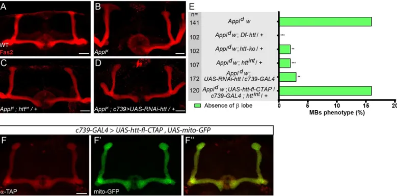
Appl null (Appld) flies are viable, fertile and display no gross structural defects in the brain [39]. However,AppldMBs display modestly-penetrant axonal defects inαβ neurons [5]. For simplicity, in our quantification of this phenotype we will focus our attention on the role ofAppl in β-branches, where Appl is required cell-autonomously [5]. From 3110Appld MBs, 451 (14.5%) showed an absence ofβ lobe phenotype (see details inMaterial and methods), in accordance with what was previously reported [5]. Two control MBs (0.26%) displayedβ lobe absence out of 759 MBs (see details inMaterial and methods). In line with our interest in potential developmental associations of neurodegenerative genes, we won-dered whetherhtt would modify the effect of Appl in MB αβ neurons. By itself, as expected from published data [12] the loss of one or even two copies ofhtt did not result in any sig-nificant MB developmental defects (S1A Fig). However, we found that mutation ofhtt potently suppresses the MBAppldmutant phenotype inαβ neurons (Fig 1). Specifically, theAppldMB phenotype was rescued by reducinghtt expression using three different genetic manipulations: 1)htt heterozygosity using two mutant alleles of htt (htt-ko or httint), 2) heterozygosity for a 55 kb chromosomal deficiency uncovering thehtt locus and most of the adjacentCG9990, and 3) htt RNAi knock down in the αβ MB neurons (Fig 1A– 1E; RNAi expression was driven in this experiment by theαβ neuron-specific GAL4 line c739). Conversely, when htt was overexpressed in the MB αβ neurons using
UAS-htt-fl-CTAP, a UAS-C-terminally TAP-tagged full length htt, it did not rescue httint/+ (16% absence ofβ lobe in Appld;UAS-htt-fl-CTAP/c739-GAL4; httint/+ vs 2% inAppld; +/+;
httint/+ and 16% inAppld) (Fig 1E). As expected from the above genetic results, htt-fl -CTAP protein, like wild type Htt, was present throughout the axon when expressed in αβ neurons (Fig 1F).
Since Dsh is a core intracellular component of the Appl-dependent Wnt/PCP pathway in the MBs, we tested the potential interactions betweenhtt and dsh. We found that remov-ing one copy ofhtt also suppressed the dsh1MB phenotype in theβ-lobe (21% absence of β lobe indsh1;+/+ vs 4% in dsh1;htt-ko /+—Fig 2A, 2B and 2Eupper bars). Moreover, hemi-zygosity for bothAppl and dsh resulted in a strong MB axon outgrowth phenotype (Appld w dsh1; 60% absence of β lobe, vs 21% in dsh1and 16% inAppld). We found that heterozy-gosity forhtt also strongly suppressed the Applddsh1phenotype (12% of absence ofβ lobe —Fig 2C–2E lower bars). Taken together, these data strongly suggest that Htt is a negative regulator of the Appl-dependent Wnt-PCP signaling pathway acting during MBβ-axon outgrowth.
Fig 1. The loss ofhtt rescues the AppldMB axon outgrowth mutant phenotype. (A) Wild-type MBα and β lobes revealed by anti-Fas2 staining. (B-D) Anti-Fas2
staining reveals the absence of theβ lobe in an Appldmutant brain (B), which is rescued by the loss of one copy of
htt (C) and is also rescued by the expression of RNAi
againsthtt driven by c739-GAL4 (D). (E) Quantitation of rescue of the MB Appldphenotype byDf-htt, htt-ko, httintandUAS-RNAi-htt driven by c739-GAL4. The
rescue ofAppldbyhttintis prevented by the overexpression ofhtt driven by c739-GAL4 indicating the functionality of the UAS-htt-fl-CTAP transgene. n = number of MBs analyzed,��P < 0.01 and���P < 0.001. Significance was calculated by a multiple comparison Fisher’s exact test (P = 5.8 10−10) followed by post-hoc Bonferroni’s
multiple comparison correction (p-values = 2.3 10−5, 0.0018, 0.0009, 0.0014 and 1). (F-F”) The expression ofUAS-htt-fl-CTAP revealed by an anti-TAP staining (F)
andUAS-mito-GFP (F’) driven by c739-GAL4 are similar in the MBs (F”). All panels correspond to adult brains. The scale bar on panels A-D and F indicates 30 μm.
Images are composite stacks to allow the visualization of axon trajectories along their entire length. Full genotypes: (A)y w67c23/ Y;c739-GAL4 UAS-mito-GFP / +. (B)
Appldw�/ Y; c739-GAL4 UAS-mito-GFP / +. (C) Appldw�/ Y; c739-GAL4 UAS-mito-GFP /+; httint/ +. (D) Appldw�/ Y;c739-GAL4 UAS-mito-GFP / UAS-RNAi-htt.
(E) top to bottom:Appldw�/ Y; c739-GAL4 UAS-mito-GFP / +. Appld
w�/ Y; c739-GAL4 UAS-mito-GFP / +; Df-htt / +. Appld
w�/ Y; c739-GAL4 UAS-mito-GFP / +; htt-ko / +. Appldw�/ Y; c739-GAL4 UAS-mito-GFP / +; httint
/ +. Appldw�/ Y; c739-GAL4 UAS-mito-GFP / UAS-RNAi-htt. Appld
w�/ Y; c739-GAL4 UAS-mito-GFP / UAS-htt-fl-CTAP; httint/ +. (F-F”)y w67c23/ Y; c739-GAL4 UAS-mito-GFP / UAS-htt-fl-CTAP.
Abl loss-of-function and gain-of-function mutants induce similar MB αβ
neuron phenotypes
Since Abl is a key component of the Appl-dependent Wnt-PCP signaling pathway required for axon growth in MBs [5] and Abl was previously shown to phosphorylate Dsh [19], we sought to clarify the potential relationship between Abl, Appl, and Dsh in MBαβ neurons. Indeed, we found that the axonal phenotypes ofAppldanddsh1MBαβ neurons can be rescued by modest enhancement of Abl expression, through transgenic expression of anAbl-GFP fusion under control ofAbl upstream genomic sequences (S1B and S1C Fig). We therefore characterized in more detail the axonal morphology phenotype inAbl loss- and gain-of-function MB αβ neurons.
The phenotype of MBαβ neurons with alterations in Abl expression has not previously been described thoroughly. We have used threeAbl alleles (Abl1,Abl2andAbl4) to characterize the requirement ofAbl function in MB morphology (S2A Fig). Each of three double heterozy-gous combinations (1/2, 2/4 and 1/4) are largely adult lethal but are viable at 48 hours after puparium formation (hAPF) enabling their effects to be investigated at that stage when theαβ neurons are present [37,40]. Interestingly, all the three allelic combinations gave similar MB phenotypes with a mixture of wild-type (WT) lobes (~20%) and MB lackingα-lobes, β-lobes, or both (~80%) (Fig 3A–3D). TheseAbl mutant phenotypes, including the adult lethality, were significantly rescued by introducing theAbl-GFP genomic transgene (from 20% WT MBs to 80%, P < 10−5;Fig 3D).Abl is known to be toxic when expressed in an unregulated fashion [21,25], and to cause axon growth and guidance defects when overexpressed in some neurons [29–31]. We therefore overexpressedAbl specifically in the MBs using the GAL4/UAS system [41]. Overexpression of WTAbl in MB neurons with the OK107-GAL4 driver produced αβ lobe loss phenotypes similar to those observed withAbl LOF alleles (Figs3EandS2B–S2E). Note that the use of a the weakerc739-GAL4 driver resulted in no lobe loss phenotype (n = 100). The penetrance of the gain-of-function (GOF) mutant phenotype was even higher than that in theAbl LOF alleles; WT MBs were never detected. In contrast, expression of a kinase dead version ofAbl failed to produce a MB mutant phenotype, although expression
Fig 2. The loss ofhtt rescues the dsh1MB axon outgrowth mutant phenotype. (A-B) Anti-Fas2 staining reveals the absence ofβ lobe in a dsh1mutant brain (A), which is rescued by the loss of one copy ofhtt (B). (C-D) Anti-Fas2 staining reveals the loss of the two β lobes in an Applddsh1mutant brain (C), which is rescued by the loss of one copy ofhtt (D). (E-F) Quantitation of the rescue of dsh1(E) and ofApplddsh1(F) phenotypes byhtt-ko. n = number of MBs analyzed and���P < 0.001
(Fisher exact test). All panels correspond to adult brains. The scale bar on panels A-D indicates 30μm. Images are composite stacks to allow the visualization of axon trajectories along their entire length. Full genotypes: (A)w dsh1. (B)w dsh1;; htt-ko / +. (C) Appldw�dsh1. (D)Appldw�dsh1;; htt-ko / +. (E) top to bottom: w dsh1.w
dsh1;; htt-ko / +. Appldw�dsh1.
Appldw�dsh1
;; htt-ko / +.
Fig 3. Either loss or overexpression ofAbl affect MB αβ neuron morphology. (A-C) Anti-Fas2 staining on wild-type (WT) brain (A) and on
Abl2/Abl4brain (B-C). In a wild-type (WT) brain, theα lobe (indicated by yellow arrowhead) projects vertically and the β lobe, indicated by pink arrowhead, projects toward the midline and stops before reaching it. The loss of theβ and α lobes (B) and of both the α and β lobes (C) is emphasized by white dashed lines.�shows the ellipsoid body. (D) Quantitation of theαβ neuron mutant phenotype in the Abl2
/Abl1mutant and rescued brains with theAbl-GFP genomic transgene. n = number of MB observed and���P < 0.001 (Fisher exact test). Transgenic expression of
levels of WT and kinase-dead Abl from transgenes were similar (S2F–S2G Fig). Taken together, these data demonstrate that the expression ofAbl and its kinase activity must be tightly controlled in the MBs in order to ensure normal MBαβ axon morphology. In order to determine if other MB neurons, in addition to theαβs, are sensitive to the overexpression of Abl we have also examined at theα’β’ and the γ MB adult neurons. The α’β’s were clearly affected displaying no WT MBs out of 93 MBs (S2 Fig) although the adultγ looked essentially wild-type (2 MBs with “round shape”γ out of 93 MBs). Therefore, both αβ and α’β’ are sensi-tive to Abl overexpression.
Abl function is required for MB axon outgrowth and is expressed in MB αβ
neurons
The absence of MB lobes can result from either growth or guidance defects [5,40]. In order to establish which of these two cellular phenomena is affected by loss ofAbl, we first produced neuroblast mutant MARCM clones, which encompass hundreds of neurons, with the three dif-ferentAbl alleles. These clones appeared to be predominantly wild-type with only between 4% to 11% showing the absent lobe phenotype (Table 1A). Our observation of highly-expressive lobe defects in the MBs of animals that are fully mutant forAbl versus the relative lack of pheno-types in MBs containingAbl mutant clones is consistent with the evidence cited above that Wnt-PCP acts non-cell autonomously for MB axon outgrowth [38], and that Abl acts down-stream of Appl to promote this mechanism. However, given the low percentage ofAbl mutant MARCM clones that displayed morphological defects we were not able to go further in studying single-cell mutant clones. We therefore produced fully mutantAbl2/Abl4animals, and in them, we generated clones that express mCD8-GFP in single cells or small groups of cells. We term these “visualization clones” since only the membrane marker is clonal, and not theAbl muta-tion. For this experiment, clones are initiated in L3 larvae, and examined at 48 hAPF (to avoid the substantial late-pupal/adult lethality of homozygousAbl mutations). Two-cell/single cell visualization clones inAbl mutant animals revealed highly-penetrant growth defects (87% n = 15) (Table 1BandFig 3F–3I’). We conclude thatAbl, like Appl, is required for MB αβ axon outgrowth. To visualize the localization of Abl protein we employed a transgenic fly bearing a genomicAbl-GFP construct, which rescues both Abl mutant lethality and the mutant MB phe-notypes (see above) and therefore is a bona fide endogenous marker for Abl.Abl-GFP is expressed broadly and homogenously in the brain from L3 to adult with elevated levels in the MBαβ axons of 48 hAPF brains (Fig 3J–3J”). Taken together, these data show thatAbl is expressed in the MBs and thatAbl function is required for MB αβ axon outgrowth. Additional
Abl-GFP rescued the morphological defect and lethality of the double heterozygous Abl2/Abl1combination, but failed to rescueAbl2homozygote lethality, indicating that this lethality is due to associated modifiers on the chromosome independent ofAbl. (E) Quantitation of the αβ neuron
mutant phenotype when a wild-type or a kinase dead form ofAbl expression is driven in the MBs by OK107-GAL4 (n = number of MB observed).
(F-F’) Two-cell WTαβ neuron MARCM clone in a WT brain (F) associated with anti-Fas2 staining in red (F’). (G-G’) Two-cell WT-looking αβ neuron clone (G) associated with anti- Fas2 staining in red (G’) in anAbl2/Abl4brain. (H-H’) A single-cellαβ neuron clone with an α branch growth defect (H) associated with anti-Fas2 staining in red (H’) in anAbl2/Abl4brain displaying an absence ofα lobe. Note the α branch which stops just after the branching point in H (yellow arrowhead). (I-I’) A single-cell αβ neuron clone with α and β branch growth defects (I) associated
with anti-Fas2 staining in red (I’) in anAbl2/Abl4brain displaying an absence ofα and β lobes. Note the small α and β branches in I. (J-J”) Expression of Abl within the MB using anAbl-GFP genomic transgene (J). α and β lobes are revealed by anti-Fas2 staining in red (J’). Merge of
GFP and anti-Fas2 staining (J”). All panels correspond to 48 hAPF brains except for E and the rescue experiment in D which are from adult brains. White arrowheads show the peduncle or common part of theαβ axon, white arrows show the αβ branch point, yellow arrowheads show theα axon branch or the α lobe and pink arrowheads show the β axon branch or the β lobe. The scale bar in panels A-C and F-J indicates 30 μm. Images are composite stacks to allow the visualization of axon trajectories along their entire length. Full genotypes: (A) wild type:y w67c23. (B and C)y w67c23;; Abl2FRT2A / Abl4FRT2A. (D) top to bottom: y w67c23;; Abl2FRT2A / Abl1FRT2A. y w67c23; Abl-GFP / +; Abl2FRT2A / Abl1FRT2A. (E) top to bottom:y w67c23/ Y; UAS-Abl / UAS-mCD8-GFP;; OK107-GAL4 / +. y w67c23/ Y; UAS-AblKD/ UAS-mCD8-GFP; TM6B,Tb1/ +; OK107-GAL4 / +. (F) w�tubP-GAL80 hs-FLP122 FRT19A / w�sn FRT19A; c739-GAL4 UAS-mCD8-GFP / UAS-mCD8-GFP. (G-H-I) w� tubP-GAL80 hs-FLP122 FRT19A / w�sn FRT19A; c739-GAL4 UAS-mCD8-GFP / UAS-mCD8-GFP; Abl2/ Abl4. (J)y w67c23; Abl-GFP / +.
neuroblast and more than two-cell visualization clones inAbl mutant animals confirmed the Abl requirement for MBαβ axon outgrowth (S3 Fig). In order to know if other MB neurons, in addition to theαβ’s, are sensitive to the lack of Abl we have also looked at the α’β’ and the γ MB neurons. We could not assess theα’β’ neurons because neither the anti-Trio antibody nor the c305a-GAL4 line labelled the MB α’β’ neurons adequately before 48 hAPF. However, anti-Fas2 revealed a clear defect inAbl2/Abl4γ neurons (S3 Fig). Therefore, at least theγ and the αβ MB neurons are sensitive to the lack of Abl function. Since bothαβ and α’β’ are sensitive to Abl overexpression (see above), all MB neurons appear to require normal levels of Abl function.
htt modifies the Abl mutant phenotype in MB axon outgrowth
As discussed above, theAppldMB phenotypes inβ axons can be rescued by UAS-driven over-expression of Abl [5], and both theAppldanddsh1β axonal phenotypes can be rescued by modest overexpression ofAbl using a genomic transgene, (S1B and S1C Fig). Taken together
Table 1.Abl mutant MARCM clones.
(A)Ablmut/AblmutMARCM clones. Neuroblast clones
Genotype WT Absence ofα lobe Absence ofβ lobe Absence ofα/β lobes n
Control 22 100% - - - - - - 22 Abl2/Abl2 43 90% 5 10% - - - - 48 Abl4/Abl4 37 82% 5 11% 3 7% - - 45 Abl1/Abl1 71 92% 3 4% 3 4% - - 77
(B) VisualizationAbl2/Abl4MARCM clones. Two-cells / Single-cell clones
Genotype WT α branch growth defect β branch growth defect α/β branch growth defects n
Control 30 100% - - - - - - 30 Abl2/ Abl4 2 13% 10 67% - - 3 20% 15 >Two-cells clones
Genotype WT Absence ofα lobe Absence ofβ lobe Absence ofα/β lobes n
Control 9 100% - - - - - - 9 Abl2/ Abl4 6 23% 14 54% 2 8% 4 15% 26 Neuroblast clones
Genotype WT Absence ofα lobe Absence ofβ lobe Absence ofα/β lobes n
Control 9 100% - - - - - - 9 Abl2/ Abl4 2 17% 7 58% - - 3 25% 12
WT: wild-type clones. n: number of clones analyzed.
Full genotypes: A) Control:y w67c23hs-FLP122 / +; c739-GAL4 UAS-mCD8-GFP / +; tubP-GAL80, FRT2A / FRT2A. Mutant:. y w67c23hs-FLP122 / +; c739-GAL4
UAS-mCD8-GFP / +; tubP-GAL80, FRT2A / Abl2FRT2A. y w67c23hs-FLP122 / +; c739-GAL4 UAS-mCD8-GFP / +; tubP-GAL80, FRT2A / Abl4FRT2A. y w67c23hs-FLP122 / +;
c739-GAL4 UAS-mCD8-GFP / +; tubP-GAL80, FRT2A / Abl1FRT2A. B) Control: w�tubP-GAL80 hs-FLP122 FRT19A / w�sn FRT19A; c739-GAL4 UAS-mCD8-GFP / UAS-mCD8-GFP. Abl2/Abl4:w�tubP-GAL80 hs-FLP122 FRT19A / w�sn FRT19A; c739-GAL4 UAS-mCD8-GFP / UAS-mCD8-GFP; Abl2/ Abl4.
with the finding thathtt suppresses both Appl and dsh MB phenotypes, we hypothesized that htt might be a suppressor of Abl action. To test this hypothesis, we conducted three sets of experiments to examine the effects of reducinghtt dosage on Abl phenotypes. First, we found that the increased severity of the MB mutant phenotype inAppld;Abl2/+ individuals was completely abolished when one copy ofhtt was also removed (31% absence of β lobe in Appld; Abl2/+ vs 14% inAppld;Abl2httint/+ compared to 14% inAppld; +/+—Fig 4Aupper and lower bars). Second, a modest but significant increase in the proportion of WT MBs inAbl2/Abl1 individuals was observed when one dose ofhtt was removed (from 20% to 32%—Figs4Cand
S4). Third, enhancement of theAbl GOF mutant phenotype, measured by the simultaneous absence of bothα and β lobes, was seen when one copy of htt was removed (from 21% to 75% —Fig 4Dupper and middle bars). Conversely, rescue of this phenotype was observed whenhtt was overexpressed usingUAS-htt-fl-CTAP driven by c739-GAL4 (from 21% to 3%—Fig 4D
upper and lower bars). This interpretation of a phenotypic rescue is further supported in this comparison by the increase in the number of wild-type MBs (0% inUAS-Abl, vs 16% in UAS-Abl; UAS—htt-fl-CTAP). The ability of htt/+ to suppress the Abl LOF phenotype or to enhance theAbl GOF phenotype, as well as the ability of htt overexpression to suppress the Abl GOF phenotype appears to correlate with the amount of kinase-competent residual pro-tein in the various mutant backgrounds (seeDiscussion). Taken together, these data strongly indicate that Htt is a repressor of Abl function during MB axon outgrowth.
Reduction of
htt increases Abl kinase activity in the developing MBs
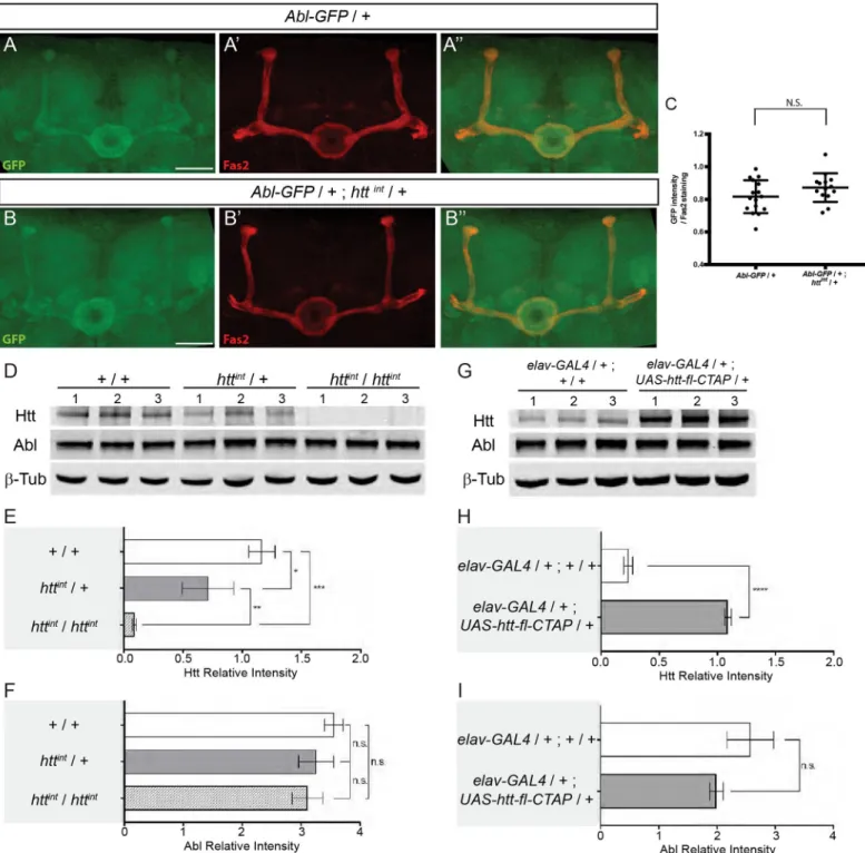
One potential explanation for the suppressor effect ofhtt on the Abl mutant MB phenotype is that Htt inhibits Abl kinase activity. To test this idea, we employed a fluorescence resonance energy transfer (FRET) biosensor probe that allows Abl kinase activity to be assayedin vivo in Drosophila [42]. Using this tool, we first showed that Abl kinase activity is detectable in the MBs by comparing the FRET activity of wild typeUAS-Abl-FRET versus the FRET activity of a mutantUAS-Y➔F Abl-FRET that lacks its phosphorylatable tyrosine (Figs5D–5FandS5). We then tested the FRET efficiency in the developing MBs of control (+/+) versushttint/+ larvae and detected a significant increase of the FRET efficiency when one copy ofhtt was removed (Fig 5E and 5G). Note that the exact value of the FRET ratio depends on various microscope and laser settings during imaging. Consequently, this number can only be compared directly between samples imaged together in a single imaging session and cannot be compared from separate experiments. In these experiments, images for validation of the FRET(WT) probe by comparison to FRET(Y->F) were collected in a single session (D,F) as were images comparing FRET activity in a wild type vshtt/+ genetic background (E,G). Quantitative PCR (qRT-PCR) analysis of control versushttint/+ larval brains revealed no difference in Abl mRNA expression (Fig 5H). Furthermore, there were no apparent differences in the overall levels of Abl protein in thehtt/+ heterozygous MBs relative to WT controls (Fig 6A–6C). The expression level of Abl protein is fairly homogenous in the larval brain rendering the quantitation of Abl in the MBs difficult. In order to reliably quantitate Abl protein levels, we measured the level of Abl protein in 48 hAPF MBs. As noted above, Abl is enriched during this stage. Finally, reduction or increase in Htt expression did not alter the total levels of neuronal Abl protein inDrosophila heads (Fig 6D–6I). Taken together, these data strongly suggest that Htt is a repressor of Abl kinase activity in the developing MB axons.
Discussion
Tumorigenesis and neurodegeneration may be two sides of the same coin [43]. Indeed, defin-ing the overlap of molecular pathways implicated in cancer and neurodegeneration may open
the door to novel therapeutic approaches for both groups of disorders [43]. Correlative studies have highlighted a decreased cancer incidence in the population with the neurodegenerative disorder Huntington’s disease and both wild-type and mutant huntingtin (Htt) have been implicated in tumor progression [44]. Interestingly, it has been proposed that, in the normal
Fig 4.htt interacts with Abl. (A) The Appldmutant phenotype is enhanced in anAbl2/+ mutant background. (B) However, theAppldmutant phenotype is not modified in anAbl2httint/+ + mutant background. (C) The Abl2/Abl1
mutant phenotype is partially rescued by the loss of one copy ofhtt as inferred by the increase of the wild-type (WT)
MBs. (D) The absence ofα and β lobe phenotype observed in UAS-Abl driven by OK107-GAL4 is strongly increased by the loss of one copy ofhtt (upper and middle bars). However, this phenotype is rescued by the overexpression of
full-lengthhtt (upper and lower bars). n = number of MBs analyzed,��P<0.01 and���P < 0.001. Significance was
calculated by a Chi2test for A-C and by a multiple comparison Fisher’s exact test (P = 3.4 10−28) followed by post-hoc Bonferroni’s multiple comparison correction (p-values = 6.3 10−12and 0.0003 for D). All panels correspond to adult
brains except for B which is from 48 hAPF brains. Full genotypes: (A) top to bottom:Appldw�/ Y; c739-GAL4 UAS-mito-GFP / +. Appldw�/ Y; c739-GAL4 UAS-mito-GFP / +; Abl2
/ +. Appldw�/ Y;c739-GAL4 UAS-mito-GFP / +; Abl2
FRT2A httint/ +. (B) top to bottom: y w67c23;; Abl2FRT2A / Abl1FRT2A. y w67c23;; Abl2FRT2A httint/ Abl1FRT2A. (C)
top to bottom:y w67c23/ Y;UAS-Abl / +; UAS-FRT-y+-FRT / +; OK107-GAL4 / +. y w67c23/ Y;Abl /
UAS-mCD8-GFP; httint/ +; OK107-GAL4 / +. y w67c23/ Y;UAS-Abl, UAS-htt-fl-CTAP / +;; OK107-GAL4 / +. Note that
UAS-FRT-y+-FRT and UAS-mito-GFP are used here as neutral UAS to adjust for 2 UAS sequences in the three
different genotypes.
Fig 5. Reducinghtt expression increases Abl-FRET biosensor phosphorylation state during MB development. (A) Schematic representation of an
imaged MB in the brain. (B) TheAbl-FRET biosensor is based on mammalian CRK protein scaffold with two additional fluorescent proteins (CFP and
YFP). ABL kinase activity induces phosphorylation ofUAS-Abl-FRET biosensor leading to its spatial rearrangement and increased FRET efficiency
[59]. (C) Representative maximal projection of the YFP and FRET signals recorded in the MB lobes of adult flies using confocal microscopy. These are presented as examples of the kind of image data that goes into the FRET ratio calculation. Scale bar: 10μm. (D) FRET and YFP signals are recorded on
physiological situation, the neurodegeneration-related Amyloid precursor protein (APP) recruits the oncogenic Abelson (Abl) kinase in order to promote axonal outgrowth [5]. It is, therefore, tempting to propose that different neurodegenerative diseases (ND) may share com-ponents and mechanisms and that Abl may also have a role in ND.
In this study, we have shown thatAbl is required for axonal growth in the MBs, a brain structure that is involved in memory. Furthermore, we show that bothAbl overexpression and lack of expression in the MBs result in similar phenotypes, indicating the need to tightly regu-late Abl activity during MB axon outgrowth. This raises the question of how Abl activity is nor-mally negatively regulated during MB axon outgrowth. We confirmed the previous
observation that overexpression ofAbl rescues the AppldMB phenotypes [5]. Furthermore, we found thatAbl overexpression also rescues the dsh1MB phenotype. These two results support the model that Appl activates Abl, which in turn phosphorylates Dsh. At the genetic level, we expected an increase of Abl activity in an individual bearing a loss-of-function mutation of a putativeAbl repressor. We therefore hypothesized that reducing the levels of an Abl repressor would result in suppression of theAppl and dsh mutant MB phenotype. We found that Htt is such an inhibitor of Abl activity in the MBs. The loss of one dose ofhtt increased the activity of the wild-type Abl still present inAbl2/+ individuals and therefore prevented the enhancement of the MB mutant phenotype. While a number of studies have concluded that Htt deficiency results in significant alterations to kinase signaling pathways [45], to our knowledge this study is the first to implicate the crucial tyrosine kinase, Abl. Together, these data demonstrate the power that neurodevelopmental studies have to reveal close functional relationships between genes implicated in different forms of neurodegenerative disease.
It was a surprise that nullhtt mutants show no obvious developmental defects in Drosophila although strong defects could have been expected from the lack of such a conserved protein [12]. One possible hypothesis is that, due to its fundamental importance, some functional redundancy has been selected to buffer against variation in the production of the Htt protein. We reasoned that altering the levels of another protein, particularly another protein known to be implicated in neurodegeneration, could reveal cryptic phenotypes ofhtt mutation during brain development. Following this reasoning, we combined a nullAppl mutant with heterozy-gous nullhtt mutations in double mutant individuals. Unexpectedly, we found that mutant htt suppressed theAppl MB axonal outgrowth defect.
Abl is a key component of the Appl signaling pathway required for axonal arborization and growth in the fly brain and the functional relationship between these two proteins is likely con-served in mammals [5,34]. While the role of APP-mediated signaling has been shown most clearly inDrosophila, a number of lines of evidence suggest that mammalian APP also fulfills a
adjacent 0.5μm confocal planes along all the anterior-posterior axis of the α and β MB lobes in adult flies. Either wild-type UAS-Abl-FRET biosensor or a mutated form (UAS-Y➔F Abl-FRET) are expressed in the αβ MB neurons using the c739-GAL4 driver. In the UAS-Y➔F Abl-FRET biosensor, the
tyrosine located in ABL target site (PYAQP) was replaced by a phenylalanine (PFAQP) impairing phosphorylation [42]. FRET efficiency is significantly reduced forUAS-Y➔F Abl-FRET relative to UAS-Abl-FRET biosensor for all confocal planes considered except for the nine most anterior and six most
posterior planes. Two-tailed Mann-Whitney tests on non-Normally distributed data. Results are mean± SEM with n � 7 MB for each confocal plane. (E) Reducinghtt expression increases FRET efficiency of UAS-Abl-FRET biosensor in third instar larval MB lobes. The UAS-Abl-FRET biosensor is
expressed in MB neurons using thec739-GAL4 driver in control (+/+) and httint/+ flies. FRET efficiency is significantly increased inhttint/+ flies for all confocal planes along the anterior-posterior axis except for the three located 3 to 4μm from the initial confocal plane. Two-tailed Mann-Whitney tests on non-Normally distributed data. Results are mean± SEM with n � 13 MB for each confocal plane. (F) FRET efficiency is globally reduced in
UAS-Y➔F Abl-FRET mutant versus UAS-Abl-FRET. FRET efficiency is averaged for all confocal planes and all along the anterior-posterior axis.
Two-tailed Mann-Whitney test with non-Normally distributed data. Results are mean± SEM with n �462;���p<0.001. (G) FRET efficiency is globally
increased whenhtt expression is reduced. FRET efficiency is averaged for all confocal planes and all along the anterior-posterior axis. Two-tailed
Mann-Whitney test on non-Normally distributed data. Results are mean± SEM with n � 1055;���p<0.001. (H)Abl mRNA expression is not changed in L3
brains followinghtt partial inactivation. Abl expression was assessed using RT-qPCR in L3 brains of httint/+ versus WT (+/+) male flies. Results show three independent biological replicates. Full genotypes: Genotypes:y w67c23/ Y;c739-GAL4/+; UAS-Abl-FRET/+. y w67c23/ Y;c739-GAL4/+; UAS-Y-F
Abl-FRET/+. y w67c23/ Y;c739-GAL4/+; httintUAS-Abl-FRET/+.
Fig 6.htt does not seem to affect the quantity of ABL in the MBs or the total levels of neuronal Abl in Drosophila. (A-B”) Expression of the Abl-GFP genomic
transgene in a WT (A) and in ahttintheterozygous mutant background (B) at 48 hAPF. Anti-Fas2 staining markedαβ neurons (A’-B’). Merge of GFP and anti-Fas2 staining (A”-B”). Note that panels A-A” are also presented inFig 3J–3J”. (C) Quantitation of the GFP expression within MBs is not significantly different (N.S.) between WT andhttintheterozygous mutant background using a Mann-WhitneyU test. The scale bar indicates 30 μm. Details of image quantification procedure and full genotypes are: (C) After having outlined the MB with the Fas2 staining, GFP and Fas2 intensities from MB shape were quantified for each slices of the stack. The GFP intensity of each slices was averaged and then normalized by the mean Fas2 intensity. Number of MB analyzed: control = 16,htt mutant = 14. Quantitation of the
GFP expression within MBs is not significantly different between WT andhttintheterozygous mutant background using a Mann-WhitneyU test. Quantitation were
done with ImaJ software. Images are composite stacks to allow the visualization of axon trajectories along their entire length. Genotypes: (A)y w67c23; Abl-GFP / +. (B)
y w67c23; Abl-GFP / +; httint/ +. (D) Western blots of whole cell lysates from adult heads from the following genotypes: +/+, httint/+ and
httint/httint. Levels of Htt, Abl, andβ-Tubulin (β-Tub) were assessed by probing blots using the indicated antibodies. 1, 2, and 3 indicate biological replicates for each line. (E and F) Quantitation of Htt (E) or Abl (F) protein levels in the indicated genotypes was assessed from western blots in (D) relative toβ-Tubulin and plotted as relative band intensity. (G) Htt was over-expressed usingelavc155-GAL4 > UAS-htt-fl-CTAP. Lysates from adult heads were subjected to western blotting to assess Htt, Abl, and β-Tubulin levels. 1, 2,
signaling role [46]. Importantly, and in line with its proto-oncogenic role,Abl tyrosine kinase activity is tightly regulated by intramolecular inhibition [22]. AlthoughAbl is clearly required as a downstream effector ofAppl in the MB axon growth, its precise role and regulation in the MBs has not been described previously.
The threeAbl alleles used in this study have all been shown to result in truncated proteins [47]. WhileAbl1retains the SH3, SH2 and TK domains of Abl,Abl2is mutated within the TK domain and only retains the SH3 and SH2 domains, whileAbl4is mutated in the SH2 domain, and only retains an intact SH3 domain. It therefore seems likely that very little or no residual Abl function remains in Abl2/Abl4andAbl4/Abl1individuals and may explain why no rescue was observed when one dose ofhtt was removed in these genetic backgrounds. In contrast, the Abl1/Abl2allelic combination is likely less severe than the other two genotypes andAbl1/Abl2 animals do accumulate truncated Abl proteins. Indeed, while significant amounts of truncated Abl1andAbl2mutant proteins are detectable, only faint protein bands are observed inAbl4 pupae [48]. Therefore, some functionally significant kinase activity could remain inAbl2/Abl1 individuals and the loss of one dose ofhtt might increase the activity of the remaining kinase activity. Finally, removing one dose ofhtt in animals overexpressing Abl would result in even more Abl activity, which in turn would exacerbate the mutant phenotype. Contrarily, over-expressing Htt, as in theUAS-Abl, UAS-htt doubly overexpressing individuals, would inhibit Abl function when compared to theUAS-Abl overexpression alone which thus might explain the observed rescue.
There are three different levels at which Htt might act to modulateAbl activity. First, Htt could either directly or indirectly affectAbl mRNA levels. Although fly Htt has been described as a cytoplasmic protein [12],htt has been shown to be a suppressor of position-effect variega-tion, suggesting a possible role in chromatin organization [13]. This hypothesis is unlikely for the MB phenotype described here since qRT-PCR analysis of third instar brains did not reveal a significant differences inAbl mRNA levels between httint/+ and control individuals. Second, Htt could play a role regulating Abl protein level in the MBs. We also consider this unlikely since the quantity of the endogenous Abl is unchanged inhttint/+ relative to control individu-als. Third, Htt could influence the kinase activity of Abl itself. Taking advantage of a FRET bio-sensor enabling Abl kinase activity to be assayed directly in the MBs, we revealed a significant increase in active Abl inhttint/+ versus control individuals. Therefore, we favor a model of Htt acting as an inhibitor of Abl kinase activity during normal MB axonal growth (Fig 7). Abl activity in axons needs to be maintained within rather narrow limits [32,33]. These two recent studies show that either increase or decrease of Abl activity cause disorganization of actin structure in the growth cone and prevent the orderly oscillation of growth cone actin that is the motor for growth cone advance and thus axon extension. Those papers also explain why Abl gain and loss can result in superficially similar mutant axon patterning phenotypes even though the molecular effects of Abl increase versus decrease are opposite.
The HEAT repeat domains of Htt are thought to function as a solenoid-like structure that acts as a scaffold and mediates inter- and intra-molecular interactions [9]. It is therefore tempting to propose that a scaffolding role of Htt could elicit repression of Abl kinase activity. At least in the MBs, a balance seems to exist in the activity of Abl, positively regulated by the membrane complex formed by the core PCP proteins and Appl, and suppressed by Htt and possibly other proteins, as well. On one hand, if Appl is absent, Abl is not optimally activated
and 3 indicate biological replicates for each line. (H and I) Quantitation of Htt (H) or Abl (I) protein levels in the indicated genotypes was assessed from western blots in (G) relative toβ-Tubulin and plotted as relative band intensity. In E and F, significance was calculated by one-way ANOVA. In H and I, significance was calculated by unpairedt-test. In E, F, H and I, errors indicate standard deviation. (n.s. not statistically different,�P<0.05,��P<0.01,���P<0.001,����P<0.0001).
leading to defects in MB axon growth. Conversely, decrease of Htt to 50% of wild type levels leads to de-repression ofAbl kinase activity, which in turn compensates for its sub-optimal level of activation in the absence ofAppl. This unexpected apparent balance of activation and inhibition ofAbl by Appl and htt, whose mutant orthologs are central players in human neuro-logical disease, may define a conserved functional interaction to maintain Abl activity in the relatively narrow window to appropriately effect axon outgrowth.
Material and methods
Drosophila stocks
All crosses were performed on standard culture medium at 25˚C. Except where otherwise stated, all alleles and transgenes have been described previously (http://flystocks.bio.indiana. edu/). The following alleles were used:Abl1,Abl2,Abl4,Appld,dsh1,httint[13],htt-ko and Df-htt [12]. The following transgenes were used:UAS-Abl (Bloomington Stock Center line (BL) #28993),UAS-Abl.K417N (from BL #8566) named here UAS-AblKDfor kinase dead, UAS-Abl-FRET andUAS-Y➔F Abl-FRET [42],UAS-RNAi-htt [49],UAS-mCD8GFP, UAS-mito-GFP, UAS-FRT-y+-FRT and the genomic transgenic Abl-GFP [50].UAS-htt-fl-CTAP was produced for this study (see Constructs). We used three GAL4 lines:c739-GAL4 and OK107-GAL4 expressed in MB neurons [51] and the pan-neuronal driverelavc155-GAL4 (BL #458). Recom-binant chromosomes were obtained by standard genetic procedures and were molecularly ver-ified when required.
Fig 7. Htt is a repressor of Abl activity required for Appl-induced axonal growth. Schematic representation of the
proposed mechanism. Htt represses Abl activity at the growth cone ofαβ neuron in MB. Appl and Htt have opposing roles in MB development, to promote and suppress Abl kinase activity, respectively, to maintain the appropriate intermediate level of Abl phosphorylation necessary for axon growth. This model is based on the proposed model of Appl signaling required for MB axon growth (Fig 7 in [5]).
Adult and pupal brain dissection and immunostaining
Adult brains were dissected in PBS after fly heads and thoraxes had been fixed for 1hr in 3.7% formaldehyde in PBS. They were then treated for immunostaining as previously described [52,53]. Pupal brains were dissected in PBS and fixed for 20min in 3.7% formaldehyde in PBS at 4˚C with gentle rocking. After washing twice in PBS with 0.5% Triton X-100 (PBT) for 15 min at room temperature, they were incubated in PBT and 5% bovine serum albumin (BSA) (blocking solution) at room temperature for 30 min, followed by overnight incubation at 4˚C with primary antibody diluted in blocking solution. Brains were then washed three times in PBS for 20 min, followed by 30 min in the blocking solution, and then addition of the second-ary antibody with incubation for 3 hr at 4˚C. Brains were then washed three times in PBS for 20 min and were mounted with Vectashield (Vector Laboratories). Antibody combinations used: anti-Fas2 (mAb 1D4 from DSHB) at 1:50 dilution followed by anti-mouse Cy3 (Jackson ImmunoResearch) at 1:300; rabbit anti-Myc (Cell Signaling) at 1:1000 followed by anti-rabbit Cy5 (Jackson ImmunoResearch) at 1:300; mouse anti-TAP (Santa Cruz Biotechnology) at 1:300 followed by anti-mouse Cy3 (Jackson ImmunoResearch) at 1:300; rabbit anti-Trio (kind gift from Barry Dickson) at 1:1000 followed by anti-rabbit Cy2 (Jackson ImmunoResearch) at 1:300. Anti-Fas2 (mAb 1D4 from DSHB) at 1:10 dilution followed by anti-mouse Alexa 647 (Jackson ImmunoResearch) at 1:300.
Quantitation of the absence of mushroom body lobes
We took particular care in order to ascertain a suppression effect from a penetrance of about 15%. The absence of lobes was assessed with the anti-Fas2 staining or with thec739-GAL4 UAS-mito-GFP marker visualized with an epi-fluorescence microscope (Leica DM 6000). In order to be certain that we were indeed measuring suppression of theAppldordsh115–20% of absence ofβ lobes and not a mere variation from this rather low phenotypic penetrance (Figs1,2,4,S1B and S1C), we followed a strict protocol. A large number (at least 50) of Appldw or w dsh1orAppldw dsh1females were collected, pooled together and crossed in groups of 25 with eithery w67c23wild-type control ory w67c23;mutant/Balancer males. In this way, there were alwaysAppldw/Y or w dsh1/Y orAppldw dsh1/Y males from the same experi-ment that show the expected mutant phenotype, and any modifier in theAppldordsh1 stocks, if it exists, should be statistically equally present in the control and in the experiment. We could therefore associate the suppression phenotype seen inAppldw/Y or w dsh1/Y or Appldw dsh1/Y;httmutant/+ males unequivocally to the presence of thehttmutantallele. Noticeably, throughout this study in a total of 26 experiments, MBs fromAppld;c739-GAL4
UAS-mito-GFP/+ males were assessed. Of 3110 MBs, 451 (14.5%) displayed an absence of β lobe phenotype (22/141; 15/106; 16/84; 15/124; 15/105; 18/152; 19/108; 14/100; 14/100; 27/156; 25/164; 12/112; 2/36; 17/102; 21/139; 14/109; 19/158; 19/155; 15/104; 30/146; 14/91; 24/138; 13/124; 13/102; 11/90; 27/164). In addition, throughout this study, we assessed the absence ofβ lobe phenotype in wild-type controls. In a total of nine different experiments (0/106; 0/102; 0/100; 1/107; 0/100; 0/50; 0/50; 0/37; 1/107), control MBs displayed two MBs with absence ofβ lobes out of 759 MBs (0.26% showing absence of β lobes and 99.74% of the MBs appearing WT). Thus, wild-type flies almost invariably have intact MB lobes as was pre-viously described [5].
MARCM clonal analysis
The MARCM technique was used to generate clones in the MB [53]. We use the term MARCM clones when homozygous mutant clones were examined in a heterozygous back-ground and visualization MARCM clones when homozygous mutant clones were examined in
a homozygous mutant background. For MARCM neuroblast clones, L1 larvae were heat-shocked at 37˚C for 1 hr and adult brains were dissected and stained. For visualization MARCM neuroblast clones, L1 larvae were heat-shocked at 37˚C for 1 hr and 48 hAPF brains were dissected and stained. For visualization single and two-cell clones, L3 larvae were heat-shocked at 37˚C for 15 min and 48 hAPF brains were dissected and stained.
Microscopy and image processing
Images were acquired at room temperature using a Zeiss LSM 780 laser scanning confocal microscope (MRI Platform, Institute of Human Genetics, Montpellier, France) equipped with a 40x PLAN apochromatic 1,3 oil-immersion differential interference contrast objective lens. The immersion oil used was Immersol 518F. The acquisition software used was Zen 2011. Contrast and relative intensities of the green (GFP), red (Cy3) and blue (Cy5) channels were processed with Fiji Software. Quantitation was performed using ImageJ software.
Constructs
pUAS-htt-fl-CTAP: A htt “mini-gene” bearing a dual C-terminal Tandem Affinity Purification tag (Protein G and a streptavidin binding peptide: GS-TAP tag) was constructed. PCR was per-formed using thedhtt “mini-gene” comprising the dhtt full length cDNA with intron 10 (as described in [13]) and the following primers: HTT-TAP-FOR: ggtaccATGGACAAATCCAG GTCCAG (KpnI site added) and HTT-TAP-REV: tctagaCAGGCACTGCAACATCCGG (XbaI site added). The resulting PCR product was digested with KpnI/XbaI and sublconed into thepUAST-CTAP(SG) vector [54]. To avoid rearrangements due todhtt instability, cul-turing conditions were used as previously described [13].pUAS-htt-fl-CTAP transgenic flies were generated and balanced using standard procedures and expression of dhtt-SG was assessed using western blots.
FRET imaging
Fly brains were dissected in 1X PBS at room temperature and collected in ice-cold PBS before being fixed in 3.6% formaldehyde for 20 min. Brains were rinsed twice in PBST 0.5X for 20 min before being mounted in Vectashield (Vector Laboratories). MBs were imaged on adja-cent 0.5μm confocal planes along the anterior-posterior axis using a LSM780 confocal micro-scope (Zeiss) at x40 with oil immersion. Cyan fluorescent protein (CFP) was excited at 405 nm and emission recorded between 454 and 500 nm. Yellow fluorescent protein (YFP) was excited at 514 nm and emission recorded between 516 and 571 nm. Fluorescence resonance energy transfer (FRET) was generated at 405 nm. To avoid CFP emission, FRET was recorded out of CFP emission range, between 587 and 624 nm.
FRET image analysis
Brains were oriented anterior-posteriorly using the peduncle as an anatomical landmark and aligned according to the first confocal plane where a signal was visible. We ensured that the same number of planes were obtained for each group (c739>Abl-FRET: 48 ± 4 planes and c739>(Y➔F) Abl-FRET: 46 ± 1.5 planes, Student t-test: P = 0,7. c739>Abl-FRET: 79,3 ± 2,3 planes andc739; httint>Abl-FRET: 75,4 ± 3,7 planes; Student t-test: P = 0,4) indicating that there were no differences due to mounting. Average YFP and FRET signals were computed using the measurement of ‘Mean Grey Value’ and the ‘Plot Z-axis Profile’ functions of ImageJ [55] into a region of interest (ROI) corresponding to the contour of the MB and for each con-focal plane. Background was corrected using the ‘Rolling Ball Background Subtraction’
function (50 px radius). For each plane and within the same ROI, FRET signal was expressed relative to YFP to account for variability in Abl-FRET biosensor expression level or differences between preparations. Only groups (i.e.Abl-FRET vs (Y➔F) Abl-FRET and Abl-FRET vs httint; Abl-FRET) crossed, collected, dissected and imaged on the same day were compared. For any given confocal plane, the FRET ratio was averaged between left and right MBs and multiple genetically identical animals.
qRT-PCR
To quantifyAbl expression, RNA was extracted from the brains of L3 males. Brains (~20/sam-ple) were dissected in PBS 1X (Sigma) and kept on ice before homogenized in Trizol reagent (Ambion). Total RNA was treated with DNAse to eliminate genomic DNA (Applied Biosys-tems). RNA was purified using phenol-chloroform extraction and first strand cDNA synthesis was performed using reverse transcriptase (Invitrogen). Primers forAbl RNA amplification were designed on each side of intron 4–5 within exon 4 and 5 respectively. These exons are present in allAbl transcripts. Abl primers were designed using Primer3Plus online software [56].Abl forward primer sequence is 5’-GCGGCCATCATGAAGGAAATG-3’ and reverse primer sequence is 5’-TTGCCGTGCGACATAAACTC-3’.Abl RNAs were quantified in real-time during amplification using incorporation of SYBRGreen (Roche) and Light Cycler (Roche). Primers efficacy was first evaluated using a range of cDNA concentrations to ensure linearity of the amplification (E = 1,944). Only a single PCR product with the expected melting temperature was obtained. The amplicon was run on a gel to verify that the size was as expected for the splicedAbl product (107 bp). A control without reverse transcription was done to ensure thatAbl amplicon was not obtained. For each sample, a technical triplicate was per-formed and averaged. Independent biological replicates were prepared for each condition and the fold change was averaged (see Statistics). The biological replicates correspond to indepen-dent dissections, extractions, reverse transcriptions and quantifications. In the experimental condition (y w67c23/Y;;httint/+),Abl expression was expressed relative to control flies (y w67c23/ Y) after normalization to internal controls,Rpl9 and Rpl32, and using the ΔΔCT method [57].
Western blotting
Lysates of adultDrosophila heads were prepared using RIPA buffer supplemented with prote-ase inhibitors (Sigma #11836170001). Antibodies used for immunoblotting with dilutions were: anti-dhtt (3526, rabbit polyclonal, 1:1,000; [13]), anti-dAbl (as above, 1:1,000; [58]), anti-β-Tubulin (mouse, DSHB E7, 1:10,000), anti-mouse and anti-rabbit IRDye secondary antibod-ies (LI-COR Biosciences, 1:10,000). Lysates were resolved on NuPAGE 3–8% gradient Tris-Acetate gels with Tris-Tris-Acetate running buffer (for Htt and Abl) or NuPAGE 4–12% gradient Bis-Tris gels with MOPS running buffer (forβ-Tubulin). After transfer to nitrocellulose mem-branes, blots were processed according to the Odyssey CLx protocol. Median band intensity was quantified using Image Studio.
Statistics
Comparisons between two groups expressing a qualitative variable were analyzed for statistical significance using the Chi2or the Fisher exact test (BiostaTGV:http://biostatgv.sentiweb.fr/? module=tests). Comparison of two groups expressing a quantitative variable was analyzed using the two-tailed Mann-WhitneyU test or the unpaired t-test. For FRET quantitation, sta-tistical analyses were performed using Prism 8.0 (GraphPad). For each confocal plane of each group, the normality of the FRET ratio was assessed using D’Agostino & Pearson normality test. Non-parametric Mann-Whitney tests were used to compare groups at each confocal
plane. For the RT-qPCR, the averaged fold change ofAbl expression was compared to the the-oretical value of 1 that would correspond to no change inAbl expression and non-parametric Wilcoxon signed-rank test for non-normally distributed data or small samples was used. For Western blots, significance was quantified using Prism 8.0 (GraphPad) and calculated by one-way ANOVA (Figs6E, 6FandS2) or by unpaired Student’st-test (Fig 6H and 6I). Values of P < 0.05 were considered to be significant.
Detailed R commands for the statistical analysis of Figs1Eand4Dare accessible at:
[https://github.com/HKeyHKey/Marquilly_et_al_2020/blob/master/README.md]
Supporting information
S1 Fig. The overexpression ofAbl rescues the Appldand thedsh1mutant phenotypes. (A)
The loss ofhtt does not produce per se any significant MB developmental defects. (B) Quanti-tation of the rescue ofAppldMB phenotype by the genomic constructAbl-GFP. (C) Quantita-tion of the rescue ofdsh1phenotype by theAbl-GFP genomic construct. n = number of MBs analyzed,�P < 0.05 and���P < 0.001 (Chi2
test). All panels correspond to adult brains. Geno-types: (A) top to bottom:y w67c23/ Y; c739-GAL4 UAS-mito-GFP / +; htt-ko / +. y w67c23/ Y; c739-GAL4 UAS-mito-GFP / +; Df-htt / +. y w67c23/ Y;; Df-htt / htt-ko. y w67c23/ Y;; httint/ +. y w67c23/ Y;;httint/ httint.y w67c23/ Y; c739-GAL4 UAS-mito-GFP / UAS-RNAi-htt. y w67c23/ Y; UAS-mCD8-GFP / UAS-RNAi-htt;; OK107-GAL4 / +. (B) top to bottom: Appldw�/ Y;
c739-GAL4 UAS-mito-GFP / +. Appldw�/ Y; c739-GAL4 UAS-mito-GFP / Abl-GFP. (C) top to
bottom:w dsh1/ Y.w dsh1/ Y;Abl-GFP / +. (TIF)
S2 Fig. Structure and overexpression of the Abl protein. (A) Molecular scheme of the Abl
protein. Abl protein is composed of conserved domains: Src Homology 3 (SH3) domain (blue), Src Homology 2 (SH2) domain (orange), Kinase Domain (red), Poly-Proline PP domain (purple) and F-Actin Binding Domain (FABD) (green). The protein produced byAbl1 mutant allele is truncated between PP and Kinase domains. The protein produced byAbl2 mutant allele is truncated within the Kinase domain. The protein produced byAbl4mutant allele is truncated within the SH2 domain [47]. (B-E) Anti-Fas2 staining showing theα and β lobes in a WT adult brain (B) and inAbl forced expression by OK107-GAL4 (C-E) with an absence ofβ lobe (C), an absence of α lobe (D) and an absence of α and β lobes (E). The loss of lobes is emphasized by white dashed lines. Note that panel B is also presented as the left MB in
Fig 1A. The scale bar indicates 30μm. Images are composite stacks. Genotypes: (B) y w67c23/ Y. (C-E)y w67c23/ Y;UAS-Abl / UAS-mCD8-GFP;; OK107-GAL4 / +. (F) The expression levels ofUAS-Abl and UAS-AblKDtransgenes are similar.UAS-Abl and UAS-AblKDwere expressed usingOK107-GAL4. Lysates from adult heads were subjected to western blotting to assess Abl andβ-Tubulin (β-Tub) levels. 1, 2, and 3 indicate biological replicates for each line. (G) Quan-titation of Abl protein levels in the indicated genotypes was assessed from western blots in (F) relative toβ-Tubulin and plotted as relative band intensity. Errors indicate standard deviation. Significance was calculated by one-way ANOVA (P < 0.0001) followed by post-hoc Bonferro-ni’s multiple comparison correction (n.s. not statistically different and����P < 0.0001).
(H-L”‘) GFP (green) labelling is showing all the lobes (α and α’ vertically, β and β’ and γ medi-ally), anti-Trio (red) staining is showing theα’ and β’ and γ lobes and anti-Fas2 (blue) staining is showing theα and β and weakly the γ lobes in a WT adult brain (H-H”‘) and in Abl forced expression byOK107-GAL4 (I-L”‘) with an absence of β’ lobe (I-I”‘), an absence of α’ lobe (J-J”‘) and an absence ofα’ and β’ lobes (K-K”‘). In H’ and H”‘ the α’ lobe, indicated by a yellow arrowhead, projects vertically and theβ’ lobe, indicated by a pink arrowhead, projects toward the midline. In I’, I”‘ and J’, J”‘ the present lobes are indicated by arrowheads although the