REVIEW
Tight junction dynamics: the role of junctional adhesion
molecules (JAMs)
S. Garrido-Urbani&P. F. Bradfield&B. A. Imhof
Received: 27 November 2013 / Accepted: 16 January 2014 / Published online: 5 March 2014 # Springer-Verlag Berlin Heidelberg 2014
Abstract Junctional adhesion molecules (JAMs) are a family of adhesion molecules localized at the tight junction of polar-ized cells and on the cell surface of leukocytes. The last 20 years of research in this field has shown that several members of the family play an important role in the regulation of cell polarity, endothelium permeability and leukocytes mi-gration. They mediate these pleiotropic functions through a multitude of homophilic and heterophilic interactions with intrafamily and extrafamily partners. In this article, we review the current status of the JAM family and highlight their functional role in tight junction dynamics and leukocyte transmigration.
Keywords Tight junction . JAM . Polarity . Leukocyte migration . Inflammation
Introduction
Epithelial and endothelial cells can assemble into cellular sheets that form a physical barrier between different tissues. To form these barriers, the cells establish and maintain several different types of intercellular junctions. Tight junctions (TJs) are the most apical type of junction, with the physical contact point bordering as a belt on the upper lateral membrane of polarized epithelial cells whereas adherens junctions, desmo-somes and gap junctions are organized in a scattered lateral distribution. At the level of the TJ, the intercellular space is
practically absent as the two cell membranes are in direct contact with each other and can appear fused.
Many studies have been conducted over the last 50 years that have specifically focused on the biology of cell junctions.
TJs were initially visualized as a fusion point or “kissing”
points between two adjacent cell membranes, thus explaining why TJs were referred to as zonula occludens (Farquhar and
Palade1963). The underlying functionality of these structures
was proposed in a later study using electron microscopy where the diffusion of a tracer was observed to be restricted across endothelial barriers in the mouse brain (Reese and
Karnovsky 1967). A few years later, it was confirmed that
occluding TJs of parenchymal brain capillaries and the epi-thelial layer of choroid plexus form the structural basis of the
blood–brain barrier (BBB) (Brightman and Reese1969). The
role of TJs in barrier formation was shown in studies that measured transepithelial electrical resistance (TER), which correlated directly with the number of TJ strands (Claude 1978; Claude and Goodenough1973). In the 1980s, the new freeze-fracture technology demonstrated that the enhanced barrier functionality of the BBB may be mediated by the extended numbers of tight junctional belts in the endothelium
of cerebral capillaries (Nagy et al.1984). In these more recent
studies, it has become clear that the number of TJ strands and the complexity of their ramification can vary significantly between cell types and plays a key role in dictating barrier function.
TJs form a molecular obstruction for solutes between the apical and the basolateral domains of epithelial and endothe-lial cells. Therefore, TJs are essential for the establishment and the maintenance of cell polarity and act in coordination with adherens junctions. However, TJs are not impermeable struc-tures, they show a certain selectivity and specificity for ions and molecules through aqueous pores and transporters
(Tsukita et al. 2001). For example, endothelial cells play a
key role in regulating the inflammatory response by
S. Garrido-Urbani (*)
:
P. F. Bradfield:
B. A. ImhofDepartment of Pathology and Immunology, CMU, University of Geneva, Geneva, Switzerland
e-mail: [email protected] B. A. Imhof
controlling permeability and leukocyte trafficking processes
(Bazzoni and Dejana2004).
Key to this function is the existence of an extensive array of intracellular signaling molecules. Cell signaling events medi-ated by cytoplasmic partners of TJ molecules have been shown to modulate endothelial function by regulating
prolif-eration and differentiation processes (Guillemot et al.2008;
Tsukita et al.2001). TJs are composed of the transmembrane
proteins called occludin, the claudins and the junctional adhe-sion molecules (JAMs) and the cytoplasmic polarity com-plex molecules including ZO-1, AF-6, MUPP-1, MAGI-1,
cingulin and PAR-3 (Guillemot et al.2008; Tsukita et al.
2001). The first proteins to be characterized were occludin
and the first two claudins (Furuse et al.1998), which are part of
a family consisting in 24 members (Angelow et al.2008). In
the cytoplasm, the ZO proteins, MUPP-1, cingulin, MAGI-1 and AF-6, form a dense plaque, which links the membrane proteins to the actin cytoskeleton and transmits intracellular signaling. Surprisingly, in occludin-deficient epithelia, the TJs are still present and functional, which questioned the precise role for occludin in the TJ structure. An essential role for this molecule was confirmed in occludin-deficient animals where impairment of epithelial barrier function was shown to lead to growth retardation, male sterility and gastritis (Saitou et al. 2000). Meanwhile, claudins were shown to be essential
mol-ecules for forming a functional barrier (Angelow et al.2008).
Indeed, exogenous expression of claudins in non-polarized cells induced the neoformation of TJs and mediated tight
cell-to-cell adhesion (Angelow et al.2008; Furuse and Tsukita
2006; Van Itallie and Anderson2006). The different claudins have a tissue specific distribution and the specificity of the TJ barrier can be attributed to the type of Claudins found at the
junctions (Furuse and Tsukita2006).
The JAMs are a family of transmembrane receptors local-ized adjacent to TJs and can form homophilic and heterophilic interactions at the adhesion contact. In contrast to Claudins, transfection of JAMs into fibroblasts did not induce TJ strand
formation (Itoh et al.2001). However, in endothelial cells, they
interact with polarity complex proteins through their PDZ binding domains and are thought to regulate cell polarity and
leukocytes migration (Liang et al.2000; Liu et al.2000). In this
review, we will discuss the current knowledge about the roles of JAM proteins in the regulation of TJ dynamics.
The JAM family: structure and interaction
The JAM family belongs to the immunoglobulin super family
(Williams and Barclay1988) and is composed of seven
mem-bers; three classical JAMs (JAM-A, B, C) and four related
proteins (JAM-4, JAM-L, CAR, ESAM) (Fig.1). JAMs are
type I transmembrane glycoproteins, composed of two immunoglobulin-like domains, one transmembrane domain
and one cytoplasmic tail of variable length containing a PDZ
domain (post-synaptic density protein (PSD95)), Drosophila
disc large tumor suppressor (Dlg1) and zonula occludens pro-tein (ZO-1) binding motif. The Ig-like domains are of V type and C2 type for JAM-B and JAM–C (Aurrand-Lions et al. 2001b). The V domain contains a J-like sequence and the C2-type domain contains an extra disulfide bridge between cyste-ines. Concerning JAM-A, the nature of the Ig domain was initially described to contain 2 V-type domains, then proposed to have two C2 domains and finally to have as JAM-B and -C, one V-type and one C2-type domain (Aurrand-Lions et al. 2001b). Interestingly, the JAMs have significant homology
with the cortical thymocyte markers for the Xenopus (CTX)
family, indicating that possible V-C2 domain ancestors of MHC or T-cell receptors (TCR) had evolved through gene duplication
of ancestral CTX family precursor genes (Chretien et al.1998;
Du Pasquier et al.1999). The three classical JAMs share 32–
38 % sequence identity (Aurrand-Lions et al.2000) while they
share only 14-18 % with the other related non-classical family members JAM-4, JAM-L, CAR or ESAM. The non-classical JAMs differ from the classical JAMs mainly by their
cytoplas-mic tail. Indeed JAM-A, -B and–C have relatively conserved
cytoplasmic sequences containing a type II PDZ binding motif
at the cytoplasmic C-terminus (Bradfield et al. 2007a). This
PDZ binding motif can interact with various intracellular
scaf-fold proteins and forms polarity complexes (Ebnet et al.2003;
Mandell and Parkos 2005). The non-classical JAMs have a
longer cytoplasmic tail at the C-terminal part containing a type
I PDZ binding motif. The molecules JAM-A and–C can be
processed by metalloproteinase such as 10 or ADAM-17, leading to shedding from endothelial cell surfaces and
release as a soluble factor (Koenen et al.2009; Rabquer et al.
2010; Salifu et al. 2007). Soluble JAMs may function as adhesion inhibitors or induce chemotactic signals for
endothe-lial cells (Rabquer et al.2010). This process has not been shown
for JAM-B and still remains an area of investigation.
JAMs can also form homophilic and heterophilic
interac-tions in trans- and cis-configurainterac-tions (Fig. 1 and Table1).
JAM-A is able to interact heterophilically with the leukocyte
integrin CD11a/CD18 (LFA-1) (Ostermann et al.2002) and the
endothelial integrinαvβ3 (Naik et al.2003) and is the receptor
of the reovirus attachment protein σ-1 (Barton et al. 2001).
JAM-B can form heterophilic interactions with JAM–C, which are of higher affinity than homophilic interactions and with the
leukocyte integrinα4β1 (VLA-4, CD49d/CD29) (Arrate et al.
2001; Cunningham et al.2002). JAM–C has even more possi-ble partners and has been shown to bind to the leukocyte
integrins αMβ2, αXβ2, αVβ3 and CAR (Lamagna et al.
2005; Li et al.2009; Mirza et al.2006; Santoso et al.2002). The non-classical family members can also form multiple interactions. The molecule JAM-L is able to bind to
non-activatedα4β1 integrin and CAR (Luissint et al.2008; Zen
et al.2005; Mirza et al.2006). Concerning JAM-4 and ESAM, no heterophilic interaction has so far been described.
The protein structure for JAM-A has been devised using X-ray crystallography studies. In these studies, it was shown that a three amino acid sequence (Val-Leu-Val) in the linker of the two immunoglobulin-like domains allows the distal Ig-like domains to bend and another three amino acid sequence (Arg-Leu-Glu) in the V type Ig-like domain to bind and dimerize (Kostrewa
et al.2001; Prota et al.2003). A dimer of JAM-A at the surface
of a cell will be able to interact in trans with another JAM-A dimer expressed by the adjacent cell and thus form a tetramer. At the apical cell border, the successive JAMs tetramer could form a kind of zipper structure surrounding the cell (Kostrewa
et al.2001). The same type of dimerization motif is found in
JAM-B (Arg-Leu-Glu) and JAM–C (Arg-Ile-Glu) suggesting that they can form dimers in the same way as JAM-A.
The cytoplasmic portion of the JAMs can mediate protein-protein interaction through their PDZ binding motif. The motif Phe-Leu-Val of JAM-A interacts with proteins contain-ing type II PDZ such as ZO-1, AF-6, CASK, PAR-3 and
MUPP-1 (Ebnet et al.2000; Hamazaki et al.2002; Itoh et al.
2001; Martinez-Estrada et al. 2001). The other JAMs also
contain a PDZ binding motif of type II (JAM-B and–C) or
a type I (CAR, ESAM, JAM-4). So far, the bindings of the
following PDZ proteins have been identified: JAM-B and–C
bind PAR-3 (Ebnet et al. 2003), CAR binds LNX2, ZO-1,
MUPP-1 and MAGI-1 (Cohen et al.2001; Coyne et al.2004;
Excoffon et al.2004; Sollerbrant et al.2003), JAM-4 binds
MAGI-1 and LNX1 (Hirabayashi et al.2003; Kansaku et al.
2006) and ESAM bind to the MAGI-1 protein (Wegmann
et al. 2004). The molecule JAM-L is the only JAM family
member that does not contain a PDZ binding motif and as a consequence is thought not to bind to any PDZ protein.
Through this extensive array of extracellular and intracel-lular interactions, it is thought that JAMs can participate in a diverse array of cell functions such as cell polarity, permeabil-ity and leukocyte migration. We will discuss some of these functions later in this review.
Identification and expression pattern of JAMs (Table2)
The first member of the JAM family to be discovered was
JAM-A (Martin-Padura et al. 1998). It was identified in a
Fig. 1 JAM family and interactions. a. The JAM family is composed of 7 members with structural homology; two extracellular Ig-like domains, a transmembrane portion and a cytoplasmic tail. In the cytoplasmic por-tion, classical JAMs (A, B and C) contain a type II PDZ binding motif. Members of the non-classical JAM family (CAR, ESAM and JAM-4)
contain a type I PDZ binding motif. Both motifs can bind multiple intracellular partners (AF-6, MUPP-1, ZO-1, PAR-3, MAGI-1, LNX and CASK). b. JAMs can form homophilic interactions via their intra-cellular domains and can also form cis- or trans-interactions with other JAM family members or integrins
Ta b le 1 Ligand and function Het er ophili c liga n d Int ra ce ll ul ar pa rt ner F unctions JAM-A α V β 3 (CD51/CD61) (Naik et al. 2003 ) α L β 2 (LF A -1, C D 1 1 a/C D 18) (O st erman n et al. 20 02 ) Z O -1 ,A F -6( E b n ete ta l. 2000 ) CA S K (M ar ti ne z-Es tr ad a et al . 2001 ) PA R -3 (I to h et al . 2001 ) M U P P -1 (H amaz aki et al. 200 2 ) Leukocyte tran smigration (C era et al. 2004 ;D el M as ch ioe ta l. 199 9 ; M artin-Padura et al . 1998 ; W oodfin et al. 200 9 ) Epithelial cell polarity (Ebnet et al. 2001 ;L ia n g et al . 2000 ;L iu et al . 20 00 ), BBB sta b ili ty (Y eung et al . 2008 ) T ran scellular leukocyte m igration (Mamdouh et al. 2009 ) R eovirus attachment (Barton et al. 2 001 ) JAM-B α 4β 1 (VLA-4) (Cunning ham et al. 200 2 ) JAM –C (A rr at e et al . 2001 ) ZO -1, P A R -3 (E bne t et al . 2003 ) L eukocyte tran smigration (Ludwig et al. 2005 , 2009 ) JAM –C JAM -B (A rr at e et al . 2001 ) α M β 2 (Mac-1 , CD1 1b/CD18) (Lamag na et al. 2005 ; Santoso et al. 2002 ) α X β 2 (Santoso et al. 2002 ) α V β 3 (CD51/CD61) (Li et al. 2 009 ) CAR (Mirza et al. 2006 ) ZO -1, P A R -3 (E bne t et al . 2003 ) L eukocyte tran smigra tion (J ohnson-Leger et al. 2002 ; B ra dfie ld et al . 2007b ; Sche iermann et al . 2009 ; W oodfin et al. 20 1 1 ) C ell pol ar ity (G liki et al. 2004 ) B B B stabi lit y (W y ss et al . 2012 ) C ell adhesion and polarity (Mandicou rt et al. 2007 ) CAR JAM –C( M ir za et al . 2006 ) JAM-L (Ze n et al. 2005 ) LN X2 (Soll erbr ant et al . 2003 ) ZO-1 (Cohen et al. 2001 ) MUP P -1 (Coyne et al. 2004 ) MA GI -1 (E xc of fon et al . 2 004 ) C oxsackie v irus and adenovirus entry (Ber gelson et al. 1997 ; M apol es et al . 1985 ). Leukocyte m igration throug h int eraction w ith JAM-L (Guo et al. 2009 ;L u is si n t et al . 2008 ; Z en et al . 2005 ) ES AM – MA GI -1 (W eg ma nn et al . 2004 ) N eutrophils diapedesis, E ndothelial junction tightness (W egmann et al. 2006 ) JAM-L α 4β 1 (VLA-4) (Luissint et al. 20 08 ) CAR (Zen et al. 2005 ) – Leukocyte m igration throug h interaction w ith C AR (Guo et al. 2009 ; L uissint et al. 2008 ; Z en et al . 2005 ) JAM-4 – M A G I-1 (H ir ab ayas hi et al . 2003 ) LN X1 (K ans aku et al . 2006 )
mouse endothelioma cell line as a surface antigen recognized by the antibody BV11 and characterized by expression at the TJs of endothelial and epithelial cells (Martin-Padura et al. 1998). In a separate study, this protein had already been characterized as a platelet receptor molecule recognized by
the antibody F11 (F11R) (Sobocka et al.2000). Soon after,
two other JAMs were identified in endothelial cells, JAM-B
(VE-JAM, human JAM2) and JAM–C (human JAM3 or
mouse JAM2) (Arrate et al. 2001; Aurrand-Lions et al.
2001a; Cunningham et al.2000; Palmeri et al.2000). In order to standardize the nomenclature, the laboratories working on JAMs agreed with the designated terms JAM-A, -B and -C in 2002 and this was published by William A. Muller in 2003
(Muller2003).
When transfected into polarized epithelial and endothelial cells, classical JAMs are found in the region of TJs. However, we found that they localized to distinct sub-compartments of the junction. Specifically, JAM–C was found to co-localize with ZO-1 at the TJs in MDCK cells, whereas JAM-A co-localized only partially with ZO-1 and JAM-B appeared to be more diffuse at the lateral membrane
(Aurrand-Lions et al.2001b). Furthermore, the heterogeneous
expression pattern of the JAM family members in different organs reflects the heterogeneity of intercellular junctions and potential tissue-specific functions. JAM-A, for example, is highly enriched on the blood–brain barrier (BBB), where the TJs are known to have a lower permeability (Aurrand-Lions
et al.2001b). Conversely, high endothelial venules (HEV) and
lymphatic endothelial cells, which can support constitutive trafficking of leukocytes, have high expression levels of JAM-B and -C but low levels of JAM-A (Aurrand-Lions
et al.2001b) (Table2).
The non-classical JAM family member ESAM was identi-fied in a hybridization screen of genes expressed by human umbilical vein endothelial (HUVEC) cells under migratory
conditions (Hirata et al. 2001). In this study, the authors
characterized the homology of the structure of ESAM and other JAM family members. Northern blot and in situ hybrid-ization revealed that ESAM expression is restricted to endo-thelium, whereas JAM-A is expressed on endothelial and epithelial cells. In murine tissue, ESAM is preferentially expressed in highly vascularized tissues such as the heart
and lung (Hirata et al. 2001). In mouse embryos, ESAM
expression is observed in blood vessels at an early stage of development (E8.5–9.5). Another study confirmed the endo-thelial specificity of ESAM expression and colocalization at
the TJ with ZO-1, occludin and claudins (Nasdala et al.2002).
In 2009, Stalker et al. (2009) identified ESAM expression on the surface of platelets confirming a broader expression profile
for this molecule (Table2).
The non-classical JAM family member CAR was original-ly characterized as a receptor for the coxsackie virus and
adenoviruses (Bergelson et al.1997; Mapoles et al.1985). It
was only in later studies that it was identified as a TJ component
colocalizing with ZO-1 in epithelial cells (Cohen et al.2001).
The expression profile for this molecule is not restricted to polarized cells, as it is also expressed by myocardial cells
(Noutsias et al.2001). Human, mouse and rat CAR mRNA
were detected mainly in the heart, brain, pancreas, kidney and
liver (Fechner et al. 1999) and in rat the protein was also
detected in epithelial cells in liver, intestines, brain, lung
and kidney (Tomko et al. 2000).
The non-classical JAM family member, JAM-4, was iden-tified in a screen searching for integral membrane proteins that interacted with MAGI-1 in a yeast two-hybrid assay using PDZ domains of human MAGI-1 as a probe (Hirabayashi
et al. 2003). JAM-4 is expressed in liver, kidney, stomach,
intestine and skeletal muscle with lower levels observed in lung. Furthermore, immunofluorescence studies have con-firmed that JAM-4 is localized at TJ in various epithelial cell populations and colocalized with ZO-1 and MAGI-1
(Hirabayashi et al.2003).
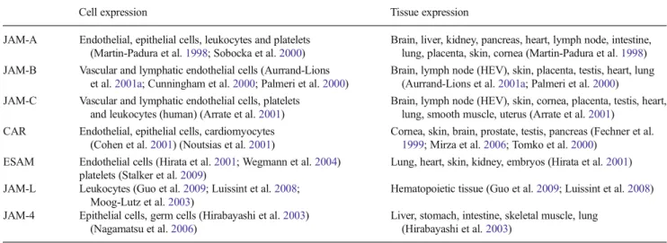
Table 2 Expression of JAMs
Cell expression Tissue expression
JAM-A Endothelial, epithelial cells, leukocytes and platelets
(Martin-Padura et al.1998; Sobocka et al.2000)
Brain, liver, kidney, pancreas, heart, lymph node, intestine,
lung, placenta, skin, cornea (Martin-Padura et al.1998)
JAM-B Vascular and lymphatic endothelial cells (Aurrand-Lions
et al.2001a; Cunningham et al.2000; Palmeri et al.2000)
Brain, lymph node (HEV), skin, placenta, testis, heart, lung
(Aurrand-Lions et al.2001a; Palmeri et al.2000)
JAM-C Vascular and lymphatic endothelial cells, platelets
and leukocytes (human) (Arrate et al.2001)
Brain, lymph node (HEV), skin, cornea, placenta, testis, heart,
lung, smooth muscle, uterus (Arrate et al.2001)
CAR Endothelial, epithelial cells, cardiomyocytes
(Cohen et al.2001) (Noutsias et al.2001)
Cornea, skin, brain, prostate, testis, pancreas (Fechner et al.
1999; Mirza et al.2006; Tomko et al.2000)
ESAM Endothelial cells (Hirata et al.2001; Wegmann et al.2004)
platelets (Stalker et al.2009)
Lung, heart, skin, kidney, embryos (Hirata et al.2001)
JAM-L Leukocytes (Guo et al.2009; Luissint et al.2008;
Moog-Lutz et al.2003)
Hematopoietic tissue (Guo et al.2009; Luissint et al.2008)
JAM-4 Epithelial cells, germ cells (Hirabayashi et al.2003)
(Nagamatsu et al.2006)
Liver, stomach, intestine, skeletal muscle, lung
As mentioned, the structure of JAM-L is different from the other JAM family members, as it does not contain a PDZ binding motif and is not expressed by endothelial or epithelial cells. It was identified by screening for retinoic acid-induced gene expression in leukemia promyelocytes (Moog-Lutz et al. 2003). JAM-L expression is found in mature hematopoietic cells and localized at cell–cell contacts with vascular
endothe-lium (Moog-Lutz et al.2003). Another study confirmed that
JAM-L expression is restricted to the leukocyte sub-populations neutrophils, monocytes and memory T-cells
(Luissint et al.2008) (Table2).
JAMs in endothelial barrier permeability Cell polarity
JAMs are associated with several cytoplasmic polarity pro-teins indicating a role in the formation of cell junctions and
cell polarity (Table1). As discussed, most of these cytoplasmic
partners contain a PDZ binding motif that can interact with the C-terminal part of JAMs. For example classical JAMs all interact with ZO-1, which also associates with the two other types of junctional molecules, occludin and the claudins (Mitic and
Anderson 1998; Tsukita et al. 2001). This is of particular
functional relevance as ZO-1 can also interact with actin
(Fanning et al. 1998). It is thought that ZO-1 may recruit
JAMs and retain them in junctions by protein stabilization. Another important cytoplasmic partner of JAMs is AF-6
(Afadin) (Ebnet et al.2000). Interaction between JAMs and
AF-6 may be important during the early stages of junction formation, as the two molecules colocalize in early nascent
spot-like junctions (Asakura et al.1999; Ebnet et al.2001).
Further studies have shown that AF-6 recruits JAM-A to junctions, as microinjection of JAM-A into epithelial cells resulted in localization of JAM-A only at sites where AF-6
is present (Ebnet et al.2000). The same type of experiments
revealed that the presence of ZO-1 at cell–cell contacts was
not sufficient to recruit JAM-A (Ebnet et al.2000; Fukuhara
et al.2002).
JAM-A is also able to interact with PAR-3 in a PDZ-dependent manner. This was demonstrated by an ectopic expression model, where induced expression of JAM-A led to recruitment of PAR-3 to cell–cell contacts (Ebnet et al. 2001; Itoh et al.2001). The molecule PAR-3 can form molec-ular complexes with atypical protein kinase C (aPKC) and PAR-6, which are thought to participate in cell polarization
(Suzuki and Ohno2006). Indeed, the overexpression of
mu-tated polarity complex proteins in cells during the course of cell polarization led to the delocalization of TJ molecules and
a disruption in endothelial function (Gao et al.2002;
Nagai-Tamai et al.2002; Suzuki et al.2001; Yamanaka et al.2001).
Therefore, JAM-A may play a critical role in maintaining cell
polarization through anchoring the PAR-3-PAR6-aPKC com-plex at the cellular junction.
Polarization of immature junctions starts with the
forma-tion of adherens juncforma-tions (Mitic and Anderson 1998). In
epithelial cells, it starts with nascent spots of contact between lamellipodia of adjacent cells containing E-cadherin, ZO-1
and JAM-A (Ebnet et al.2001; Suzuki et al.2002). JAM-A
then, through the activation of cdc42 and/or Rac1, recruits the polarity complex to the junction (PAR-3-PAR6-aPKC)
(Yamanaka et al.2001) (Fig.2).
As discussed, blocking JAM-A antibodies can increase transendothelial or transepithelial permeability through dis-ruption of JAM-A distribution at the TJ. However, some junctional integrity is maintained as it does not interfere with E-cadherin and ZO-1 recruitment to the contact sites (Liang
et al.2000; Liu et al.2000). A disruption in junction formation
is also observed when blocking the PAR-3-PAR6-aPKC com-plex where only immature adherens junctions can be formed. Similar to JAM-A, JAM-B and -C can also associate with PAR-3 and ZO-1 in a PDZ-domain-dependent manner (Ebnet
et al. 2003). Neither JAM-B nor -C has been identified in
epithelial cells suggesting that the potential existence of a JAMB/C-PAR3 complex may be restricted to endothelial cells
(Ebnet et al. 2003). However, JAM-C was shown to be
essential for spermatid polarity as it participates in the recruit-ment of polarity proteins such as PAR6, cdc42, PKCλ and
PATJ (Gliki et al.2004). Interestingly, PAR-3 is highly
spe-cific for the classical JAMs, as ESAM and CAR do not interact with this polarity complex factor, nor do they with other components of TJs such as occludin and the claudins
(Ebnet et al.2003; Ebnet et al.2001). Clustering of JAM-A at
TJs was also responsible for recruitment of molecules in-volved in cell polarity formation such as PAR-3, 1,
ZO-2, PDZ-GEF1 and GTPase Rap2c (Monteiro et al.2013).
Barrier properties and inflammation
JAM-A also regulates tight epithelial morphology through the
modulation of integrin β1 by the small GTPase molecule
Rap1 (Mandell et al. 2005). In this in vitro study, JAM-A
knockdown by siRNA increased epithelial cell permeability. A similar observation was made with the expression of a mutant JAM-A lacking the homodimerization domain, sug-gesting that JAM-A functions on epithelial cell morphology
and TJs only as a homodimer (Mandell et al.2005). Recently,
using epithelial or fibroblast cells transfected with JAMA or -C mutant, it was shown that serine phosphorylation brings JAM-A and -C into TJs and as a result reduces permeability
of the cell layer (Iden et al.2012; Mandicourt et al.2007). At
least for JAM-A, it can be concluded that phosphorylation was due to the activity and colocalization of aPKC at Serine 285, as a knockdown of this enzyme by siRNA reversed the effect of JAM-A localization in TJs and increased cell permeability.
In line with this finding, increased permeability of the gut epithelial layers was observed in a colitis model with
JAM-A-deficient mice (Khounlotham et al. 2012). Furthermore, in
corneal endothelium, JAM-A, -C and CAR are expressed but only JAM-A is localized at the TJ and colocalized with ZO-1 and AF-6. In a calcium depletion and recovery assay, an anti-JAM-A antibody was able to induce corneal swelling due to impaired barrier function. Thus, this study has shown an essential role for JAM-A in the functionality of the corneal
endothelial barrier (Mandell et al.2006).
Tissue or systemic inflammation induces an increased per-meability of blood vessels and epithelium allowing leukocytes to migrate to the site of inflammation (Johnson-Leger and
Imhof2003). As JAMs are important regulators of epithelial
and endothelial cell polarity, it is clear they can play a pivotal role in the inflammatory process. As discussed previously, an inflammatory stimulus can alter the subcellular localization of JAMs and affect tissue permeability. For example, in vitro stimulation of endothelial cells by TNF-α/IFN-γ or FGF2 has been shown to induce JAM-A redistribution to the luminal
side (Fig.3) (Naik et al.2003; Ostermann et al.2002; Ozaki
et al. 1999). JAM-C is also partially redistributed to the
luminal side of endothelial cells upon oxLDL stimulation
(Keiper et al.2005) (Fig.3). As already mentioned,
JAM-A-deficient mice exhibit a defect in permeability on the intestinal lining. Under steady state conditions, the barrier shows an almost normal morphology but under inflammatory condi-tions, infiltration of PMN and protective lymphocytes can massively increase due to a reduced barrier function
(Khounlotham et al.2012; Laukoetter et al.2007). Although
these immune cells exert a protective function against infec-tion, the clinical score of JAM-A-deficient mice is worse while the mucosa presents less injury and more proliferation
(Khounlotham et al.2012; Laukoetter et al.2007).
In quiescent microvascular endothelial cells, JAM-C is mostly localized in the cytoplasm and is recruited to the junction upon VEGF stimulation. Under these conditions, JAM-C can regulate paracellular permeability through modu-lation of actomyosin-dependant contractility and
VE-cadherin-mediated cell–cell contact (Lamagna et al. 2005;
Orlova et al. 2006). Vascular permeability is increased in
thrombin-stimulated endothelial cells that overexpress
JAM-C and this might be due to coupling of JAM-JAM-C and β3
integrins (Li et al.2009).
Blood–brain barrier (BBB)
The BBB plays an essential role in maintaining the homeo-stasis of the central nervous system (CNS). The barrier properties are maintained by the TJs and are characterized by very low rates of vesicle transport and a marked reduction
in permeability (Engelhardt and Ransohoff 2012).
Interestingly, brain capillaries in the early embryo are perme-able to small molecules that cannot normally enter the adult brain. However, the BBB starts to develop an enhanced barrier function at around E13–E15 that is not completed until after
birth (Wolburg and Lippoldt 2002). Several studies have
shown a role for astrocytes in the establishment of the BBB that requires direct contact with the blood vessels (Tao-Cheng
et al.1987) or by secretion of soluble factors in close
proxim-ity to the vessel (Arthur et al. 1987; Wolburg et al. 1994).
Whilst this micro-environmental niche maintained by astro-cytes seems to be necessary to induce and maintain the BBB, other factors are also required to sustain the functionality of
this structure (Rubin et al.1991; Wolburg et al.1994).
A breakdown in the BBB function can lead to serious damage within the CNS and a perturbation in brain function. Several diseases can lead to BBB damage through partial Fig. 2 Role of JAMs in cell polarization. JAM-A is known to be present
in the early contact points between epithelial cells, along with E-cadherin. It is stabilized at the junction being formed by interacting with ZO-1 and AF-6, which links JAM-A to the actin cytoskeleton. To form a mature junction, the polarity complex PAR-3/PAR-6/aPKC has to be recruited.
JAM-A participates through the activation of cdc42 and Rac1. JAM-A also contributes to PDZ-GEF1 and Rap2c recruitment to the junction. In the later stage of junction formation and stabilization, the TJ molecules occludin and the claudins are recruited to stabilize and form mature cell-cell junctions
impairment or total breakdown, such as the development of tumors, stroke, multiple sclerosis, hypertension and
encepha-litis (Wong et al.2013).
We mentioned the BBB here, since JAM-A, -B and -C are often localized and function at the intercellular junctions of
BBB capillaries (Aurrand-Lions et al.2001b; Martin-Padura
et al.1998; Padden et al.2007; Vorbrodt and Dobrogowska
2004). Interestingly, a homozygotic JAM-C mutation was recently found in a consanguineous family in the United Arab Emirates that has provided an insight into the role of this molecule in humans. Some members of this family de-velop an autosomal-recessive syndrome leading to brain hem-orrhage, subependymal calcification and cataracts (Mochida
et al. 2010). In this study, they mapped the disease locus
on the chromosome 11q25. Further sequence analysis of genes revealed a mutation in the intron 5 of JAM3. RT-PCR analysis of patient cells confirmed abnormal splicing, which results in an early termination of translation. This suggests an important role for JAM-C in maintaining the integrity of the
BBB (Mochida et al.2010). Moreover, a study using
JAM-C-deficient mice revealed that mice backcrossed onto a C57BL6 background develop hydrocephalus characterized by enlarged ventricles and disrupted brain fluid circulation (Wyss et al. 2012).
Recently, JAM-A has been implicated in regulating leuko-cyte trafficking across the BBB in HIV infection models
(Williams et al. 2013). This study demonstrated that upon
HIV infection, CD14+CD16+ monocyte transmigration across the BBB is facilitated by increased expression of JAM-A, ALCAM, CD99 and CD31. In addition, this study confirmed that antibodies against JAM-A reduced the number of transmigrating monocytes irrespective of HIV infection status, indicating a broader role for this molecule in leukocyte
trafficking (Williams et al.2013). Furthermore, in a model of
rat cortical cold-injury, JAM-A expression was lost at the lesion site 12 h after injury, while 2 days later the level of expression was back to normal. This loss of expression was concomitant to the BBB breakdown and demonstrates an Fig. 3 JAMs regulation upon inflammatory condition. In resting
endo-thelium, JAMs are localized at the lateral membrane in the junctional
zone. Upon inflammatory stimuli with TNF-α, IFN-γ, or OxLDL, JAMs
are relocalized onto the luminal surface of the endothelial membrane
where they can interact with circulating leukocytes or be processed into soluble forms by metalloproteinases. This relocalization of JAM-A in-creases endothelium permeability and is characterized by a decrease in electrical resistance
essential role for JAM-A in supporting BBB integrity (Yeung
et al.2008).
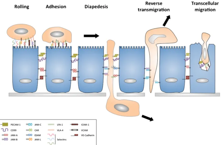
JAMs in leukocytes transmigration
Leukocyte trafficking across an epithelial or endothelial bar-rier is an essential process in inflammation and the immuno-surveillance. This process involves several distinct steps that include rolling on the luminal endothelial layer, which is followed by firm adhesion and diapedesis (Garrido-Urbani
et al.2008). The last step can use a paracellular route along
adjoining endothelial cell–cell contacts or by transcytosis, where the leukocytes pass through the body of endothelial
cells by forming a channel (Fig.4). The process of transcytosis
appears to be a minor route of access and has been mostly observed in vitro with neutrophils and in brain vessels in vivo
(Muller2011). An inflammatory stimulus can lead to the rapid
redistribution of JAMs on endothelial and epithelial cells, which has important implications in leukocyte transmigration.
In the following part of this review, we will discuss the role of each JAM in the process of transmigration, with a particular
focus on the endothelium (Fig.4).
JAM-A
The laboratory of Elisabetha Dejana, which discovered JAM-A through its interaction with the antibody BV11, has studied the role of this molecule in leukocytes transmigration. In an air-pouch model, they observed that the BV11 antibody inhibited recruitment of leukocytes to the site of inflammation
(Martin-Padura et al. 1998). In vitro, they have also shown
that the antibody inhibited monocyte transmigration in a static
migration assay (Martin-Padura et al. 1998). This antibody
specifically recognized JAM-A dimers but not monomers, indicating that a dimeric form of JAM-A is required for
transmigration (Bazzoni et al.2000). Moreover, intravenous
injection of the BV11 antibody reduced leukocytes infiltration into the cerebrospinal fluid and cytokine-induced BBB
per-meability (Del Maschio et al.1999). A later study has gone
Fig. 4 Implication of JAMs in leukocyte transmigration. Migration of leukocytes across endothelium can be separated into distinct steps, rolling, adhesion, diapedesis and reverse transmigration. Under inflam-matory conditions, JAMs are localized onto luminal surfaces of the endothelium and are able to interact with integrins on leukocyte surfaces. Along with PECAM-1 and CD99, JAM-A contributes to the migration of
leukocytes in-between endothelial cells by diapedesis. In some cases where leukocytes cannot pass through the basal membrane, the leukocyte can exit the ablumenal compartment by reverse transmigration. This process is regulated by JAM-C. Leukocytes are able also to pass across the endothelium using a transcellular route. This process is regulated by CD99, PECAM-1, ICAM-1 and JAM-A
further and shown that JAM-A controls leukocytes transendothelial migration through a direct interaction be-tween LFA-1 (CD11a/CD18) and the C2 domain of JAM-A
(Ostermann et al.2002). In this study, it was observed that
endothelial cells stimulated with PMA or the chemokine CXCL12 led to a reduction of T-lymphocyte and monocyte arrest on the endothelial layer in the presence of JAM-A
blocking antibodies (Ostermann et al.2002). The heterophilic
interaction between JAM-A and LFA-1 has a higher affinity than homophilic JAM-A/JAM-A interaction (Ostermann et al. 2002). It has been proposed that, under resting conditions, JAM-A engages in homophilic interactions at cell–cell con-tacts but upon inflammation JAM-A relocalizes to the apical cell membrane where it can then interact with LFA-1 of
neutrophils (Ostermann et al. 2002). This mechanism may
promote the recruitment of leukocytes across the vascular barrier by enhancing cell adhesion events at the endothelial
surface (Ozaki et al. 1999). However, not all studies are
consistent with this finding, as several other anti-JAM-A antibodies are unable to block leukocyte transmigration
(Lechner et al.2000; Liu et al.2000). Furthermore, the
stim-ulation of endothelium with inflammatory cytokines has been shown to have no effect on JAM-A-dependent leukocyte
transmigration under flow conditions (Shaw et al.2001).
Other results obtained with JAM-A-deficient mice support these in vitro data where neutrophils trans-endothelial
migra-tion was reduced (Cera et al.2004). In a model of acute lung
injury, migration of neutrophils to the alveolar space is re-duced in JAM-A-deficient animals or in the presence of
anti-JAM-A antibodies (Cera et al. 2004). However, in another
study, it was shown that blocking of JAM-A does not neces-sarily correlate with reduced lung injury (Lakshmi et al. 2012). Surprisingly, the migration of dendritic cells (DC) to lymph nodes was shown to increase in JAM-A-deficient mice. This was shown to be due to a promigratory phenotype of DCs lacking JAM-A and was associated with enhanced contact
hypersensitivity (Cera et al. 2004). Consistent with these
findings, another study also described an increased migration of JAM-A-deficient DCs and T-cells to tumor sites (Murakami
et al.2010).
Intravital microscopy using mouse venules in cremaster muscle of different gene-deficient animal strains, has demon-strated a sequential action of ICAM-2, JAM-A and PECAM-1 during transendothelial migration of neutrophils after IL-1
stimulation (Woodfin et al.2009). Intriguingly, when
inflam-mation was stimulated by TNF-α, these molecules are not
required for leukocyte transmigration (Woodfin et al.2009).
Further experiments confirmed a differential effect and usage of molecular mechanisms by transendothelial migrating neu-trophils. Specifically, a reduction in migration was observed in JAM-A-deficient mice (or in the presence of an anti-JAM-A (BV11) antibody) when a tissue compartment was stimulated with IL-1, or in ischemia and reperfusion models but this was
not the case after stimulation by leukotriene or PAF (Woodfin
et al.2007). This is consistent with the redistribution of
JAM-A being dependent on the type of stimuli and is supported by observations where JAM-A is redistributed with TNF-α
stim-ulation but not with phorbol ester (Shaw et al.2004). A more
recent study has shown a role for endothelial JAM-A in monocyte recruitment into atherosclerotic plaques, using endothelium-specific JAM-A-deficient mice bred with
ApoE-deficient mice (Schmitt et al.2013). The
endothelium-specific JAM-A deficiency led to reduced monocyte migra-tion through the arterial wall, which contrasted with leukocyte-specific JAM-A deficiency, which exhibited in-creased vascular permeability and lesion formation.
Apart from the role of JAM-A in paracellular transmigration of leukocytes, it has also been implicated in regulating the transcellular migratory route through an endothelial cell. In an in vitro study with HUVEC cells, it has been shown that JAM-A is part of a molecular complex that surrounds the leukocyte (neutrophils or monocytes) migrating through the membrane channel formed by the endothelial cells, together with CD31
and CD99 (Mamdouh et al.2009). This role appears not to be
essential, as JAM-A blockade does not affect this process,
which is in contrast to CD31 and CD99 (Mamdouh et al.2009).
JAM-B
As discussed, vascular JAM-B interacts with JAM-C and the
integrinα4β1. This integrin has previously been described as
mediating memory T-lymphocyte adhesion to inflamed
endothelium through VCAM-1 (Butcher and Picker 1996).
At present, it is not clear whether the integrin can simulta-neously engage VCAM-1 and JAM-B through different bind-ing sites. However, it is also possible that JAM-B and VCAM-1 may interact laterally and obtain integrin reinforcement by conformational changes. This may explain why JAM-B/α4β1 interactions require JAM-B to engage with JAM-C in cis prior
to this interaction (Cunningham et al. 2002). Other recent
studies have proposed an independent role of JAM-B in leukocyte transmigration. Ludwig et al. (2005) demonstrated that anti-JAM-B and anti-JAM-C antibodies had an additive effect on blocking leukocytes infiltration in a model of allergic contact dermatitis. In a later study using a model of DNFB-induced contact hypersensitivity, JAM-B was shown to have a role in rolling and firm adhesion of T-lymphocytes through an
interaction withα4β1 (Ludwig et al.2009).
JAM-C
As mentioned previously, JAM-C is expressed on endothelial cells and human lymphocytes and can play a role in regulating leukocyte transmigration. The first study supporting this hy-pothesis came by enforced expression of JAM-C on endothe-lioma cell lines. These cells could support increased rates of
leukocyte transmigration and this could be abrogated using
anti-JAM-C antibodies (Johnson-Leger et al.2002). This
ef-fect may be due to a homophilic interaction between multimers of JAM-C and JAM-B in trans or in cis formations
(Santoso et al.2002; (Arrate et al.2001; Liang et al.2002).
Several other groups have implicated JAM-C in regulating leukocyte migration in different disease contexts. In rheuma-toid arthritis patients and animal models, JAM-C overexpres-sion has been observed in endothelial cells and has been proposed to play a role in leukocyte adhesion and retention
within the inflamed synovium (Rabquer et al.2008). In a
diabetes-type I model (RIP-LCMV), JAM-C has been impli-cated in the recruitment of pathogenic T-lymphocytes into
Langerhans islets (Christen et al. 2013). These transgenic
m i c e e x p r e s s i n g a p r o t e i n o f t h e l y m p h o c y t i c
choriomeningitis virus (LCMV) inβ-cells, became diabetic
after infection with LCMV virus. The authors found that, upon infection, JAM-C protein was upregulated near the islets. Moreover, JAM-C blockade with a neutralizing anti-JAM-C antibody was able to reduce the incidence of type 1 diabetes. Similarly, in an ischemia and reperfusion model, JAM-C has been shown to regulate leukocyte transmigration
(Scheiermann et al.2009). In this study, they described that
soluble JAM-C injected into mice as a pretreatment is able to inhibit leukocyte migration into zones of ischemia of the kidney and the cremaster muscle. Furthermore, leukocyte adhesion and transmigration, analyzed by intravital microsco-py of the cremaster muscle model, was suppressed in JAM-C-deficient mice and enhanced in mice overexpressing JAM-C
specifically in the endothelium (Scheiermann et al.2009).
However, a role for JAM-C beyond that of simple transmi-gration has been supported by a number of recent publica-tions. A study in 2007 using blocking antibodies and JAM-C overexpression showed no effect of JAM-C on neutrophil
transmigration across HUVECs (Sircar et al. 2007). This
implies that the effect of JAM-C blockade on leukocyte traf-ficking in vivo may extend beyond that of the initial recruit-ment phase. This is supported by an extended in vitro study in which JAM-C has been shown to play a role in the reverse transmigration of monocytes, confirming that post-transmigratory events can be regulated by molecules such as
JAM-C (Bradfield et al.2007b). This process was first
ob-served in vitro and has now been confirmed in vivo by 3D
intravital microscopy (Woodfin et al. 2011). Both studies
demonstrated that a proportion of transmigrated neutrophils or monocytes will reverse transmigrate back onto the luminal surfaces and return to the vascular compartment. In the study using monocytes, JAM-C-specific antibodies or JAM-C defi-ciency increased the number of leukocytes that reverse
transmigrated (Bradfield et al. 2007b). This function may
serve to block an overactive inflammatory response. However, this regulatory feedback loop may lead to reverse transmigrated cells, identified by high ICAM-1 expression,
accumulating in the lungs where the secretion of reactive oxygen species can lead to the induction of lung edema
(Woodfin et al. 2011). In conclusion, JAM-C promotes the
unidirectional transmigration of leukocytes as disrupting this process by antibodies, or a reduction in gene expression, leads to increased reverse transmigration and a return to blood flow. Related JAMs
In an intravital microscopy study performed on ESAM-deficient mice, neutrophil diapedesis was inhibited by 50 % but the first steps of rolling and firm adhesion remained
unaffected (Wegmann et al. 2006). It has been shown that
ESAM is responsible for the maintenance of the tightness of endothelial junctions and VEGF-induced vascular permeabil-ity is affected by the absence of ESAM in knock-out animals
(Wegmann et al.2006). A recent study has shown that ESAM
is also important for regulating monocyte migration during
atherosclerosis (Inoue et al. 2010). As monocytes do not
express ESAM, this result suggests that ESAM has another ligand that has yet to be elucidated.
The interaction of JAM-L/CAR is involved in regulating neutrophil, monocyte and T-cell transendothelial migration
across the inflamed endothelium (Guo et al. 2009; Luissint
et al.2008; Zen et al.2005). It has been shown that JAM-L is
expressed by granulocytes and monocytes and is a ligand of CAR, which is present on the surface of endothelial cells (Guo
et al.2009; Luissint et al.2008; Zen et al.2005). However,
JAM-L/CAR interactions can only be observed when
leuko-cytes either do not express the integrin α4β1 or when this
integrin is activated, otherwise JAM-L will form a complex
with the non-activated integrin α4β1 at the surface of
leukocytes.
Concluding remarks
The study of the JAM family has revealed these molecules as having functional duality: controlling the immune cell re-sponse through recruitment of leukocytes to sites of inflam-mation and regulating the forinflam-mation of cellular junctions in epithelial and endothelial cells. Indeed, the JAMs participate in the transmigration of leukocytes from blood to inflamed tissue, by favoring the opening of the endothelial barrier for crossing leukocytes. JAM-C controls even one-way leukocyte traffic across the vascular barrier. At the same time, however, JAMs are important for the formation and the maintenance of endothelial junctions. Thus, there is clearly a dual role for these molecules. Nevertheless, many questions remain unan-swered. For example, in the classical JAM family (JAM-A, -B and -C), several extracellular ligands for each JAM have been identified, which is not the case for the non-classical JAM family members. The molecule CAR has only two
ligands and ESAM and JAM-4 have only been confirmed in forming homophilic interactions.
Whilst the crystal structure of JAM-A has been fully elu-cidated, which revealed a bending site in the linker domain and a dimerization site in the V domain, this has not been confirmed for the other JAMs. The molecules JAM-B and -C present homology in these domains and a common mecha-nism of multimerization has been proposed. However, no study has as yet been conducted to confirm this binding process, even though JAM-B/-C heterodimerisation is known to take place.
Acknowledgments Our laboratory is supported by Swiss National
Science Foundation grants (31003AB_135701) and ONCOSUISSE grants (KFS 2914-02-2012) to Prof B.A. Imhof.
References
Angelow S, Ahlstrom R, Yu AS (2008) Biology of claudins. Am J
Physiol Ren Physiol 295:F867–F876
Arrate MP, Rodriguez JM, Tran TM, Brock TA, Cunningham SA (2001) Cloning of human junctional adhesion molecule 3 (JAM3) and its identification as the JAM2 counter-receptor. J Biol Chem 276: 45826–45832
Arthur FE, Shivers RR, Bowman PD (1987) Astrocyte-mediated induc-tion of tight juncinduc-tions in brain capillary endothelium: an efficient in vitro model. Brain Res 433:155–159
Asakura T, Nakanishi H, Sakisaka T, Takahashi K, Mandai K, Nishimura M, Sasaki T, Takai Y (1999) Similar and differential behaviour between the nectin-afadin-ponsin and cadherin-catenin systems dur-ing the formation and disruption of the polarized junctional align-ment in epithelial cells. Genes Cells 4:573–581
Aurrand-Lions MA, Duncan L, Pasquier LD, Imhof BA (2000) Cloning of JAM-2 and JAM-3: an emerging junctional adhesion molecular family? Curr Top Microbiol Immunol 251:91–98
Aurrand-Lions M, Duncan L, Ballestrem C, Imhof BA (2001a) JAM-2, a novel immunoglobulin superfamily molecule, expressed by endo-thelial and lymphatic cells. J Biol Chem 276:2733–2741
Aurrand-Lions M, Johnson-Leger C, Wong C, Pasquier LD, Imhof BA (2001b) Heterogeneity of endothelial junctions is reflected by dif-ferential expression and specific subcellular localization of the three
JAM family members. Blood 98:3699–3707
Barton ES, Forrest JC, Connolly JL, Chappell JD, Liu Y, Schnell FJ, Nusrat A, Parkos CA, Dermody TS (2001) Junction adhesion
mol-ecule is a receptor for reovirus. Cell 104:441–451
Bazzoni G, Dejana E (2004) Endothelial cell-to-cell junctions: molecular
organization and role in vascular homeostasis. Physiol Rev 84:869–901
Bazzoni G, Martinez-Estrada OM, Mueller F, Nelboeck P, Schmid G, Bartfai T, Dejana E, Brockhaus M (2000) Homophilic interaction of
junctional adhesion molecule. J Biol Chem 275:30970–30976
Bergelson JM, Cunningham JA, Droguett G, Kurt-Jones EA, Krithivas A, Hong JS, Horwitz MS, Crowell RL, Finberg RW (1997) Isolation of a common receptor for Coxsackie B viruses and adenoviruses 2 and
5. Science 275:1320–1323
Bradfield PF, Nourshargh S, Aurrand-Lions M, Imhof BA (2007a) JAM family and related proteins in leukocyte migration (Vestweber series).
Arterioscler Thromb Vasc Biol 27:2104–2112
Bradfield PF, Scheiermann C, Nourshargh S, Ody C, Luscinskas FW, Rainger GE, Nash GB, Miljkovic-Licina M, Aurrand-Lions M,
Imhof BA (2007b) JAM-C regulates unidirectional monocyte
transendothelial migration in inflammation. Blood 110:2545–2555
Brightman MW, Reese TS (1969) Junctions between intimately apposed
cell membranes in the vertebrate brain. J Cell Biol 40:648–677
Butcher EC, Picker LJ (1996) Lymphocyte homing and homeostasis.
Science 272:60–66
Cera MR, Del Prete A, Vecchi A, Corada M, Martin-Padura I, Motoike T, Tonetti P, Bazzoni G, Vermi W, Gentili F, Bernasconi S, Sato TN, Mantovani A, Dejana E (2004) Increased DC trafficking to lymph nodes and contact hypersensitivity in junctional adhesion
molecule-A-deficient mice. J Clin Invest 114:729–738
Chretien I, Marcuz A, Courtet M, Katevuo K, Vainio O, Heath JK, White SJ, Pasquier LD (1998) CTX, a Xenopus thymocyte receptor, de-fines a molecular family conserved throughout vertebrates. Eur J
Immunol 28:4094–4104
Christen S, Coppieters K, Rose K, Holdener M, Bayer M, Pfeilschifter JM, Hintermann E, von Herrath MG, Aurrand-Lions M, Imhof BA, Christen U (2013) Blockade but not overexpression of the junctional adhesion molecule C influences virus-induced type 1 diabetes in mice. PLoS ONE 8:e54675
Claude P (1978) Morphological factors influencing transepithelial per-meability: a model for the resistance of the zonula occludens. J Membr Biol 39:219–232
Claude P, Goodenough DA (1973) Fracture faces of zonulae occludentes from "tight" and "leaky" epithelia. J Cell Biol 58:390–400 Cohen CJ, Shieh JT, Pickles RJ, Okegawa T, Hsieh JT, Bergelson JM
(2001) The coxsackievirus and adenovirus receptor is a transmem-brane component of the tight junction. Proc Natl Acad Sci U S A 98: 15191–15196
Coyne CB, Voelker T, Pichla SL, Bergelson JM (2004) The coxsackievirus and adenovirus receptor interacts with the multi-PDZ domain protein-1 (MUPP-1) within the tight junction. J Biol Chem 279:48079–48084
Cunningham SA, Arrate MP, Rodriguez JM, Bjercke RJ, Vanderslice P, Morris AP, Brock TA (2000) A novel protein with homology to the junctional adhesion molecule. Characterization of leukocyte inter-actions. J Biol Chem 275:34750–34756
Cunningham SA, Rodriguez JM, Arrate MP, Tran TM, Brock TA (2002) JAM2 interacts with alpha4beta1. Facilitation by JAM3. J Biol Chem 277:27589–27592
Del Maschio A, De Luigi A, Martin-Padura I, Brockhaus M, Bartfai T, Fruscella P, Adorini L, Martino G, Furlan R, De Simoni MG, Dejana E (1999) Leukocyte recruitment in the cerebrospinal fluid of mice with experimental meningitis is inhibited by an antibody to
junc-tional adhesion molecule (JAM). J Exp Med 190:1351–1356
Du Pasquier L, Courtet M, Chretien I (1999) Duplication and MHC linkage of the CTX family of genes in Xenopus and in mammals.
Eur J Immunol 29:1729–1739
Ebnet K, Schulz CU, Meyer Zu Brickwedde MK, Pendl GG, Vestweber D (2000) Junctional adhesion molecule interacts with the PDZ domain-containing proteins AF-6 and ZO-1. J Biol Chem 275:
27979–27988
Ebnet K, Suzuki A, Horikoshi Y, Hirose T, Meyer Zu Brickwedde MK, Ohno S, Vestweber D (2001) The cell polarity protein ASIP/PAR-3 directly associates with junctional adhesion molecule (JAM).
EMBO J 20:3738–3748
Ebnet K, Aurrand-Lions M, Kuhn A, Kiefer F, Butz S, Zander K, Meyer zu Brickwedde MK, Suzuki A, Imhof BA, Vestweber D (2003) The junctional adhesion molecule (JAM) family members JAM-2 and JAM-3 associate with the cell polarity protein PAR-3: a possible role
for JAMs in endothelial cell polarity. J Cell Sci 116:3879–3891
Engelhardt B, Ransohoff RM (2012) Capture, crawl, cross: the T cell
code to breach the blood–brain barriers. Trends Immunol 33:579–
589
Excoffon KJ, Hruska-Hageman A, Klotz M, Traver GL, Zabner J (2004) A role for the PDZ-binding domain of the coxsackie B virus and
adenovirus receptor (CAR) in cell adhesion and growth. J Cell Sci
117:4401–4409
Fanning AS, Jameson BJ, Jesaitis LA, Anderson JM (1998) The tight junction protein ZO-1 establishes a link between the transmembrane protein occludin and the actin cytoskeleton. J Biol Chem 273:
29745–29753
Farquhar MG, Palade GE (1963) Junctional complexes in various
epi-thelia. J Cell Biol 17:375–412
Fechner H, Haack A, Wang H, Wang X, Eizema K, Pauschinger M, Schoemaker R, Veghel R, Houtsmuller A, Schultheiss HP, Lamers J, Poller W (1999) Expression of coxsackie adenovirus receptor and alphav-integrin does not correlate with adenovector targeting in vivo
indicating anatomical vector barriers. Gene Ther 6:1520–1535
Fukuhara A, Irie K, Yamada A, Katata T, Honda T, Shimizu K, Nakanishi H, Takai Y (2002) Role of nectin in organization of tight junctions in
epithelial cells. Genes Cells 7:1059–1072
Furuse M, Tsukita S (2006) Claudins in occluding junctions of humans
and flies. Trends Cell Biol 16:181–188
Furuse M, Sasaki H, Fujimoto K, Tsukita S (1998) A single gene product,
claudin-1 or −2, reconstitutes tight junction strands and recruits
occludin in fibroblasts. J Cell Biol 143:391–401
Gao L, Macara IG, Joberty G (2002) Multiple splice variants of Par3 and of a novel related gene, Par3L, produce proteins with different binding properties. Gene 294:99–107
Garrido-Urbani S, Bradfield PF, Lee BP, Imhof BA (2008) Vascular and epithelial junctions: a barrier for leucocyte migration. Biochem Soc Trans 36:203–211
Gliki G, Ebnet K, Aurrand-Lions M, Imhof BA, Adams RH (2004) Spermatid differentiation requires the assembly of a cell polarity complex downstream of junctional adhesion molecule-C. Nature 431:320–324
Guillemot L, Paschoud S, Pulimeno P, Foglia A, Citi S (2008) The cytoplasmic plaque of tight junctions: a scaffolding and signalling center. Biochim Biophys Acta 1778:601–613
Guo YL, Bai R, Chen CX, Liu DQ, Liu Y, Zhang CY, Zen K (2009) Role of junctional adhesion molecule-like protein in mediating monocyte transendothelial migration. Arterioscler Thromb Vasc Biol 29:75–83 Hamazaki Y, Itoh M, Sasaki H, Furuse M, Tsukita S (2002) Multi-PDZ domain protein 1 (MUPP1) is concentrated at tight junctions through its possible interaction with claudin-1 and junctional
adhe-sion molecule. J Biol Chem 277:455–461
Hirabayashi S, Tajima M, Yao I, Nishimura W, Mori H, Hata Y (2003) JAM4, a junctional cell adhesion molecule interacting with a tight
junction protein, MAGI-1. Mol Cell Biol 23:4267–4282
Hirata K, Ishida T, Penta K, Rezaee M, Yang E, Wohlgemuth J, Quertermous T (2001) Cloning of an immunoglobulin family adhe-sion molecule selectively expressed by endothelial cells. J Biol
Chem 276:16223–16231
Iden S, Misselwitz S, Peddibhotla SS, Tuncay H, Rehder D, Gerke V, Robenek H, Suzuki A, Ebnet K (2012) aPKC phosphorylates JAM-A at Ser285 to promote cell contact maturation and tight junction
formation. J Cell Biol 196:623–639
Inoue M, Ishida T, Yasuda T, Toh R, Hara T, Cangara HM, Rikitake Y, Taira K, Sun L, Kundu RK, Quertermous T, Hirata K (2010) Endothelial cell-selective adhesion molecule modulates atheroscle-rosis through plaque angiogenesis and monocyte-endothelial
inter-action. Microvasc Res 80:179–187
Itoh M, Sasaki H, Furuse M, Ozaki H, Kita T, Tsukita S (2001) Junctional adhesion molecule (JAM) binds to PAR-3: a possible mechanism for
the recruitment of PAR-3 to tight junctions. J Cell Biol 154:491–497
Johnson-Leger C, Imhof BA (2003) Forging the endothelium during inflammation: pushing at a half-open door? Cell Tissue Res 314:
93–105
Johnson-Leger CA, Aurrand-Lions M, Beltraminelli N, Fasel N, Imhof BA (2002) Junctional adhesion molecule-2 (JAM-2) promotes
lym-phocyte transendothelial migration. Blood 100:2479–2486
Kansaku A, Hirabayashi S, Mori H, Fujiwara N, Kawata A, Ikeda M, Rokukawa C, Kurihara H, Hata Y (2006) Ligand-of-Numb protein X is an endocytic scaffold for junctional adhesion molecule 4.
Oncogene 25:5071–5084
Keiper T, Al-Fakhri N, Chavakis E, Athanasopoulos AN, Isermann B, Herzog S, Saffrich R, Hersemeyer K, Bohle RM, Haendeler J, Preissner KT, Santoso S, Chavakis T (2005) The role of junctional adhesion molecule-C (JAM-C) in oxidized LDL-mediated
leuko-cyte recruitment. FASEB J 19:2078–2080
Khounlotham M, Kim W, Peatman E, Nava P, Medina-Contreras O, Addis C, Koch S, Fournier B, Nusrat A, Denning TL, Parkos CA (2012) Compromised intestinal epithelial barrier induces adaptive immune
compensation that protects from colitis. Immunity 37:563–573
Koenen RR, Pruessmeyer J, Soehnlein O, Fraemohs L, Zernecke A, Schwarz N, Reiss K, Sarabi A, Lindbom L, Hackeng TM, Weber C, Ludwig A (2009) Regulated release and functional modulation of junctional adhesion molecule A by disintegrin metalloproteinases.
Blood 113:4799–4809
Kostrewa D, Brockhaus M, D'Arcy A, Dale GE, Nelboeck P, Schmid G, Mueller F, Bazzoni G, Dejana E, Bartfai T, Winkler FK, Hennig M (2001) X-ray structure of junctional adhesion molecule: structural basis for homophilic adhesion via a novel dimerization motif. EMBO J 20:4391–4398
Lakshmi SP, Reddy AT, Naik MU, Naik UP, Reddy RC (2012) Effects of JAM-A deficiency or blocking antibodies on neutrophil migration and lung injury in a murine model of ALI. Am J Physiol Lung Cell Mol Physiol 303:L758–L766
Lamagna C, Meda P, Mandicourt G, Brown J, Gilbert RJ, Jones EY, Kiefer F, Ruga P, Imhof BA, Aurrand-Lions M (2005) Dual inter-action of JAM-C with JAM-B and alpha(M)beta2 integrin: function in junctional complexes and leukocyte adhesion. Mol Biol Cell 16: 4992–5003
Laukoetter MG, Nava P, Lee WY, Severson EA, Capaldo CT, Babbin BA, Williams IR, Koval M, Peatman E, Campbell JA, Dermody TS, Nusrat A, Parkos CA (2007) JAM-A regulates permeability and inflammation in the intestine in vivo. J Exp Med 204:3067–3076 Lechner F, Sahrbacher U, Suter T, Frei K, Brockhaus M, Koedel U,
Fontana A (2000) Antibodies to the junctional adhesion molecule cause disruption of endothelial cells and do not prevent leukocyte influx into the meninges after viral or bacterial infection. J Infect Dis
182:978–982
Li X, Stankovic M, Lee BP, Aurrand-Lions M, Hahn CN, Lu Y, Imhof BA, Vadas MA, Gamble JR (2009) JAM-C induces endothelial cell permeability through its association and regulation of {beta}3
integrins. Arterioscler Thromb Vasc Biol 29:1200–1206
Liang TW, DeMarco RA, Mrsny RJ, Gurney A, Gray A, Hooley J, Aaron HL, Huang A, Klassen T, Tumas DB, Fong S (2000) Characterization of huJAM: evidence for involvement in cell-cell contact and tight junction regulation. Am J Physiol Cell Physiol 279:
C1733–C1743
Liang TW, Chiu HH, Gurney A, Sidle A, Tumas DB, Schow P, Foster J, Klassen T, Dennis K, DeMarco RA, Pham T, Frantz G, Fong S (2002) Vascular endothelial-junctional adhesion molecule (VE-JAM)/JAM 2 interacts with T, NK, and dendritic cells through
JAM 3. J Immunol 168:1618–1626
Liu Y, Nusrat A, Schnell FJ, Reaves TA, Walsh S, Pochet M, Parkos CA (2000) Human junction adhesion molecule regulates tight junction
resealing in epithelia. J Cell Sci 113(Pt 13):2363–2374
Ludwig RJ, Zollner TM, Santoso S, Hardt K, Gille J, Baatz H, Johann PS, Pfeffer J, Radeke HH, Schon MP, Kaufmann R, Boehncke WH, Podda M (2005) Junctional adhesion molecules (JAM)-B and -C contribute to leukocyte extravasation to the skin and mediate
cuta-neous inflammation. J Investig Dermatol 125:969–976
Ludwig RJ, Hardt K, Hatting M, Bistrian R, Diehl S, Radeke HH, Podda M, Schon MP, Kaufmann R, Henschler R, Pfeilschifter JM, Santoso S, Boehncke WH (2009) Junctional adhesion molecule (JAM)-B
supports lymphocyte rolling and adhesion through interaction with
alpha4beta1 integrin. Immunology 128:196–205
Luissint AC, Lutz PG, Calderwood DA, Couraud PO, Bourdoulous S (2008) JAM-L-mediated leukocyte adhesion to endothelial cells is regulated in cis by alpha4beta1 integrin activation. J Cell Biol 183:
1159–1173
Mamdouh Z, Mikhailov A, Muller WA (2009) Transcellular migration of leukocytes is mediated by the endothelial lateral border recycling
compartment. J Exp Med 206:2795–2808
Mandell KJ, Parkos CA (2005) The JAM family of proteins. Adv Drug
Deliv Rev 57:857–867
Mandell KJ, Babbin BA, Nusrat A, Parkos CA (2005) Junctional adhe-sion molecule 1 regulates epithelial cell morphology through effects
on beta1 integrins and Rap1 activity. J Biol Chem 280:11665–11674
Mandell KJ, Holley GP, Parkos CA, Edelhauser HF (2006) Antibody blockade of junctional adhesion molecule-A in rabbit corneal endo-thelial tight junctions produces corneal swelling. Investig
Ophthalmol Vis Sci 47:2408–2416
Mandicourt G, Iden S, Ebnet K, Aurrand-Lions M, Imhof BA (2007) JAM-C regulates tight junctions and integrin-mediated cell adhesion and migration. J Biol Chem 282:1830–1837
Mapoles JE, Krah DL, Crowell RL (1985) Purification of a HeLa cell receptor protein for group B coxsackieviruses. J Virol 55:560–566 Martinez-Estrada OM, Villa A, Breviario F, Orsenigo F, Dejana E,
Bazzoni G (2001) Association of junctional adhesion molecule with calcium/calmodulin-dependent serine protein kinase (CASK/LIN-2) in human epithelial caco-2 cells. J Biol Chem 276:9291–9296 Martin-Padura I, Lostaglio S, Schneemann M, Williams L, Romano M,
Fruscella P, Panzeri C, Stoppacciaro A, Ruco L, Villa A, Simmons D, Dejana E (1998) Junctional adhesion molecule, a novel member of the immunoglobulin superfamily that distributes at intercellular junctions and modulates monocyte transmigration. J Cell Biol 142: 117–127
Mirza M, Hreinsson J, Strand ML, Hovatta O, Soder O, Philipson L, Pettersson RF, Sollerbrant K (2006) Coxsackievirus and adenovirus receptor (CAR) is expressed in male germ cells and forms a complex with the differentiation factor JAM-C in mouse testis. Exp Cell Res 312:817–830
Mitic LL, Anderson JM (1998) Molecular architecture of tight junctions. Annu Rev Physiol 60:121–142
Mochida GH, Ganesh VS, Felie JM, Gleason D, Hill RS, Clapham KR, Rakiec D, Tan WH, Akawi N, Al-Saffar M, Partlow JN, Tinschert S, Barkovich AJ, Ali B, Al-Gazali L, Walsh CA (2010) A homozygous mutation in the tight-junction protein JAM3 causes hemorrhagic destruction of the brain, subependymal calcification, and congenital
cataracts. Am J Hum Genet 87:882–889
Monteiro AC, Sumagin R, Rankin CR, Leoni G, Mina MJ, Reiter DM, Stehle T, Dermody TS, Schaefer SA, Hall RA, Nusrat A, Parkos CA (2013) JAM-A associates with ZO-2, afadin, and PDZ-GEF1 to activate Rap2c and regulate epithelial barrier function. Mol Biol
Cell 24:2849–2860
Moog-Lutz C, Cave-Riant F, Guibal FC, Breau MA, Di Gioia Y, Couraud PO, Cayre YE, Bourdoulous S, Lutz PG (2003) JAML, a novel protein with characteristics of a junctional adhesion molecule, is induced during differentiation of myeloid leukemia cells. Blood
102:3371–3378
Muller WA (2003) Leukocyte-endothelial-cell interactions in leukocyte transmigration and the inflammatory response. Trends Immunol 24:
327–334
Muller WA (2011) Mechanisms of leukocyte transendothelial migration.
Annu Rev Pathol 6:323–344
Murakami M, Francavilla C, Torselli I, Corada M, Maddaluno L, Sica A, Matteoli G, Iliev ID, Mantovani A, Rescigno M, Cavallaro U, Dejana E (2010) Inactivation of junctional adhesion molecule-A enhances antitumoral immune response by promoting dendritic cell
and T lymphocyte infiltration. Cancer Res 70:1759–1765
Nagai-Tamai Y, Mizuno K, Hirose T, Suzuki A, Ohno S (2002) Regulated protein-protein interaction between aPKC and PAR-3 plays an es-sential role in the polarization of epithelial cells. Genes Cells 7:
1161–1171
Nagamatsu G, Ohmura M, Mizukami T, Hamaguchi I, Hirabayashi S, Yoshida S, Hata Y, Suda T, Ohbo K (2006) A CTX family cell adhesion molecule, JAM4, is expressed in stem cell and progenitor cell populations of both male germ cell and hematopoietic cell
lineages. Mol Cell Biol 26:8498–8506
Nagy Z, Peters H, Huttner I (1984) Fracture faces of cell junctions in cerebral endothelium during normal and hyperosmotic conditions.
Lab Investig 50:313–322
Naik MU, Mousa SA, Parkos CA, Naik UP (2003) Signaling through JAM-1 and alphavbeta3 is required for the angiogenic action of bFGF: dissociation of the JAM-1 and alphavbeta3 complex. Blood
102:2108–2114
Nasdala I, Wolburg-Buchholz K, Wolburg H, Kuhn A, Ebnet K, Brachtendorf G, Samulowitz U, Kuster B, Engelhardt B, Vestweber D, Butz S (2002) A transmembrane tight junction protein selectively expressed on endothelial cells and platelets. J Biol Chem 277:16294–16303
Noutsias M, Fechner H, de Jonge H, Wang X, Dekkers D, Houtsmuller AB, Pauschinger M, Bergelson J, Warraich R, Yacoub M, Hetzer R, Lamers J, Schultheiss HP, Poller W (2001) Human coxsackie-adenovirus receptor is colocalized with integrins alpha(v)beta(3) and alpha(v)beta(5) on the cardiomyocyte sarcolemma and upregu-lated in diupregu-lated cardiomyopathy: implications for cardiotropic viral infections. Circulation 104:275–280
Orlova VV, Economopoulou M, Lupu F, Santoso S, Chavakis T (2006) Junctional adhesion molecule-C regulates vascular endothelial per-meability by modulating VE-cadherin-mediated cell-cell contacts. J Exp Med 203:2703–2714
Ostermann G, Weber KS, Zernecke A, Schroder A, Weber C (2002) JAM-1 is a ligand of the beta(2) integrin LFA-1 involved in transendothelial migration of leukocytes. Nat Immunol 3:151–158 Ozaki H, Ishii K, Horiuchi H, Arai H, Kawamoto T, Okawa K, Iwamatsu
A, Kita T (1999) Cutting edge: combined treatment of TNF-alpha and IFN-gamma causes redistribution of junctional adhesion mole-cule in human endothelial cells. J Immunol 163:553–557 Padden M, Leech S, Craig B, Kirk J, Brankin B, McQuaid S (2007)
Differences in expression of junctional adhesion molecule-A and beta-catenin in multiple sclerosis brain tissue: increasing evidence for
the role of tight junction pathology. Acta Neuropathol 113:177–186
Palmeri D, van Zante A, Huang CC, Hemmerich S, Rosen SD (2000) Vascular endothelial junction-associated molecule, a novel member of the immunoglobulin superfamily, is localized to intercellular
boundaries of endothelial cells. J Biol Chem 275:19139–19145
Prota AE, Campbell JA, Schelling P, Forrest JC, Watson MJ, Peters TR, Aurrand-Lions M, Imhof BA, Dermody TS, Stehle T (2003) Crystal structure of human junctional adhesion molecule 1: implications for
reovirus binding. Proc Natl Acad Sci USA 100:5366–5371
Rabquer BJ, Pakozdi A, Michel JE, Gujar BS, Haines GK 3rd, Imhof BA, Koch AE (2008) Junctional adhesion molecule C mediates leuko-cyte adhesion to rheumatoid arthritis synovium. Arthritis Rheum 58:
3020–3029
Rabquer BJ, Amin MA, Teegala N, Shaheen MK, Tsou PS, Ruth JH, Lesch CA, Imhof BA, Koch AE (2010) Junctional adhesion molecule-C is a
soluble mediator of angiogenesis. J Immunol 185:1777–1785
Reese TS, Karnovsky MJ (1967) Fine structural localization of a blood–
brain barrier to exogenous peroxidase. J Cell Biol 34:207–217
Rubin LL, Hall DE, Porter S, Barbu K, Cannon C, Horner HC, Janatpour M, Liaw CW, Manning K, Morales J et al (1991) A cell culture
model of the blood–brain barrier. J Cell Biol 115:1725–1735
Saitou M, Furuse M, Sasaki H, Schulzke JD, Fromm M, Takano H, Noda T, Tsukita S (2000) Complex phenotype of mice lacking occludin, a