*Drs Bibli and Hu contributed equally. The full author list is available on page 112.
Key Words: atherosclerosis ◼ biomarkers ◼ cell adhesion molecules ◼ hydrogen sulfide ◼ RNA binding proteins
Sources of Funding, see page 113
BACKGROUND: Hydrogen sulfide (H2S), generated by cystathionine γ lyase (CSE), is an important endogenous regulator of vascular function. The aim of the present study was to investigate the control and consequences of CSE activity in endothelial cells under physiological and proatherogenic conditions.
METHODS: Endothelial cell CSE knockout mice were generated, and lung
endothelial cells were studied in vitro (gene expression, protein sulfhydration, and monocyte adhesion). Mice were crossed onto the apolipoprotein E– deficient background, and atherogenesis (partial carotid artery ligation) was monitored over 21 days. CSE expression, H2S bioavailability, and amino acid profiling were also performed with human material.
RESULTS: The endothelial cell–specific deletion of CSE selectively increased the expression of CD62E and elevated monocyte adherence in the absence of an inflammatory stimulus. Mechanistically, CD62E mRNA was more stable in endothelial cells from CSE-deficient mice, an effect attributed to the attenuated sulfhydration and dimerization of the RNA-binding protein human antigen R. CSE expression was upregulated in mice after partial carotid artery ligation and in atheromas from human subjects. Despite the increase in CSE protein, circulating and intraplaque H2S levels were reduced, a phenomenon that could be attributed to the serine phosphorylation (on Ser377) and inhibition of the enzyme, most likely resulting from increased interleukin-1β. Consistent with the loss of H2S, human antigen R sulfhydration was attenuated in atherosclerosis and resulted in the stabilization of human antigen R–target mRNAs, for example, CD62E and cathepsin S, both of which are linked to endothelial cell activation and atherosclerosis. The deletion of CSE from endothelial cells was associated with the accelerated development of endothelial dysfunction and atherosclerosis, effects that were reversed on treatment with a polysulfide donor. Finally, in mice and humans, plasma levels of the CSE substrate
L-cystathionine negatively correlated with vascular reactivity and H2S levels,
indicating its potential use as a biomarker for vascular disease.
CONCLUSIONS: The constitutive S-sulfhydration of human antigen R (on
Cys13) by CSE-derived H2S prevents its homodimerization and activity, which attenuates the expression of target proteins such as CD62E and cathepsin S. However, as a consequence of vascular inflammation, the beneficial actions of CSE-derived H2S are lost owing to the phosphorylation and inhibition of the enzyme.
Sofia-Iris Bibli, PhD* Jiong Hu, PhD* et al
Cystathionine
γ Lyase Sulfhydrates the
RNA Binding Protein Human Antigen R to
Preserve Endothelial Cell Function and Delay
Atherogenesis
http://doc.rero.ch
Published in "Circulation 139(1): 101–114, 2019"
which should be cited to refer to this work.
D
espite intensive therapy, the clinical manifesta-tions of atherosclerosis remain the primary cause of morbidity and mortality worldwide. Athero-sclerotic plaque formation occurs mainly at susceptible sites of major arteries in which disturbed flow can ac-tivate endothelial cells, leading to altered expression of genes involved in inflammatory process and the devel-opment of vascular disease.1,2 A number of stimuli andfactors have been found to be important in maintain-ing endothelial cells in a quiescent and antiatherogenic state, including the shear stress–stimulated generation of nitric oxide (NO). Apart from NO, other gasotrans-mitters are thought to play a significant role in vascular homeostasis, including carbon monoxide and hydrogen sulfide (H2S). H2S is generated mainly via the reverse transsulfuration pathway in reactions catalyzed by 2 pyridoxal phosphate-dependent enzymes, cystathionine β synthase (CBS) and cystathionine γ lyase (CSE), and 1 pyridoxal phosphate-independent enzyme, 3-mercap-topyruvate sulfurtransferase.3,4 The production of H
2S
in the vasculature is attributed largely to the activity of CSE, which generates H2S from L-cysteine and L-cysta-thionine and has been implicated in the modulation of angiogenesis and vascular tone.5 However, the
mecha-nisms regulating the expression of CSE in endothelial cells and the molecular targets of CSE-derived H2S re-main to be elucidated.
Although partially contradictory phenotypes on blood pressure changes and hyperhomocysteinemia have been observed that seem to depend on the ge-netic background of the mice studied,6,7 it seems clear
that CSE-derived H2S can exert antiatherosclerotic ef-fects.8–10 In mice lacking CSE, both elevated adhesion
molecule levels and enhanced leukocyte adherence have been described,8 whereas the overexpression of
CSE was found to reduce atherosclerotic plaque size and circulating lipid levels.11 In humans, CSE has been
detected in atherosclerotic plaques,12,13 but little is
known about its consequences on disease development or outcome. The aims of this study, therefore, were to determine the mechanisms by which CSE expression and activity are regulated in endothelial cells and to unravel its potential role in the initiation and develop-ment of atherosclerosis in mice and humans. Moreover, because at the molecular level H2S signals through the persulfidation or sulfhydration of target cysteine resi-dues,14,15 we set out to identify physiologically relevant
sulfhydration targets that may be altered during, and contribute to, disease development.
METHODS
Detailed methods are available in the online-only Data Supplement. Requests by researchers to access the data, analytical methods, and study materials for the purposes of reproducing the results or replicating procedures can be made to the corresponding author, who manages the information.
Animals
Apolipoprotein E–deficient (ApoE−/−) mice were purchased
from Charles River Laboratories (Sulzfeld, Germany). Floxed CSE (CSEfl/fl) mice were generated as described7 and crossed
with tamoxifen-inducible Cdh5-CreERT2 mice16 in the C57/
BL6J background or with Cdh5-Cre mice in the ApoE−/−
back-ground. Mice were housed in conditions that conform to the Guide for the Care and Use of Laboratory Animals pub-lished by the US National Institutes of Health (publication No. 85-23). Animals received the usual laboratory diet, and all studies were approved by the animal research ethics com-mittees in Athens (790/13-02-2014) and Darmstadt (FU1177 and FU1189). Littermates of both sexes were used. To induce robust Cre activity, animals were treated with tamoxifen (75 mg/kg IP, Sigma‐Aldrich) for 5 days.
Human Samples
Carotid plaques were prospectively collected from 24 random patients who had internal carotid artery stenosis of 75% to 90% and underwent carotid endarterectomy (Table I in the online-only Data Supplement). Arteriographical evaluation of the carotid bifurcation stenosis was performed and the degree of luminal stenosis was determined according to NASCET (North American Symptomatic Carotid Endarterectomy Trial)
Clinical Perspective
What Is New?
• Hydrogen sulfide generated by cystathionine γ lyase constitutively sulfhydrates the RNA binding protein human antigen R to prevent its homodimerization and to attenuate its activity in the vessel wall. • The sulfhydration of human antigen R prevents its
binding to and stabilization of target mRNAs—for example, CD62E (E-selectin)—ensuring their low expression.
• Inflammation results in the phosphorylation and inhibition of cystathionine γ lyase, which reduces hydrogen sulfide production and thus alleviates the inhibition of human antigen R.
• Endothelial ablation of cystathionine γ lyase results in increased CD62E expression and acceler-ated development of endothelial dysfunction and atherosclerosis.
• Plasma levels of the cystathionine γ lyase substrate L-cystathionine correlate with impaired vascular reactivity in humans.
What Are the Clinical Implications?
• Oral supplementation with polysulfide donors (eg, SG1002) may serve as a therapeutic approach to attenuate atherosclerosis development in humans.
• Circulating L-cystathionine levels can serve as a bio-marker for endothelial dysfunction.
criteria. Peak systolic velocity was monitored with a Philips HD11 ultrasound platform (Philips, Best, the Netherlands). Eight additional samples of healthy thyroid arteries were used as the control group. Thyroid arteries were chosen from age-matched subjects without additional comorbidities (Table I in the online-only Data Supplement). Samples were collected postmortem and evaluated by a pathologist for the possibility of atherosclerotic lesions. Arteries that showed no pathologi-cal characteristics were snap-frozen for additional analysis. Tissue samples were either frozen and used for biochemical analyses or embedded in paraffin for immunostaining. Plasma from another 70 patients with internal carotid artery steno-sis of 75% to 90% before carotid endarterectomy and 32 age-matched healthy donors was used for amino acid profil-ing, H2S measurements, and assay of interleukin (IL)–1β levels
(Table II in the online-only Data Supplement). All studies fol-lowed the Code of Ethics of the World Medical Association (Declaration of Helsinki). The study protocols were approved by the Institutional Ethics Committee (Scientific and Ethic Committee of Hipokrateion University Hospital, PN1539), and all patients gave their informed consent.
Statistics
Data are expressed as mean±SEM. Statistical evaluation was performed with the Student t test for unpaired data. The Mann-Whitney test was used if the sample size was <8 or popula-tions followed non-Gaussian distribution. One-way ANOVA followed by the Newman-Keuls test and 2-way ANOVA with a Bonferroni posttest were used when appropriate. The Pearson correlation coefficient was used to measure asso-ciations between continuous variables. Repeated-measures ANOVA with a Bonferroni posttest was used when appropri-ate. Statistical tests are described in the figure legends for each experiment. Central tendency and dispersion of the data were examined for replicates <6. Values of P<0.05 were con-sidered statistically significant. MetaboAnalyst17 was used to
construct the heat map and to perform hierarchical clustering based on amino acid profile.
RESULTS
Link Between Fluid Shear Stress, CSE
Expression, and CD62E Expression
Given that shear stress is a key player in endothelial cell homeostasis and atherosclerosis development, the ex-pression of CSE in endothelial cells along the aorta was assessed, concentrating on potential changes in areas normally associated with high shear stress/laminar flow versus low shear stress/disturbed flow. In contrast to the changes described for the endothelial NO synthase,18,19
the expression of CSE was higher in the lesser curvature and arterial bifurcations and lower in the descending aorta (Figure 1A and 1B), implying that CSE expression was negatively regulated by shear stress. In line with these observations, the application of fluid shear stress to cultured human endothelial cells resulted in a time-dependent decrease in CSE protein levels (Figure 1C
and 1D), as well as H2S production (Figure 1D). Shear stress did not alter the expression of the other H2 S-gen-erating enzymes—that is, CBS and 3-mercaptopyruvate sulfurtransferase—in endothelial cells (Figure IA in the online-only Data Supplement).
The importance of CSE in endothelial cell homeo-stasis was studied in inducible endothelial cell–specific CSE knockout (CSEiΔEC) mice. Treating CSEiΔEC mice with
tamoxifen abrogated CSE expression in endothelial cells and resulted in an ≈65% reduction in H2S produc-tion (Figure 1A and Figure IB and IC in the online-only Data Supplement). Although intracellular adhesion molecule-1 and vascular cell adhesion molecule-1 were barely detectable in cells from wild-type and CSEiΔEC
mice (Figure IIA in the online-only Data Supplement
and Figure 1E), the expression of CD62E (E-selectin) was elevated in CSEiΔEC endothelial cells in the absence
of any inflammatory stimulus. Similar effects were ob-served in freshly isolated aortas from wild-type and CSEiΔEC mice (Figure IIB in the online-only Data
Supple-ment). The increased CD62E in endothelial cells from CSEiΔEC mice was functional because it was expressed
on the cell surface (Figure IIC in the online-only Data Supplement) and correlated with an increase in mono-cyte adhesion to CSE-deficient endothelial cells under basal conditions (Figure IID in the online-only Data Supplement). Moreover, the increase in monocyte ad-herence was abrogated in cells treated with a CD62E-neutralizing antibody (Figure IIE in the online-only Data Supplement). Rescue experiments, in which CSE was reintroduced into CSE-deficient endothelial cells, sup-pressed the abnormal CD62E expression and abolished monocyte adhesion (Figure IIF and IIG in the online-only Data Supplement). These effects were unrelated to changes in NO production because the differences between wild-type and CSE-deficient endothelial cells were unaffected by the addition of an NO synthase in-hibitor (Figure IIH in the online-only Data Supplement). Cell stimulation with IL-1β increased intracellular ad-hesion molecule-1, vascular cell adad-hesion molecule-1, and CD62E in cells from wild-type mice but failed to further increase CD62E levels in CSEiΔEC mice
(Fig-ure 1E), even though vascular cell adhesion molecule-1 and intracellular adhesion molecule-1 expression and monocyte adherence increased as expected (Figure IIA and IIB in the online-only Data Supplement).
Sulfhydration of Human Antigen R by
CSE-Derived H
2S
The selective upregulation of CD62E in endothelial cells lacking CSE suggested that CD62E may be directly targeted by CSE-derived H2S. However, it was not pos-sible to demonstrate the sulfhydration of CD62E in CSE-expressing endothelial cells, indicating that the ef-fect was indirect. Therefore, to identify potential H2S
targets, CSE was immunoprecipitated from murine en-dothelial cells and coprecipitated proteins identified by mass spectrometry. This procedure revealed that CSE interacts with a number of proteins under basal con-ditions (Table III in the online-only Data Supplement), including the RNA-binding protein human antigen R (HuR; also known as ELAV-like protein 1; Figure IIIA in the online-only Data Supplement). This was relevant inasmuch as CD62E mRNA levels are reportedly regu-lated by HuR,20 and CD62E mRNA was more stable in
CSE-deficient cells than in CSE-expressing cells treated with actinomycin D (Figure IIIB in the online-only Data Supplement). The association of HuR with CSE could be confirmed by immunoprecipitating the enzyme from CSE-overexpressing endothelial cells (Figure 2A).
HuR is an attractive candidate for sulfhydration be-cause it possesses 3 cysteines, one of which (Cys13) is predicted to be highly nucleophilic and unlikely to re-main as a free cysteine (http://clavius.bc.edu/≈clotelab/ DiANNA; http://propka.org).21–23 A biotin-thiol
label-ing assay revealed the specific, dithiothreitol-sensitive
sulfhydration of HuR in cells from wild-type mice but not in cells from CSEiΔEC mice (Figure 2B). Similar results
were obtained with a modified in situ biotin switch– coupled proximity ligation assay (Figure 2C). To identify which cysteine was targeted by H2S, a series of mutants was generated in which Cys13, Cys245, and Cys284 were replaced by alanine. When introduced into CSE-expressing HEK-293 cells, the wild-type HuR and the Cys245Ala and Cys284Ala mutants were sulfhydrated, whereas the Cys13Ala mutant was not (Figure 2D).
One possible consequence of HuR sulfhydration is an alteration in conformation because the dimerization of the 2 RNA recognition motifs within the HuR protein requires a disulfide bond on Cys13.24 In agreement with
this, the ability of the Cys13Ala HuR mutant to form di-mers was impaired (Figure 2E). Moreover, in endothelial cells from wild-type mice, HuR was detected in its mo-nomeric (inactive) and dimeric (active) forms, but only as a dimer in cells from CSEiΔEC mice (Figure 2F),
indicat-ing increased HuR activity in CSE-deficient cells. Indeed, substantially more CD62E mRNA bound to HuR
immu-Figure 1. Cystathionine γ lyase (CSE) regulation and effects in endothelial cells (ECs).
A, En face staining showing the expression of CSE (red) and CD144 (green) in different areas of aorta: (1) lesser curvature, (2) subclavian artery, (3) carotid artery, (4) descending aorta, and (5) thoracic artery branch point. Samples from CSEiΔEC mice were included as a negative control. DAPI (gray), bar=5μm. B,
Quantifica-tion of CSE expression, n=6 mice per group (ANOVA, Newman-Keuls). C, Time course of the changes in CSE and endothelial nitric oxide synthase (eNOS) protein expression in human ECs exposed to fluid shear stress (12 dynes/cm2) for up to 48 hours. CSE-expressing HEK-293 cells were included as a positive control (PC).
D, Time course of the relative (Rel.) changes in CSE and eNOS protein expression (quantification of data in B) and H2S production in human ECs exposed to fluid shear stress (12 dynes/cm2) for up to 48 hours. All values are relative to levels in cells maintained under static conditions; n=6 to 12 experiments with 5 to 7
differ-ent cell batches (2-way ANOVA, Bonferroni, vs static/0 hr). E, Expression of CSE and CD62E in cultured ECs from wild-type (WT) and CSEiΔEC (iΔEC) mice treated
with solvent or interleukin (IL)–1β (30 ng/mL) for 3 hours. The graphs summarize n=6 to 9 experiments from 6 different batches of ECs (ANOVA, Newman-Keuls). **P<0.01. ***P<0.001. DAPI indicates 4’,6-diamidine-2’-phenylindole dihydrochloride; H2S, hydrogen sulfide.
noprecipitated from CSE-deficient than CSE-expressing endothelial cells (Figure 2G). This relationship seemed to be causal because the small interfering RNA-mediated knockdown of HuR in CSE-deficient murine endothelial cells decreased CD62E protein expression to basal levels (Figure 2H). Taken together, our data indicate that the sulfhydration of HuR by CSE-derived H2S reduces its abil-ity to bind to its target mRNAs (eg, CD62E).
Consequences of Disturbed Flow and
Vascular Inflammation on CSE Activity
To study the influence of CSE on the induction of athero-sclerosis associated with disturbed flow and low shear stress, ApoE−/− mice were subjected to partial ligation ofthe left carotid artery.25 Carotid artery ligation elicited a
clear time-dependent (over 3 weeks) increase in CSE
ex-Figure 2. Link between cystathionine γ lyase (CSE)–derived hydrogen sulfide and the sulfhydration of HuR on Cys13.
A, Coprecipitation of human antigen R (HuR) with CSE from murine endothelial cells (ECs) transduced with green fluorescent protein (GFP) or CSE adenoviruses (ad). Results are representative of a further 4 cell batches. B, Sulfhydrated HuR (S-SH) and total HuR in endothelial cells from wild-type (WT) and CSEi∆EC mice.
DTT was included to demonstrate specificity by quenching the signal. Comparable results were obtained in an additional 5 experiments (Student t test). C, HuR sulfhydration (HuR-S-SH; red) in endothelial cells from WT and CSEi∆EC (i∆EC) mice detected with a biotin switch–coupled proximity ligation assay. Blue indicates
phalloidin; and cyan, DAPI. Nonbiotinylated WT cells were used as negative control (NC). Similar results were observed with 3 additional batches of cells; bar=10 μm. D, Sulfhydration of HuR in HEK-293 cells expressing the WT HuR or the different cysteine mutants. Similar results were obtained in 3 additional experiments. E, HuR monomers (m) and dimers (d) in HEK-293 cells expressing the WT HuR or the different cysteine mutants. Blots are representative of an additional 4 to 5 experiments. F, HuR monomers and dimers in endothelial cells from WT and CSEi∆EC mice. Representative blot from n=6 different cell batches. G, CD62E RNA
im-munoprecipitated with HuR from endothelial cells from WT and CSEiΔEC mice; n=6 experiments with 4 different cell batches (Student t test). Blots demonstrate the
equivalent immunoprecipitation (IP) of HuR and are representative of 3 additional experiments. H, Expression of CD62E in endothelial cells from WT and CSEiΔEC
(iΔEC) mice treated with a control small interfering RNA (siRNA) (siCTL) or siRNA directed against HuR (siHuR); n=6 experiments using 4 different cell batches (2-way ANOVA, Bonferroni). ***P<0.001. DAPI indicates 4’,6-diamidine-2’-phenylindole dihydrochloride; DTT, dithiothreitol; Sup, supernatant after IP.
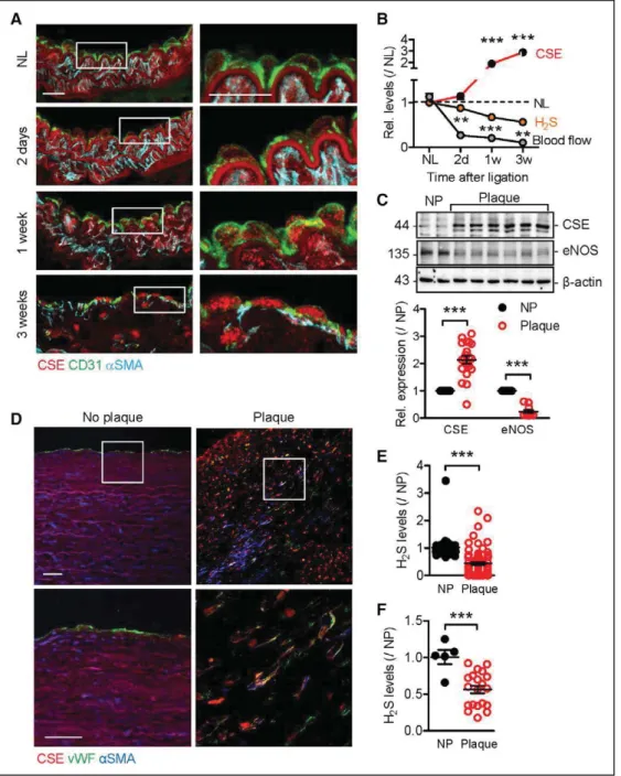
pression in CD31-positive cells followed by an increase in vascular smooth muscle cells (Figure 3A). Surprisingly, this was associated with a decrease rather than an in-crease in H2S production (Figure 3B). CSE expression was also clearly elevated in atherosclerotic plaques from indi-viduals who had undergone endarterectomy because of 75% to 90% internal carotid artery stenosis (Figure 3C and 3D). Similar to the situation in mice, circulating
(Fig-ure 3E) and intra-arterial (Figure 3F) levels of H2S were reduced in human subjects with atherosclerosis.
The decrease in CSE activity was not a consequence of substrate deficiency because levels of the CSE sub-strate L-cysteine were increased in plasma from subjects with atherosclerosis compared with healthy donors (Figure 4A and Figure IV and Table IV in the online-only Data Supplement). In addition, levels of L-cystathionine,
Figure 3. Cystathionine γ lyase (CSE) expression in the vascular wall.
A, Expression of CSE (red), CD31 (green), and α-smooth muscle actin (αSMA; blue) in a nonligated (NL) carotid artery from an apolipoprotein E–deficient mouse vs samples collected 2 days or 1 or 3 weeks after partial carotid artery ligation. White boxes mark the area magnified in the right panels. Bar=20 μm. B, Time course of the relative (Rel.) changes in CSE expression, blood flow, and plasma H2S levels in the same samples as in A; n=4 per group (each sample representing a pool of 3 animals) for protein and hydrogen sulfide (H2S) levels and n=6 to 8 animals per group for blood flow (2-way ANOVA, Bonferroni). C, Comparison of CSE and endothelial nitric oxide synthase (eNOS) expression in plaque-free (NP; n=5) arteries and carotid plaques (n=20) from human subjects. D, Representative staining of CSE (red), von Willebrand factor (vWF; green), αSMA (blue), and DAPI (gray) in aortas without atherosclerotic plaque vs carotid atherosclerotic plaques. Similar results were observed in 5 additional subjects per group. Bar=50 μm E, Plasma H2S levels in samples from NP individuals (n=32) or subjects with atherosclerosis (Plaque; n=70; Mann-Whitney). F, Arterial H2S levels in arterial tissue from NP individuals (n=5) or subjects with atherosclerosis (n=20) (Mann-Whitney). **P<0.01. ***P<0.001. DAPI indicates 4’,6-diamidine-2’-phenylindole dihydrochloride.
which is selectively converted to L-cysteine by CSE, were also markedly increased in plasma from the atheroscle-rosis group. Circulating levels of the CBS substrate L -homocysteine and its product L-serine were not signifi-cantly different between the 2 groups.
Next, a link between inflammation and altered CSE activity was addressed. We focused on IL-1β because it was significantly elevated in patients with atherosclero-sis compared with healthy donors (Figure 4B) and gradu-ally increased during the development of atherosclerosis in mice (Figure 4C). In in vitro studies, the stimulation of endothelial cells with IL-1β elicited the phosphorylation of CSE on serine and tyrosine but not threonine residues (Figure VA–VC in the online-only Data Supplement). Of the conserved potentially phosphorylatable amino
ac-ids, the mutation of Tyr60 (human sequence) or Tyr114 (to Phe) partially inhibited CSE activity. The mutation of Ser282 (to either Ala or Asp) was without effect, where-as the mutation of Ser377 to Asp abrogated CSE activity (Figure VD and VE in the online-only Data Supplement). Although IL-1β attenuated H2S production in HEK-293
cells transfected with the wild-type CSE, the Ser377Ala CSE mutant was resistant to the cytokine (Figure 4D).
Using an antibody that specifically recognized CSE phosphorylated on Ser377, we could demonstrate the serine phosphorylation of a V5-CSE fusion protein im-munoprecipitated from IL-1β–treated human endo-thelial cells, which coincided with a decrease in H2S production (Figure VF and VG in the online-only Data Supplement). The phosphorylation of CSE on Ser377
A
B
E
F
C D
Figure 4. Consequences of inflammation on cystathionine γ lyase (CSE) activity in later stages of atherosclerosis.
A, Circulating levels of L-cysteine, L-cystathionine, L-homocysteine, and L-serine in plasma from n=26 to 32 plaque-free (NP) donors and n=64 to 70 patients with
atherosclerosis (Plaque; Mann-Whitney). B, Circulating levels of interleukin (IL)–1β in samples from 12 NP subjects and 30 subjects with plaques (Mann-Whitney). C, Circulating levels of IL-1β in samples from apolipoprotein E–deficient mice 2 days or 1 or 3 weeks after partial carotid artery ligation; n=4 samples per time point (each sample representing a pool of 3 animals; ANOVA, Newman-Keuls). D, Hydrogen sulfide (H2S) production detected in HEK-293 cells expressing the wild-type (WT) CSE or the S377A and S377D CSE mutants and treated with either solvent (Sol) or IL-1β (30 ng/mL, 18 hours); n=6 independent experiments (ANOVA, New-man-Keuls). E, CSE phosphorylation (pCSE) on Ser377 in nonligated (NL) or ligated carotid arteries 2 days and 1 and 3 weeks after operation. Graph summarizes relative (Rel) levels from 4 independent experiments, with each experiment being a pool of 3 animals (ANOVA, Newman-Keuls). F, Serine phosphorylation of CSE in human arteries detected by proximity ligation assay (red) with phospho-serine and CSE antibodies. Cyan indicates DAPI. Right, Bright-field images merged with DAPI. Similar results were observed in 5 additional subjects per group. Bar=50 μm. ***P<0.001. DAPI indicates 4’,6-diamidine-2’-phenylindole dihydrochloride.
was also increased in murine carotid arteries 1 and 3 weeks after ligation (Figure 4E), as well as in human atherosclerotic plaques (Figure 4F and Figure VH in the online-only Data Supplement). IL-1β also slightly at-tenuated the activity of CBS but did not affect that of 3-mercaptopyruvate sulfurtransferase (Figure VIA in the online-only Data Supplement). The contribution of CBS to endothelial cell H2S production, however, was small because IL-1β had only a minor effect (≈6% de-crease) on H2S production in endothelial cells lacking CSE (Figure VIB in the online-only Data Supplement). Taken together, these data suggest that vascular in-flammation elicits the phosphorylation of CSE and its inactivation.
Consequences of Endothelial Cell–Specific
Deletion of CSE on Atherogenesis
To assess the role of endothelial cell CSE on athero-genesis in a model associated with disturbed flow, endothelial cell–specific CSE knockout mice (CSEΔEC
mice) were crossed onto the ApoE−/− background and
subjected to partial ligation of the left carotid artery. Twenty-one days after ligation, the lumen area was clearly reduced in arteries from ApoE−/− mice;
how-ever, the effects were much more pronounced in carotid arteries from ApoExCSEΔEC mice (Figure 5A).
Microcomputed tomography analyses confirmed the extensive atherosclerotic plaque formation and a de-crease in lumen area along the length of the carotid artery in animals lacking endothelial cell CSE (Fig-ure 5B).
Using a biotin switch–coupled proximity ligation assay, we could demonstrate HuR sulfhydration in ca-rotid artery endothelial cells in situ (Figure 5C and 5D). However, this signal rapidly decreased (within 2 days) after partial carotid artery ligation. With disease pro-gression, a significant reduction in HuR sulfhydration in vascular smooth muscle cells also became apparent. In line with the decrease in H2S levels in plasma and tissue from humans with atherosclerosis, the sulfhydration of HuR was also decreased in plaque material compared with healthy arteries (Figure 5E). Unfortunately, the CSE target studied in murine endothelial cells, CD62E, was not detected in the available human atheroscle-rotic plaque material, but a second HuR-regulated pro-tein, cathepsin S (CTSS),26 was increased in the human
plaque versus nonplaque material (Figure VIIA in the online-only Data Supplement). Indeed, when HuR was immunoprecipitated from CSE-overexpressing endo-thelial cells, its binding to the CTSS 3′ untranslated re-gion was decreased (Figure VIIB in the online-only Data Supplement). Overexpression of the phosphomimetic Ser377Asp CSE mutant, however, increased the bind-ing of HuR to CTSS mRNA (Figure VIIC in the online-only Data Supplement).
Effect of the Polysulfide Donor SG1002
on the Development of Atherosclerosis
To demonstrate the importance of H2S in the process of atherogenesis, rescue experiments were performed with sodium polysulthionate (SG1002), a slow-releas-ing polysulfide donor27,28 that more closelyrecapitu-lates the endogenous production of H2S than fast-releasing sulfur salts (Figure 6A). In endothelial cells from CSEiΔEC mice, the compound was able to restore
HuR sulfhydration (Figure 6B), to decrease CD62E protein levels (Figure 6C), and to attenuate mono-cyte adherence (Figure 6D). In mice, the addition of SG1002 to the drinking water increased circulating H2S levels by ≈60% within 48 hours (Figure 6E), a lev-el that remained stable over the observation period. Plaque formation in ApoExCSEΔEC mice 21 days after
partial carotid artery ligation was significantly attenu-ated in the animals that received SG1002 (Figure 6F and 6G). In addition, RNA immunoprecipitation stud-ies revealed reduced binding of HuR to CTSS mRNA in ligated arteries from the SG1002-treated mice (Fig-ure 6H). There was no direct effect of SG1002 on CSE activity because plasma levels of its substrates were unaffected (Figure VIIIA and VIIIB and Table V in the online-only Data Supplement). The effects of SG1002 were also independent of an increase in NO synthase activity because nitrite levels did not differ compared with the vehicle-treated animals (Figure VIIIC in the online-only Data Supplement). In fact, plasma levels of L-arginine increased and levels of L-citrulline de-creased, which would be more indicative of NO syn-thase inhibition.
L
-Cystathionine as a Biomarker of
Endothelial Cell Dysfunction
Given that our data highlighted the importance of CSE activity in maintaining vascular homeostasis, we next addressed the possibility that changes in plasma lev-els of the CSE substrate L-cystathionine could be used as a surrogate marker of CSE activity and endotheli-al cell function. Amino acid profiling of plasma from wild-type, CSEiΔEC, and globally CSE-deficient mice
re-vealed that >80% of the circulating L-cystathionine was seemingly metabolized by endothelial cells (Figure 7A). Moreover, the attenuated acetylcholine-induced relax-ation of aortic rings from ApoExCSEΔEC versus ApoE−/−
mice (Figure 7B) was coincident with a clear increase in circulating L-cystathionine but a decrease in H2S levels (Figure 7C). A similar reciprocal relationship between L-cystathionine and H2S levels was also noted between wild-type and CSEiΔEC mice.
In a small human cohort, noninvasive recording of vascular reactivity revealed impaired endothelial function (flow-mediated dilatation) in patients with
atherosclerosis (Figure 7D and Table VI in the online-only Data Supplement). In this group, endothelial dys-function was also linked to increased plasma L-cysta-thionine levels (Figure 7E). In a larger collective, the situation was clearer, and donors without atheroscle-rosis could be classified as a L-cystathioninelow/H
2S high
population, whereas the subjects with atherosclero-sis were generally classed as L-cystathioninehigh/H
2S low
(Figure 7F). Although the reciprocal relationship be-tween L-cystathionine, H2S, and the health condition
(no plaque versus plaque) was not fully characterized and a prediction model for L-cystathionine levels in the diseased population was not presented, our data indicate that, in the pathological conditions in which
CSE fails to generate H2S, the levels of its substrate, L-cystathionine, increase in the circulation.
DISCUSSION
The results of the present investigation revealed that CSE is the major source of endogenous H2S in native endothelial cells and that its expression and activity are tightly regulated by fluid shear stress and inflam-mation. The basal activity of CSE was important to maintain low arterial levels of CD62E and to minimize monocyte adhesion at sites of low or disturbed flow via a mechanism involving the sulfhydration of the RNA-binding protein HuR. In inflammatory conditions,
Figure 5. Characterization of atherosclerosis development in endothelial cell (EC)–specific cystathionine γ lyase (CSE) knockout mice.
A and B, Effect of partial ligation on carotid arteries from ApoE−/− and ApoExCSEΔEC (ΔEC) mice. A, Representative cross-sections showing Oil Red O staining in
nonligated and partially ligated murine carotid arteries. B, Carotid artery lumen evaluated by microcomputed tomography scanning (ligated arteries in blue) and quantification of lumen area; n=7 to 8 animals per group. Bar=200 µm (ANOVA for repeated measurements). C, Human antigen R (HuR) sulfhydration (S-SH) and total HuR levels in carotid arteries from ApoE−/− mice with no ligation (NL) and 2 days or 1 or 3 weeks after partial carotid artery ligation. The results are
representa-tive of 4 additional samples (each sample representing a pool of 3 animals) per group. D, S-SH (red), CD31 (green) and DAPI (blue) in cross-sections from carotid arteries from ApoE−/− mice with no ligation and 2 days or 1 week after partial ligation. Cyan indicates DAPI. Bar=20 μm. Graphs summarize sulfhydration events
per EC or smooth muscle cell (SMC); n = 6 animals per group (ANOVA, Newman-Keuls). E, Sulfhydration of HuR in nonplaque (NP) material and in atherosclerotic plaques (Plaque) from human subjects. Results are representative of n=4 NP and n=12 plaque samples per group. **P<0.01. ***P<0.001. ApoE indicates apolipo-protein E; DAPI, 4’,6-diamidine-2’-phenylindole dihydrochloride; DTT, dithiothreitol; and LCA, left carotid artery.
however, this protective mechanism was lost because of the phosphorylation (on Ser377) and inactivation of CSE. The loss of H2S and attenuated sulfhydration of HuR resulted in an increase in HuR dimerization and activity and the stabilization of the HuR-target mRNAs CD62E and CTSS, both of which have previously been linked to endothelial cell activation and atherosclerosis (Figure IX in the online-only Data Supplement). All of the observations made in a murine model of athero-genesis could be confirmed in a human cohort, making a strong case for a functional link between CSE inacti-vation and accelerated disease progression. Moreover, at least in mice, an H2S donor was able to decrease HuR binding to CTSS and decelerate atherogenesis. Finally, in both mice and humans, circulating L-cystathionine
levels were inversely correlated with H2S levels and en-dothelial function, highlighting its potential usefulness as a biomarker of vascular disease.
Since the initial report that CSE is expressed in endo-thelial cells,6 it has been attributed a major role in
gen-erating endothelial cell–derived H2S.29 It was possible
to confirm this assumption by generating mice lacking CSE specifically in endothelial cells, in which circulat-ing H2S levels were attenuated by ≈65%. Moreover, fluid shear stress was found to be a major negative regulator of CSE expression in situ and in vitro, so that the expression of the enzyme was elevated at sites of low or disturbed blood flow, which are predilection sites for the development of atherosclerosis. It seems that the CSE expressed at these sites exerts a
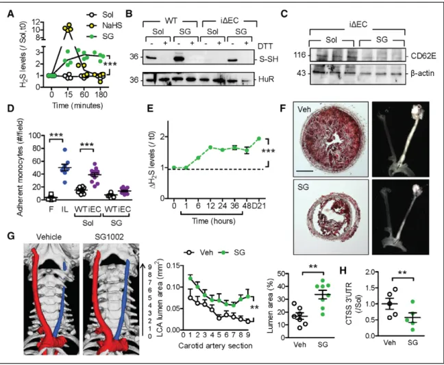
protec-Figure 6. Effect of SG1002 (SG) on the phenotype of ApoExCSEΔEC mice.
A, Hydrogen sulfide (H2S) release from murine endothelial cells (ECs) treated with solvent (Sol), NaHS (100 µmol/L), or SG (1 µmol/L); n=4 independent cell batches (2 way ANOVA, Bonferroni). B, Human antigen R (HuR) sulfhydration (S-SH) in ECs from wild-type (WT) and CSEiΔEC (iΔEC) mice treated with solvent or SG. Results
are representative of an additional 4 cell batches. C, CD62E levels in ECs from CSEiΔEC mice treated with solvent or SG for 60 minutes. Results are representative
of an additional 5 cell batches per group. D, Monocyte adherence to ECs from WT and CSEiΔEC mice treated with solvent or SG for 60 minutes. Adherence on
fibronectin (F) was included as a negative control and interleukin-1β (IL; 30 ng/mL, 3 hours) as a positive control; n=12 experiments from 6 different cell batches (ANOVA, Newman-Keuls). E, H2S levels in plasma from ApoE−/− mice given SG (400–600 ng/d) in drinking water; n=6 animals per group (ANOVA, Newman-Keuls).
F and G, Effect of SG on the development of atherosclerosis. Representative images of Oil Red O staining of left carotid arteries (LCAs) from ApoExCSEΔEC mice 21
days after partial ligation. Mice were treated with vehicle (Veh) or SG starting 1 day before partial ligation (F). Carotid artery lumen evaluated by microcomputed tomography scanning (blue indicates ligated; red, nonligated) and quantification of lumen area 21 days after carotid artery ligation in ApoExCSEΔEC mice treated
with vehicle or SG; n=7 to 8 animals per group (ANOVA for repeated measurements, Dunn) (G). H, RNA immunoprecipitation showing the association of the CTSS (cathepsin S) 3′ untranslated region (UTR) with HuR in carotid arteries from the animals shown in F and G; n=5 per group (Student t test). *P<0.05. **P<0.01. ***P<0.001. ApoE indicates apolipoprotein E; CSE, cystathionine γ lyase; and DTT, dithiothreitol.
tive function that can be attributed at least partly to repressing the expression of CD62E. However, be-cause CSE-derived H2S did not directly target CD62E, an indirect approach was taken to identify potential sulfhydration targets that could in turn account for the effects observed. Reasoning that CSE should as-sociate with H2S targets, coprecipitation studies iden-tified the RNA-binding protein HuR as part of the CSE interactome. HuR was an attractive candidate for sulf-hydration because it possesses 3 cysteines, 1 of which is predicted to be highly nucleophilic. Indeed, it was possible to detect the sulfhydration of HuR in wild-type endothelial cells, which was barely detectable in cells lacking CSE. The finding that Cys13 was targeted by H2S hinted at a possible molecular mechanism be-cause Cys13 is essential for the formation of the disul-fide bonds that stabilize the active HuR homodimer.24
The sulfhydration of HuR in CSE-expressing endothe-lial cells meant that HuR was detected largely as an inactive monomer that was unable to bind its target mRNA, CD62E.
To evaluate the pathophysiological relevance of these findings, the expression and activity of CSE were studied in a murine model of atherogenesis linked to disturbed flow.25 Consistent with the in vitro
observa-tions, CSE was significantly upregulated 1 week after partial carotid artery ligation. More important, in
sam-ples from patients with carotid artery stenosis (75%– 85%) that demonstrated disturbed or low flow, CSE levels were clearly upregulated. Somewhat unexpect-edly, however, the clear increase in arterial CSE expres-sion contrasted with the marked reduction in circulat-ing and tissue levels of H2S, indicating that endothelial dysfunction and atherosclerosis were linked to the in-hibition of CSE activity. In agreement with this obser-vation, partial ligation of the carotid artery in mice led to a time-dependent decrease in the sulfhydration of HuR. The latter effects could not be attributed to CSE inactivity resulting from substrate deficiency because the amino acid profile of human plasma and plaque material revealed that levels of the CSE-specific sub-strate L-cystathionine were actually elevated. Given the link between vascular inflammation and atherosclero-sis, the consequences of inflammatory cytokines on CSE activity were assessed. IL-1β was chosen for these studies because its levels were increased in the mouse model and in the available human samples. Indeed, IL -1β elicited the serine phosphorylation of CSE, and in agreement with a previous report,30 it was possible to
demonstrate that the serine phosphorylation of CSE on Ser377 resulted in enzyme inactivation. Making the link back to atherosclerosis, the serine phosphorylation of CSE was detectable in carotid arteries from the mu-rine model of atherosclerosis and in plaque material
Figure 7.L-cystathionine as a biomarker of cystathionine γ lyase (CSE) activity and endothelial cell (EC) dysfunction in humans.
A, L-cystathionine levels in plasma from wild-type (WT), CSEiΔEC (iΔEC), and inducible CSE knockout (iKO) mice; n=6 to 12 animals per group (ANOVA, New-man-Keuls). B, Acetylcholine (ACh)–induced relaxation (% maximum) in aortic rings from ApoE−/− and ApoExCSEΔEC mice fed a high-fat diet for 21 days; n=8
animals per group (2-way ANOVA, Bonferroni). C, Linear correlation of circulating L-cystathionine and hydrogen sulfide (H2S) levels in WT, CSEiΔEC, ApoE−/−, and
ApoExCSEΔEC mice, n=6 animals per group (Pearson). D, Flow-mediated dilatation (FMD) and flow-mediated constriction (FMC) in healthy volunteers (n=6) and
patients (n=6) with atherosclerotic plaques. E, Linear correlation of FMD and circulating L-cystathionine levels in healthy subjects (n=6) without atherosclerosis (NP)
and patients (n=6) with atherosclerosis (Plaque) and carotid stenosis between 75% and 90% (Pearson). F, Linear correlation of circulating L-cystathionine and H2S levels in samples from human subjects without plaques (n=32) and with atherosclerosis (n=70) (Pearson). **P<0.01. ***P<0.001. ApoE indicates apolipoprotein E.
from individuals with atherosclerosis. The inactivation of CSE in humans could be linked to decreased sulf-hydration of HuR and increased expression of the HuR target CTSS. Of course, a number of additional, recent-ly identified targets of H2S donors could contribute to the phenotype observed, including sirtuin-1,31 which is
also stabilized by HuR.20,32 A number of additional
pro-teins are physically associated with CSE in endothelial cells, including collagen and 2 pacsin proteins, which are implicated in vesicle trafficking,33,34 and it will be
interesting to determine whether the sulfhydration of these proteins can also affect vascular homeostasis.
Animal models of atherosclerosis are generally based on plaque formation resulting from a choles-terol-rich/Western-type diet, in combination with the manipulation of genes involved in cholesterol metabo-lism. Although these mouse models have provided a wealth of insight into disease pathogenesis, each of them comes with its own limitations.35 The ApoE−/−
model of partial carotid artery ligation studied here was chosen because of the initial link to disturbed flow and the accelerated development of atherosclerotic le-sions. Given the potential limitations of this model, we placed particular emphasis on comparing changes ob-served in the animal model with samples from humans with and without atherosclerosis. By taking this dual approach, we could confirm the altered sulfhydration of HuR, the upregulation of CSE expression in athero-sclerotic plaques, the phosphorylation of CSE, a sub-sequent decrease in H2S levels, and alterations in the amino acid profile that correlated with CSE activity in human material.
To determine the importance of endothelial cell CSE in atherogenesis, studies were repeated in mice specifically lacking CSE in endothelial cells. Consistent with a key role of the endothelium in atherosclerosis initiation, plaque size was greater and carotid lumen size smaller in partially ligated carotid arteries from ApoExCSEΔEC mice versus ApoE−/− mice. Thus, although
H2S has been proposed to freely diffuse across mem-branes,3 it seems that the H
2S produced in smooth
muscle cells cannot substitute for the loss of endo-thelial cell–derived H2S. Given the apparent important antiatherosclerotic role of CSE, the next step was to assess the consequences of exogenous H2S applica-tion on the development of atherosclerosis. The res-cue proved to be efficient in vitro because the H2S do-nor SG1002 largely restored the sulfhydration of HuR and decreased the expression of CD62E and monocyte adhesion in CSE-deficient endothelial cells. Moreover, the in vivo administration of SG1002 to ApoExCSEΔEC
mice resulted in a maintained elevation in plasma H2S levels and a pronounced reduction in atherosclerosis. Preclinical studies have demonstrated that SG1002 is also able to attenuate cardiac dysfunction, to protect against pressure overload–induced heart failure (see
review27), and to restore plasma H
2S and NO levels
to normal in patients with congestive heart failure.36
However, there was no indication of elevated NO pro-duction in our studies as assessed by amino acid profil-ing and assay of circulatprofil-ing nitrite levels, thus suggest-ing that the antiatherosclerotic actions of SG1002 can be attributed directly to H2S.
The amino acid profiling studies also suggested that high plasma levels of L-cystathionine mirrored de-creased CSE activity. Therefore, in a final step, we set out to determine whether circulating levels of L -cysta-thionine could be used as a biomarker of endothelial dysfunction. By comparing plasma from the available wild-type, globally CSE-deficient, and endothelial cell– specific CSE knockout mice, we were able to demon-strate that altering endothelial cell CSE activity had a major impact on circulating L-cystathionine levels. High plasma L-cystathionine levels also correlated with impaired endothelial function in ApoExCSEΔEC versus
ApoE−/− mice. Similarly, circulating L-cystathionine
lev-els were negatively correlated with H2S production in patients with atherosclerosis compared with healthy subjects. Moreover, in a small cohort of subjects, it was possible to classify those patients with atheroscle-rosis and attenuated flow-induced vasodilatation as being L-cystathioninehigh/H
2S
low. All of these data
indi-cate that plasma levels of L-cystathionine and/or H2S
could be useful biomarkers of endothelial dysfunction in nondiagnosed, asymptomatic individuals with early vascular disease.
Taken together, the results of this study have high-lighted the importance of CSE in endothelial cells for the prevention of atherosclerosis development, identi-fied HuR as a primary molecular target of endogenous H2S that can account for proatherosclerotic changes in the vessel wall after CSE inactivation, and provided evi-dence that L-cystathionine is an effective biomarker of impaired endothelial dysfunction.
Authors
Sofia-Iris Bibli, PhD; Jiong Hu, PhD; Fragiska Sigala, MD; Ilka Wittig, PhD; Juliana Heidler, PhD; Sven Zukunft, PhD; Diamantis I. Tsilimigras, MD; Voah-anginirina Randriamboavonjy, PhD; Janina Wittig, BSc; Baktybek Kojonazarov, PhD; Christoph Schürmann, PhD; Mauro Siragusa, PhD; Daniel Siuda, PhD; Bert Luck, MSc; Randa Abdel Malik, PhD; Konstantinos A. Filis, MD; George Zografos, MD; Chen Chen, MD; Dao Wen Wang, MD; Josef Pfeilschifter, MD; Ralf P. Brandes, MD; Csaba Szabo, PhD; Andreas Papapetropoulos, PhD; Ingrid Fleming, PhD
Correspondence
Ingrid Fleming, PhD, Institute for Vascular Signalling, Centre for Molecular Medicine, Goethe University, Theodor Stern Kai 7, 60596 Frankfurt am Main, Germany. Email fl[email protected]
Affiliations
Institute for Vascular Signalling, Centre for Molecular Medicine (S.-I.B., J. Hu, S.Z., V.R., J.W., M.S., D.S., B.L., R.A.M., I.F.), Functional Proteomics, SFB 815 Core Unit (J.W., J. Heidler), Institute for Cardiovascular Physiology (C. Schürmann, R.P.B.), and Pharmacentre Frankfurt/Zentrum für Arzneimittel-forschung, Entwicklung und –Sicherheit (J.P.), Goethe University, Frankfurt am Main, Germany. German Center of Cardiovascular Research, Partner site RheinMain, Frankfurt am Main, Germany (S.-I.B., J. Hu, I.W., J. Heidler, S.Z., V.R., J.W., C. Schürmann, M.S., D.S., B.L., R.A.M., R.P.B., I.F.). First Prope-deutic Department of Surgery, Vascular Surgery Division, National and Ka-podistrian University of Athens Medical School, Greece (F.S., D.I.T., K.A.F., G.Z.). Department of Internal Medicine, Universities of Giessen and Marburg Lung Center, Justus Liebig University, Germany (B.K.). Division of Cardiology, Department of Internal Medicine and Gene Therapy Center, Tongji Hospi-tal, Tongji Medical College, Huazhong University of Science and Technology, Wuhan, China (C.C., D.W.W.). Laboratory of Pharmacology, Department of Medicine, University of Fribourg, Switzerland (C. Szabo). Laboratory of Phar-macology, Department of Pharmacy, National and Kapodistrian University of Athens, Greece (A.P.). Clinical, Experimental Surgery and Translational Re-search Center, Biomedical ReRe-search Foundation of the Academy of Athens, Greece (A.P.).
Acknowledgments
The authors are indebted to Isabel Winter, Mechtild Piepenbrock-Gyamfi, and Katharina Herbig for expert technical assistance and to Andersen Clark from the University of Texas Medical Branch for help with statistical analy-ses. Drs Bibli, Hu, and Fleming designed and guided research. Drs Bibli, Hu, Heidler, Randriamboavonjy, Tsilimigras, Wittig, and Malik performed ex-periments. Drs Bibli, Hu, and Fleming analyzed data. Drs Wittig and Heidler performed the proteomic analyses. Dr Zukunft performed the amino acid profiling and analysis. Dr Siuda performed amino acid analyses. Dr Kojonaz-arov performed the echocardiography. Dr Siragusa measured the circulating nitrite levels. Drs Luck and Zukunft analyzed H2S levels. Drs Chen and Wang provided immunohistochemistry stainings in human material. Dr Pfeilschift-er genPfeilschift-erated the floxed CSE mouse. Dr Szabo raised the phospho-specific Ser377 CSE specific antibody. Drs Sigala and Tsilimigras provided human samples and performed the vascular function studies in humans. Drs Si-gala, Filis, and Zografos analyzed the human vascular function studies and provided the echocardiography human data. Dr Papapetropoulos provided viruses, mutants, and activity data for H2S-generating enzymes. Drs Schür-mann and Brandes performed the microcomputed tomography analyses. Drs Sigala, Szabo, and Papapetropoulos provided conceptual advice. Drs Bibli and Fleming wrote the manuscript.
Sources of Funding
This work was supported by the European Society of Cardiology (Basic Re-search Fellowship 2016 and ReRe-search Grant R-2016–054), the Schering Stif-tung (Young Investigator Fund for Innovative Research 2017, to Dr Bibli), and the Deutsche Forschungsgemeinschaft (SFB 815/A16 to Dr Fleming, SFB 815/A7 to Dr Pfeilschifter, and Exzellenzcluster 147 “Cardio-Pulmonary Systems”).
Disclosures None.
REFERENCES
1. Ross R, Glomset JA. Atherosclerosis and the arterial smooth muscle cell.
Science. 1973;180:1332. doi: 10.1126/science.180.4093.1332
2. Davies PF, Civelek M, Fang Y, Fleming I. The atherosusceptible en-dothelium: endothelial phenotypes in complex haemodynamic shear stress regions in vivo. Cardiovasc Res. 2013;99:315–327. doi: 10.1093/cvr/cvt101
3. Wang R. Physiological implications of hydrogen sulfide: a whiff ex-ploration that blossomed. Physiol Rev. 2012;92:791–896. doi: 10.1152/physrev.00017.2011
4. Shibuya N, Koike S, Tanaka M, Ishigami-Yuasa M, Kimura Y, Ogasawara Y, Fukui K, Nagahara N, Kimura H. A novel pathway for the production of hydrogen sulfide from D-cysteine in mammalian cells. Nat Commun. 2013;4:1366. doi: 10.1038/ncomms2371
5. Szabo C, Papapetropoulos A. International Union of Basic and Clinical Pharmacology, CII: pharmacological modulation of H2S levels: H2S donors and H2S biosynthesis inhibitors. Pharmacol Rev. 2017;69:497–564. doi: 10.1124/pr.117.014050
6. Yang G, Wu L, Jiang B, Yang W, Qi J, Cao K, Meng Q, Mustafa AK, Mu W, Zhang S, Snyder SH, Wang R. H2S as a physiologic vasorelaxant: hy-pertension in mice with deletion of cystathionine gamma-lyase. Science. 2008;322:587–590. doi: 10.1126/science.1162667
7. Syhr KMJ, Boosen M, Hohmann SW, Longen S, Köhler Y, Pfeilschifter J, Beck KF, Geisslinger G, Schmidtko A, Kallenborn-Gerhardt W. The H2S-producing en-zyme CSE is dispensable for the processing of inflammatory and neuropathic pain. Brain Res. 2015;1624:380–389. doi: 10.1016/j.brainres.2015.07.058 8. Mani S, Li H, Untereiner A, Wu L, Yang G, Austin RC, Dickhout JG, Lhoták
Š, Meng QH, Wang R. Decreased endogenous production of hydrogen sulfide accelerates atherosclerosis. Circulation. 2013;127:2523–2534. doi: 10.1161/CIRCULATIONAHA.113.002208
9. Li H, Mani S, Cao W, Yang G, Lai C, Wu L, Wang R. Interaction of hydro-gen sulfide and estrohydro-gen on the proliferation of vascular smooth muscle cells. PLoS One. 2012;7:e41614. doi:10.1371/journal.pone.0041614 10. Li H, Mani S, Wu L, Fu M, Shuang T, Xu C, Wang R. The interaction of estrogen
and CSE/H2S pathway in the development of atherosclerosis. Am J Physiol
Heart Circ Physiol. 2017;312:H406–H414. doi: 10.1152/ajpheart.00245.2016
11. Cheung SH, Kwok WK, To KF, Lau JY. Anti-atherogenic effect of hydrogen sulfide by over-expression of cystathionine gamma-lyase (CSE) gene. PLoS
One. 2014;9:e113038. doi: 10.1371/journal.pone.0113038
12. van den Born JC, Mencke R, Conroy S, Zeebregts CJ, van Goor H, Hil-lebrands JL. Cystathionine γ-lyase is expressed in human atheroscle-rotic plaque microvessels and is involved in micro-angiogenesis. Sci Rep. 2016;6:34608. doi: 10.1038/srep34608
13. Sigala F, Efentakis P, Karageorgiadi D, Filis K, Zampas P, Iliodromitis EK, Zografos G, Papapetropoulos A, Andreadou I. Reciprocal regulation of eNOS, H2S and CO-synthesizing enzymes in human atheroma: correlation with plaque stability and effects of simvastatin. Redox Biol. 2017;12:70– 81. doi: 10.1016/j.redox.2017.02.006
14. Yu B, Zheng Y, Yuan Z, Li S, Zhu H, De La Cruz LK, Zhang J, Ji K, Wang S, Wang B. Toward direct protein S-persulfidation: a prodrug approach that directly delivers hydrogen persulfide. J Am Chem Soc. 2018;140:30–33. doi: 10.1021/jacs.7b09795
15. Paul BD, Snyder SH. Protein sulfhydration. Methods Enzymol. 2015;555:79–90. doi: 10.1016/bs.mie.2014.11.021
16. Monvoisin A, Alva JA, Hofmann JJ, Zovein AC, Lane TF, Iruela-Arispe ML. VE-cadherin-CreERT2 transgenic mouse: a model for inducible recombination in the endothelium. Dev Dyn. 2006;235:3413–3422. doi: 10.1002/dvdy.20982 17. Xia J, Sinelnikov IV, Han B, Wishart DS. MetaboAnalyst 3.0–making me-tabolomics more meaningful. Nucleic Acids Res. 2015;43:W251–W257. doi: 10.1093/nar/gkv380
18. Ziegler T, Silacci P, Harrison VJ, Hayoz D. Nitric oxide synthase expres-sion in endothelial cells exposed to mechanical forces. Hypertenexpres-sion. 1998;32:351–355. doi: 10.1161/01.HYP.32.2.351
19. Ziegler T, Bouzourene K, Harrison VJ, Brunner HR, Hayoz D. Influence of oscillatory and unidirectional flow environments on the expression of en-dothelin and nitric oxide synthase in cultured endothelial cells. Arterioscler
Thromb Vasc Biol. 1998;18:686–692. doi: 10.1161/01.ATV.18.5.686
20. Ceolotto G, De Kreutzenberg SV, Cattelan A, Fabricio AS, Squarcina E, Gion M, Semplicini A, Fadini GP, Avogaro A. Sirtuin 1 stabilization by HuR represses TNF-α- and glucose-induced E-selectin release and endothelial cell adhesiveness in vitro: relevance to human metabolic syndrome. Clin
Sci (Lond). 2014;127:449–461. doi: 10.1042/CS20130439
21. Olsson MH, Søndergaard CR, Rostkowski M, Jensen JH. PROPKA3: con-sistent treatment of internal and surface residues in empirical pKa predic-tions. J Chem Theory Comput. 2011;7:525–537. doi: 10.1021/ct100578z 22. Ferrè F, Clote P. DiANNA: a web server for disulfide connectiv-ity prediction. Nucleic Acids Res. 2005;33:W230–W232. doi: 10.1093/nar/gki412
23. Ferre F, Clote P. DiANNA 1.1: an extension of the DiANNA web server for ternary cysteine classification. Nucleic Acids Res. 2006;34:W182–W185. doi: 10.1093/nar/gkl189
24. Benoit RM, Meisner NC, Kallen J, Graff P, Hemmig R, Cèbe R, Os-termeier C, Widmer H, Auer M. The x-ray crystal structure of the first RNA recognition motif and site-directed mutagenesis suggest a possible HuR redox sensing mechanism. J Mol Biol. 2010;397:1231–1244. doi: 10.1016/j.jmb.2010.02.043
25. Nam D, Ni CW, Rezvan A, Suo J, Budzyn K, Llanos A, Harrison D, Gid-dens D, Jo H. Partial carotid ligation is a model of acutely induced
turbed flow, leading to rapid endothelial dysfunction and atheroscle-rosis. Am J Physiol Heart Circ Physiol. 2009;297:H1535–H1543. doi: 10.1152/ajpheart.00510.2009
26. Stellos K, Gatsiou A, Stamatelopoulos K, Perisic Matic L, John D, Lunella FF, Jaé N, Rossbach O, Amrhein C, Sigala F, Boon RA, Fürtig B, Manavski Y, You X, Uchida S, Keller T, Boeckel JN, Franco-Cereceda A, Maegdefes-sel L, Chen W, Schwalbe H, Bindereif A, Eriksson P, Hedin U, Zeiher AM, Dimmeler S. Adenosine-to-inosine RNA editing controls cathepsin S ex-pression in atherosclerosis by enabling HuR-mediated post-transcriptional regulation. Nat Med. 2016;22:1140–1150. doi: 10.1038/nm.4172 27. Wallace JL, Vaughan D, Dicay M, MacNaughton WK, de Nucci G.
Hydro-gen sulfide-releasing therapeutics: translation to the clinic. Antioxid Redox
Signal. 2018;28:1533–1540. doi: 10.1089/ars.2017.7068
28. Chappell DC, Varner SE, Nerem RM, Medford RM, Alexander RW. Oscil-latory shear stress stimulates adhesion molecule expression in cultured human endothelium. Circ Res. 1998;82:532–539.
29. Polhemus DJ, Lefer DJ. Emergence of hydrogen sulfide as an endog-enous gaseous signaling molecule in cardiovascular disease. Circ Res. 2014;114:730–737. doi: 10.1161/CIRCRESAHA.114.300505
30. Yuan G, Vasavda C, Peng YJ, Makarenko VV, Raghuraman G, Nanduri J, Gadalla MM, Semenza GL, Kumar GK, Snyder SH, Prabhakar NR. Protein kinase G-regulated production of H2S governs oxygen sensing. Sci Signal. 2015;8:ra37. doi: 10.1126/scisignal.2005846
31. Du C, Lin X, Xu W, Zheng F, Cai J, Yang J, Cui Q, Tang C, Cai J, Xu G, Geng B. Sulfhydrated sirtuin-1 increasing its deacetylation activity is an es-sential epigenetics mechanism of anti-atherogenesis by hydrogen sulfide.
Antioxid Redox Signal. 2018; doi:10.1089/ars.2017.7195
32. Abdelmohsen K, Pullmann R Jr, Lal A, Kim HH, Galban S, Yang X, Blethrow JD, Walker M, Shubert J, Gillespie DA, Furneaux H, Gorospe M. Phosphorylation of HuR by Chk2 regulates SIRT1 expression. Mol Cell. 2007;25:543–557. doi: 10.1016/j.molcel.2007.01.011
33. Kessels MM, Qualmann B. The syndapin protein family: linking membrane trafficking with the cytoskeleton. J Cell Sci. 2004;117(pt 15):3077–3086. doi: 10.1242/jcs.01290
34. Dorland YL, Malinova TS, van Stalborch AM, Grieve AG, van Geemen D, Jansen NS, de Kreuk BJ, Nawaz K, Kole J, Geerts D, Musters RJ, de Rooij J, Hordijk PL, Huveneers S. The F-BAR protein pacsin2 inhibits asymmetric VE-cadherin internalization from tensile adherens junctions. Nat
Com-mun. 2016;7:12210. doi: 10.1038/ncomms12210
35. Emini Veseli B, Perrotta P, De Meyer GRA, Roth L, Van der Donckt C, Marti-net W, De Meyer GRY. Animal models of atherosclerosis. Eur J Pharmacol. 2017;816:3–13. doi: 10.1016/j.ejphar.2017.05.010
36. Polhemus DJ, Li Z, Pattillo CB, Gojon G Sr, Gojon G Jr, Giordano T, Krum H. A novel hydrogen sulfide prodrug, SG1002, promotes hydrogen sulfide and nitric oxide bioavailability in heart failure patients. Cardiovasc Ther. 2015;33:216–226. doi: 10.1111/1755-5922.12128