HAL Id: tel-02941976
https://hal.univ-lorraine.fr/tel-02941976
Submitted on 17 Sep 2020
HAL is a multi-disciplinary open access
archive for the deposit and dissemination of
sci-entific research documents, whether they are
pub-lished or not. The documents may come from
teaching and research institutions in France or
abroad, or from public or private research centers.
L’archive ouverte pluridisciplinaire HAL, est
destinée au dépôt et à la diffusion de documents
scientifiques de niveau recherche, publiés ou non,
émanant des établissements d’enseignement et de
recherche français ou étrangers, des laboratoires
publics ou privés.
microcirculation
Philippe Guerci
To cite this version:
Philippe Guerci. Current and new therapies for the critically injured microcirculation. Human
health and pathology. Université de Lorraine; Universiteit van Amsterdam, 2020. English. �NNT :
2020LORR0053�. �tel-02941976�
AVERTISSEMENT
Ce document est le fruit d'un long travail approuvé par le jury de
soutenance et mis à disposition de l'ensemble de la
communauté universitaire élargie.
Il est soumis à la propriété intellectuelle de l'auteur. Ceci
implique une obligation de citation et de référencement lors de
l’utilisation de ce document.
D'autre part, toute contrefaçon, plagiat, reproduction illicite
encourt une poursuite pénale.
Contact : ddoc-theses-contact@univ-lorraine.fr
LIENS
Code de la Propriété Intellectuelle. articles L 122. 4
Code de la Propriété Intellectuelle. articles L 335.2- L 335.10
http://www.cfcopies.com/V2/leg/leg_droi.php
BioSE
Défaillance
cardiovasculaire
aigue et chronique,
Unité INSERM 1116
University of Amsterdam, Amsterdam, The Netherlands
Thèse
Présentée et soutenue publiquement pour l’obtention du titre de
DOCTEUR DE l’UNIVERSITE DE LORRAINE
Mention : SCIENCES DE LA VIE ET DE LA SANTÉ
par
Philippe GUERCI
Sous la direction de Bruno LEVY
CURRENT AND NEW THERAPIES FOR THE
CRITICALLY INJURED MICROCIRCULATION
16 Juin 2020
Membres du jury :
Directeur(s) de thèse :
Mr Can INCE Professeur, Docteur, Universiteit van Amsterdam (UvA),
Amsterdam, The Netherlands
Mr Bruno LEVY Professeur, Docteur, Université de Lorraine, Nancy, France
Co-Directeur de thèse :
Mme Marie-Reine LOSSER Professeure, Université de Lorraine, Nancy, France
Rapporteurs :
Examinateurs :
Mr A.J. VERHOEVEN Professeur, Docteur, Universiteit van Amsterdam (UvA),
Amsterdam, The Netherlands
Mr E.T. van BAVEL Professeur, Docteur, Universiteit van Amsterdam (UvA),
Amsterdam, The Netherlands
Mr A.P.J. VLAAR Docteur, Universiteit van Amsterdam (UvA),
Amsterdam, The Netherlands
Mr J. BAKKER Professeur, Docteur, Erasmus Universiteit Rotterdam,
Rotterdam, The Netherlands Mr Didier PAYEN DE LA
GARANDERIE
Professeur, Docteur, Université Paris 7 Denis Diderot, Paris, France
Membres invités :
Mr L. VOGT Docteur, Universiteit van Amsterdam (UvA), Amsterdam,
OR THE CRITIC
ALL
Y INJURED MICROCIRCULA
TION
PHILIPPE GUERCI
FOR THE CRITICALLY INJURED
MICROCIRCULATION
CURRENT AND NEW THERAPIES FOR THE
CRITICALLY INJURED MICROCIRCULATION
CURRENT AND NEW THERAPIES FOR THE CRITICALLY INJURED MICROCIRCULATION © Philippe Olivier Guerci, 2020
ISBN: 978-94-6380-819-4
Cover by Bregje from Proefschriftontwerp.nl Printed by ProefschriftMaken || Proefschriftmaken.nl
CURRENT AND NEW THERAPIES FOR THE
CRITICALLY INJURED MICROCIRCULATION
ACADEMISCH PROEFSCHRIFT
ter verkrijging van de graad van doctor aan de Universiteit van Amsterdam
op gezag van de Rector Magnificus prof. dr. ir. K.I.J. Maex
ten overstaan van een door het College voor Promoties ingestelde commissie, in het openbaar te verdedigen in de Agnietenkapel
op dinsdag 16 juni 2020, te 14.00 uur door
Philippe Olivier Guerci geboren te Nancy
Promotiecommissie
Promotores: prof. dr. ir. C. Ince AMC-UvA
prof. dr. B. Levy Université de Lorraine
Copromotor: prof. dr. M.R. Losser Université de Lorraine
Overige leden: prof. dr. A.J. Verhoeven AMC-UvA
prof. dr. E.T. van Bavel AMC-UvA
dr. A.P.J. Vlaar AMC-UvA
dr. L. Vogt AMC-UvA
prof. dr. D.M. Payen Université Paris Diderot
prof. dr. J. Bakker Erasmus Universiteit Rotterdam
Faculteit der Geneeskunde
Universiteit van Amsterdam en de Université de Lorraine met als doel het behalen van een gezamenlijk doctoraat. Het proefschrift is voorbereid in de Faculteit der Geneeskunde van de Universiteit van Amsterdam en de Faculté de Médecine de Nancy van de Université de Lorraine.
This thesis was prepared within the partnership between the University of Amsterdam and the Université de Lorraine with the purpose of obtaining a joint doctorate degree. The thesis was prepared in the Faculty of Medicine at the University of Amsterdam and in the Faculté de Médecine de Nancy at the Université de Lorraine.
Chapter 1 General introduction 7
Chapter 2 The macro- and the microcirculation of the kidney 13
Chapter 3 Endothelial dysfunction of the kidney in sepsis 39
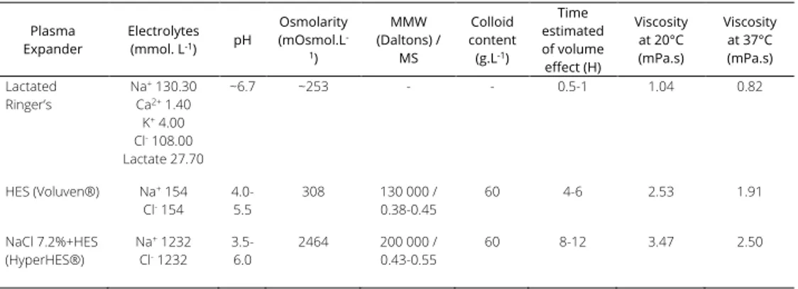
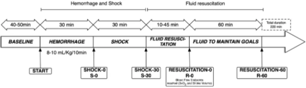
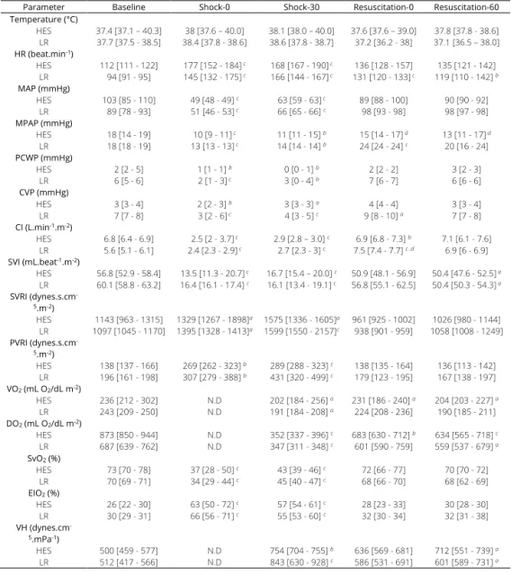
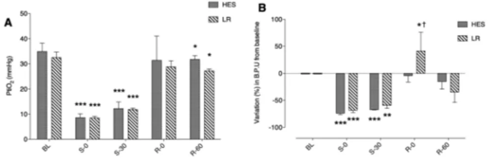
Chapter 4 Impact of fluid resuscitation with hypertonic-hydroxyethyl starch
versus lactated ringer on hemorheology and microcirculation in hemorrhagic shock
65
Chapter 5 Glycocalyx degradation is independent of vascular barrier
permeability increase in non-traumatic hemorrhagic shock in rats
89
Chapter 6 Glycocalyx shedding during stepwise hemodilution and
microvascular permeability
119
Chapter 7 A led-based phosphorimeter for measurement of microcirculatory
oxygen pressure
141
Chapter 8 The role of bicarbonate precursors in balanced fluids during
haemorrhagic shock with and without compromised liver function
167
Chapter 9 Effects of N-acetylcysteine (NAC) supplementation in resuscitation
fluids on renal microcirculatory oxygenation, inflammation, and function in a rat model of endotoxemia
189
Chapter 10 Effect of PEGylated-carboxyhemoglobin on renal microcirculation in
a rat model of hemorrhagic shock
215
Chapter 11 Resuscitation with pegylated-carboxyhemoglobin preserves the
renal function and tissue oxygenation during endotoxemia
249
Chapter 12 Summary & Conclusion
Samenvatting en Conclusies
277
Appendices Acknowledgments/Dankwoord 287
Portfolio 290
1
G
ENERAL INTRODUCTION
The microcirculation is a core element of humans and terrestrial mammals. Microcirculation is a complex and heterogenous network comprising arterioles, capillaries, venules but also lymphatics. The main functions of the microcirculation are to ensure the delivery of oxygen to tissue and cells while providing nutrients and removal of the carbon dioxide produced by the cell metabolism, thus, to meet metabolic demands.
The regulatory mechanisms controlling the microcirculatory flow and perfusion are various, redundant and elaborated. The regulation of flow to and in the microcirculation is determined by: (i) at general level, organ perfusion pressure, cardiac pump output, and (ii) at a microscopic scale by myogenic responses to flow (strain and stress), metabolic demand and acid/base
balance (local O2, CO2, H+ and lactate levels) and neurohormonal responses. The
microcirculatory blood flow is regulated using autocrine and paracrine interactions to meet the metabolic demand, mainly oxygen requirements of tissue cells. Thus, microcirculatory oxygen level is of critical importance.
From a pragmatic perspective, the structure of the microcirculation comprises three major components: the container defined by the different layers of the vascular wall, the contents representing the flowing plasma and its formed elements and the extraluminal surrounding tissue. In disease, each of these components can be altered separately or simultaneously. The endothelial cells form the interface between the content of the inner lumen of vessels and the surrounding environment. The endothelium is remarkably heterogenous in structure and function and differs from organ to organ. It plays a crucial role in the regulation of macromolecular trafficking, vasomotor tone, hemostasis, immunological functions, and the secretion of molecules by sensing through mechanotransductors, which subsequently initiate transcellular and intra-cellular signaling and activation. Endothelial cells are linked together by transcellular components, including gap junctions for electrical communication for upstream vascular regulation and intercellular tight junctions for maintaining vascular barriers. The glycocalyx is a thin layer lining the luminal membrane and contributes to microvascular permeability of the vascular wall. It is a highly interactive scaffold capable of sensing blood flow and houses many endothelial receptors that interacts with leukocytes and other formed elements. Although, the glycocalyx participates to vascular barrier permeability, its relative contribution to the transcapillary macromolecular and fluid escapes is incompletely elucidated. Regarding the formed elements flowing in the microcirculation, red blood cells and leukocytes are among the most studied, mainly because their size allows readily identification with the help
1
of a handheld intravital microscope. However, the amount of oxygen carried out by red bloodcells and released within the microcirculation cannot be assessed by this technique.
In many instances, the microcirculation can be altered or even jeopardized in critical settings. The kidney remains one of the most sensitive organs to injury, especially in case of circulatory shock, or ischemia/reperfusion conditions, leading to acute kidney injury, a leading cause of mortality and morbidity in critically ill patients. Therefore, our research is mainly focused on investigating the renal microcirculation, oxygenation and renal function. The objectives of this thesis were to: (i) provide an overview of the damages of the different components of the microcirculation in various settings such as hemorrhagic shock, sepsis, ischemia/reperfusion injury, (ii) depict the effects of past and current therapies on these constituents and (iii) assess new molecules having capabilities of carrying oxygen and anti-inflammatory actions.
Chapter 2 and chapter 3 review the structural and functional mechanisms of the renal microcirculation in both states of healthy and sepsis. A comprehensive review of the complex regulatory mechanisms governing the microcirculation are presented.
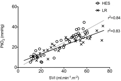
The alterations of the different structural components considered as having a central role in the regulation microcapillary blood flow and vascular barrier permeability were investigated in the following chapters. Plasma viscosity is among one of the major shear stress-induced modulator of functional capillary density. In chapter 4, the impacts of 2 strategies of fluid resuscitation following hemorrhagic shock in pigs, on the plasma viscosity were evaluated. In this study, we demonstrated that plasma viscosity decreased regardless of the strategy used for fluid resuscitation (low volume and hypertonic colloids versus lactated ringer’s). Moreover, no differences were found between the groups for other rheological variables, microcirculatory flow or tissue oxygen tension. In chapter 5 and chapter 6, we undertook a physiological integrative assessment of the contributive role of the glycocalyx to vascular barrier permeability in a rodent model following hemorrhagic shock resuscitation and severe hemodilution, respectively. The observed results challenged the contributive importance of glycocalyx to the vascular barrier permeability. Indeed, although the glycocalyx may be damaged in these settings, it did not impair vascular barrier competence but rather caused microcirculatory disturbances and vascular tone alterations probably through diminished mechanotransduction. Considered together these results stress the complexity of vascular barrier permeability regulation in the context of hemorrhagic shock and identify a gradation of vascular injury.
To accurately determine the microcirculatory oxygen pressure within the cortical renal structure, a new LED-based device relying on time-resolved phosphorescence quenching method was developed and presented in chapter 7. This device has several advantages over previous flash lamps-based devices, which had a plasma glow that persists for tens of microseconds after the primary discharge. The benefits of using LEDs as a light excitation source are presented.
Various treatments have been considered to treat injured or compromised microcirculation due to a critical condition. Fluid therapy remains cornerstone of the management of shock states. Studies on comparative microcirculatory effects with regards to renal microcirculatory oxygen pressure are limited. In chapter 8, we investigated the role of bicarbonate precursors in balanced fluids, used in everyday practice, during hemorrhagic shock with and without compromised liver function A 70% partial liver resection was performed to mimic compromised liver function in a rat model. While the balanced fluids are widely used, no data was available on the fate of anions composing them. Interestingly, acetate-buffered balanced fluids showed superior buffering effects with improve compared with Ringer’s lactate or saline, and that gluconate did not contribute to acid–base control.
Antioxidant therapies have been largely evaluated as microcirculatory resuscitation strategies. A large body of evidence is available regarding N-acetylcysteine (NAC) use and its antioxidant, vasodilatory and renal protective effects. In chapter 9, we studied the effect of NAC administration in conjunction with fluid resuscitation on renal oxygenation and function in a rat model of endotoxemia, hypothesizing that dampening inflammation within the kidney would improve the renal microcirculatory oxygenation and limit the onset of acute kidney injury. We showed that the addition of NAC improved renal oxygenation and attenuated microvascular dysfunction and acute kidney injury. The effects may be partially mediated by a decrease in renal nitric oxide and hyaluronic acid levels.
In the various models investigated, the renal microcirculatory oxygen pressure cannot be fully restored compared to control. Thus, there are still unmet needs regarding the resuscitation of the microcirculation. The so-called concept of “pink fluid resuscitation” emerged based on therapies that should combine a volume expander effect with antioxidant and oxygen carrying capacities. In this idea, new generation of hemoglobin-based oxygen carrier with additional antioxidant properties, such as PEGylated carboxyhemoglobin, is arguably a valuable option to investigate. In chapter 10, we investigated the effectiveness of this latter compound on the renal cortical microcirculatory oxygen pressure and the role of its embedded carbon monoxide
1
molecules on the mitigation of acute kidney injury during low volume resuscitation ofhemorrhagic shock. Although, it could not compete against blood transfusion in terms of renal cortical oxygenation, the compound limited tubular damage while a lower volume was required to sustain macrocirculation. In chapter 11, on the contrary, resuscitation with PEGylated carboxyhemoglobin enhanced renal cortical oxygenation while restoring skeletal muscle microcirculatory flow in previously non-flowing capillaries in an endotoxemia murine model without dampening short term sepsis-induced acute kidney injury.
2
T
HE MACRO
-
AND
MICROCIRCULATION OF THE
KIDNEY
Philippe Guerci,1,2,3 Bulent Ergin,3,4 and Can Ince3,4
1. Department of Anesthesiology and Critical Care Medicine, University Hospital of Nancy, France
2. INSERM U1116, University of Lorraine, Vandoeuvre-Les-Nancy, France 3. Department of Translational Physiology, Academic Medical Centre,
Amsterdam, The Netherlands
4. Department of Intensive Care Medicine, Erasmus MC, University Medical Centre, Rotterdam, The Netherlands
ABSTRACT
Acute kidney injury (AKI) remains one of the main causes of morbidity and mortality in the intensive care medicine today. Its pathophysiology and progress to chronic kidney disease is still under investigation. In addition, the lack of techniques to adequately monitor renal function and microcirculation at the bedside makes its therapeutic resolution challenging. In this article we review current concepts relating to renal hemodynamics compromise as being the event underlying AKI. In doing so, we discuss the physiology of the renal circulation as well as the effects of alterations in systemic hemodynamics leading the way to renal injury specifically in the context of reperfusion injury and sepsis. The ultimate key culprit of AKI leading to failure is the dysfunction of the renal microcirculation. The cellular and subcellular components of the renal microcirculation are discussed and how their injury contribute to AKI.
Keywords: Acute kidney injury, microcirculation, hemodynamic coherence, renal tissue oxygenation, renal blood flow
2
Introduction
Acute kidney injury (AKI) is among the most common organ failure observed in patients
admitted or hospitalized in intensive care units.1 The AKI morbidity and mortality remains high
despite early identification and increasing ability to support organ function.1,2 As opposed to
previously thought, the occurrence of AKI in these patients not only increase their mortality rates, but they will sooner progress towards chronic kidney disease even after apparent fully
recovery of the kidney function after the insult.3,4 The pathophysiological mechanisms behind
AKI in critically ill patients is still under investigation but it is now quite clear that it is much more complex than a simple decrease in renal blood flow subsequent to low cardiac output and/or
hypotension.5
The hallmark feature of AKI is a reduction in glomerular filtration rate (GFR) which implies an underlying impairment in hemodynamic regulation in the kidney. However, this hemodynamic “dysregulation” may occur at both, macro- and microcirculatory levels. In this review, we focus on the role of the macro- and microcirculatory changes in the pathophysiology of AKI. We discuss the respective role of renal blood flow (RBF) and the microcirculation and its interactions with inflammation.
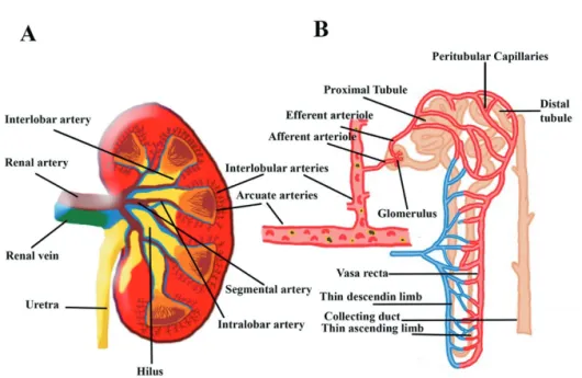
The macrocirculation of the kidney
The kidney is the most vascularized organ of the body. The arteriolar system is rich, tightly regulated and supplies 2 different types of microcirculation in the cortex and in the medulla,
each one exerting different functions.6 The kidneys receive about 20 to 25% of the cardiac
output, which in adults is equivalent to 1000 to 1200 ml of blood per minute. Twenty percent of the plasma flow – approximately 120 to 140 ml/minute – is filtered at the glomerulus in the cortex and passes into the Bowman capsule. The remaining 80% (about 480 ml/minute) of plasma flows through the efferent arterioles to the peritubular capillaries in the medulla to ensure solute exchange and water resorption. It is estimated that only 5–15% of total RBF is directed to the medulla, with the outer medulla having higher blood flow (130–340 mL/100 g
tissue/min) than the inner medulla (22–69 mL/100 g tissue/min).7,8
In the kidney, a local mechanism of autoregulation keeps fairly constant the glomerular perfusion and therefore the GFR over a wide range of arterial pressures between 80 to 180
mmHg. Intravascular volume and mean arterial pressure vary along the day as a result of physiological adaptation to the environment without affecting much the GFR. Pressure in the glomeruli will remain stable by the means of the activation of various regulatory process. The RBF is regulated by intrinsic autoregulatory mechanisms, neural regulation such as the renal sympathetic nerve activation (RSNA), and hormonal regulation such as the renin-angiotensin-aldosterone system (RAAS).
Autoregulation of renal blood flow in physiology
The vascular tone of afferent and efferent arterioles of the glomeruli is able to change to preserve GFR and to prevent fluctuations in systemic arterial pressure from being transmitted to glomerular capillaries, reducing the alterations in solute and water excretion.
Several components are involved in this autoregulatory response in the kidney. The myogenic response is one of the primary and most responsive mechanisms. This mechanism mainly concerns the afferent artery and is driven by the stretch on arteriolar smooth muscle. When arterial pressure increases, the stretch also increases, and the arteriole constricts causing a decrease in glomerular perfusion. Conversely, when the arterial pressure drops, the stretch is less important, and the arteriole relaxes to increase glomerular pressure. It has been shown that the afferent arteriole exhibits a very short delay in activation (200-300 ms) and rapid constriction kinetics when exposed to a sudden elevation of blood pressure and the renal vasculature responds passively to pressure signals presented at rates exceeding the myogenic
operating frequency (0.2-0.3 Hz).9
The tubuloglomerular feedback (TGF) is the second mechanism involved for keeping GFR constant. Briefly, the macula densa cells in the distal tubule sense the increasing or decreasing amounts of filtered sodium. When the sodium filtration decreases for instance, the macula densa cells drive afferent arteriolar vasodilation in the glomeruli to increase GFR. The vascular tone changes are also themselves under influence of nitric oxide release, adenosine triphosphate and prostaglandin E2 mediators.
Renal Sympathetic Nerve Activation (RSNA)
The diameter of arterioles of the kidney is also regulated by the sympathetic system (adrenergic/noradrenergic fibers). Both afferent and efferent arterioles are richly innervated. The RSNA is mediated through feedback from systemic baroreceptor such as from the carotid
2
artery or aortic arch. When the baroreflex is activated, a vasoconstriction of the afferentarterioles through activation of α1-adrenoreceptors occurs. Subsequent decrease in GFR will
lead to a decreased water and salt excretion boosting the increase in blood volume. Thus, blood pressure will be increased. Electrical stimulation of the renal nerves has been shown to de-
crease single nephron GFR.10,11
Other physiological or pathophysiological conditions can trigger the RSNA such as exercise or hypoxia. Severe hypoxia, apart from stimulating hypoxia inducible factor and erythropoietin production, mediates the activation of chemoreceptors of the carotid arteries and decreases
RBF by means of RSNA.12
Renin-Angiotensin-Aldosterone System (RAAS)
The RAAS is a major hormonal regulator of RBF by increasing sodium reabsorption and
causing a subsequent rise in arterial blood pressure. The TGF is also activated by the RAAS.13
Increased levels of angiotensin-2 (Ang-2) leads to a reduction in GFR by causing vasoconstriction of the afferent and efferent arterioles in the glomerulus. The activation of TGF then decreases hydrostatic pressure in the glomerulus and reduces the GFR.
Renin is an enzyme formed and stored in granular cells of the afferent arterioles of the juxta-glomerular apparatus. A decreased blood pressure in the afferent arterioles, a RSNA with stimulation of β-adrenergic receptors on the juxtaglomerular cells, a decreased sodium concentration in the distal convoluted tubule, and a release of prostaglandins are considered triggers for the release of renin. The renin mediates angiotensin-1 (Ang-1) and in the presence of the angiotensin converting enzyme (ACE), Ang-1 converts to Ang-2 to stimulate aldosterone (in the adrenal cortex). Aldosterone causes an increase in sodium reabsorption in the collecting ducts, loop of Henle and in the distal tubules and also stimulates the anti-diuretic hormone.
The renal blood flow and the GFR are tightly regulated parameters with numerous, complex, and redundant physiological systems to ensure a relative independency of the GFR to the local and systemic environment. Of course, these systems can be overwhelmed, leading to a dysregulated TGF and inappropriate secretions and may contribute to the occurrence of AKI.
What happens with the macrocirculation during AKI?
Although we extended the overall understanding of regulation of renal hemodynamics in physiology, little is known of the interactions between systemic, renal and glomerular
hemodynamics and the renal microcirculation during AKI. The reduction in GFR is the hallmark of AKI. It is unclear whether autoregulation mechanisms are still functioning and efficient or if
the RBF and GFR become more dependent on the cardiac output.14–16 In a population of critically
ill oliguric patients, although the majority were not cardiac responder to fluid challenge, half of
them responded in terms of urine output.14 Urine sodium concentration was shown to be a
poor predictor of fluid responsiveness, translating the variable degree of RAAS activation.14
Consequently, hemodynamic interventions such as fluid loading or vasopressor infusion aimed at restoring systemic hemodynamics (and RBF) in the hope of limiting insults to the kidney or
improving recovery from AKI, is not tailored to the kidney.15 Traditionally, global renal ischemia
secondary to compromised renal perfusion has been advocated to be the common factor leading to development of AKI in ICU patients.
During AKI, the observed changes in macrocirculation of the kidney are largely heterogeneous among experimental models of shock depending on the type of insult (ischemia/reperfusion), the animal considered, the time course of the study and time of analysis, and the design of the study itself. In addition, human clinical studies – in apparent homogeneous population – showed inconsistent results in terms of RBF. To date, the evolution of RBF is largely unpredictable because of the interplay of numerous regulators aforementioned that may or may not be significantly altered.
The techniques of investigation of RBF also differ in experimental, where a transonic Doppler flow probe in easily placed around the renal artery (with adjusted diameter to the artery), and in human clinical practice, where noninvasive surrogates such as ultrasound-based renal blood flow velocity or Contrast Enhanced Ultrasonography (CEUS), are mostly used at bedside.
Numerous experimental studies focused on the evolution of RBF during sepsis (sepsis-associated AKI) or after renal ischemia-reperfusion secondary to surgery or hemorrhage/hypovolemia. Apart from the very typical situations, in some selected ICU patients presenting arterial or venous thrombosis for instance, in which the RBF is totally interrupted (thrombosis for instance), the RBF can be either decreased, unchanged or increased depending on the type of insult and time of observation.
Renal macrocirculation during sepsis-associated AKI
The total RBF during sepsis-associated AKI has been largely studied in both experimental and clinical settings and yielded to inconsistent results. The common accepted theory is that
2
sepsis causes a decrease in cardiac output and a drop-in blood pressure leading to a decreasein total RBF, with subsequent renal arteriolar vasoconstriction and hypoperfusion. Secondary
renal ischemia is believed to occur with acute tubular necrosis and loss of renal function.17
However, little, if any, human evidence supports this hypothesis because RBF measurement in
the septic critically ill patient is scarce.18 Actually, animal studies have confirmed that
sepsis-associated AKI can develop despite no change or even an increase in RBF.19 The early phase of
sepsis is generally “hyperdynamic”, meaning that cardiac output is typically increased whereas
AKI is already developing or established.16,20,21 In post-mortem studies, inconsistencies in
histopathological findings were noted.22,23 In a sheep model of septic shock induced by
intravenous infusion of Escherichia coli, was not associated with changes in RBF, oxygen delivery,
nor histological appearance although animals developed AKI.24 Systematic reviews of
experimental and clinical histopathological findings in sepsis-associated AKI revealed mostly
nonspecific morphologic changes and acute tubular necrosis was relatively uncommon.25,26
Therefore, these observations challenge the previous paradigm of global-hypoperfusion secondary to decrease in RBF as the primary cause of sepsis-associated AKI and lead to a new
understanding of the pathogenesis of AKI.27 In this regard, because RBF is preserved while GFR
may be lost, the concept of hemodynamic coherence emerged and explains why the
microcirculation is disassociated from the macrocirculation.28
Large multicenter randomized clinical trials failed to demonstrate a benefit of early
goal-directed therapy for reducing mortality and AKI occurrence in patients with septic shock.29–31 In
other words, hemodynamic optimization with aggressive fluid resuscitation and vasopressors administration for early achievement of the recommended targets are of no benefit to prevent or hasten recovery from AKI in this population. A restored RBF – if altered – does not warrant
per se a recovery of renal function and GFR. On the contrary, fluid resuscitation may promote
intrarenal shunting and heterogeneity with a decreased capillary density and enhance intrarenal
medullary hypoxia.27,28,32 Two pitfalls of these large trials should be however considered. First,
patients were in established septic shock, in whom AKI was already present. Secondly, the target of mean arterial pressure in not tailored to patient’s characteristics, thus, far from the
personalized hemodynamic management.33 A large trial targeting a high versus low
blood-pressure in patients with septic shock, resulted in similar mortality at 28 and 90 days.34 However,
a subgroup of patients with chronic hypertension required less renal replacement therapy when treated in high blood-pressure management. This might be the first step of a tailored systemic hemodynamics approach regarding the kidney function.
AKI in hypovolemia or ischemia/reperfusion injury
In the setting of hypovolemia or ischemia/reperfusion injury, it is simpler to count for the changes in RBF. The decrease in RBF follows cardiac output in hypovolemia. During ischemia, the RBF is nearly interrupted, therefore blood and oxygen supply to the kidney is shut down. Changes also occurs in microcirculation after the RBF is re-established, but the magnitude of microcirculatory alterations and related inflammation are dependent on the length of the insult.
The renal microcirculation
The renal microcirculation is complex and plays a major role in the oxygen supply to the kidney and ensures plasma filtration, electrolyte exchange and water reabsorption. Renal microcirculation involves two capillary systems: the glomerular capillary system and peritubular capillary system. The glomerular capillary system is found within the glomerulus that responsible for the glomerular filtration (GFR) which is driven by the filtration pressure (within the Bowman’s capsule and hydrostatic pressure). The glomerular capillary system ends with the efferent arteriole. The peritubular capillary bed emerges from this arteriole and surrounds all the tubules. In the medulla, it becomes the vasa recta in order to create the counter-current exchange system along with the parallel positioned Henle loop. The role of counter-current exchange system is to maintain the cortico-medullary osmotic gradient for concentrating the
2
Figure 1: Representative image of the renal preglomerular vessels (Panel A), glomerular vessels and tubules (Panel B).
Renal tissue oxygenation
To achieve all its complex functions, the kidney needs an adequate amount of the oxygen to maintain oxygen-dependent adenosine triphosphate (ATP) production. Both oxygen delivery
(DO2) and consumption (VO2) determine renal tissue oxygenation, which is not only necessary
for cell survival and function but also function of Na/K ATPase pumps. There are two
mechanisms to maintain normal renal function in case of renal hypoxia; (i) the increment of DO2
acts to increase tissue oxygenation, (ii) reduced VO2 that may protect tissues from hypoxia
during moderate to severe arterial hypoxemia. Evans et al., demonstrated that even 55%
reduction of arterial oxygen content did not lead to significant impairment of VO2 because of
the adaptive reduction of oxygen demand following the decrease in tubular sodium
reabsorption.8 Thus, prevailing experimental evidence supports the concept that renal VO
2 is
Under normal conditions, oxygen partial pressure measured by oxygen-dependent phosphorescence quenching methods was found to be 50-80 mmHg and 20-45 mmHg in the
rat kidney cortex and medulla respectively.8,36 Thus, there is a gradient in oxygen concentration
in parallel to the countercurrent exchange system. The largest part of renal oxygen consumption
is used to drive Na/K ATPase in the proximal tubules and medullary thick ascending limp.37
Various clinical conditions may lead to decreased renal oxygenation, such as
hemodilution,38 sepsis induced microcirculatory heterogeneity,39 mismatch between oxygen
consumption and delivery, hypoxemia or severe hypotension.8,40 A protective mechanism that
maintains the global oxygenation homeostasis and functions as an anti-oxidant defense mechanism through arterial-to-venous (AV) oxygen shunting in the renal cortex and shunting of
oxygen from descending to ascending vasa recta in the renal medulla exists.41,42
The renal endothelium
The endothelium of the kidney is remarkably heterogeneous in structure and function and
varies according to the type of microcirculation (glomerular and peritubular).6,7 Indeed, various
endothelial cells arrangements grant different permeability properties. In the glomeruli, the capillaries are lined by fenestrated endothelial cells and covered by specialized epithelial cells known as podocytes. In the cortex, the endothelium is exposed to almost normal oxygen partial
pressure (PO2) and osmolarity whereas the one in the medullar microcirculation functions in an
osmolarity of up to 1,200 mosmol.L-1 and a PO
2 as low as 20 mmHg. Glomerular endothelial
cells are unusually thin; around capillary loops, and present large fenestrated areas constituting 20–50% of the entire endothelial surface. Glomerular microcirculation functions via continuous and fenestrated endothelium with no diaphragm, whereas it is more continuous and nonfenestrated in the descending vasa recta in peritubular microcirculation. Because of their different role in the microcirculation, these endothelial cells are dissimilarly sensitive to injury.
The glycocalyx
Endothelial cells are lined by a gel-like thin layer, the glycocalyx which comprises glycoproteins, glycosylated glycoproteins and proteoglycans. The glycocalyx contributes to vascular barrier permeability however it is not clear to what extent. This highly interactive scaffold is capable of sensing blood flow and facilitating protein interactions with their receptors
2
or other proteins, and it houses many EC receptors and compounds important for maintaininghemostasis (antithrombotic properties), homeostasis and providing anti-inflammatory defense to the parenchymal cells.
Interplay of the microcirculation with local mediators
Hundreds of mediators interact with the microcirculation in the kidney and it is almost impossible to list their detailed effects in physiology and even more complex in the pathophysiology of AKI. We present here the relevant mediators interacting directly with the vasculature.
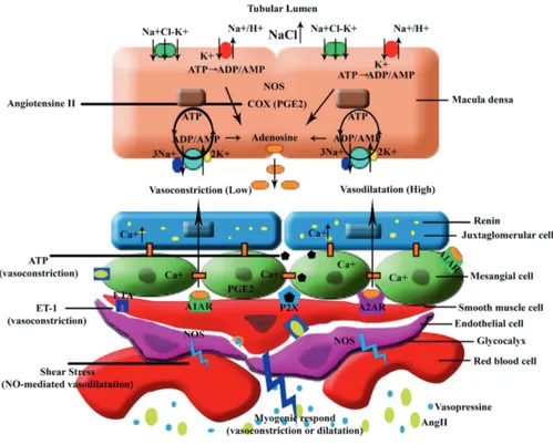
Adenosine
Adenosine is an ATP breakdown product causing vasodilatation in most vessels and contributes to the metabolic control of kidney perfusion to provide a balance between the oxygen demand and delivery. It was originally postulated that adenosine is generated in macula densa cells by dephosphorylation of ATP when an increase in transcellular NaCl transport and
the cellular energy demand.43 In the renal vasculature, adenosine can produce vasoconstriction
in afferent arteriole, especially arteriolar segment the closest the glomerulus, through A1 adenosine receptor (A1AR) at lower concertation. On the contrary, it accompanies with a renal vasodilator response as a result of A2AR-mediated vasorelaxation in most parts of the renal vasculature, including larger renal arteries, juxtamedullary afferent arterioles, efferent arterioles,
and medullary vessels with the plasma concentration above normal.44 However, because the
kidney vasculature is sufficiently heterogeneous, while adenosine causes vasoconstriction in one part of the renal vascular bed and vasodilatation in another, it is difficult to account for the real changes induced by adenosine in the renal vasculature. Recent studies confirmed the biphasic effects of adenosine exerts vasoconstriction via A1 receptors (A1AR) at a lower concentration and vasodilation via A2AR at higher concentration of adenosine. Lately, it has been reported that A1A3 receptor is expressed in the afferent arteriole and activation of this receptor leads to dilate the afferent arteriole.
ATP
Extracellular ATP activates a variety of purinergic receptors – ionotropic P2X receptors and metabotropic G-protein coupled P2Y receptors – since both receptor families are present in
renal vascular, glomerular, mesangial and tubular epithelial cells.45 Intrarenal infusion of α, ß,
𝛾-methylene ATP reduced both cortical and medullary blood flow in rabbit kidneys in vivo,46 which
implicates P2X receptors in the regulation of regional renal blood flow. Intra-arterial infusion of ATP or α, ß-methylene ATP increased perfusion pressure in a dose dependent fashion in isolated
rat kidneys and increase in renal vascular resistance.47
Endothelin
Endothelin (ET-1,2,3) is one of the most potent renal vasoconstrictors. Endothelin plays an essential role in the regulation of renal blood flow, glomerular filtration, sodium and water transport, and acid-base balance. ET production by the kidney is much higher than any other
organ in especially inner medulla.48 The high ET-1 expression levels in the renal inner medulla
suggest a potentially important role for ET-1 in regulating sodium and water balance under
physiological conditions and perhaps in regulating medullary blood flow.49 The physiological or
pathophysiological effects of ET are mediated through activation of two ET receptors as ETA and
ETB.48 Moreover, Gellai et al. reported that the relative expression of ETA and ETB in the rat
kidney was equal in cortex but expression (ratio) of ETB was found much higher in medulla
(70:30).50 ET-1 causes a marked and prolonged renal vasoconstriction manifested by profound
reductions in renal blood flow and glomerular filtration rate.51,52 ET-1 leads to vasoconstriction
in both afferent and efferent arterioles, however which segment exhibits the greatest sensitivity to ET-1 remains controversial. In isolated and perfused afferent and efferent arterioles from ETB deficient mice ET-1 induced a greater vasoconstriction of afferent arterioles than efferent arterioles (Figure 2).
2
Figure 2: Mechanism of the tubule-glomerular feedback and regulation of glomerular microcirculation through the vasoactive substances.
Ang-II: Angiotensin-II, ATP: adenosine triphosphate, ADP: adenosine diphosphate, AMP: adenosine monophosphate, ET: endothelin, A1AR/A2AR: A1/A2 Adenosine Receptor, COX: cyclo-oxygenase, NO: Nitric oxide, NOS: Nitric Oxide Synthase, P2X: purinergic receptor, PGE 2: Prostaglandin E2
The study also showed that the vasoconstriction caused by ET-1 in the afferent arterioles is due primarily to ETA, while ET-1-mediated vasoconstriction of the efferent arteriole requires both ETA and ETB; the efferent arteriole response is influenced by ETB-mediated release of nitric
oxide.53 In addition, the intense vasoconstriction exhibited by descending vasa recta in response
to ET-1 suggests that ET-1 might play an important role in the development of renal injury in conditions where the renal ET system is upregulated, such as ischemia-reperfusion-induced
Nitric oxide (NO)
Nitric oxide (NO) is a gas with biological and regulatory properties, produced from arginine by the way of nitric oxide synthase (NOS). It has a very short half-life (few seconds). NO involves
extensive metabolic, vascular and cellular effects,54 with a deep involvement in the renal
function. NO exerts its vasodilating effect through activation of the soluble guanylate cyclase and the synthesis of 3’,5’-cyclic guanosine monophosphate (cGMP), the final effector of vasodilatation. NO is currently considered an important determinant of vascular integrity through the regulation of many biological effects, such as vascular tone and permeability, leukocytes-endothelial interaction, cell proliferation, maintenance of the antithrombotic properties of the endothelium, neurotransmission, gene transcription, mRNA translation, and
posttranslational modifications of proteins.55 The endothelial NOS (eNOS) isoform is strongly
expressed in the renal vascular endothelium and in the tubules.56 Whereas neuronal NOS
(nNOS) is highly expressed in the macula densa. In contrast, inducible NOS (iNOS) is weakly
expressed in renal tissues under normal conditions.56,57 NO dilates both the afferent and the
efferent arterioles, increases the glomerular filtration rate (GFR), and affects renal sodium handling in the tubules, from the thick ascending limb to the distal tubule and the collecting duct (Figure 2).
The renal microcirculation: a cornerstone in the
pathophysiology of AKI
All the mechanisms aforementioned play a crucial role to maintain the regulation of renal microcirculation that finally provides adequate oxygen, nutrient, immune cell, hormones and effective vast removal for renal function. Different type of insults may result in similar
mechanisms taking part to subsequent development of AKI.26,58,59 The two main triggers are: (i)
renal tissue hypoxia and (ii) activation of inflammatory pathways. These phenomena are present in either ischemia/reperfusion injury, hypoxemia or septic shock and lead to microcirculatory
dysfunction. Different types of microcirculatory dysfunction have been identified.28,60
However, the common pattern observed is a decrease in functional capillary density. In sepsis-associated AKI, the microcirculatory dysfunction is characterized by heterogeneous abnormalities, in which some capillaries are under-perfused (sluggish or stopped flow/plugged
2
vessels) while others have normal or high blood flow (hyperdynamic).61 In a septic sheep model,
while the cortical perfusion and oxygenation were preserved, renal medullary hypoxia caused by intrarenal blood flow redistribution may contribute to the development of septic acute kidney
injury.62 In addition, the authors found that treatment with norepinephrine led to further
reduction of the medullary perfusion and oxygenation with a sustained renal and systemic
hemodynamics.62 In dogs, Gullichsen et al. also reported that endotoxemia is associated with
renal hypoperfusion and hypoxia in the renal cortex despite an increase in venous PO2.63 Thus,
it has been suggested that oxygen shunts may also contribute to kidney hypoxia during sepsis. Besides the systemic hypotension, the increase in renal vascular resistance in sepsis was associated with global and regional blood flow in kidney impairment and a decrease in renal
cortical and medullary microvascular oxygenation.64,65 Johannes et al. demonstrated that
sepsis-induced AKI was associated with renal cortical and medullar perfusion heterogeneity and
overexpression of iNOS.66 Increase in renal vascular resistance which is suggested an important
macrohemodynamic factor to induce AKI, can be induced by arteriolar vasoconstriction, microcirculatory disturbances such as a leaky endothelium, tissue edema, leukocyte adhesion
and microthrombosis.67 Finally, in a rat model of endotoxemia, Legrand et al. demonstrated that
early fluid resuscitation partially improved intrarenal perfusion besides reducing renal
inflammation but could not prevent reduced renal microvascular oxygenation.68
In sepsis, activation of the RAAS is often part of host response with high levels of Ang II and vasopressin. Although it has been suggested that afferent arteriole dilatation cause a loss of
intraglomerular filtration pressure resulting a decrease in GFR,69 local production of Ang II was
found to lead to a reduction in the GFR due to vasoconstriction of the glomerular afferent and efferent arterioles.
Microvascular dysfunction also involves imbalance between the endothelium-dependent vasorelaxation and overresponses to vasoconstrictive agents. NO plays an important role to regulate the vascular tone and is a major contributor to endothelial dysfunction. For instance, lack of the eNOS activity is directly related to the organ perfusion. Indeed, it was shown that while blocking of eNOS activity exacerbate organ ischemia, high eNOS expression may improve
sepsis-induced AKI.70 Moreover, Schwartz et al. pointed out that loss of the renal function is due
to local inhibition of eNOS by elevated iNOS activation, possibly via NO autoinhibition.71 Recently,
a study found that expression of all NOS subtypes is significantly increased in the renal cortex
hypothesized that overexpression of NOS isoforms in the cortex may induce a shunt from the
medulla to the cortex, leading to medullary ischemia.26
Sepsis-induced endothelial leukocyte transmigration, together with the activated coagulation system, endothelial swelling, and enhanced vasoconstriction of the arterioles, also
results in a compromised renal microcirculation.72 In this case, the upregulation of endothelial
adhesion molecules, production of cytokines, chemokines and NO gives rise to increase in endothelial-leukocyte interactions causing leukocyte transmigration toward renal interstitial area, further enhancing epithelial inflammation and vascular endothelial barrier dysfunction through the production of cytokines, reactive oxygen (ROS) and nitrogen species (RNS). During ischemic AKI, compromise of the endothelial cell–cell junctions and changes in the endothelial
glycocalyx increase microvascular permeability and contribute to microcirculatory dysfunction.73
In case of septic AKI, diabetic nephropathy, or AKI following ischemia/reperfusion injury, the
glycocalyx is disrupted, leading to vascular hyperpermeability and albuminuria.74,75
Renal microvascular hypoxia during AKI
The occurrence of hypoxia plays also a central role in the development of AKI and is directly linked to microcirculatory alterations. After shock or ischemia/reperfusion injury, persistent oxygen extraction deficit may persist despite the apparent adequate recovery of systemic
hemodynamic and oxygen-derived variables.76 Hypoxic inducible factor (HIF) is widely expressed
in renal epithelial cells, endothelial cells, and renal interstitial fibroblast-like cells and plays crucial
role in the hypoxic adaptation through the extracellular formation of adenosine.77 Adenosine
signals through binding to 1 of the 4 adenosine receptors of which are expressed by activated
immune cells 78 and nephron segments, including the glomerular epithelium, renal vasculature,
proximal tubules, and collecting ducts.79
Hemodilution or anemia, in addition to their hemorheological effects, cause a reduction in the oxygen carrier capacity of blood and in organ perfusion and oxygenation. Recently, Koning
et al. investigated the effects of hemodilution with or without cardiopulmonary bypass (CPB) in
which they concluded that despite transient impairment of microcirculation after hemodilution alone, hemodilution with CPB may cause a further impairment of renal microcirculation,
inflammation and renal injury in rat.80 Moreover, another study showed that CPB impairs renal
2
blood flow away from the kidney during and after cardiopulmonary bypass, accompanied bydecreased renal oxygen delivery, increased oxygen extraction and release of a tubular injury
marker.81 Results from experimental studies showed that acute normovolemic hemodilution
(ANH) led to a reduction of perfused vessel density,82 increased endothelial activation,83 renal
edema and impaired renal oxygenation.84 In addition, an increase in hematocrit by red blood
cell transfusions during CPB improved microcirculatory perfusion in cardiosurgical patients,85,86
It has been suggested that the renal oxygen supply of the kidney became critical already in an early stage of ANH causing an increase in intrarenal oxygen shunts that contribute to the
development of renal hypoxia.38
During the hemorrhagic shock and reperfusion are reported a reduction of systemic perfusion and local perfusion that can lead to multiple-organ failure, including AKI because of ischemia and hypoxia, dysfunction in the microcirculation, release of reactive oxygen species,
and endogenous apoptotic pathways associated with mitochondrial disorders.58,87 In a review
Kang et al. highlighted the direct correlation between renal hypoxia and the loss of microvasculature with the in stimulation of fibrogenesis and development of glomerular and
tubulointestitial scarring.88 The central role of renal microcirculatory hypoxia to AKI was shown
by Zafrani and co-workers in a sepsis model in rats where they demonstrated that blood transfusions was successful in improving kidney microcirculatory oxygen levels and in resolving
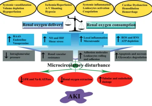
Figure 3: Key mechanisms involved in the development of acute kidney injury (AKI)
GFR: Glomerular Filtration Rate, HIF: Hypoxia Inducible Factor, NO: Nitric oxide, ROS: Reactive Oxygen Species, RAAS: Renin Angiotensin Aldosterone System
Conclusion
The relative contribution of total renal blood flow in the pathophysiology of AKI remains to be determined. A unified theory for the development of sepsis-associated AKI has been recently
proposed.90 Although, the pathophysiological aspects of AKI following ischemia/reperfusion
injury are different, it shares common pathways with the sepsis-associated AKI 58 in which the
alterations in microcirculation, renal endothelium/inflammation and renal oxygenation are the critical determinants of the severity of AKI (Figure 3). Therefore, future targeted therapies aiming at preventing or hastening recovery from injured kidney should take into consideration these 3 main aspects in the resuscitation process.
2
Practice points
• An integrative evaluation of the functional state of the circulation is required to assess the
risk for developing AKI and assessing the success of therapy.
• Fluid therapy inappropriately used may contribute to the development of AKI.
• Tissue hypoxia and renal microcirculation compromise should be regards as a key
component of the pathogenesis of AKI
• Anemia, either previously present or induced by should be regarded as a major risk factor
contributing to AKI.
• Reperfusion injury and sepsis causing renal microcirculatory injury are defining in the
Research agenda
• Innovative diagnostic techniques need to be developed to non-invasively monitor renal
hemodynamics.
• Sublingual microcirculatory alterations measured by hand-held vital microscopes should
be investigated for use as a surrogate for renal microcirculatory dysfunction.
• Contrast enhance ultrasound is a promising technique for non-invasive monitoring of the
kidney microcirculation but needs to be further developed taking into account the special properties of compromised renal hemodynamics
• Anti-inflammatory medication as a prophylactic intervention should be considered as a
method to protect the kidney at risk for injury.
Improvement of oxygen availability in the injured kidney by blood transfusions or even hemoglobin-based oxygen carriers should be investigated as a new therapeutic strategy to resuscitate the injured kidney.
Conflict of interest
Prof Can Ince is a member of the editorial board of Intensive Care Medicine Experimental and has participated to the Acute Dialysis Quality Initiative ADQI XIV workgroup. Prof. Ince has received grants and consultant fees from Fresenius Kabi, Baxter Healthcare, BBraun and AM Pharma. Dr. Philippe Guerci was supported by a grant from the French Society of Anesthesiology and Intensive Care Medicine (SFAR).
2
References
1. Hoste EAJ, Bagshaw SM, Bellomo R, et al. Epidemiology of acute kidney injury in critically
ill patients: the multinational AKI-EPI study. Intensive Care Med. 2015;41(8):1411-1423.
2. Rewa O, Bagshaw SM. Acute kidney injury-epidemiology, outcomes and economics. Nat
Rev Nephrol. 2014;10(4):193-207.
3. Heung M, Chawla LS. Acute kidney injury: gateway to chronic kidney disease. Nephron
Clin Pract. 2014;127(1-4):30-34.
4. Forni LG, Darmon M, Ostermann M, et al. Renal recovery after acute kidney injury.
Intensive Care Med. 2017;43(6):855-866.
5. Basile DP, Anderson MD, Sutton TA. Pathophysiology of Acute Kidney Injury. Compr
Physiol. 2012;2(2):1303-1353.
6. Pallone TL, Silldorff EP, Turner MR. Intrarenal blood flow: microvascular anatomy and
the regulation of medullary perfusion. Clin Exp Pharmacol Physiol. 1998;25(6):383-392.
7. Pallone TL, Edwards A, Mattson DL. Renal medullary circulation. Compr Physiol.
2012;2(1):97-140.
8. Evans RG, Ince C, Joles JA, et al. Haemodynamic influences on kidney oxygenation:
clinical implications of integrative physiology. Clin Exp Pharmacol Physiol. 2013;40(2):106-122.
9. Loutzenhiser R, Bidani A, Chilton L. Renal myogenic response: kinetic attributes and
physiological role. Circ Res. 2002;90(12):1316-1324.
10. Pelayo JC. Renal adrenergic effector mechanisms: glomerular sites for prostaglandin
interaction. Am J Physiol. 1988;254(2 Pt 2):F184-190.
11. Hermansson K, Larson M, Källskog O, Wolgast M. Influence of renal nerve activity on
arteriolar resistance, ultrafiltration dynamics and fluid reabsorption. Pflugers Arch. 1981;389(2):85-90.
12. Leonard BL, Malpas SC, Denton KM, Madden AC, Evans RG. Differential control of
intrarenal blood flow during reflex increases in sympathetic nerve activity. Am J Physiol Regul
Integr Comp Physiol. 2001;280(1):R62-68.
13. Just A. Mechanisms of renal blood flow autoregulation: dynamics and contributions. Am
J Physiol Regul Integr Comp Physiol. 2007;292(1):R1-17.
14. Legrand M, Le Cam B, Perbet S, et al. Urine sodium concentration to predict fluid
responsiveness in oliguric ICU patients: a prospective multicenter observational study. Crit Care. 2016;20(1):165.
15. Legrand M, Dupuis C, Simon C, et al. Association between systemic hemodynamics and
septic acute kidney injury in critically ill patients: a retrospective observational study. Crit Care. 2013;17(6):R278.
16. Langenberg C, Bellomo R, May C, Wan L, Egi M, Morgera S. Renal blood flow in sepsis.
Crit Care. 2005;9(4):R363-374.
17. Schrier RW, Wang W. Acute renal failure and sepsis. N Engl J Med. 2004;351(2):159-169.
18. Prowle JR, Ishikawa K, May CN, Bellomo R. Renal blood flow during acute renal failure in
19. Chvojka J, Sykora R, Krouzecky A, et al. Renal haemodynamic, microcirculatory, metabolic and histopathological responses to peritonitis-induced septic shock in pigs. Crit Care. 2008;12(6):R164.
20. Langenberg C, Wan L, Egi M, May CN, Bellomo R. Renal blood flow and function during
recovery from experimental septic acute kidney injury. Intensive Care Med. 2007;33(9):1614-1618.
21. Di Giantomasso D, May CN, Bellomo R. Vital organ blood flow during hyperdynamic
sepsis. Chest. 2003;124(3):1053-1059.
22. Lerolle N, Nochy D, Guérot E, et al. Histopathology of septic shock induced acute kidney
injury: apoptosis and leukocytic infiltration. Intensive Care Med. 2010;36(3):471-478.
23. Takasu O, Gaut JP, Watanabe E, et al. Mechanisms of cardiac and renal dysfunction in
patients dying of sepsis. Am J Respir Crit Care Med. 2013;187(5):509-517.
24. Maiden MJ, Otto S, Brealey JK, et al. Structure and Function of the Kidney in Septic
Shock. A Prospective Controlled Experimental Study. Am J Respir Crit Care Med. 2016;194(6):692-700.
25. Kosaka J, Lankadeva YR, May CN, Bellomo R. Histopathology of Septic Acute Kidney
Injury: A Systematic Review of Experimental Data. Crit Care Med. 2016;44(9):e897-903.
26. Langenberg C, Gobe G, Hood S, May CN, Bellomo R. Renal histopathology during
experimental septic acute kidney injury and recovery. Crit Care Med. 2014;42(1):e58-67.
27. Bellomo R, Kellum JA, Ronco C, et al. Acute kidney injury in sepsis. Intensive Care Med.
2017;43(6):816-828.
28. Ince C. Hemodynamic coherence and the rationale for monitoring the microcirculation.
Crit Care. 2015;19 Suppl 3:S8.
29. ProCESS Investigators, Yealy DM, Kellum JA, et al. A randomized trial of protocol-based
care for early septic shock. N Engl J Med. 2014;370(18):1683-1693.
30. Mouncey PR, Osborn TM, Power GS, et al. Trial of early, goal-directed resuscitation for
septic shock. N Engl J Med. 2015;372(14):1301-1311.
31. ARISE Investigators, ANZICS Clinical Trials Group, Peake SL, et al. Goal-directed
resuscitation for patients with early septic shock. N Engl J Med. 2014;371(16):1496-1506.
32. Legrand M, Ince C. Intravenous Fluids in AKI: A Mechanistically Guided Approach. Semin
Nephrol. 2016;36(1):53-61.
33. Saugel B, Eschermann K, Hoffmann R, et al. Stenotrophomonas maltophilia in the
respiratory tract of medical intensive care unit patients. Eur J Clin Microbiol Infect Dis. 2012;31(7):1419-1428.
34. Asfar P, Meziani F, Hamel J-F, et al. High versus low blood-pressure target in patients
with septic shock. N Engl J Med. 2014;370(17):1583-1593.
35. Pallone TL, Turner MR, Edwards A, Jamison RL. Countercurrent exchange in the renal
medulla. Am J Physiol Regul Integr Comp Physiol. 2003;284(5):R1153-1175.
36. Evans RG, Gardiner BS, Smith DW, O’Connor PM. Intrarenal oxygenation: unique
challenges and the biophysical basis of homeostasis. Am J Physiol Renal Physiol. 2008;295(5):F1259-1270.
37. Brezis M, Agmon Y, Epstein FH. Determinants of intrarenal oxygenation. I. Effects of
2
38. Johannes T, Mik EG, Nohé B, Unertl KE, Ince C. Acute decrease in renal microvascular
PO2 during acute normovolemic hemodilution. Am J Physiol Renal Physiol. 2007;292(2):F796-803.
39. Johannes T, Mik EG, Nohé B, Raat NJH, Unertl KE, Ince C. Influence of fluid resuscitation
on renal microvascular PO2 in a normotensive rat model of endotoxemia. Crit Care. 2006;10(3):R88.
40. Evans RG, Goddard D, Eppel GA, O’Connor PM. Factors that render the kidney
susceptible to tissue hypoxia in hypoxemia. Am J Physiol Regul Integr Comp Physiol. 2011;300(4):R931-940.
41. O’Connor PM, Anderson WP, Kett MM, Evans RG. Renal preglomerular arterial-venous
O2 shunting is a structural anti-oxidant defence mechanism of the renal cortex. Clin Exp
Pharmacol Physiol. 2006;33(7):637-641.
42. Zhang W, Edwards A. Oxygen transport across vasa recta in the renal medulla. Am J
Physiol Heart Circ Physiol. 2002;283(3):H1042-1055.
43. Ren Y, Garvin JL, Liu R, Carretero OA. Role of macula densa adenosine triphosphate
(ATP) in tubuloglomerular feedback. Kidney Int. 2004;66(4):1479-1485.
44. Hansen PB, Schnermann J. Vasoconstrictor and vasodilator effects of adenosine in the
kidney. Am J Physiol Renal Physiol. 2003;285(4):F590-599.
45. Bailey MA, Hillman KA, Unwin RJ. P2 receptors in the kidney. J Auton Nerv Syst.
2000;81(1-3):264-270.
46. Eppel GA, Ventura S, Evans RG. Regional vascular responses to ATP and ATP analogues
in the rabbit kidney in vivo: roles for adenosine receptors and prostanoids. Br J Pharmacol. 2006;149(5):523-531.
47. Inscho EW. P2 receptors in regulation of renal microvascular function. Am J Physiol Renal
Physiol. 2001;280(6):F927-944.
48. Guan Z, VanBeusecum JP, Inscho EW. Endothelin and the renal microcirculation. Semin
Nephrol. 2015;35(2):145-155.
49. Kohan DE, Inscho EW, Wesson D, Pollock DM. Physiology of endothelin and the kidney.
Compr Physiol. 2011;1(2):883-919.
50. Gellai M, DeWolf R, Pullen M, Nambi P. Distribution and functional role of renal ET
receptor subtypes in normotensive and hypertensive rats. Kidney Int. 1994;46(5):1287-1294.
51. Evans RG, Madden AC, Oliver JJ, Lewis TV. Effects of ET(A) - and ET(B)-receptor
antagonists on regional kidney blood flow, and responses to intravenous endothelin-1, in anaesthetized rabbits. J Hypertens. 2001;19(10):1789-1799.
52. Pollock DM, Opgenorth TJ. Evidence for endothelin-induced renal vasoconstriction
independent of ETA receptor activation. Am J Physiol. 1993;264(1 Pt 2):R222-226.
53. Schildroth J, Rettig-Zimmermann J, Kalk P, et al. Endothelin type A and B receptors in
the control of afferent and efferent arterioles in mice. Nephrol Dial Transplant. 2011;26(3):779-789.
54. Moncada S, Higgs A. The L-arginine-nitric oxide pathway. N Engl J Med.
1993;329(27):2002-2012.
55. Majid DS, Navar LG. Nitric oxide in the control of renal hemodynamics and excretory
56. Tessari P. Nitric oxide in the normal kidney and in patients with diabetic nephropathy.
J Nephrol. 2015;28(3):257-268.
57. Mount PF, Power DA. Nitric oxide in the kidney: functions and regulation of synthesis.
Acta Physiol (Oxf). 2006;187(4):433-446.
58. Bonventre JV, Yang L. Cellular pathophysiology of ischemic acute kidney injury. J Clin
Invest. 2011;121(11):4210-4221.
59. Basile DP. The endothelial cell in ischemic acute kidney injury: implications for acute
and chronic function. Kidney Int. 2007;72(2):151-156.
60. Arnemann P, Seidel L, Ertmer C. Haemodynamic coherence - The relevance of fluid
therapy. Best Pract Res Clin Anaesthesiol. 2016;30(4):419-427.
61. Le Dorze M, Legrand M, Payen D, Ince C. The role of the microcirculation in acute kidney
injury. Curr Opin Crit Care. 2009;15(6):503-508.
62. Lankadeva YR, Kosaka J, Evans RG, Bailey SR, Bellomo R, May CN. Intrarenal and urinary
oxygenation during norepinephrine resuscitation in ovine septic acute kidney injury. Kidney Int. 2016;90(1):100-108.
63. Gullichsen E, Nelimarkka O, Halkola L, Niinikoski J. Renal oxygenation in endotoxin
shock in dogs. Crit Care Med. 1989;17(6):547-550.
64. Ergin B, Zafrani L, Kandil A, et al. Fully Balanced Fluids do not Improve Microvascular
Oxygenation, Acidosis and Renal Function in a Rat Model of Endotoxemia. Shock. 2016;46(1):83-91.
65. Ergin B, Kapucu A, Demirci-Tansel C, Ince C. The renal microcirculation in sepsis.
Nephrol Dial Transplant. 2015;30(2):169-177.
66. Johannes T, Ince C, Klingel K, Unertl KE, Mik EG. Iloprost preserves renal oxygenation
and restores kidney function in endotoxemia-related acute renal failure in the rat. Crit Care Med. 2009;37(4):1423-1432.
67. Bouglé A, Duranteau J. Pathophysiology of sepsis-induced acute kidney injury: the role
of global renal blood flow and renal vascular resistance. Contrib Nephrol. 2011;174:89-97.
68. Legrand M, Bezemer R, Kandil A, Demirci C, Payen D, Ince C. The role of renal
hypoperfusion in development of renal microcirculatory dysfunction in endotoxemic rats.
Intensive Care Med. 2011;37(9):1534-1542.
69. Verma SK, Molitoris BA. Renal endothelial injury and microvascular dysfunction in acute
kidney injury. Semin Nephrol. 2015;35(1):96-107.
70. Souza ACCP de, Volpini RA, Shimizu MH, et al. Erythropoietin prevents sepsis-related
acute kidney injury in rats by inhibiting NF-κB and upregulating endothelial nitric oxide synthase.
Am J Physiol Renal Physiol. 2012;302(8):F1045-1054.
71. Schwartz D, Mendonca M, Schwartz I, et al. Inhibition of constitutive nitric oxide
synthase (NOS) by nitric oxide generated by inducible NOS after lipopolysaccharide administration provokes renal dysfunction in rats. J Clin Invest. 1997;100(2):439-448.
72. Ince C. The microcirculation is the motor of sepsis. Crit Care. 2005;9 Suppl 4:S13-19.
73. Sutton TA, Mang HE, Campos SB, Sandoval RM, Yoder MC, Molitoris BA. Injury of the
renal microvascular endothelium alters barrier function after ischemia. Am J Physiol Renal Physiol. 2003;285(2):F191-198.
![Table 3. Hemorheological variables Hemorhreological variable BL S-30 R-0 R-60 PV (mPa.s) HES 1.32 [1.31 - 1.32] 1.30 [1.29 - 1.34] 1.21 [1.19 - 1.21] c 1.20 [1.17 - 1.21] c LR 1.36 [1.32 - 1.38] 1.30 [1.28 - 1.31] 1.20 [1.19 - 1.22]](https://thumb-eu.123doks.com/thumbv2/123doknet/14414743.512432/84.755.96.659.164.525/table-hemorheological-variables-hemorhreological-variable-bl-pv-hes.webp)
