HAL Id: hal-01899178
https://hal.sorbonne-universite.fr/hal-01899178
Submitted on 19 Oct 2018
HAL is a multi-disciplinary open access
archive for the deposit and dissemination of
sci-entific research documents, whether they are
pub-lished or not. The documents may come from
teaching and research institutions in France or
abroad, or from public or private research centers.
L’archive ouverte pluridisciplinaire HAL, est
destinée au dépôt et à la diffusion de documents
scientifiques de niveau recherche, publiés ou non,
émanant des établissements d’enseignement et de
recherche français ou étrangers, des laboratoires
publics ou privés.
Distributed under a Creative Commons Attribution| 4.0 International License
regulon in marine heterotrophic bacteria
Elizabeth Ficko-Blean, Aurelie Prechoux, François Thomas, Tatiana Rochat,
Robert Larocque, Yongtao Zhu, Mark Stam, Sabine Génicot, Murielle Jam,
Alexandra Calteau, et al.
To cite this version:
Elizabeth Ficko-Blean, Aurelie Prechoux, François Thomas, Tatiana Rochat, Robert Larocque, et al..
Carrageenan catabolism is encoded by a complex regulon in marine heterotrophic bacteria. Nature
Communications, Nature Publishing Group, 2017, 8, pp.1685. �10.1038/s41467-017-01832-6�.
�hal-01899178�
Carrageenan catabolism is encoded by a complex
regulon in marine heterotrophic bacteria
Elizabeth Ficko-Blean
1
, Aurélie Préchoux
1
, François Thomas
1
, Tatiana Rochat
2
, Robert Larocque
1
,
Yongtao Zhu
3
, Mark Stam
4
, Sabine Génicot
1
, Murielle Jam
1
, Alexandra Calteau
4
, Benjamin Viart
4
,
David Ropartz
5
, David Pérez-Pascual
2
, Gaëlle Correc
1
, Maria Matard-Mann
1
, Keith A. Stubbs
6
,
Hélène Rogniaux
5
, Alexandra Jeudy
1
, Tristan Barbeyron
1
, Claudine Médigue
4
, Mirjam Czjzek
1
,
David Vallenet
4
, Mark J. McBride
3
, Eric Duchaud
2
& Gurvan Michel
1
Macroalgae contribute substantially to primary production in coastal ecosystems. Their
biomass, mainly consisting of polysaccharides, is cycled into the environment by marine
heterotrophic bacteria using largely uncharacterized mechanisms. Here we describe the
complete catabolic pathway for carrageenans, major cell wall polysaccharides of red
mac-roalgae, in the marine heterotrophic bacterium Zobellia galactanivorans. Carrageenan
cata-bolism relies on a multifaceted carrageenan-induced regulon, including a non-canonical
polysaccharide utilization locus (PUL) and genes distal to the PUL, including a susCD-like pair.
The carrageenan utilization system is well conserved in marine Bacteroidetes but modi
fied in
other phyla of marine heterotrophic bacteria. The core system is completed by additional
functions that might be assumed by non-orthologous genes in different species. This complex
genetic structure may be the result of multiple evolutionary events including gene
duplica-tions and horizontal gene transfers. These results allow for an extension on the definition of
bacterial PUL-mediated polysaccharide digestion.
DOI: 10.1038/s41467-017-01832-6
OPEN
1Sorbonne Universités, UPMC Univ Paris 06, CNRS, UMR 8227, Integrative Biology of Marine Models, Station Biologique de Roscoff, CS 90074 Roscoff,
Bretagne, France.2VIM, INRA, Université Paris-Saclay, 78350 Jouy-en-Josas, France.3Department of Biological Sciences, University of
Wisconsin-Milwaukee, 53201 Wisconsin-Milwaukee, WI, USA.4UMR 8030, CNRS, Université Évry-Val-d’Essonne, CEA, Institut de Génomique - Genoscope, Laboratoire
d’Analyses Bioinformatiques pour la Génomique et le Métabolisme, F-91000 Évry, France.5INRA, UR1268 Biopolymers Interactions Assemblies, F-44316
Nantes, France.6School of Molecular Sciences, The University of Western Australia, Crawley, WA 6009, Australia. Elizabeth Ficko-Blean and Aurélie
Préchoux contributed equally to this work. Correspondence and requests for materials should be addressed to G.M. (email:gurvan.michel@sb-roscoff.fr)
123456789
C
arrageenans, alongside agars, are the main cell wall
poly-saccharides of red macroalgae and play vital roles in the
development and physiology of these photosynthetic
eukaryotes. These complex polymers consists of
D-galactose based
units alternatively linked by
1,4 and α-1,3 linkages. The
β-linked unit is either a
D-galactose-6-sulfate or a 3,6-anhydro-
D-galactose, a bicyclic sugar unique to red macroalgae
1.
Carragee-nan structures are further modified by the presence of various
substituents (sulfate, methyl and pyruvate groups). For more than
70 years, carrageenans have been widely used as ingredients in
food, personal care, and cosmetic industries due to their gelling
and emulsifying properties. In addition, these polysaccharides
and their derived oligosaccharides have multiple biological
properties and are promising molecules for blue biotechnology
2.
In marine ecosystems carrageenans constitute a huge biomass and
thus a precious carbon source for marine heterotrophic bacteria
(MHB). However, carrageenan catabolism is largely
unchar-acterized and only a few enzymes specific for these sulfated
galactans are known
3–7.
Bacteroidetes
are
considered
key
recyclers
of
marine
polysaccharides
8–10and notably of carrageenans
2. Bacteria from
this phylum are particularly suitable models for efficiently
char-acterizing complete carbohydrate degradation pathways. Indeed,
they have developed multi-component protein systems tailored
for sensing, binding, transporting, and degrading specific glycans,
as described for the starch-utilization system (Sus)
11,12. The genes
encoding these proteins are adjacent and co-regulated; these
regions are referred to as Polysaccharide Utilization Loci (PUL
(singular) or PULs (plural)). Tandem susD-like and susC-like
genes, which encode a carbohydrate-binding lipoprotein and a
TonB-dependent transporter (TBDT) respectively, are considered
as a hallmark of PULs
13, and the presence of these susCD-like
gene pairs is used to identify PULs in Bacteroidetes genomes
14.
Several PULs have been extensively characterized in human
intestinal Bacteroidetes
15–18, describing the complex molecular
mechanisms behind the glycoside hydrolase (GH) enzymes and
carbohydrate-binding proteins involved in the digestion of
common dietary polysaccharides. Some previous studies have
described the partial characterization of PULs targeting marine
polysaccharides, for instance, alginate-specific PULs were shown
to be genuine operons and to encode the enzymes responsible for
the bioconversion of alginate into
2-keto-3-deoxy-6-phos-phogluconate
19. The transcription of a PUL in Bacteroides
ple-beius was shown to be induced by porphyran. The PUL-encoded
enzymes responsible for the initial endolytic degradation of this
sulfated agar (family GH16 and GH86
β-porphyranases) were
biochemically and structurally characterized
20. However, such
studies, focusing on macroalgal polysaccharides, have not yet
reached the level of integrative characterization observed in the
field of PULs from human gut Bacteroidetes.
The marine Bacteroidetes Zobellia galactanivorans Dsij
T,
ori-ginally isolated from a healthy red macroalga, is a bacterial model
for the bioconversion of algal polysaccharides
10. Notably, this
bacterium can utilize kappa-carrageenan (KC), iota-carrageenan
(IC), and lambda-carrageenan as sole carbon sources and its
kappa-carrageenase (CgkA, CAZy family GH16, http://www.cazy.
org/
21) and iota-carrageenases (CgiA1, CgiA2, and CgiA3, CAZy
family GH82) have been studied
22–24.
Here we describe in Z. galactanivorans the discovery and the
integrative characterization of the complete pathway for
utiliza-tion of the kappa family carrageenans (containing 3,6-anhydro-
D-galactose units, mainly kappa-, iota- and beta-carrageenans
25),
which is carried out by a complex regulon including a dedicated
atypical PUL, lacking the susCD-like pair, and
carrageenan-induced loner genes including distal susCD-like pairs. The core of
this complex system is conserved in MHB from different phyla.
These carrageenan utilization systems appear to display a
remarkable plasticity, likely resulting from diverse evolutionary
events such as horizontal gene transfers and gene duplications.
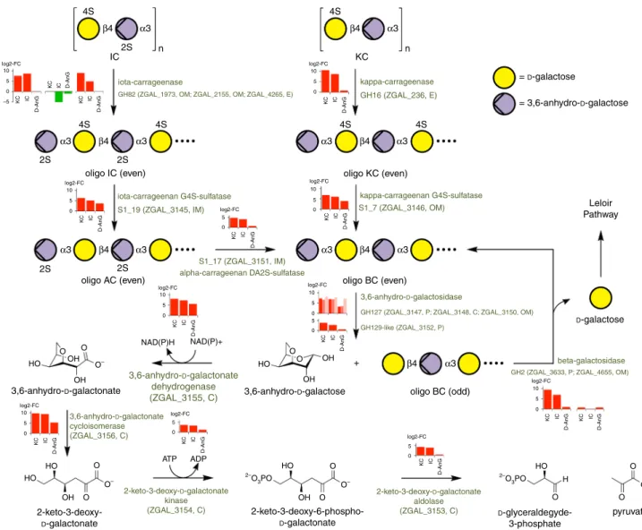
Results
The ZGAL_3145-3159 cluster encodes carrageenan-speci
fic
enzymes. The annotation of the genome sequence of Z.
galacta-nivorans
10revealed a cluster of genes encoding three GH127
enzymes (ZGAL_3147, ZGAL_3148, and ZGAL_3150), one
enzyme assigned to the GH129 family
21(ZGAL_3152), and three
sulfatases belonging to different S1 subfamilies (ZGAL_3145,
ZGAL_3146, and ZGAL_3151 belonging to the subfamilies
S1_19, S1_7, and S1_17, respectively, http://abims.sb-roscoff.fr/
sulfatlas/
26), suggesting this gene cluster could be a PUL specific
for a sulfated algal polysaccharide (Fig.
1
). This 15-gene locus
notably encodes 4 additional carbohydrate-related enzymes
(ZGAL_3153-3156), three cytoplasmic membrane transporters
and an araC family transcription factor (ZGAL_3159), but does
not include a susCD-like gene pair. The GHs have distant
homologs in enteric bacteria: BT1003 (BtGH127) from
Bacter-oides thetaiotaomicron is a 3-C-carboxy-5-deoxy-
L-xylose (aceric
acid)-hydrolase involved in rhamnogalacturonan-II
depolymer-ization
18; BlGH127 from Bifidobacterium longum is an exo-β-
L-arabinofuranosidase that acts on the plant glycoproteins
exten-sins
27; NagBb (BbGH129) from Bifidobacterium bifidum
hydro-lyzes alpha-linked N-acetyl-
D-galactosamine from intestinal
mucin
28. To the best of our knowledge L-aceric acid,
β-L-arabi-nofuranose and
α-N-acetyl-
D-galactosamine are not known
components of red algae
29, suggesting that the GHs from Z.
galactanivorans may have new substrate specificities
(Supple-mentary Discussion). To test these hypotheses, we
first cloned
and overproduced the 11 enzyme-coding genes of this cluster in
Escherichia coli BL21(DE3). The resulting recombinant proteins
were all soluble and were purified by affinity chromatography
(Supplementary Fig.
1
), allowing examination of their activities.
In vitro assays of the GH127 and GH129-like enzymes showed
no activity on poly- or oligo-saccharides of agarose (uncharged
substrate), agars, porphyrans, KC or IC (sulfated substrates).
GH129-like dagB 3152 3151 3150 3149 3148 3147 3146 3145 S1_19 cgsA S1_17 cgsC S1_7 cgsB GH127-1 dagA1 GH127-2 dagA2 GH127-3 dagA3
dauD dauC dauA dauB
AraC family regulator
cgrA DMT MFS GNTP 3159 3158 3157 3156 3155 3154 3153
Fig. 1 Z. galactanivorans PUL involved in carrageenan catabolism. Genes are annotated according to their CAZy family (GH127, GH129-like), sulfatase
subfamily (S1_19, S1_7, and S1_17) or enzymatic activity. The names of the genes and their functions are given in Table1. The acronyms for the gene names
However, the presence of sulfatases in the gene cluster suggested
that these GHs could act after the sulfatases and thus require
de-sulfated substrates. Since we had already tested agarose, we
searched for a natural source of de-sulfated carrageenans.
Furcellaria lumbricalis is a red macroalga with beta-carrageenan
(BC) in its cell wall, primarily composed of kappa-carrabiose and
uncharged beta-carrabiose motifs
30. This hybrid polysaccharide is
commonly referred to as furcellaran. No GH activity was detected
on furcellaran using
fluorophore-assisted carbohydrate
electro-phoresis (FACE)
31. In order to produce oligosaccharides,
furcellaran was treated with the kappa-carrageenase from
Pseudoalteromonas carrageenovora which cleaves the
β-1,4 bond
within kappa-carrabiose motifs in an endolytic manner
3. The
product oligosaccharides have a neutral 3,6-anhydro-
D-galactose
(D-AnG) on the non-reducing end and a
D-galactose-4-sulfate on
the reducing end. These oligosaccharides were purified by
size-exclusion chromatography and the fraction containing a majority
of hexasaccharides was tested with the GHs (Fig.
2
). All the GHs
showed activity on the furcellaran hexasaccharide, as
demon-strated using FACE. Nonetheless, there appears to be some as yet
undetermined differences in specificity between the three GH127
enzymes as the FACE gel shows different intensities and banding
patterns (Fig.
2
b). ZGAL_3150 (GH127-3) and ZGAL_3152
(GH129-like) were the most active enzymes and appear
indistinguishable biochemically based on FACE patterns (Fig.
2
c).
MALDI-TOF-MS analysis of these enzymatic digests indicated
the release of the terminal neutral monosaccharide D-AnG and of
a pentasaccharide (Fig.
2
e). Thus, these enzymes are exo-lytic
α-1,3-(3,6-anhydro)-
D-galactosidases which cleave the
α-1,3 linkage
between D-AnG and
D-galactose on the non-reducing end,
releasing D-AnG and odd-DP (degree of polymerization)
oligocarrageenans (Fig.
3
). Overall, this describes a novel
enzymatic activity, long predicted to be present in nature, but
for the
first time described here in two families of
non-homologous enzymes. The genes of these new GHs have been
named:
dagA1
(ZGAL_3147
encoding
GH127-1),
dagA2
(ZGAL_3148 encoding GH127-2), dagA3 (ZGAL_3150 encoding
GH127-3), and dagB (ZGAL_3152 encoding GH129-like).
Furthermore,
this
discovery
strongly
supports
that
the
ZGAL_3145-3159 gene cluster is carrageenan-specific.
ZGAL_3152 No enzyme
No enzyme ZGAL_3152 ZGAL_3150 ZGAL_3150 ZGAL_3148 ZGAL_3147 pFO4
negative control
a
b
c
D-AnG DP5 DP6 D-AnG DP5 DP6 D-AnG DP5 DP6d
e
f
D-AnG ZGAL_3152_E531Q ZGAL_3152_E517Q ZGAL_3152_D486N ZGAL_3152_D202N ZGAL_3152 No enzyme DP6 furcellaran Empty pFO4 vectorZGAL_3147 ZGAL_3148 ZGAL_3150 DP6 furcellaran No enzyme ZGAL_3152 1117.16 1015.22 1117.17 1015.23 973.13 871.20 871.22 973.14 1015.25 1117.19 800 900 1000 1100 m/z 973.14 871.20 1117.19 1015.25 1117.12 1015.19 † β-κ-κ - D-AnG β-κ-κ - D-AnG-SO3 Na β-κ-κ - SO3 Na β-κ-κ 973.13 1117.16 871.16 941.11 1015.22 * * † †
Fig. 2 Biochemical characterization of the GH127 and GH129-like enzymes. GH127 enzymes (DagA1, DagA2, DagA3; ZGAL_3147, ZGAL_3148,
ZGAL_3150) and GH129-like enzymes (DagB, ZGAL_3152).a FACE gel depicting the reaction products after furcellaran (beta-carrageenan)
oligosaccharides were treated with pure ZGAL_3152.b FACE gel depicting the reaction products after furcellaran oligosaccharides were treated with
soluble lysate of E. coli BL21 (DE3) that were transformed with the pFO4 vector alone (negative control) or with the genes of interest cloned into the pFO4
vectorc FACE gels depicting the reaction products after furcellaran oligosaccharides were treated with IMAC-purified ZGAL_3152 and ZGAL_3150. d FACE
gels depicting the reaction products after furcellaran oligosaccharides were treated with ZGAL_3152 and four conservative active site mutants.e MALDI
MS spectra of the DP6 beta-kappa-kappa oligosaccharide obtained in negative ionization mode after incubation with soluble lysates of BL21(DE3) cells that
were transformed with the pFO4 vector only and ZGAL_3147, ZGAL_3148, ZGAL3150.f Spectrum obtained for DP6 beta-kappa-kappa oligosaccharide
incubated with no enzyme and the spectrum of the same sample after treatment with IMAC purified ZGAL_3152. Fragments annotated with a † correspond
to a sulfate loss induced by the ionization process. Peaks annotated with a * correspond to HEPES adducts on matrix clusters. DP stands for degree of polymerization
ZGAL_3145 (S1_19), ZGAL_3146 (S1_7), and ZGAL_3151
(S1_17) were
first shown to be active sulfatases using the artificial
substrate 4-methylumbelliferyl sulfate. A combination of
anion-exchange chromatography (HPLC) and
1H-NMR was used to
determine the natural substrates and the regioselectivity of these
sulfatases (Supplementary Discussion). As predicted, all these
sulfatases were active on carrageenans (Supplementary Fig.
2
A,
B). ZGAL_3146 (gene named cgsB1) is active on kappa-carrabiose
motifs, removing the 4-linked sulfate group from
D-galactose to
generate beta-carrabiose motifs (Supplementary Fig.
2
C).
ZGAL_3145 (cgsA) removes the 4-linked sulfate group from the
galactose moiety of iota-carrabiose motifs, generating
alpha-carrabiose motifs (Supplementary Figs.
3
,
4
). ZGAL_3151 (cgsC)
acts subsequently on the alpha-carrabiose motifs, removing the
2-linked sulfate group from D-AnG to generate beta-carrabiose
motifs (Supplementary Fig.
4
). To the best of our knowledge, this
is the
first time this sulfatase activity has been demonstrated.
The original annotations of the four remaining enzymes were
not obviously connected to carrageenan:
2-dehydro-3-deoxy-6-phosphogalactonate
aldolase
(ZGAL_3153),
2-dehydro-3-deoxygalactonokinase (ZGAL_3154), lactaldehyde dehydrogenase
(ZGAL_3155), and aldonic acid dehydratase (ZGAL_3156).
However, recent discovery of two enzymes from Vibrio sp.
EJY3 that convert 3,6-anhydro-
L-galactose (agar component) into
2-dehydro-3-deoxygalactonate
32provided new insights. Indeed,
Vibrio 3,6-anhydro-
L-galactose dehydrogenase (VEJY3_09240)
and 3,6-anhydro-
L-galactonate cycloisomerase (VEJY3_09370)
are distantly related to ZGAL_3155 and ZGAL_3156 (35% and
31% sequence identity, respectively). We thus hypothesized that
ZGAL_3155 and ZGAL_3156 could catalyze similar reactions but
on the D enantiomer of 3,6-anhydrogalactose. As predicted,
ZGAL_3155 oxidizes D-AnG into 3,6-anhydro-
D-galactonate in
the presence of NAD
+and NADP
+, with a 5–6-fold preference
for NAD
+(Supplementary Fig.
5
A, B). ZGAL_3155 is inactive on
D-galactose and is therefore a specific 3,6-anhydro-
D-galactose
dehydrogenase (gene named dauA). The resulting 3,6-anhydro-
D-galactonate is converted to 2-keto-3-deoxy-
D-galactonate by
ZGAL_3156, as measured through the thiobarbituric acid
(TBA) assay (Supplementary Fig.
5
C). ZGAL_3156 did not
demonstrate any activity on D-AnG alone (Supplementary
4S O O O O O 2–O 3PO 2–O 3PO HO HO ATP ADP HO HO O O OH OH H O– O– O– HO HO O O OH OH OH OH + O O O– 2S 2S 2S 2Soligo IC (even) oligo KC (even)
oligo BC (even) Leloir Pathway = D-galactose = 3,6-anhydro-D-galactose D-galactose oligo BC (odd) 2-keto-3-deoxy-D-galactonate aldolase (ZGAL_3153, C) 2-keto-3-deoxy-D-galactonate kinase (ZGAL_3154, C) 3,6-anhydro-D-galactonate dehydrogenase (ZGAL_3155, C) S1_17 (ZGAL_3151, IM) alpha-carrageenan DA2S-sulfatase S1_19 (ZGAL_3145, IM) iota-carrageenan G4S-sulfatase S1_7 (ZGAL_3146, OM) kappa-carrageenan G4S-sulfatase kappa-carrageenase GH16 (ZGAL_236, E) iota-carrageenase
GH82 (ZGAL_1973, OM; ZGAL_2155, OM; ZGAL_4265, E)
3,6-anhydro-D-galactosidase
GH127 (ZGAL_3147, P; ZGAL_3148, C; ZGAL_3150, OM)
GH2 (ZGAL_3633, P; ZGAL_4655, OM) beta-galactosidase GH129-like (ZGAL_3152, P) 3,6-anhydro-D-galactonate cycloisomerase (ZGAL_3156, C) 3,6-anhydro-D-galactose 3,6-anhydro-D-galactonate 2-keto-3-deoxy-D-galactonate 2-keto-3-deoxy-6-phospho-D-galactonate D -glyceraldegyde-3-phosphate pyruvate NAD(P)H NAD(P)+ oligo AC (even) 4S 4S 4S 4S 4S 2S IC KC log2-FC 10 5 0 KC IC D-AnG log2-FC 10 5 –5 0 KC IC D-AnG log2-FC 10 5 0 KC IC D-AnG log2-FC 10 5 0 KC IC D-AnG log2-FC 10 5 0 KC IC D-AnG log2-FC 5 0 KC IC D-AnG log2-FC 5 0 KC IC D-AnG log2-FC 5 0 KC IC D-AnG log2-FC 10 5 5 0 0 KC IC D-AnG log2-FC 10 5 0 KC IC D-AnG log2-FC 10 5 0 KC IC D-AnG KC IC D-AnG KC IC D-AnG KC IC D-AnG n n α3 α3 α3 α3 β4 α3 β4 α3 α3 β4 α3 α3 β4 α3 β4 α3 β4 β4
Fig. 3 Kappa-carragenan and iota-carrageenan catabolic pathways in Z. galactanivorans. Enzymes performing each reaction step are mentioned, together with their encoding genes. For each step, bar plots depict the relative expression of the corresponding gene(s) in kappa-carrageenan (KC),
iota-carrageenan (IC), or 3,6-anhydro-D-galactose (D-AnG) compared toD-galactose, as measured by RNA-seq (values are mean of log2-fold change, n= 3).
Predicted enzyme localization is shown next to the gene number: OM, outer membrane; IM, inner membrane; E, extracellular; C, cytoplasmic; P, periplasm. AC stands for alpha-carrageenan and BC for beta-carrageenan. In this schematic, alpha- and beta-carrabiose motifs are considered as intermediary compounds. Nonetheless, alpha- and beta-carrageenans can be naturally found in some red algal species and the catabolic pathway described here is also valid for these natural carrageenans
Fig.
5
D) and is thus a 3,6-anhydro-
D-galactonate cycloisomerase
(dauB). In the presence of ATP, ZGAL_3154 phosphorylated the
2-keto-3-deoxy-
D-galactonate to 2-keto-3-deoxy-6-phospho-
D-galactonate. The activity of this 2-keto-3-deoxy-
D-galactonate
kinase (dauC) was indirectly measured as a function of the
oxidation
of
NADH
(Supplementary
Fig.
5
E).
Finally,
ZGAL_3153 catalyzed the conversion of
2-keto-3-deoxy-6-phospho-
D-galactonate into
D-glyceraldehyde-3-phosphate and
pyruvate. The activity of this 2-keto-3-deoxy-
D-galactonate
aldolase (dauD) was measured both in the forward and reverse
direction using the TBA assay (Supplementary Fig.
5
F, G). In
parallel, and in support of our
findings, Lee et al.
33recently
biochemically characterized homologs of these four enzymes in
other marine bacteria.
Crystal
structure
of
two
3,6-anhydro-
D-galactose-related
enzymes. Most recombinant proteins were put into crystal trials
60Å
b
c
d
e
a
40Å 60Å Trp455 Asp202 Cys198 His347 Trp382 Ile565 Leu536 Glu531 Tyr422 Glu517 Glu486 Asn218 Lys564 Gln566 Lys208 Tris2 (+2) Tris1 (–1) MPD (+1)Fig. 4 X-ray crystal structure of theα-1,3-(3,6-anhydro)-D-galactosidase DagB.a Secondary structure representation of DagB (ZGAL_3152, GH129-like)
showing the depth of the active site crevasse for ZGAL_3152 (yellow and pink secondary structure representation for each monomer, pdb id 5opq). Active
site residues, colored by element, are shown in light blue for one monomer and white for the other.b Secondary structure representation showing the
width of the active site crevasse.c Surface structure representation of the model orientation shown in a. d Surface structure representation of the model
orientation shown in B.e Active site substructure of theα-1,3-(3,6-anhydro)-D-galactosidase ZGAL_3152 (DagB). Three putative subsites are shown as
represented by bound Tris1 (subsite–1), MPD (subsite +1) and Tris2 (subsite +2). Four potential catalytically active acidic residues are shown in light blue
belonging to two different monomers of ZGAL_3152. Their site-directed mutagenesis leads to inactive enzymes. The amino acids, colored by element, interact with the Tris and MDP molecules and are conserved at 80% or more within the closest DagB homologs
in order to deepen our understanding of the structure/function
relationship of these new enzymes. We were successful in solving
the structures of the
first α-1,3-(3,6-anhydro)-
D-galactosidase
(DagB, ZGAL_3152, GH129-like) and of the
first 3,6-anhydro-
D-galactonate cycloisomerase (DauB, ZGAL_3156) (pdb id 5opq
and 5olc, Supplementary Table
1
).
ZGAL_3152 forms a homodimer and the monomer has a
complex architecture with an N-terminal distorted beta-sandwich
domain (Pro35-Asp299), a central TIM barrel (Tyr300-Lys620)
and a C-terminal immunoglobulin-like domain (Glu621-Asp693)
(Fig.
4
a–d, Supplementary Fig.
6
). The dimeric interface is mainly
formed by the interaction of long loops from the TIM-barrel
(342–357 and 430–459) with the two helices protruding from the
N-terminal domain (η3 and α2) of the other monomer. The
dimer is also stabilized by the swapping of the C-terminal strand
β36. There are two predicted active sites at either side of the base
a
b
c
d
e
f
g
h
Lys166 Asp194 Mg2+ Glu220 Glu246 His296 Lys166 Asp194 Glu220 Glu246 His296 Ca2+ Lys166 Trp298 His296 Asp87 Tyr49 Tyr88 Met86 lys166 Trp298 Ser23 Met24 Arg86 Asp87 Gln88 Leu49 His296 His296Fig. 5 X-ray crystal structure of the 3,6-anhydro-D-galactonate cycloisomerase DauB.a Cartoon representation of the octamer of DauB (ZGAL_3156; pdb id
5olc), each chain being colored with a different color.b Cartoon representation of the monomer of ZGAL_3156 (chain A) colored according to a blue-red
gradient from N- to C-terminal.c Cartoon representation of the low-resolution structure (2.0 Å) of the monomer of theD-galactaro-1,4-lactone
cycloisomerase AtGCI from Agrobacterium tumefaciens (pdb id 4ggb).Theα-helices and β-strands are colored in magenta and cyan, respectively. d Cartoon
representation of the high-resolution structure (1.6 Å) of the monomer of AtGCI (pdb id 4hpn) with the entire lid domain visible.e and f Zoom view of the
catalytic machinery of ZGAL_3156 and AtGCI (pdb id 4hpn), respectively.g and h Zoom view of the substrate binding pocket of ZGAL_3156 and AtGCI
(pdb id 4hpn), respectively. The chainsh (in orange) and b (cyan) of ZGAL_3156 are shown g. Two equivalent chains of the AtGCI (Cyan and green,
respectively) are shownh. The crystal corresponding to 4hpn contains only one molecule per asymmetric unit and the biological octamer of AtGCI was
of an impressive crevasse that is 60 Å long and 40 Å deep (Fig.
4
a,
b). Numerous trials for obtaining the structure of ZGAL_3152
complexes were performed, but they were unsuccessful; however,
buffer molecules found in the active site of each ZGAL_3152
monomer likely mimic monosaccharide units. In chain C, a Tris,
an MDP and a second Tris are bound in the potential subsites
−1,
+1, and +2, respectively (Fig.
4
e). The residues interacting with
these buffer molecules (Cys198, Lys208, Asn218, His347, Trp455,
Leu536, and Gln566) are well conserved in ZGAL_3152
homologs (Supplementary Fig.
7
), suggesting their implication
in substrate recognition. Four acidic residues are candidate
catalytic residues. Three are located in one monomer from the
homodimer (Asp486, Glu517, and Glu531), while Asp202
originates from helix
α2 of the second monomer. Asp202,
Asp486, and Glu517 are strictly conserved. Site-directed
muta-genesis of the four candidates yielded soluble inactive enzymes
(Fig.
2
d); these residues are thus involved in the catalytic
machinery, although the structure of a substrate-enzyme complex
would be needed to determine their respective roles. Surprisingly,
among the two catalytic residues identified in the GH129
alpha-N-acetylgalactosaminidase NagBb
28, only Asp435 is conserved in
ZGAL_3152 homologs (Asp486 in ZGAL_3152, Supplementary
Fig.
7
). ZGAL_3152 and NagBb display only 16% sequence
identity and therefore, considering this extreme sequence
divergence and the non-conservation of the catalytic residues,
we propose that ZGAL_3152 homologs do not belong to the
GH129 family but rather constitute a new GH family.
ZGAL_3156 folds as a (β/α)
7β TIM-barrel (amino acids
137–340) with an α/β lid domain (amino acids1–136 and
341–377). The crystal structure reveals an octamer, not
uncommon in the enolase superfamily (Fig.
5
a, Supplementary
Fig.
8
). A size-exclusion column analysis confirmed that
ZGAL_3156 constitutes an octamer in solution (Supplementary
Fig.
9
). In the enolase superfamily, the active site is located at the
interface of the two domains, the lid domain shielding the
catalytic machinery from bulk solvent. Two disordered regions
(17–26 and 138–143) were not modeled. They are close spatially
and constitute the tip of the lid domain (Fig.
5
b). Equivalent
regions are similarly disordered in the low-resolution structure of
the
D-galactaro-1,4-lactone cycloisomerase AtGCI from
Agrobac-terium tumefaciens (41% identity, PDB: 4ggb, Fig.
5
c), speaking to
the
flexibility of the lid region in these enzymes
34. The residues
involved in the coordination of the catalytic cation (Asp194,
Glu220, and Glu246) and the predicted general base (Lys166) and
acid (His296) of AtGCI are strictly conserved between AtGCI and
ZGAL_3156 and have similar spatial orientations (Fig.
5
e, f). This
cation was modeled as an Mg
2+in ZGAL_3156 and as a Ca
2+in
AtGCI. The residues shaping the substrate-binding pocket in
AtGCI originate from the TIM-barrel and the lid domain of one
monomer and from the loop between helices
α2 and α3 of the
neighboring monomer (Fig.
5
h). While Asp87 and Trp298 are
conserved in both enzymes, the other residues are substituted and
are thus likely involved in 3,6-anhydro-
D-galactonate recognition
(Fig.
5
g).
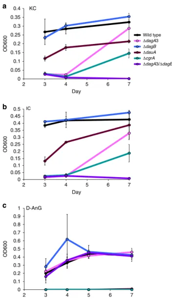
PUL-encoded genes are essential for the in vivo utilization of
carrageenans. To investigate in vivo gene function in Z.
galac-tanivorans, we recently developed a genetic technique to
con-struct deletion mutants in this bacterium
35. Four single deletion
mutants [ΔdagA3 (Δzgal_3150, Δgh127-3), ΔdagB (Δzgal_3152,
Δgh129-like), ΔdauA (Δzgal_3155, Δ3,6-anhydro-D-galactose
dehydrogenase),
ΔcgrA (Δzgal_3159, ΔaraC family regulator)]
and a double deletion mutant (ΔdagA3/ΔdagB) were constructed.
They all grew comparably to the wild-type strain in Zobell
medium and minimum medium supplemented with agar or
galactose (Supplementary Fig.
10
A, B). The
ΔdagB mutant had
little effect on growth relative to wild type in minimal media
containing either KC or IC; however, the
ΔdagA3 mutant showed
a significant growth delay and the double ΔdagA3/ΔdagB mutant
abolished growth altogether, confirming the importance of these
α-1,3-(3,6-anhydro)-
D-galactosidases in carrageenan degradation
(Fig.
6
a, b). This sharply contrasts with the biochemical
char-acterization of ZGAL_3150 (DagA3, GH127-3) and ZGAL_3152
(DagB, GH129-like), where there was no discernible difference in
specificity (Fig.
2
c). We attribute the difference in deletion effect
may be due to either an unknown difference in specificity
between the enzymes or different cellular localizations of the
enzymes. Subtle differences between the GH127 and GH129-like
enzymes would not be surprising considering the complex, hybrid
structure of carrageenans whose regular structures are masked by
multiple substituents (e.g., sulfate, methyl, pyruvate). For
0 0.05 0.1 0.15 0.2 0.25 0.3 0.35 0.4 2 3 4 5 6 7 OD600 Day 0 0.05 0.1 0.15 0.2 0.25 0.3 0.35 0.4 0.45 0.5 2 3 4 5 6 7 OD600 Day 0 0.1 0.2 0.3 0.4 0.5 0.6 0.7 0.8 0.9 1 2 3 4 5 6 7 OD600 Day Wild type ΔdagA3 ΔdagB ΔdauA ΔcgrA ΔdagA3/ΔdagB KC
a
b
c
IC D-AnGFig. 6 Average growth of wild-type Z. galactanivorans and deletion mutants.
Growth ona kappa-carrageenan, b iota-carrageenan and c 3,6-anhydro-D
-galactose (D-AnG) as sole carbon source over a 7-day period. Error bars represent standard error of the mean between three replicates. Legend:
dagA3,α-1,3-(3,6-anhydro)-D-galactosidase (zgal_3150, gh127); dagB,
α-1,3-(3,6-anhydro)-D-galactosidase (zgal_3152, gh129-like); dauA,
3,6-anhydro-D-galactose dehydrogenase (zgal_3155); crgA, araC family transcription
instance, such variations in substrate specificities are known in
GH16
β-agarases and β-porphyranases which act on other red
algal sulfated galactans
36. Bioinformatic analyzes predict that
ZGAL_3150 and ZGAL_3152 are anchored into the outer
membrane and secreted in the periplasm, respectively (Fig.
3
).
The more likely scenario is that ZGAL_3150 is oriented toward
the periplasm. Thus, both enzymes would be localized within the
periplasm in order to sequentially degrade carrageenan
oligo-saccharides to produce the monosaccharide D-AnG for uptake by
the bacterium. Finally, this deletion effect indicates that the two
remaining GH127 enzymes are unable to compensate for the loss
of ZGAL_3150 or ZGAL_3152. The
ΔdauA mutant did not grow
on D-AnG and complementation experiments restored growth
on this sugar (Fig.
6
c, Supplementary Fig.
10
C). This indicates
that neither ZGAL_4659 (orthologue of the 3,6-anhydro-
L-galactose dehydrogenase VEJY3_09240, 68% identity) nor any
other sugar dehydrogenases from Z. galactanivorans could
degrade D-AnG and that ZGAL_3155 (DauA, 3,6-anhydro-
D-galactose dehydrogenase) is essential for D-AnG catabolism.
ΔdauA had diminished growth on both KC and IC (Fig.
6
a, b),
suggesting that this mutant is still capable of using the
D-galactose
units released by carrageenan degradation as a carbon source. The
deletion mutant of the AraC family regulator ZGAL_3159
(ΔcgrA) showed a significant lag phase and reduced growth on
carrageenans (Fig.
6
a, b), confirming its importance on the
positive regulation of the ZGAL_3145-3159 cluster function.
Complementation experiments restored growth of
ΔcgrA on KC
(Supplementary Fig.
10
D). In both sets of complementation
experiments the growth was improved relative to the wild-type
strain (Supplementary Fig.
10
C, D). This is likely because the
complemented genes are under the Flavobacterium johnsoniae
OmpA promoter and therefore under less stringent
transcrip-tional control than the carrageenan-specific PUL. Phenotyping of
ΔcgrA on KC or IC solid media indicates a complex mode of
regulation (Supplementary Fig.
10
E–L, Supplementary
Discus-sion). Finally, the cgrA deletion had no effect on growth with
agar, indicating that this transcription factor is specific for
car-rageenan catabolism (Supplementary Fig.
10
A).
RNA-seq analysis unravels a complex carrageenan-related
regulon. RNA-seq expression profiling was performed on Z.
galactanivorans grown in minimal media containing D-AnG, KC,
and IC, relative to the growth on
D-galactose (Table
1
,
Supple-mentary Data
1
–
9
). The entire carrageenan-specific PUL was
strongly upregulated in both KC and IC with the exception of the
transcriptional regulator cgrA in IC which was at the border of
significance. Growth on D-AnG induced 10 genes from the PUL
suggesting this monosaccharide, unique to red algae, acts as an
effector in regulation. The genes that were induced by D-AnG
were specific for oligosaccharide and monosaccharide utilization;
no genes were induced for the degradation of carrageenan
Table 1 Selection of carrageenan-induced genes in Z. galactanivorans
Locus_tag Description (gene name/acronym, family) KC/D-gal IC/D-gal D-AnG/D-gal
log2-FC* padj log2-FC padj log2-FC padj
ZGAL_181 Sulfatase (cgsB2, S1_7) 8.4 7.47E-94 8.0 5.81E-85 5.1 3.66E-34
ZGAL_236 Kappa-carrageenase (cgkA, GH16) 10.6 2.68E-163 8.7 7.26E-110 0.8 1
ZGAL_1973 Iota-carrageenase (cgiA3, GH82) 7.6 7.15E-56 8.7 1.12E-72 0.1 1
ZGAL_3145 Sulfatase (cgsA, S1_19) 6.3 1.7E-65 5.1 1.69E-41 3.7 3.48E-20
ZGAL_3146 Sulfatase (cgsB1, S1_7) 6.8 1.2E-117 6.0 1.04E-91 3.9 1.20E-33
ZGAL_3147 α-1,3-(3,6-anhydro)-D-galactosidase (dagA1,
GH127)
7.2 7.4E-135 6.9 5.12E-125 3.3 2.67E-25
ZGAL_3148 α-1,3-(3,6-anhydro)-D-galactosidase (dagA2,
GH127)
6.6 2.7E-61 6.8 5.52E-65 3.5 6.80E-16
ZGAL_3149 Sugar permease (MFS) 6.7 3.0E-65 6.2 9.61E-55 4.7 9.94E-31
ZGAL_3150 α-1,3-(3,6-anhydro)-D-galactosidase (dagA3,
GH127)
8.1 2.9E-108 7.8 3.02E-99 7.1 3.78E-82
ZGAL_3151 Sulfatase (cgsC, S1_17) 4.8 5.4E-44 4.1 1.50E-31 0.7 1
ZGAL_3152 α-1,3-(3,6-anhydro)-D-galactosidase (dagB,
new GH)
3.9 6.5E-23 2.8 5.72E-11 0.6 1
ZGAL_3153 2-keto-3-deoxy-D-galactonate aldolase (dauD) 4.2 9.9E-44 3.8 2.07E-35 0.4 1
ZGAL_3154 2-keto-3-deoxy-D-galactonate kinase (dauC) 3.6 2.4E-30 3.3 1.84E-25 1.2 0.1516
ZGAL_3155 3,6-anhydro-D-galactose dehydrogenase
(dauA)
8.1 1.3E-80 7.3 4.01E-65 5.6 1.40E-37
ZGAL_3156 3,6-anhydro-D-galactonate cycloisomerase
(dauB)
9.7 2.0E-89 9.3 1.21E-81 5.2 1.07E-23
ZGAL_3157 Sugar/H+symporter (DMT) 8.4 5.8E-140 8.0 3.68E-125 7.0 3.85E-95
ZGAL_3158 High-affinity sugar transporter 4.3 8.1E-39 3.0 4.15E-17 4.1 8.61E-34
ZGAL_3159 Transcriptional regulator (cgrA, AraC family) 2.3 1.8E-14 1.2 0.0504 −0.5 1
ZGAL_3580 SusD-like lipoprotein (cgtB) 9.1 2.04E-124 8.3 5.65E-104 4.2 1.17E-24
ZGAL_3581 SusC-like TonB-dependent receptor (cgtA) 9.6 1.93E-103 9.5 1.31E-99 4.3 4.99E-19
ZGAL_3629 Sulfatase (S1_30) 8.5 8.26E-55 4.9 2.82E-16 −0.1 1
ZGAL_3630 Sulfatase(S1_28) 7.1 5.54E-48 4.3 6.27E-16 0.8 1
ZGAL_3631 Polygalacturonase, (GH28) 7.8 1.43E-36 3.9 1.70E-07 −0.4 1
ZGAL_3632 Polygalacturonase, (GH28) 7.8 8.43E-20 4.3 1.66E-04 −0.3 1
ZGAL_3633 Beta-galactosidase (GH2) 9.4 1.85E-66 6.9 2.66E-35 1.0 1
ZGAL_3634 Alpha-L-fucosidase (GH29) 8.3 3.04E-28 4.8 9.25E-08 1.8 1
ZGAL_3637 SusC-like TonB-dependent transporter (TBDT) 9.2 1.81E-51 5.1 1.14E-14 0,7 1
ZGAL_3638 SusD-like lipoprotein (SGBP) 9.9 3.92E-53 5.3 8.26E-14 −0,1 1
ZGAL_4265 Iota-carrageenase (cgiA1, GH82) 8.7 2.10E-56 4.7 6.12E-15 0.0 1
polymers. Unexpectedly, numerous stress-related proteins were
also induced (e.g., small heat shock protein, universal stress
protein, peptide methionine sulfoxide reductase). Together with
the observation that the growth with D-AnG induced cell
aggregation, this suggests that free D-AnG is not frequent in the
natural environment of Z. galactanivorans and that this sugar is
normally only an intracellular degradation intermediate and
unlikely to be transported by a specific outer membrane
transporter.
Numerous genes outside the ZGAL_3145-3159 cluster were
strongly upregulated by carrageenans. Significantly, the most
induced gene when grown on KC was the kappa-carrageenase
cgkA (zgal_236, gh16)
22; this gene was also heavily induced by
growth on IC (Table
1
, Supplementary Data
1
–
9
). The
iota-carrageenase genes cgiA1 (zgal_4265, gh82-1) and cgiA3
(zgal_1973, gh82-3)
23,24were upregulated in both KC and IC.
Interestingly, cgiA2 (zgal_2155, gh82-2) was the most expressed
iota-carrageenase gene in
D-galactose but was downregulated in
KC and IC. Thus ZGAL_2155 could act as a constitutive sentinel
enzyme involved in the initial degradation step releasing signal
oligosaccharides inducing the carrageenolytic system. The
wild-type kappa-carrageenase CgkA and iota-carrageenase CgiA1 were
previously shown to be extracellular enzymes
22,23. Thus, the role
of these enzymes is probably to generate oligosaccharides for
transport by Z. galactanivorans’ SusCD-like transport system.
The family S1–7 sulfatase, ZGAL_181, was also among the genes
most induced by carrageenans. The corresponding protein
displays 65% identity with ZGAL_3146 (CgsB1, S1_7 sulfatase)
which desulfates the alpha-carrabiose into beta-carrabiose motifs,
suggesting that ZGAL_181 (gene named cgsB2) catalyzes the
same reaction. These genes are probably the result of relatively
recent gene duplication. Two major players missing in the
carrageenan-specific PUL are the archetypal susC-like and
susD-like genes. The most induced gene when grown on IC was the
SusC-like TBDT zgal_3581; this gene was also highly induced by
growth on KC and to a lesser degree by growth on D-AnG. The
adjacent gene encodes a SusD-like lipoprotein (ZGAL_3580)
which was also substantially upregulated in all three conditions.
This susCD-like gene pair, distal to the PUL, is a good candidate
for the outer membrane transport system associated with the
ZGAL_3145-3159 cluster. This hypothesis is supported by
genomic comparative analyzes, mutant phenotyping and
bio-chemical experiments (see below). Finally, a second PUL is
strongly induced in both KC and IC but not D-AnG
(ZGAL_3629-3638). This gene cluster encodes four glycoside
hydrolases (1 GH2, 1 GH29, and 2 GH28), two sulfatases (S1_28
and S1_30 subfamilies) and a SusCD-like pair.
Characterization of key genes distant from the carrageenan
PUL. Complete carrageenan catabolism requires the hydrolysis of
the beta-1,4-linkage at the non-reducing end of odd-DP
oligo-carrageenans produced by the action of the 3,6-anhydro-
D-galactosidases. After beta-galactosidase hydrolysis the resulting
even-DP oligosaccharides become again substrates for the
3,6-anhydro-
D-galactosidases, and so on until the complete
degra-dation into free
D-galactose and 3,6-anhydro-
D-galactose. We
hypothesized that at least one of the 8 predicted GH2
beta-galactosidases encoded in the genome of Z. galactanivorans was
capable of this activity. ZGAL_3633 stood out as the most
probable candidate since its gene expression was induced
sig-nificantly in both iota- and kappa-carrageenans (Table
1
). Thus,
we cloned ten GH2 constructs from Z. galactanivorans, including
ZGAL_3633. All these recombinant enzymes displayed
beta-galactosidase activity on an artificial substrate (pNP-beta-
D-galactopyranoside, Supplementary Fig.
11
). As demonstrated
using HPLC (Fig.
7
, Supplementary Discussion), two of the
GH2s, ZGAL_3633 and ZGAL_4655, are active on odd-DP
oligo-carrageenans (furcellaran hydrolyzed by kappa-carrageenase
fol-lowed
by
hydrolysis
by
the
3,6-anhydro-
D-galactosidase
ZGAL_3152). Both ZGAL_3633 and ZGAL_4655 are
con-stitutively expressed at low levels in Z. galactanivorans in
mini-mum medium supplemented with
D-galactose (Supplementary
10.0 11.0 12.0 13.0 14.0 15.0 16.0 17.0 18.0 19.0 20.0 25.0 30.0 Conductivity ( μ S) oligo-KC standards oligo-furcellaran (furcellaran treated with kappa-carrageenase to produce even-DP oligosaccharides) oligo-furcellaran + GH129-like ZGAL_3152 oligo-furcellaran + GH129-like ZGAL_3152 + GH2 ZGAL_3633 oligo-furcellaran + GH129-like ZGAL_3152 + GH2 ZGAL_4655 DP4 KC DP2 KC Furcellaran-trioses GH2 effects 0.0 5.0 10.0 15.0 DP6 KC DP8 KC
Elution time (min)
P11P5 P6 P7 P8 P9 P10
P4 P3 P2 P1
Fig. 7 Sequential digestion of furcellaran oligosaccharides by DagB and GH2 enzymes. HPLC results for the ZGAL_3152 (DagB, GH129-like) and GH2 (ZGAL_3633, ZGAL_4655) sequential enzyme digests on furcellaran oligosaccharides. Oligo-kappa-carrageenan standards (DP2, DP4, DP6, and DP8) are shown in black dotted line on the chromatogram. First, oligo-furcellaran (blue line) was treated with ZGAL_3152 (purple line). This reaction was then stopped by heating at 90 °C for 10 min. Following the inactivation of ZGAL_3152 the oligosaccharides were treated with either ZGAL_3633 (orange line) or ZGAL_3655 (green line)
Data
1
–
9
); however, ZGAL_4655 is not induced by kappa- and
iota-carrageenans, suggesting that ZGAL_3633 is the key
carrageenan-specific beta-galactosidase. Thus, Z. galactanivorans
has all the enzyme activities necessary for the complete
degra-dation of kappa family carrageenans, most of them encoded by
the ZGAL_3145-3159 cluster and the others by remote,
carrageenan-induced
genes
(GH16:
ZGAL_236;
GH82:
ZGAL_1973, ZGAL_4265; GH2: ZGAL_3633; S1–7: ZGAL_181).
The carrageenan gene cluster lacks the susCD-like gene pair
found within canonical PULs; however, the susCD-like gene pairs
zgal_3580/zgal_3581 and zgal_3637/zgal_3638 are upregulated
when grown on iota- and kappa-carrageenans suggesting these
are good outer membrane candidates for oligo-carrageenan
transport. We succeeded in producing and purifying soluble
ZGAL_3580 and ZGAL_3638 (SusD-like proteins) and probed
their interaction with red algal cell wall polysaccharides using
affinity gel electrophoresis (Fig.
8
a). In the native gel without
polysaccharide, ZGAL_3580 migrates as a single band while
ZGAL_3638 forms a smear. There are changes in the intensity of
the different bands corresponding to ZGAL_3638 on all of the
polysaccharides tested, suggesting that the polysaccharides may
have an effect on the quaternary structure of the protein, but
there is no obvious delay in the migration and ZGAL_3638 did
not appear to significantly interact with the ligands tested. The
absence of ZGAL_3638 specificity for carrageenans and the
presence of two putative GH16 beta-porphyranases, ZGAL_3628
(PorD) and ZGAL_3640 (PorE) encoded in the ZGAL_3628-3640
locus suggest that this locus is more likely dedicated to the
degradation of another sulfated galactan distinct from
carragee-nans (e.g., sulfated agars). In contrast, migration of ZGAL_3580 is
retarded (as a clear band) by kappa-carrageenan and furcellaran
and only slightly by iota carrageenan. No shift is apparent in agar,
porphyran or lambda-carrageenan. Therefore, ZGAL_3580
inter-acts with kappa family carrageenans. Phenotyping experiments
on Z. galactanivorans deletion mutant
Δzgal_3580/Δzgal_3581
indicate pronounced inhibition of growth relative to wild type on
both KC and IC and moderate inhibition of growth on furcellaran
(Fig.
8
b). The biochemical (Fig.
8
a) and the genetic (Fig.
8
b)
experiments confirm the affinity of ZGAL_3580 and ZGAL_3581
for family kappa carrageenans, but with a difference in preference
depending on the chosen method. However, the genetic approach
evaluates
the
combined
properties
of
ZGAL_3580
and
ZGAL_3581, whereas the gel shift assay only characterized
ZGAL_3580. This suggests that the TBDT ZGAL_3581 has a
strong affinity for iota-carrageenan, which compensates for the
reduced affinity of the SusD-like protein ZGAL_3580 for this
polysaccharide relative to KC. Overall, these results are consistent
with ZGAL_3580/ZGAL_3581 being responsible for carrageenan
oligosaccharide import, forming part of the carrageenan regulon
and thus acting as the distal susCD-like gene pair for the
carrageenan PUL. The genes have been named cgtA for the
SusC-like TBDT (zgal_3581) and cgtB for the SusD-SusC-like lipoprotein
(zgal_3580).
Based on the biochemical, genetic, and transcriptomic evidence
presented here, we have demonstrated for the
first time a
complete scheme for the catabolism of kappa-, iota- and
beta-carrageenans in Z. galactanivorans, from the initial action of the
GH16
kappa-carrageenase
(CgkA)
22and
GH82
iota-carrageenases (CgiA1-3)
23,24to the conversion in four steps of
D-AnG into
D-glyceraldehyde-3-phosphate and pyruvate (Fig.
3
);
these latter compounds presumably then enter glycolysis and the
citric acid cycle, respectively. The released
D-galactose residues are
expected to be further converted by the Leloir pathway. The
majority of the molecular actors in this pathway and in its
regulation are encoded by a discrete genetic locus; however, other
key genes are localized remotely on the genome and are part of a
carrageenan-induced gene network (Table
1
). These genes are
thus part of a complex regulon (referred to as the carrageenan
utilization system) and the key genes have been named (Table
1
).
Plasticity of carrageenan utilization systems amends the notion
of PUL. We searched the Genbank database for bacteria
pos-sessing a potential carrageenan-specific PUL, using BlastP with
key enzymes as queries (ZGAL_3150 (DagA3, GH127-3),
ZGAL_3155 (DauA, 3,6-anhydro-
D-galactose dehydrogenase)).
After manual verification of each genomic region, we identified
29 species with a homologous carrageenan-specific PUL
(including Tenacibaculum jejuense whose genome sequence has
been sequenced by Eric Duchaud’s group and deposited at EMBL
in the context of this study). These bacteria belong to four phyla:
Bacteroidetes, Proteobacteria, Planctomycetes, and Firmicutes. All
these microorganisms originate from marine ecosystems: mostly
free-living bacteria isolated from seawater, marine sediments or
isolated at the surface of macroalgae, but also gut bacteria from
animals feeding on macroalgae (surgeon
fish, sea urchin, and
abalone). The limits of each PUL were manually refined and
clusters of orthologous genes from this PUL were subsequently
determined (Supplementary Data
10
,
11
, Supplementary Fig.
12
).
Homologs of selected carrageenan-induced genes from Z.
galac-tanivorans were also searched in the 29 bacterial genomes
(Supplementary Data
10
,
11
) with the conservation of these genes
evaluated by a Heatmap; based on these conservation profiles, the
bacterial species clustered into 5 main clades (Fig.
9
). Clades 1
and 2 include only Bacteroidetes (from different classes) and their
PUL organizations are the most similar to that of Z.
galactani-vorans. Strikingly, several carrageenan-induced genes remote
from the Z. galactanivorans carrageenan-specific PUL are found
within the carrageenan-specific PULs of other Bacteroidetes. This
is notably the case of ZGAL_3581 (cgtA) and ZGAL_3580 (cgtB),
which is consistent with the function in Z. galactanivorans of this
SusCD-like pair in the import of carrageenan
degradation-products. Z. galactanivorans possesses two S1_7 sulfatases (65%
sequence identity), one located in the carrageenan PUL
(ZGAL_3146) and the other distal to the PUL (ZGAL_181) but
forming part of the carrageenan regulon (Table
1
). In contrast,
the PULs of several Bacteroidetes species contains both
ortholo-gous genes of the S1_7 sulfatases (Supplementary Fig.
12
),
con-sistent with the hypothesis of recent gene duplication. The
GH127 genes are likely another example of gene duplication,
since their number varies from 1 to 3 paralogous genes depending
on the species. Other Bacteroidetes PULs contain GH16 genes
distantly related to zgal_236 (cgkA) and most likely forming a
new GH16 subfamily. Within clades 1 and 2 we can define a core
carrageenan utilization system which includes dauA, dauB, dauC,
dauD, dagA, cgsB, cgrA, cgtA, and cgtB. Unexpectedly, the GH16
kappa-carrageenases and the GH82 iota-carrageenases are not
part of the core system. Indeed some species harbor only one type
of carrageenase, while others are deprived of any known
carra-geenases, suggesting that these latter bacteria may have new
carrageenase families or that they can only degrade predigested
oligosaccharides. Whereas the GH127 genes (dagA) are strictly
conserved in the PUL, the GH129-like gene (dagB) is only found
in a few species, suggesting that the ancestral
α-1,3-(3,6-anhy-dro)-
D-galactosidase activity was due to the GH127 family.
In clades 3, 4, and 5, the structure of the carrageenan-specific
PUL is significantly modified. When considering the 30 bacterial
species, the core system is restricted to the genes responsible for
the release of D-AnG (dagA) and for its catabolism (dauA, dauB,
dauD). This may be due to horizontal gene transfer (HGT) events
from Bacteroidetes to other phyla. Somewhat surprising is the lack
of dauC in some species. Such bacteria may use a
non-phosphorylative variant of the Entner-Doudoroff pathway to
degrade 2-keto-3-deoxy-
D-galactonate, as observed in the
archaeon Picrophilus torridus
37. The susD-like genes are unique
to the Bacteroidetes
8and transfer of the PUL to species belonging
to other phyla has resulted in the loss of the susCD-like pair. This
phenomenon is observed in the clade 3 Gammaproteobacteria
(Fig.
9
) and was previously identified in the case of alginolytic
operons
19. However, this does not mean that TBDT are absent in
the gene clusters of other phyla. For instance, all clade 3
Gammaprotobacteria species have a TBDT gene in their cluster
(CATDS2_v1220055
in
Catenovulum
agarivorans
DS-2;
JRKG01_v1_110122 in Pseudoalteromonas sp. PLSV; Patl_0887
in P. atlantica T6c; H978DRAFT_1909 in Alteromonas sp.
ALT199). They are only distantly related to ZGAL_3581, but
their location within the cluster strongly suggests that they play a
similar role in the import of the carrageenan degradation product.
Native KC IC Lambda Agar Porphyran Furcellaran
BSA 3580 3638 BSA 3580 3638 BSA 3580 3638 BSA 3580 3638 BSA 3580 3638 BSA 3580 3638 BSA 3580 3638
0.00 0.10 0.20 0.30 0.40 0.50 0.60 0.70 0.80 0 50 100 150 200 OD600 Time (h) 0.00 0.05 0.10 0.15 0.20 0.25 0.30 0.35 0.40 0.45 0 50 100 150 200 OD600 Time (h) Zobell Agar 0.00 0.05 0.10 0.15 0.20 0.25 0 50 100 150 200 OD600 Time (h) 0.00 0.10 0.20 0.30 0.40 0.50 0.60 0.70 0.80 0.90 0 50 100 150 200 OD600 Time (h) KC IC 0.00 0.20 0.40 0.60 0.80 1.00 1.20 0 50 100 150 200 OD600 Time (h) D-galactose 0.00 0.02 0.04 0.06 0.08 0.10 0.12 0.14 0.16 0.18 0.20 0 50 100 150 200 OD600 Time (h) Furcellaran
b
a
Fig. 8 Affinity gel electrophoresis for the SusD-like proteins and phenotyping for the susCD-like mutant ΔcgtA-cgtB. a BSA and recombinant ZGAL_3580
(CgtB, SusD-like) and ZGAL_3638 (SusD-like) migrated at 60 V over 5 h in native-PAGE gels or in gels containing different red algal cell wall polysaccharides. The delay of migration for ZGAL_3580 (red line) and ZGAL_3638 (green line) was compared between the native gels and the
polysaccharide containing gels by comparison with BSA (black line).b Growth curves for wild-type Z. galactanivorans (black curves) andΔcgtA-cgtB (red
Such clusters containing polysaccharide-related TBDT genes have
already been described in Gammaproteobacteria
38. The presence
of carrageenan-specific PULs in gut bacteria from marine
herbivorous animals is also reminiscent of the horizontal
acquisition of porphyran/agar-related genes in animal and human
intestinal symbionts
39. This is quite clear for the surgeon
fish
symbiont Epulopiscium sp. (Fig.
9
, clade 3), which is also known
to have horizontally acquired GH16 porphyranase and GH117
genes from marine
flavobacteria
40. Phylogenetic analysis of the
GH127 family unraveled another likely HGT case
(Supplemen-tary Fig.
13
, Supplementary Data
12
). Whereas ZGAL_3148
(DagA2, GH127-2) and ZGAL_3150 (DagA3, GH127-3) cluster
only with homologs from marine bacteria harboring the
carrageenan-specific PUL, ZGAL_3147 (DagA1, GH127-1) is at
the root of a clade including GH127 enzymes from human gut
Bacteroides species. These dagA1-like genes are within PULs
including GH genes at
first sight unrelated to carrageenans (e.g.
GH78, GH95, GH105; Supplementary Fig.
14
). Thus, these
Bacteroides GH127 genes have a marine origin, but have most
likely evolved in specificity after their horizontal acquisition.
Conclusion. Here we have shown that PUL-like structures
(lacking the susCD-like pair but maintaining other
carrageenan-related genes) are found in bacterial phyla other than
Bacter-oidetes. Furthermore, the carrageenan utilization system is not
static and can be characterized by gene losses and gene
acquisi-tions with a dedicated core 3,6-anhydro-
D-galactose metabolism
that is conserved within carrageenolytic bacteria. This core system
is essential but not sufficient for carrageenan utilization. Missing
functions (e.g., carrageenases, some specific sulfatases) may be
assumed by non-orthologous genes in different bacterial species.
Therefore, polysaccharide utilization pathways are not always
conferred by a single locus, even in Bacteroidetes, and may consist
}
}
}
}
}
} } } Kappa-carrageenase Iota-carrageenase Clade 2 Clade 1 Clade 3 Clade 4 Clade 5 Aquimarina sp. RZW4-3-2 (Ba-Fl) Algibacter lectus JCM 1930 (Ba-Fl) Saccharicrinis fermentans DSM 9555 (Ba-Fl) Rhodopirellula sp. SWK7 (Pl-Pm) Tenacibaculum jejuense CNURICO (Ba-Fl) Aquimarina latercula DSM 2041 (Ba-Fl) Flammeovirga sp. SJP92 PUL1 (Ba-Cy) Flammeovirga sp. OC4 PUL1 (Ba-Cy) Flammeovirga sp. OC4 PUL2 (Ba-Cy) Flammeovirga sp. SJP92 PUL2 (Ba-Cy) Flammeovirga pacifica WPAGA1 (Ba-Cy) Persicobacter sp. JZB09 (Ba-Cy)Catenovolum agarivorans DS_2 PUL2 (Pr-Gp) Catenovolum agarivorans DS_2 PUL1 (Pr-Gp) Alteromonas sp. ALT199 (Pr-Gp)
Pseudoalteromonas atlantica T6c (Pr-Gp) Pseudoalteromonas sp. PLSV (Pr-Gp) Epulopiscium sp. N.t. morphotype B (Fi-Cl) Cellulophaga sp. E6 (Ba-Fl)
Cellulophaga baltica 13 (Ba-Fl) Cellulophaga algicola DSM 14237 (Ba-Fl) Cellulophaga sp. 18 (Ba-Fl)
Cellulophaga baltica NN016038 (Ba-Fl) Cellulophaga lytica DSM 7489 (Ba-Fl) Cellulophaga lytica HI1 (Ba-Fl) Cellulophaga geogensis KL-A (Ba-Fl) Flexithrix dorotheae DSM 6795 (Ba-Cy) Maribacter thermophilus HT7-2 (Ba-Fl) Lewinella persica DSM 23188 (Ba-Sp) Echinicola pacifica DSM 19836 (Ba-Cy) Zobellia galactanivorans DsiJT (Ba-Fl)
CGSA CGSB CGSC DAGA DAGB DAUA DAUB DAUC DAUD
CGTA CGTB MFS DMT GNTP CGRA GH16_KC GH16_NC GH82 GH2 GH28 GH29 GH42 S1_28 S1_30 S1_NC ZGAL_3637 ZGAL_3638 AA3 FR\SR ADH OXR A1E SK GNTR-like LUXR-like
UNK1 UNK2 UNK3 UNK4 UNK5
Carrageenan-specific transcriptional regulator Cytoplasmic sugar transporters
Carrageenan-specific SusCD-like pair 3,6-anhydro-D-galactose bioconversion
3,6-anhydro-D-galactose release Carrageenan desulfation
Fig. 9 Conservation of the carrageenolytic PUL organization among bacterial genomes. Putative homologs of selected carrageenan-induced genes from Z. galactanivorans were found in 29 other bacterial genomes with the conservation of these genes evaluated by a heatmap. Based on these conservation
profiles, the bacterial species cluster into 5 main clades. Red coloring indicates the genes are conserved in a PUL, yellow indicates the genes are conserved
elsewhere in the genome and blue indicates the genes are absent in the genome. Gene names for the Z. galactanivorans carrageenan-utilization locus are
found in Table1and the legend of Fig.1. Abbreviations: CGTA, CarraGeenan Transport A (SusC-like, TonB-dependent transporter); CGTB, CarraGeenan
Transport B (SusD-like lipoprotein); GH, glycoside hydrolase; S1, family 1 sulfatase; AA3, auxiliary activity 3; FR/SR, fumarate reductase/succinate dehydrogenase; ADH, aldehyde dehydrogenase; OXR, isoquinoline 1-oxidoreductase alpha subunit; A1E, aldose 1-epimerase; SK, sugar kinase; GNTR-like and LUXR-like, transcription factor families GNTR and LUXR; UNK, unknown function. The phylum and class are indicated beside each organism in parentheses: Ba, Bacteroidetes; Pl, Planctomycetes; Pr, Proteobacteria; Fi, Firmicutes; Fl, Flavobacteriia; Pm, Planctomycetia; Cy, Cytophagia; Gp,