HAL Id: hal-03108208
https://hal.archives-ouvertes.fr/hal-03108208
Submitted on 13 Jan 2021
HAL is a multi-disciplinary open access
archive for the deposit and dissemination of
sci-entific research documents, whether they are
pub-lished or not. The documents may come from
teaching and research institutions in France or
abroad, or from public or private research centers.
L’archive ouverte pluridisciplinaire HAL, est
destinée au dépôt et à la diffusion de documents
scientifiques de niveau recherche, publiés ou non,
émanant des établissements d’enseignement et de
recherche français ou étrangers, des laboratoires
publics ou privés.
Reconstruction of partially sampled STEM-EELS images
with atomic resolution
Etienne Monier, Thomas Oberlin, Nathalie Brun, Nicolas Dobigeon
To cite this version:
Etienne Monier, Thomas Oberlin, Nathalie Brun, Nicolas Dobigeon. Reconstruction of partially
sam-pled STEM-EELS images with atomic resolution. Workshop on Signal Processing with Adaptative
Sparse Structured Representations (SPARS 2019), Nicolas Dobigeon, Toulouse INP, France; Cédric
Févotte, CNRS, France, Jul 2019, Toulouse, France. �hal-03108208�
Reconstruction of partially sampled STEM-EELS images
with atomic resolution
Etienne Monier(1), Thomas Oberlin(1), Nathalie Brun(2) and Nicolas Dobigeon(1)
(1)University of Toulouse, IRIT/INP-ENSEEIHT, Toulouse
(2)Laboratoire de Physique des Solides, CNRS UMR 8502, Univ. Paris-Sud, Univ. Paris-Saclay
[email protected], [email protected]
Abstract—Electron microscopy has shown to be a powerful tool to ana-lyze chemical composition of samples. However, acquiring a high quality image is hard due to radiation damages which limit the signal-to-noise ratio. One solution, considered in this work, consists in spatially partially acquiring the multi-band image and reconstructing it afterwards. We propose a reconstruction algorithm, referred to as Fourier sparsity in 3D (FS3D), based on a regularization specifically tailored for atomically resolved images. Experiments show that the proposed FS3D method leads to state-of-the-art results with a significantly lighter computational cost.
I. INTRODUCTION
In a scanning transmission electron microscope (STEM), an elec-tron beam is used as the illumination source and is focused as a probe which is moved over the sample area of interest. Among the commonly collected signals is the electron energy loss spectrum (EELS) [1], which provides multi-band images, usually referred to as spectrum-images. A classical encountered problem is that the electron beam induces damages for sensitive materials [2] such as organic components. Indeed, standard acquisition schemes operate sequen-tially, line-by-line, and thus concentrate electrons in contiguous areas. A common solution consists in reducing the electron dose (number of electrons per unit surface), which significantly lowers the signal-to-noise ratio (SNR) and the overall image quality. Recent works propose another strategy which consists in a random sampling scheme for reducing cumulative damage on successive pixels while keeping admissible SNR and spatial resolution [3]. This approach has several advantages as it allows adaptive studies to be envisioned. Such a random sampling has been implemented on the STEM VG HB 501 microscope in the Lab. Physique des Solides (Orsay, France) [4].
Our approach is based on a partial and random acquisition scheme. It requires computational reconstruction schemes to recover the full data from the partial measurements within a post-processing task. In image processing this is commonly referred to as inpainting, a particular inverse problem which can be solved by considering appropriate spatial and spectral regularizations. Capitalizing on our recent work [5], this paper presents an algorithm particularly suit-able to reconstruct atomic scale STEM-EELS images. In particular, we introduce a simple yet relevant regularization which promotes periodic patterns frequently encountered in images of this resolution. We compare the resulting performance to the ones obtained by a state-of-the-art method.
II. METHODS
Recent works in remote sensing and microscopy aims at recon-structing multi-band images for data compression, corrupted image inpainting or sample preservation (e.g., in the specific case of microscopy). In this context, Beta Process Factor Analysis (BPFA) [6] is a widely used reconstruction algorithm and can be considered as a state-of-the-art reconstruction technique (see [7]). This approach is based on dictionary learning, which has been at the core of efficient denoising and reconstruction approaches in the last decades. Besides, by adopting a Bayesian formalism, several critical hyperparameters can be included into the Bayesian model and jointly estimated with
the reconstructed image. In particular, it automatically adjusts the size of the dictionary and the size of the atoms. However, this algorithm remains computationally expensive due to Monte Carlo sampling and its usage is limited to off-line reconstruction.
Conversely, particular applicative scenarios require fast image re-construction for online and/or embedded analysis. As a consequence, BPFA could not be used and more computationnally efficient methods should be considered [3]. In the specific context of fast reconstruction of atomic-scale spectrum-images, we propose an alternative recon-struction algorithm which exploits the particular spatial content of those images. More precisely, the spectrum-image to be recovered is assumed to be spatially sparse in an appropriate space, e.g., resulting from a discrete cosine transform, since the sample is composed of interleaved periodic nets of atoms. The proposed method, called Fourier Sparsity in 3D (FS3D), relies on a spectral dimension reduction induced by a principal component representation. Then, in the identified lower-dimensional space, the reconstruction task is formulated as the minimization of an energy function composed of `2 data fidelity term and a group sparse regularization of the
band-by-band DCT coefficients.
III. RESULTS
To compare the proposed FS3D approach and BPFA, a synthetic spectrum-image of 70×120 pixels with 1435 bands has been gen-erated. It mimics an atomic-scale sample composed of four distinct materials. This image has been sub-sampled randomly with 20% ac-quired pixels. Both algorithms are applied to reconstruct the unknown image in the 4-dimensional subspace identified by PCA. Note that BPFA could be applied directly on the 4-band image by adopting a 3D-patch representation or, alternatively, band-by-band by adopting a 2D-patch representation. Yet, the 3D-implementation of BPFA can only be applied with sub-optimal parameters (reduced patch size and overlap) due to its computational burden. Similar comparisons have been conducted with a real image, whose dimention has been reduced to 9 bands by principal component analysis.
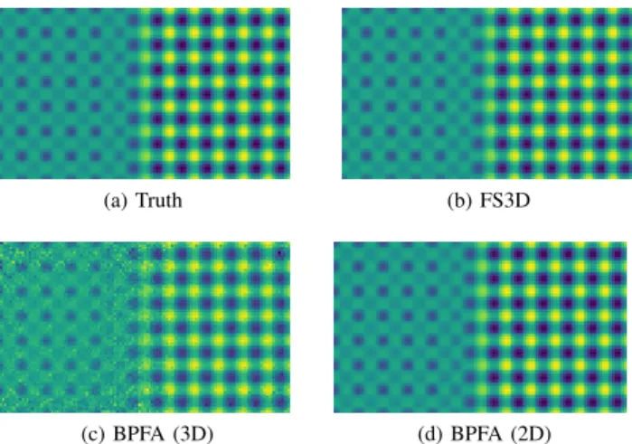
Figure 1 (resp., 2) shows the 4th band of the synthetic (resp., real) ground truth and reconstructed spectrum-images. The corresponding SNR, averaged spectral angle disctance (aSAD) [8] and execution time are reported in Table I (resp. Table II) for synthetic (resp., real) spectrum-image. These results show that in the particular case of atomic-scale spectrum-images, FS3D gives results comparable to the state-of-the-art technique BPFA with a much reduced execution time. Note also that BPFA gives better results as a 2D algorithm than as a 3D algorithm due to a sub-optimal parameter choice. However the 2D implementation is much more time-consuming. These results show that FS3D is an efficient method which is fast and efficient in the considered applicative context.
(a) Truth (b) FS3D
(c) BPFA (3D) (d) BPFA (2D)
Fig. 1: Comparison of the 4th (least powerful) band of the synthetic image for the compared methods.
TABLE I: Metrics for the synthetic image.
Method SNR aSAD (×10−3) Time Time (w.r.t. FS3D)
FS3D 43.73 1.05 4.25s 1
BPFA (3D) 43.85 3.46 12min30s 176
BPFA (2D) 55.39 0.48 2h59min 2527
(a) Truth (b) FS3D
(c) BPFA (3D) (d) BPFA (2D)
Fig. 2: Comparison of the 4thband of the real image for the compared methods.
TABLE II: Metrics for the real image.
Method SNR aSAD (×10−2) Time Time (w.r.t. FS3D)
FS3D 32.43 1.33 0.44s 1
BPFA (3D) 29.48 1.87 28min19s 3861 BPFA (2D) 34.11 1.27 3h37min 2.959 × 10⁴
REFERENCES
[1] R. F. Egerton, Electron Energy-Loss Spectroscopy in the Electron Micro-scope. New York: Springer, 2011.
[2] R. F. Egerton, P. Li, and M. Malac, “Radiation damage in the TEM and SEM,” Micron, vol. 35, no. 6, pp. 399–409, 2004.
[3] A. B´ech´e, B. Goris, B. Freitag, and J. Verbeeck, “Development of a fast electromagnetic beam blanker for compressed sensing in scanning transmission electron microscopy,” Appl. Phys. Lett., vol. 108, no. 9, pp. 093 103–1–093 103–5, Feb. 2016.
[4] A. Tararan, M. Tenc´e, N. Brun, M. Kociak, and A. Zobelli, “Random scanning mode for the spectroscopy of sensitive materials,” in Proc. Eur. Microsc. Congr., 2016.
[5] E. Monier, T. Oberlin, N. Brun, M. Tenc´e, M. de Frutos, and N. Dobigeon, “Reconstruction of partially sampled multi-band images – Application to EELS microscopy,” IEEE Trans. Computational Imaging, vol. 4, no. 4, pp. 585–598, Dec. 2018.
[6] Z. Xing, M. Zhou, A. Castrodad, G. Sapiro, and L. Carin, “Dictionary learning for noisy and incomplete hyperspectral images,” SIAM J. Imag. Sci., vol. 5, no. 1, pp. 33–56, 2012.
[7] A. Stevens, H. Yang, L. Carin, I. Arslan, and N. D. Browning, “The potential for bayesian compressive sensing to significantly reduce electron dose in high-resolution stem images,” Microscopy, vol. 63, no. 1, pp. 41– 51, 2013.
[8] N. Keshava, “Distance metrics and band selection in hyperspectral pro-cessing with applications to material identification and spectral libraries,” IEEE Trans. Geosci. Remote Sens., vol. 42, no. 7, pp. 1552–1565, 2004.