HAL Id: hal-03222929
https://hal-amu.archives-ouvertes.fr/hal-03222929
Submitted on 10 May 2021
HAL is a multi-disciplinary open access
archive for the deposit and dissemination of
sci-entific research documents, whether they are
pub-lished or not. The documents may come from
teaching and research institutions in France or
abroad, or from public or private research centers.
L’archive ouverte pluridisciplinaire HAL, est
destinée au dépôt et à la diffusion de documents
scientifiques de niveau recherche, publiés ou non,
émanant des établissements d’enseignement et de
recherche français ou étrangers, des laboratoires
publics ou privés.
Distributed under a Creative Commons Attribution| 4.0 International License
Brain SPECT perfusion and PET metabolism as
discordant biomarkers in major depressive disorder
Maud Tastevin, Laurent Boyer, Theo Korchia, Guillaume Fond, Christophe
Lançon, Raphaëlle Richieri, Eric Guedj
To cite this version:
Maud Tastevin, Laurent Boyer, Theo Korchia, Guillaume Fond, Christophe Lançon, et al.. Brain
SPECT perfusion and PET metabolism as discordant biomarkers in major depressive disorder.
EJN-MMI Research, SpringerOpen, 2020, 10 (1), �10.1186/s13550-020-00713-2�. �hal-03222929�
PRELIMINARY RESEARCH
Brain SPECT perfusion and PET metabolism
as discordant biomarkers in major depressive
disorder
Maud Tastevin
1, Laurent Boyer
2,3,4, Theo Korchia
1, Guillaume Fond
1,2,3, Christophe Lançon
1,2,
Raphaëlle Richieri
1,5and Eric Guedj
6*Abstract
Background: Brain SPECT perfusion and PET metabolism have been, most often interchangeably, proposed to study the underlying pathological process in major depressive disorder (MDD). The objective of this study was to specify similarities and inconsistencies between these two biomarkers according to global characteristics of the disease. We conducted a retrospective study in 16 patients suffering from treatment-resistant MDD who underwent, during the
same current episode, a cerebral perfusion SPECT with 99mTc-HMPAO and a metabolic PET with 18F-FDG. Whole-brain
voxel-based SPM(T) maps were generated in correlation with the number of depressive episodes and in correlation with the depression duration, separately for the two exams (p-voxel < 0.001 uncorrected, k > 20).
Results: No significant correlations were found between brain metabolism and either the number of depressive episodes or the duration of the disease, even at an uncorrected p-voxel < 0.005. On the other hand, the increased number of depressive episodes was correlated with decreased perfusion of the right middle frontal cortex, the right anterior cingulum cortex, the right insula, the right medial temporal cortex and the left precuneus. The increased depression duration was correlated with decreased perfusion of the right anterior cingulum cortex.
Conclusions: This preliminary study demonstrates more significant results with brain perfusion compared with glucose metabolism in treatment-resistant MDD, highlighting the value of brain SPECT despite less favourable instru-mentation detection compared to PET.
Keywords: Treatment-resistant depression, Major depressive disorder, PET, SPECT, Biomarker
© The Author(s) 2020. Open Access This article is licensed under a Creative Commons Attribution 4.0 International License, which permits use, sharing, adaptation, distribution and reproduction in any medium or format, as long as you give appropriate credit to the original author(s) and the source, provide a link to the Creative Commons licence, and indicate if changes were made. The images or other third party material in this article are included in the article’s Creative Commons licence, unless indicated otherwise in a credit line to the material. If material is not included in the article’s Creative Commons licence and your intended use is not permitted by statutory regulation or exceeds the permitted use, you will need to obtain permission directly from the copyright holder. To view a copy of this licence, visit http://creat iveco mmons .org/licen ses/by/4.0/.
Background
Major depressive disorder (MDD) is a common men-tal health disorder. It is now the first cause of disability worldwide and a major contributor to the overall global burden of diseases according to the World Health Organ-ization. It has been estimated that 15–30% of patients present with treatment-resistant depression (TRD),
which is resistant to antidepressants and cognitive behav-ioural treatment [1].
The current diagnosis and course evaluation of MDD rely on clinical examination. To date, the risk of misdi-agnosis remains present because of the lack of non-invasive and quantifiable assessments of all depression dimensions [2]. Several promising treatments have been proposed (pharmacological, non-invasive and invasive neuromodulation) in the case of antidepressant resist-ance. Nonetheless, the pathophysiology of depression and the neural and biological mechanisms of treatment efficacy are still not fully understood. In this context, neuroimaging biomarkers are needed for diagnosing,
Open Access
*Correspondence: eric.guedj@ap-hm.fr
6 Nuclear Medicine Department, APHM, CNRS, Centrale Marseille, Institut
Fresnel, Timone Hospital, CERIMED, Aix Marseille University, Marseille, France
Page 2 of 7 Tastevin et al. EJNMMI Res (2020) 10:121
predicting the course of the disorder and guiding the choice of therapy, as well as monitoring the response to these therapies.
Since 2008, with the development of the “Rdoc Research” programme, brain SPECT with 99mTc-ECD or 99mTc-HMPAo and 18F-FDG PET have been proposed
to, respectively, study regional blood flow and glucose metabolism in a range of psychiatric disorders, including MDD [3]. Despite distinct underlying mechanisms (per-fusion vs. metabolism), both biomarkers are supposed to be coupled with the global synaptic activity [4]. They have been used to help in the neurological differential diagno-sis of depression, and more recently considered to better understand the underlying pathological process in MDD and to predict response and non-response outcomes to neuromodulation therapies, such as repetitive transcra-nial magnetic stimulation (rTMS) or deep brain stimula-tion (DBS) [5, 6]. In this line, some brain regions seem particularly involved (e.g. the frontal cortex and more broadly the limbic system), especially in treatment-resist-ant cohorts. Nevertheless, the genuine overlap across PET/SPECT studies is globally more limited and slows down the clinical integration of these biomarkers into the patient’s evaluation. The main explanations of these discrepancies are the small size and clinical heterogene-ity of inclusions, as well as the variabilheterogene-ity of neuroimag-ing techniques, radiotracers and statistical models [7]. One alternative hypothesis would be that neuroimaging biomarkers are not as equivalent as anticipated in MDD, especially for perfusion and metabolism.
The objective of this study was to specify similarities and inconsistencies between brain perfusion and brain glucose metabolism according to the global characteris-tics of the disease (i.e. the number of previous depressive episodes and the depression duration) in a single group of patients with MDD.
Methods
Subjects
We conducted a retrospective study. The database included patients with psychiatric follow-up at Sainte Marguerite University Hospital (Marseille, France) from January 2011 through July 2019. Our inclusion criteria were patients over the age of 18 suffering from TRD who underwent a cerebral 99mTc-HMPAo SPECT and a 18
F-FDG PET with an interval delay of less than 18 months during a same current major depressive episode (accord-ing to DSM-IV criteria). These SPECT examinations had been performed to initially explore differential diagnoses [8]. The selected patients were subsequently included in the HrTMS trial, which included a metabolic PET imag-ing evaluation before treatment with ethical and regula-tory authorizations (ClinicalTrials.gov: NCT02559466;
Registry Identifier ID RCB: 2015-A00345-44) [6, 9]. Patients with bipolar depression, schizophrenia or neuro-logical comorbidities were excluded.
Data collection
The sociodemographic characteristics recorded included gender, age, marital status and education level. Clinical data included illness duration, number of depressive epi-sodes, and melancholic and psychotic characteristics, as well as pharmaco-resistance and global severity accord-ing to the DSM-IV (SCID-IV) structured clinical inter-view [10]. Data concerning severity scales, such as the MADRS or Beck, were not available in all patients for the two evaluation time points. Treatment data recorded included all antidepressants, antipsychotics and mood stabilizers, as well as invasive and non-invasive brain stimulation (electroconvulsive therapy, rTMS and deep brain stimulation and vagal nerve stimulation).
SPECT and PET acquisitions
SPECT and PET scans were performed for all subjects, with the same SPECT and PET cameras, and under the same conditions. PET was performed using an integrated PET/CT camera (Discovery ST, GE Healthcare, Wauke-sha, WI, USA). Patients were required to fast for at least 6 h before undergoing the scan, with a control of normal blood glucose level. They were maintained in neurosen-sory resting 10 min before and 30 min after injection.
18F-FDG was injected intravenously at the activity of
150 MBq, and PET images were then acquired over a period of 15 min. Iterative reconstruction was performed on a matrix of 192 × 192, with correction of attenuation using CT acquisition. SPECT acquisition was performed using a double-headed rotating gamma camera (ECAM, Siemens) equipped with a fan beam collimator. Patients were maintained in neurosensory resting 10 min before and 20 min after intravenous injection of 740 MBq of
99mTc-HMPAO. The total scan time was of 25 min with
sixty projections per head of 25 s, collected in 128 × 128 format. Tomographic 3D reconstruction was performed using a filtered back-projection algorithm.
Images were initially converted from the DICOM to the NifTi format using MRIcro (www.mricr o.com) and then transferred to SPM. Whole-brain statistical analy-sis was performed at the voxel level using SPM8 software (Wellcome Department of Cognitive Neurology, Uni-versity College, London, UK) after spatial normalization (the Montreal Neurological Institute atlas) and smooth-ing with a Gaussian filter (8 mm full-width at half-maxi-mum) to blur individual variations in gyral anatomy and to increase the signal-to-noise ratio. Whole-brain voxel-based SPM(T) maps were generated in correlation with the number of depressive episodes and in correlation
with the depression duration, separately for the two exams (p-voxel < 0.001 uncorrected, k > 20), using propor-tional scaling with a grand mean scaled value fixed at 50.
Statistical analysis
The Shapiro–Wilk test was used to confirm the normal-ity of the two variables tested at PET and SPECT time examination [i.e. the depression duration (p = 0.017 and p = 0.011, respectively) and the number of episodes (p < 0.001)]. Data were presented in proportions or means and standard deviations. Characteristics were compared for each patient between PET and SPECT acquisition dates using Student’s t test or the Mann–Whitney U test for continuous variables and the chi-square test or Fisher’s exact test for categorical variables. Mean PET/ SPECT values were extracted using MARSBAR software (https ://marsb ar.sourc eforg e.net/) at the individual level for significant cluster(s) to calculate Spearman’s correla-tions. The statistical significance level was set at p < 0.05 in a two-sided test.
Results
Baseline characteristics
A total of 16 subjects were included in the study. Patient characteristics are described in Table 1. Patients were mostly women with a mean age of approximately 50 years old. Our sample included 4 men and 12 women; 13 patients were single, and 10 had less than 12 years of education. They all presented with TRD with a mean number of depressive episodes estimated at 2.31 ± 1.9. Among this group, 13 patients suffered from a severe current episode, and melancholic criteria were present in 6 patients. The mean interval delay between PET and SPECT acquisitions was 4.37 ± 4.89 months (20 days to 13 months). No significant changes were found between these two time point evaluations, especially for clinical characteristics and treatments. Moreover, no treatment interruption occurred during the interval delay. However, age and disease duration did show statistically significant differences, with a respective difference of 5.28 months (p = 0.02) and 1.44 months (p = 0.023), respectively;
Table 1 Demographic and clinical characteristics of patients at PET and SPECT day acquisitions (n = 16)
Significant differences are in bold
SCID structured clinical interview for DSM-IV, SSRI selective serotonin reuptake inhibitor, SNRI serotonin–norepinephrine reuptake inhibitor, MAOI monoamine oxidase
inhibitors
At PET day acquisition At SPECT day acquisition p value
Demographic characteristics
Age (years, mean ± SD) 50.50 ± 16.3 50.06 ± 16.7 0.020
Education 1.000 > 12 years 5 5 1.000 ≤ 12 years 10 10 Marital status 1.000 Single 13 13 1.000 Couple 3 3 Clinical characteristics
Depression duration (months) 136.44 ± 120 132.56 ± 121.48 0.023
Number of depressive episodes 2.31 ± 1.9 2.31 ± 1.9 1.000
SCID severity Mild 2 2 1.000 Moderate 1 1 1.000 Severe 13 13 1.000 Melancholic features 6 6 1.000 Treatments SSRIs 5 7 0.500 SNRIs 6 5 0.300 Tricyclics 2 2 1.000 MAOIs 1 1 1.000 Pramipexole 1 1 1.000
Antipsychotics, first generation 1 1 1.000
Antipsychotics, second generation 3 4 0.607
Mood stabilizers 0 0 1.000
Page 4 of 7 Tastevin et al. EJNMMI Res (2020) 10:121
these limited but significant differences were expected, since SPECT was systematically performed before PET in these patients.
Correlation with metabolism and perfusion
No significant SPM(T) results were found for metabolism in correlation with either the number of depressive epi-sodes or the depression duration, even for a less stringent threshold at an uncorrected p-voxel < 0.005.
On the other hand, significant correlations were found with perfusion at p-voxel < 0.001 and k > 20. The increased number of depressive episodes was correlated with decreased perfusion of the right middle frontal cor-tex (T-voxel max = 5.50, k = 106), the right anterior cin-gulum cortex (ACC) (T-voxel max = 5.13, k = 75), the right medial temporal cortex (T-max = 4.93, k = 79), the left precuneus (T-voxel max = 4.80, k = 66) and the right insula (T-voxel max = 4.52, k = 61). The increased depres-sion duration was correlated with the perfudepres-sion of the right ACC (T-voxel max = 5.31, k = 58).
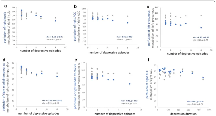
Spearman’s correlations on extracted clusters con-firmed a negative correlation between the number of depressive episodes and perfusion of the right medial temporal cortex (rho = − 0.84, p = 0.00002), the left precuneus (rho = − 0.50, p = 0.02), the right ACC (rho = − 0.49, p = 0.02) and the right insula (rho = − 0.58,
p = 0.01). Negative correlations were also confirmed
between depression duration and perfusion of the right ACC (rho = − 0.61, p = 0.01). These same clusters were extracted for brain glucose metabolism without signifi-cant correlation.
These results are presented in Figs. 1 and 2.
Discussion
We conducted a retrospective study in a sample of 16 patients suffering from TRD who all underwent a cere-bral perfusion SPECT with 99mTc-HMPAO and a
meta-bolic PET with 18F-FDG. No relevant clinical changes
were found upon evaluation at these two time points, especially for disease characteristics and treatments. Whole-brain voxel-based analysis revealed distinct
Fig. 1 Anatomical localization of significant perfusion SPECT findings (p-voxel < 0.001 uncorrected, k > 20). The increased number of depressive
episodes is correlated with decreased perfusion of: a the right anterior cingulate cortex (ACC) (T-voxel max = 3.85, k = 75), b the right insula (T-voxel max = 3.85, k = 26), c the left precuneus (T-voxel max = 3.85, k = 62), d the right medial temporal cortex (T-max = 3.85, k = 66) and e the right middle frontal cortex (T-voxel max = 3.85, k = 106). f The increased depression duration is correlated with decreased perfusion of the right ACC (T-voxel max = 5.31, k = 236)
results between perfusion and glucose metabolism; this could, at least partly, explain the previous variability of findings in the literature for these two biomarkers [7]. Significant negative correlations were found between number of episodes and perfusion of the right middle frontal cortex, the right ACC, the right insula, the right medial temporal cortex and the left precuneus, as well as between the depression duration and right ACC per-fusion, while no relationship was obtained for glucose metabolism.
Number of episodes and illness duration are considered as risk factors of pharmaco-resistance and are involved in depression recurrence [11, 12]. They also may signal risk of residual symptoms, such as sleep disturbances, execu-tive impairments and anxiety [13]. They are considered by a group of experts as great indicators of prognosis and severity of TRD [14–16]. Identifying their relation-ship with cerebral functioning could improve resistance management and selection of potential cerebral thera-peutic targets. Correlations with these two variables have already been described in a few previous neuroimaging studies. A voxel-based study of 127 subjects suffering from TRD showed a weak negative correlation between
the duration of illness and brain 99mTc-ECD SPECT
per-fusion in bilateral cingulate and orbital cortices [17]. A recent MRI meta-analysis including morphometry stud-ies found significant grey matter reduction in the rostral part of the anterior cingulum related to illness dura-tion and repeated depressive episodes [18]. Moreover, our findings are concordant with another 18F-FDG PET
study. In 18 hospitalized patients with unipolar depres-sion, Mayberg et al. [19] revealed no significant relation-ship between brain metabolism and illness chronicity.
The brain regions revealed in this study are known to be involved in TRD [7, 20]. The insula is particularly involved in emotional identification and in the affective state in response to a stimulus. Resting-state hyperactiv-ity of the insula has been linked, in MDD, to pathologi-cal self-focused mental ruminative behaviours [21]. On the other hand, the ACC modulates the link between ventral and dorsal networks involved in regulation of emotion. The dorsal ACC is specifically implicated in executive functions through the cognitive control net-work, and its subgenual subdivision is focused on emo-tional experience and processing [22]. Furthermore, the insula and ACC both belong to the salience network,
0 10 20 30 40 50 60 70 80 90 0 2 4 6 8 10
perfusion of right insula metabolis
mo
f righ
t insula
number of depressive episodes
0 10 20 30 40 50 60 70 80 90 100 0 2 4 6 8 10
perfusion of right ACC metabolism of right AC
C
number of depressive episodes
0 20 40 60 80 100 120 140 0 2 4 6 8 10 perfusion of le ft pr ecuneu s metabolis mo f le ft pr ecuneu s
number of depressive episodes c f 0 10 20 30 40 50 60 70 80 0 2 4 6 8 10 perfusion of right medial tempor al cx metabolism of right medial tempor al cx
number of depressive episodes
0 10 20 30 40 50 60 70 0 100 200 300 400 500 perfusion of right AC C metabolism of right AC C depression duration 0 20 40 60 80 100 120 0 2 4 6 8 10 perfusion of right middle fr ontal cx metabolism of right middle fr ontal cx
number of depressive episodes
rho = -0.58, p=0.01
rho =-0.23, p=0.40 rho =-0.49, p=0.02rho =-0.21, p=0.20 rho =-0.50, p=0.02rho =0.20, p=0.77
rho = -0.84, p= 0.00002
rho = -0.23, p= 0.20 rho = -0.49, p= 0.02rho = 0.16, p= 0.55 rho = -0.61, p= 0.01rho = 0.08, p= 0.76
a b
d e
Fig. 2 Scatter plot of Spearman’s correlations on extracted clusters. The increased number of depressive episodes is correlated with decreased
perfusion of: a the right insula (rho = − 0.58, p = 0.01), b the right anterior cingulate cortex (ACC) (rho = − 0.49, p = 0.02), c the left precuneus (rho = − 0.50, p = 0.02), d the right medial temporal cortex (rho = − 0.84, p = 0.00002) and e the right middle frontal cortex (rho = − 0.49, p = 0.02). This correlation is not significant with the metabolism of: a the right insula (rho = − 0.23, p = 0.40), b the right ACC (rho = − 0.21, p = 0.20), c the left precuneus (rho = 0.20, p = 0.77), d the right medial temporal cortex (rho = − 0.23, p = 0.20) and e the right middle frontal cortex (rho = 0.16, p = 0.55). f The increased depression duration is correlated with decreased perfusion of the right ACC (rho = − 0.61, p = 0.01). This correlation is not significant with the metabolism of the right ACC (rho = 0.08, p = 0.76)
Page 6 of 7 Tastevin et al. EJNMMI Res (2020) 10:121
which participates in judgement alteration and negative thoughts in MDD [23]. They also both interact with the default mode network and contribute to the alteration of attentional system and to the introspection in MDD [24]. The precuneus, medial frontal and medial temporal cortices have been mainly implicated through a default mode network and could also contribute to low self-esteem and ruminations [25]. The number of episodes and depression duration seem to alter brain perfusion, especially in cerebral areas linked to emotional and cog-nitive symptoms of depression. It could suggest their key role in pharmaco-resistance and recurrence by enhanc-ing the cortico-limbic dysregulation described in unipo-lar depression.
PET is one of the main neuroimaging techniques evalu-ated in recent psychiatry research [20]. Indeed, PET is usually preferred to SPECT because of better spatial resolution. Cerebral glucose metabolism and perfusion have been considered as coupled for a long time, because the brain consumes approximately 20% of total body oxy-gen and 25% of total body glucose. The most important energy source for the brain is adenosine triphosphate (ATP), which is produced almost entirely by the oxida-tive metabolism of glucose [26]. However, their consist-ent correlations are presconsist-ently questioned and in part justified by other mechanisms of blood flow regulation, which could produce a different cartography of cerebral perfusion from the metabolic one. This has already been highlighted in healthy subjects [27]. Furthermore, uncou-pling between glucose and oxygen metabolism, via oxy-gen depletion and induction of downstream hypoxia response pathways, could play a key role in neurodegen-erative diseases [28]. The underlying mechanisms of drug resistance in depression remain misunderstood in the current literature [29]. Nevertheless, it is not excluded that psychiatric disease could involve the same uncou-pling mechanisms. Functional compensation, more spe-cifically involving brain metabolism, may also occur. Further fundamental studies are necessary to specify the contribution of these mechanisms in depression.
The main limitations of the study were a retrospective design and a small sample size. Our results concerned patients with TRD, and they are not generalizable to all patients suffering from MDD. Subjects were mostly women with a mean age above 50 years old, which are expected sociodemographic characteristics; advanced age and female gender are risk factors for TRD [30, 31]. Moreover, the interval delay between the two acquisitions was significant and could impact the findings. This differ-ence was expected because of the sequdiffer-ence of explora-tion. Indeed, the order was fixed (PET after SPECT), with inevitably a difference using a rank test. Nevertheless, the number of depression episodes is strictly the same
at the two times of the study, and the difference between depression duration was weak among the groups (136.44 ± 120 months vs. 132.56 ± 121.48 months). Treat-ments and modifications of therapy between PET and SPECT explorations could have also impacted our find-ings, possibly more the metabolism than the perfusion. Nevertheless, these changes were limited and statistically non-significant. In detail, a selective serotonin reuptake inhibitor was changed for two patients, including one for a serotonin–norepinephrine reuptake inhibitor, and one antidepressant was potentialized by an antipsychotic during the interval delay, which constitute common changes in the therapeutic strategy of depression. Moreo-ver, no treatment interruption occurred between the two examinations. To our knowledge, this is nevertheless the first study that focuses on a single group of patients to compare perfusion and metabolism in MDD. Finally, the perfusion and metabolic measurements were semiquan-titative, as used in clinical practice. Further studies with absolute quantification could better explain the differ-ences observed in this preliminary report.
Conclusions
This preliminary study demonstrates that the clinical characteristics of TRD are more significantly associated with brain perfusion than with glucose metabolism, high-lighting the value of brain SPECT despite its less favour-able spatial resolution and image quality compared to PET. These findings warrant other comparative studies between these two imaging techniques to provide a more frequent use of brain perfusion in future psychiatric research.
Acknowledgements
Not applicable.
Authors’ contributions
MT analysed and interpreted the clinical and PET/SPECT data, drafted the work and gave final approval. LB analysed and interpreted the clinical and PET/ SPECT data, revised the work and gave final approval. TK acquired the clinical data, revised the work and gave final approval. GF analysed and interpreted the clinical and PET/SPECT data, revised the work and gave final approval. CL contributed to conception and design of the study, acquired the clinical data, revised the work and gave final approval. RR contributed to conception and design of the study, acquired, analysed and interpreted the clinical data, drafted the work and gave final approval. EG contributed to conception and design of the study, acquired, analysed and interpreted the PET/SPECT data, drafted the work and gave final approval.
Funding
This work has been carried out within DHU-Imaging with the support of the A*MIDEX Project (ANR-11-IDEX-0001-02) funded by the “Investissements d’Avenir” French Government programme managed by the French National Research Agency (ANR), the Public Assistance Marseille Hospitals (AORC junior 2014) and a research grant from a French foundation for health research and innovation “Fondation de l’Avenir”.
Availability of data and materials
The data that support the findings of this study are available from the cor-responding author upon reasonable request.
Ethics approval and consent to participate
This study has been performed in accordance with the Declaration of Helsinki, with written consent of patients and the approval of the “CPP-Sud Méditer-ranée 1” ethics committee (ClinicalTrials.gov: NCT02559466; Registry Identifier ID RCB: 2015-A00345-44).
Consent for publication
Not applicable.
Competing interests
The authors declare that they have no competing interests.
Author details
1 Department of Psychiatry, Sainte Marguerite University Hospital, Assistance
Publique- Hôpitaux de Marseille, Marseille, France. 2 CEReSS-Health Service
Research and Quality of Life Centre, Aix Marseille University, Marseille, France.
3 Department of Medical Information and Public Health, APHM, Marseille,
France. 4 Department of Epidemiology and Health Economics, Assistance
Publique-Hôpitaux de Marseille, Marseille, France. 5 CNRS, Centrale Marseille,
Institut Fresnel, Aix Marseille University, Marseille, France. 6 Nuclear Medicine
Department, APHM, CNRS, Centrale Marseille, Institut Fresnel, Timone Hospital, CERIMED, Aix Marseille University, Marseille, France.
Received: 7 May 2020 Accepted: 24 September 2020
References
1. Thase ME. Treatment-resistant depression: prevalence, risk factors, and treatment strategies. J Clin Psychiatry. 2011;72:e18.
2. Fried EI, Epskamp S, Nesse RM, Tuerlinckx F, Borsboom D. What are ‘good’ depression symptoms? Comparing the centrality of DSM and non-DSM symptoms of depression in a network analysis. J Affect Disord. 2016;189:314–20.
3. Zhang K, Zhu Y, Zhu Y, Wu S, Liu H, Zhang W, et al. Molecular, functional, and structural imaging of major depressive disorder. Neurosci Bull. 2016;32:273–85.
4. Magistretti PJ, Pellerin L. Cellular mechanisms of brain energy metabolism and their relevance to functional brain imaging. Philos Trans R Soc Lond B Biol Sci. 1999;354:1155–63.
5. Fonseka TM, MacQueen GM, Kennedy SH. Neuroimaging biomarkers as predictors of treatment outcome in major depressive disorder. J Affect Disord. 2018;233:21–35.
6. Tastevin M, Richieri R, Boyer L, Fond G, Lançon C, Guedj E. Brain PET meta-bolic substrate of TMS response in pharmaco-resistant depression. Brain Stimul Basic Transl Clin Res Neuromodul. 2020;13:683–5.
7. Fitzgerald PB, Laird AR, Maller J, Daskalakis ZJ. A meta-analytic study of changes in brain activation in depression. Hum Brain Mapp. 2008;29:683–95.
8. Amen DG, Krishnamani P, Meysami S, Newberg A, Raji CA. Classifica-tion of depression, cognitive disorders, and co-morbid depression and cognitive disorders with perfusion SPECT neuroimaging. J Alzheimers Dis. 2017;57:253–66.
9. Tastevin M, Baumstarck K, Groppi F, Cermolacce M, Lagrange G, Lançon C, et al. Double cone coil rTMS efficacy for treatment-resistant depression: a prospective randomized controlled trial. Brain Stimul. 2019. https ://doi. org/10.1016/j.brs.2019.09.009.
10. First Michael B, Williams Janet BW, Spitzer Robert L, Gibbon M. Structured clinical interview for DSM-IV-TR Axis I disorders, Clinical Trials Version (SCID-CT); 2007.
11. Bennabi D, Aouizerate B, El-Hage W, Doumy O, Moliere F, Courtet P, et al. Risk factors for treatment resistance in unipolar depression: a systematic review. J Affect Disord. 2015;171:137–41.
12. Buckman JEJ, Underwood A, Clarke K, Saunders R, Hollon SD, Fearon P, et al. Risk factors for relapse and recurrence of depression in adults and how they operate: a four-phase systematic review and meta-synthesis. Clin Psychol Rev. 2018;64:13–38.
13. Serafini G, Nebbia J, Cipriani N, Conigliaro C, Erbuto D, Pompili M, et al. Number of illness episodes as predictor of residual symptoms in major depressive disorder. Psychiatry Res. 2018;262:469–76.
14. Bennabi D, Charpeaud T, Yrondi A, Genty J-B, Destouches S, Lancrenon S, et al. Clinical guidelines for the management of treatment-resistant depression: French recommendations from experts, the French Associa-tion for Biological Psychiatry and Neuropsychopharmacology and the foundation FondaMental. BMC Psychiatry. 2019;19:262. https ://doi. org/10.1186/s1288 8-019-2237-x.
15. Gelenberg AJ, Freeman MP, Markowitz JC, Rosenbaum JF, Thase ME, Trivedi MH et al. American psychiatric association practice guideline for the treatment of patients with major depressive disorder, third edition. Am J Psychiatry. 2010;167:1–152.
16. Ruhé HG, van Rooijen G, Spijker J, Peeters FPML, Schene AH. Staging methods for treatment resistant depression. A systematic review. J Affect Disord. 2012;137:35–45.
17. Richieri R, Boyer L, Faget-Agius C, Farisse J, Mundler O, Lançon C, et al. Determinants of brain SPECT perfusion and connectivity in treatment-resistant depression. Psychiatry Res. 2015;231:134–40.
18. Bora E, Fornito A, Pantelis C, Yücel M. Gray matter abnormalities in major depressive disorder: a meta-analysis of voxel based morphometry stud-ies. J Affect Disord. 2012;138:9–18.
19. Mayberg HS, Brannan SK, Mahurin RK, Jerabek PA, Brickman JS, Tekell JL, et al. Cingulate function in depression: a potential predictor of treatment response. NeuroReport. 1997;8:1057–61.
20. Gong B, Naveed S, Hafeez DM, Afzal KI, Majeed S, Abele J, et al. Neuro-imaging in psychiatric disorders: a bibliometric analysis of the 100 most highly cited articles. J Neuroimaging. 2019;29:14–33.
21. Sliz D, Hayley S. Major depressive disorder and alterations in insular cortical activity: a review of current functional magnetic imaging research. Front Hum Neurosci. 2012;6:323. https ://doi.org/10.3389/fnhum .2012.00323 .
22. Bush G, Luu P, Posner MI. Cognitive and emotional influences in anterior cingulate cortex. Trends Cogn Sci. 2000;4:215–22.
23. Manoliu A, Meng C, Brandl F, Doll A, Tahmasian M, Scherr M, et al. Insular dysfunction within the salience network is associated with severity of symptoms and aberrant inter-network connectivity in major depressive disorder. Front Hum Neurosci. 2014;7:930. https ://doi.org/10.3389/fnhum .2013.00930 .
24. Bessette KL, Jenkins LM, Skerrett KA, Gowins JR, DelDonno SR, Zubieta J-K, et al. Reliability, convergent validity and time invariance of default mode network deviations in early adult major depressive disorder. Front Psychiatry. 2018;9:244. https ://doi.org/10.3389/fpsyt .2018.00244 . 25. Fox MD, Snyder AZ, Vincent JL, Corbetta M, Van Essen DC, Raichle ME.
The human brain is intrinsically organized into dynamic, anticorrelated functional networks. Proc Natl Acad Sci USA. 2005;102:9673–8. 26. Reilly P. Head injury: pathophysiology and management of severe closed
injury. London: Chapman and Hall Medical; 1997.
27. Henriksen OM, Vestergaard MB, Lindberg U, Aachmann-Andersen NJ, Lisbjerg K, Christensen SJ, et al. Interindividual and regional relationship between cerebral blood flow and glucose metabolism in the resting brain. J Appl Physiol. 2018;125:1080–9.
28. Watts ME, Pocock R, Claudianos C. Brain energy and oxygen metabo-lism: emerging role in normal function and disease. Front Mol Neurosci. 2018;11:216. https ://doi.org/10.3389/fnmol .2018.00216 .
29. Voineskos D, Daskalakis ZJ, Blumberger DM. Management of treatment-resistant depression: challenges and strategies. Neuropsychiatr Dis Treat. 2020;16:221–34.
30. Holtzmann J, Richieri R, Saba G, Allaïli N, Bation R, Moliere F, et al. How to define treatment-resistant depression? Presse Med. 2016;45:323–8. 31. Gronemann FH, Jorgensen MB, Nordentoft M, Andersen PK, Osler M.
Socio-demographic and clinical risk factors of treatment-resistant depression: a Danish population-based cohort study. J Affect Disord. 2020;261:221–9.
Publisher’s Note
Springer Nature remains neutral with regard to jurisdictional claims in pub-lished maps and institutional affiliations.