HAL Id: hal-02379346
https://hal.archives-ouvertes.fr/hal-02379346
Submitted on 12 Nov 2020HAL is a multi-disciplinary open access archive for the deposit and dissemination of sci-entific research documents, whether they are pub-lished or not. The documents may come from teaching and research institutions in France or abroad, or from public or private research centers.
L’archive ouverte pluridisciplinaire HAL, est destinée au dépôt et à la diffusion de documents scientifiques de niveau recherche, publiés ou non, émanant des établissements d’enseignement et de recherche français ou étrangers, des laboratoires publics ou privés.
Structural analysis of premolar roots in Middle
Pleistocene hominins from China
Lei Pan, Jean Dumoncel, Arnaud Mazurier, Clément Zanolli
To cite this version:
Lei Pan, Jean Dumoncel, Arnaud Mazurier, Clément Zanolli. Structural analysis of premolar roots in Middle Pleistocene hominins from China. Journal of Human Evolution, Elsevier, 2019, 136, pp.102669. �10.1016/j.jhevol.2019.102669�. �hal-02379346�
Structural analysis of premolar roots in Middle Pleistocene hominins from China
Abstract:
This study investigates permanent maxillary and mandibular premolar root structural organization in East
Asian Middle Pleistocene hominins. In addition to analyzing the linear and volumetric properties of the roots,
we used a landmark-free approach to both qualify and quantify in 3D premolar root shape variation of Middle
Pleistocene hominins in East Asia. Moreover, we focus on some mid- to late East Asian Middle Pleistocene
hominin specimens whose taxonomic attribution is unclear. We find considerable cementum in this sample
of hominins, similar to other fossil groups, but clearly different from modern humans which have a very
small amount of cementum. Additionally, a smaller root pulp cavity is found in later Homo (Neanderthals
and modern humans). Our analyses on the crown-root surface area ratio show that East Asian Middle
Pleistocene H. erectus as well as one late Middle Pleistocene Homo sp. specimen (PA 81 P4 from Changyang
site) are distinguished from other fossil and extant groups by relatively larger root surface, stout root branches
and thick cementum deposits. This may represent a distinct East Asian H. erectus dental pattern. Geometric
morphometric analyses on the external root surface reveal a general trend of shape simplification along the
Homo lineage examined here, and distinguish Early Pleistocene Homo, Middle Pleistocene H. erectus,
Neanderthals and modern human morphologies. The late Middle Pleistocene teeth from Changyang site (PA
76 P3 and PA 81 P4) areclose to East Asian H. erectus and Neanderthals, while the mid-Middle Pleistocene
P3 from Panxian Dadong falls within the modern human distribution. Combined with dental crown
morphology and root number/form reported in previous studies, our results show that the external root shape
human morphology is observed in part of the East Asian Middle Pleistocene specimens, while a retention of
primitive, H. erectus-like features is expressed in some late Middle Pleistocene specimens, supporting a
multi-lineage and discontinuous scenario of human settlements in East Asia.
Key words: East Asian Middle Pleistocene Homo; permanent premolar; external root surface; crown-root
tissue proportions
1. Introduction
Hominin postcanine tooth root and canal anatomy is highly variable (e.g., Sert and Bayirli, 2004;
Prado-Simón et al., 2012a; Kupczik et al., 2019). Using direct observation of the external roots (Turner, 1981;
Spencer, 2003; Moggi-Cecchi et al., 2010), 2D radiography (Sperber, 1974; Abbott, 1984; Wood et al., 1988;
Wood and Engleman, 1988), and medical/micro-tomography (Kupczik et al., 2003, 2005; Kupczik and Dean,
2008; Moore et al., 2013, 2015, 2016), studies have demonstrated that tooth roots preserve information about
dietary adaptation, taxonomy and phylogeny (Abbott, 1984; Wood et al., 1988; Wood and Engleman, 1988;
White et al., 2000; Le Cabec et al., 2013; Moore et al., 2016). In Australopithecus, Paranthropus and early
Homo the plesiomorphic great-ape pattern is expressed by (1) three-rooted maxillary premolars with three
canals, being supported by one lingual and two buccal roots, and (2) double-rooted mandibular premolars
with three (P3) or four (P4) canals (Abbott, 1984; Wood et al., 1988; Emonet, 2009; Moore et al., 2013, 2015,
2016; Emonet et al., 2014). The derived modern human pattern is a reduced premolar root number and
simplified root form, predominately represented by single-rooted premolars, even if variants such as
double-rooted maxillary premolars and Tomes’-double-rooted mandibular premolars (Tomes’ root refers to some lower
premolars being C-shaped in cross section, which resulted from the radicular or developmental grooves of
exist in modern human populations (e.g., Tomes, 1923; Loh, 1998; Sert and Bayirli, 2004; Shields, 2005;
Dou et al., 2017; Scott et al., 2018).
in addition to quantitative investigations involving standard metrics such as root length, root surface area,
cervical area and root volume (Kupczik and Hublin, 2010; Le Cabec et al., 2012, 2013; Moore et al., 2013,
2015, 2016; Kupczik et al., 2019), advances in X-ray microtomographic (micro CT) techniques have helped
to characterize premolar root phenotypes and reveal previously undocumented variation in fossil hominins
(e.g., Prado-Simón et al., 2012b; Kaifu et al., 2015; Moore et al., 2016; Liu et al., 2017).. Studies on primate
postcanine roots reveal that the proportion of crown and root surface areais a useful proxy for both functional
and phylogenetic assessments. For example, among Gorilla gorilla, Pan troglodytes and P. pygmaeus, G.
gorilla has the largest relative postcanine root surface area, resulting in a larger attachment area that provides a functional advantage as it consumes mechanically resistant foods, and closely related species have similar
crown-root area proportions (Kupczik et al., 2005, 2009; Kupczik and Dean, 2008). In contrast,
landmark-based, 3D geometric morphometric (GM) analyses of root and cervix shape (Emonet et al., 2012; Moore et
al., 2013; Kupczik et al., 2018) support a strong association between hominin taxonomy, phylogeny and root
morphology. In spite of the fact that GM can quantify the 3D shape of the root better than standard linear
and angular metrics, premolar root number and shape is highly variable, with few homologous landmarks
that can be confidently placed (Emonet et al., 2012). To overcome such obstacles and explore 3-D root
morphology, the present study employs a recently developed landmark-free approach (Durrleman et al.,
2012a; Dumoncel et al., 2014; Beaudet et al., 2016; Zanolli et al., 2018; Braga et al., 2019) to quantify the
entire external shape of the root in fossil hominins.
Chinese Middle Pleistocene human dental remains, the expression of root/canal form could be roughly
aligned in a chronological sequence -- the earliest specimens show an archaic morphology shared with H.
erectus in Asia while the mid- to late Middle Pleistocene (296–194 Ka BP) group assigned to “archaic H.
sapiens” shows a modern-like morphology (premolars exhibit symmetrical occlusal contour, simplified
occlusal conformations, lack of cingulum, and gracile roots), while some archaic features are only weakly
expressed (Liu et al., 2013; Xing et al., 2019). East Asian mid-Middle Pleistocene H. erectus populations
show closer affinity to other Early and Middle Pleistocene hominins in Eurasia than to East African early
Homo (Pan and Zanolli, 2019). The results of this previous study supported the hypothesis that at least some
of the Early Pleistocene hominin groups in Eurasia may have contributed to modern human populations
(Kaifu et al., 2005; Martinón-Torres et al., 2007). However, despite the growing evidence from tooth
structural organization—including aspects on the outer enamel surface (OES) and enamel dentine junction
(EDJ) morphology (e.g., Xing et al., 2016, 2018; Liu et al., 2017; Martinón-Torres et al., 2018; Zanolli et al.,
2018)—it is still difficult to satisfactorily link a number of late Middle Pleistocene dental specimens to known
hominin lineages (Chia, 1957; Bailey and Liu, 2010; Liu et al., 2013). To resolve this problem, morphological
variation and evolutionary trends in the Chinese Middle Pleistocene fossil record needs to be further assessed.
By using micro-CT scanning and 3D image processing techniques, this study aims at (1) report premolar root
metrics, quantify crown-root tissue proportions, and investigate root geometric morphometrics of Middle
Pleistocene hominins in East Asia; (2) evaluate the taxonomic and phylogenetic utility of external root shape
in hominins, and assess evolutionary trends of premolar root morphology; and (3) , compare the few
specimens with no clear taxonomic attribution (previously categorized as “archaic H. sapiens”) to early to
human groups from Africa and Eurasia, in order to shed light on their taxonomic status and eventually assess
if multiple human lineages may have been living in East Asia during the Middle Pleistocene. Finally, this
study will contribute to the discussion of the spatio-temporal distribution of the East Asian hominins.
2. Materials and Methods
2.1 Study sample
The Chinese Middle Pleistocene hominins consist of 11 premolars coming from six different localities
and spanning the Middle Pleistocene (Table 1). This group is comprised of two samples. The first is Middle
Pleistocene H. erectus (HEC, N=8) derived from Zhoukoudian and Chenjiawo sites and Xichuan county,
with a chronological range from 800–282 Ka BP. Up until now, these specimens were attributed to H. erectus
with no controversy (Wu and Poirier, 1995; Xing, 2012; Xing et al., 2018; Zanolli et al., 2018). We note here
that the two specimens (PA 525 P4 and PA 526 P3) from Xichuan county, Hubei Province lack accurate
stratigraphic context, but their morphological signature a priori places them close to Zhoukoudian H. erectus
(Wu and Wu, 1982). Specifically, compared with H. sapiens, the Xichuan premolars display large crown size
and robust roots, the foveae posterior are larger than the foveae anterior (see comparative studies by Wu and
Wu (1982) and Wu and Poirier (1995)). Later studies on the dental inner structure based on micro-CT
technology further acknowledged a close affinity between the Xichuan specimens and Zhoukoudian H.
erectus (Xing et al., 2018). The second sample is mid- to late Middle Pleistocene (296–194 Ka BP) hominin
specimens with no clear taxonomic attribution (here referred as Homo sp. from China, HSPC, N=3)., Previous
studies either allocated them to post-erectus Homo or to archaic H. sapiens (Wu and Poirier, 1995). They
overlap slightly in time with the HEC sample. The taxonomic status of HSPC sample needs to be further
morphology has not been extensively studied in a comparative context (e.g., PA1578 P3 from Panxian Dadong,
see Liu et al., 2013).
The comparative sample consists of 66 fossil and extant human premolars (Table 1). More precisely,
we used microCT scans of Early Pleistocene H. erectus (HEJ, N=4) specimens from Sangiran, Java (Zanolli
et al., 2018); late Early Pleistocene Homo from North Africa (HNA, N=6) (Zanolli and Mazurier, 2013);
Neanderthals (NEA, N=27) derived from the sites Krapina, La Chaise (Abri Bourgeois-Delaunay) and
Regourdou (NESPOS database, 2019); and recent modern humans (RMH, N=29) from Africa and Eurasia
(Pan et al., 2017). The dental position and label of each specimen were provided in Supplemental Online
Material (SOM) Tables S1 and S2.
As our study requires a complete external root surface model, only premolars with intact roots were
selected for the comparative sample. For three Chinese H. erectus teeth (M3887, PA525 and PA526), minor
reconstructions were conducted when the roots are lightly damaged as can be seen in Figure S1. For the entire
study sample the occlusal wear ranges from stage 1 to 3 following Molnar (1971).
2.2 Micro-tomographic scanning
Ten of the eleven East Asian specimens were imaged using a 225kV μ-CT scanner at the Institute of
Vertebrate Paleontology and Paleoanthropology of the CAS, Beijing, with the following parameters: 120–
140 kV voltage, 100–120 mA current, angular step of 0.5° over a scan angle of 360°. The final volumes were
reconstructed with an isotropic voxel size ranging from 40 to 70 µm. The fossil premolar PMU M3887
et al., 2018). Information on the crown outer and inner morphology were available for some specimens in
previous publications (see Table 1). Root and pulp canal form of these specimens was evaluated using
cross-sectional analyses (Pan and Zanolli, 2019).
A semi-automatic threshold-based segmentation was conducted using Avizo 8.0 (FEI Visualization
Sciences Group,), following an adaptation of the half-maximum height method (Fajardo et al., 2002; Spoor
et al., 2003; Coleman and Colbert, 2007). Cementum deposits (if any) were separated from the root dentine
surface and were not included in the GM analysis. Considering that cementum helps maintain the integrity
of the root grows throughout life (Selvig, 1965; Bosshardt and Selvig, 1997; Yamamoto et al., 2016), and is
probably correlated with severe attrition and stress (e.g., Trinkaus et al., 2008), as distinct from dentine tissue,
the removal of cementum deposit is warrented. After the segmentation, a surface model was generated using
the “unconstrained smoothing” parameter in Aviso. The 3D models of the right teeth were flipped so as to
for comparison with the rest of the sample.
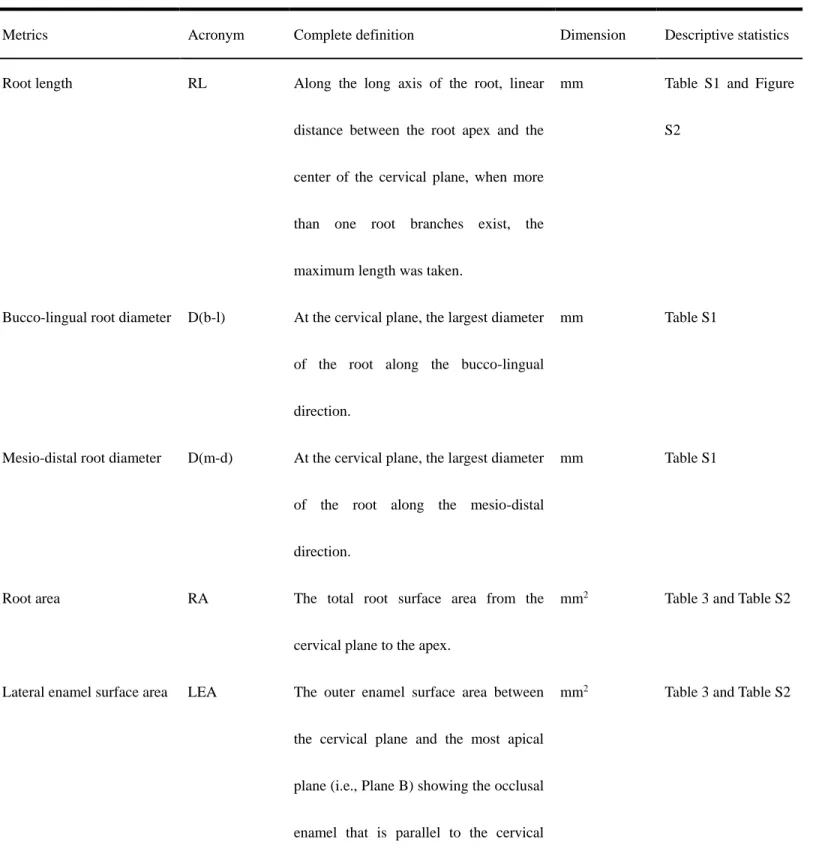
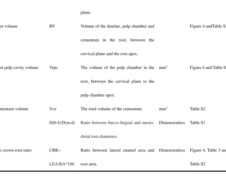
2.3 Root metrics and statistical tests
Using Avizo 8.0, premolar root metrics were assessed using published methods (Kupczik and Dean,
2008; Kupczik and Hublin, 2010; Le Cabec et al., 2013; Moore et al., 2013; Kupczik et al., 2019). Seven
variables were collected (Fig. 1 and Table 2): root length (RL, in mm, the maximum length was taken in
multi-rooted teeth), root diameter (the mesio-distal as well as the bucco-lingual diameters, in mm), lateral
enamel surface area (LEA, mm2), root surface area (RA, mm2), root volume (RV, mm3), root pulp cavity
volume (Vrpc, mm3) and cementum volume when present (Vce, mm3). Two indices were derived from the
mesio-distal root diameters (D(b-l)/D(m-d)).
For separating root(s) from the crown, we defined 10
–
20 landmarks along the cervical line (Fig. 1) andused the “slice—points to fit” option in Aviso to generate a cervical plane (i.e., the best-fit plane of the
cervical line): the crown and root(s) were then sectioned through the cervical plane (plane A). Root length is
the (maximum) distance between root apex to plane A, root diameters were measured based on plane A. Root
volume and the volume of the root pulp chamber were also measured based on the cervical plane (see Fig. 1
and Table 2 for detailed definition of parameters). Plane A was then moved gradually to the occlusal side of
the tooth, until the most apical plane (plane B) showing the occlusal enamel was located. The enamel area
between planes A and B is the lateral enamel surface area (LEA, mm2), the root surface area from plane A to
the root apex is the root area (RA, mm2). Differences in the distribution of LEA, RA and CRR between
groups were tested using the rank-based Kruskal-Wallis test with Conover's post hoc comparisons (see Table
S3 for details).
2.4 Geometric morphometric analyses
We applied a recently developed geometric morphometric (GM) landmark-free approach to compare
the root external surface shape (see previous applications on primate teeth in Beaudet et al., 2016; Zanolli et
al., 2018). This method relies on the construction of group-average surface models and their deformation to
the investigated surfaces (Durrleman et al., 2012a, b; Dumoncel et al., 2014). The surfaces are represented
by a set of oriented faces and the comparison does not assume a point-to-point correspondence between
samples, as in classical landmark-based GM analyses. First, the unscaled root surfaces of each tooth class
were aligned together using the “Align Surface” module in Avizo 8.0. Then deformations between surfaces
smooth, invertible, and with a smooth inverse, which is particularly appropriate for comparing overall shapes
and local orientation in the field of computational anatomy (Glaunes and Joshi, 2006; Durrleman et al., 2014).
From a set of surfaces, together with a set of initial control points located near its most variable parts, a mean
shape and a set of momenta parameterizing the deformations of the mean shape to each individual were
estimated (Durrleman et al., 2012a, b). Using the packages ade4 1.7-13 (Dray and Dufour, 2007) and Morpho
v.2.6 (Schlager et al., 2018) for R v.3.5.1 (R Development Core Team, 2018), a between-group principal
component analysis (bgPCA) (Mitteroecker and Bookstein, 2011) was performed. We first conducted
principal component analysis (PCA) using deformation momenta to reduce the dimensions of our dataset,
and computed the bgPCA from a subset of PCA scores (8~10) which accumulates around 90% of the overall
shape variation. The three Chinese Homo sp. specimens were projected a posteriori in the bgPCA plot. The
residual of allometric signals were tested using the coefficient of determination (R2) of a multiple regression
(Bookstein, 1991) in which the explicative variable is the root area as proxy for size and the dependent
variables are the bgPC scores.
Results
3.1 Comparisons of external root morphology
Distal and lingual views of each East Asian fossil maxillary and mandibular premolar are shown in
Figures 2 and 3, together with representatives of the comparative sample. Most of the East Asian Middle
Pleistocene upper premolars show relatively derived features compared with East African early Homo (Wood
et al., 1988). Whereas the latter usually appear to be double or triple-rooted, the East Asian Middle
2A, C and I). This is similar to Early Pleistocene H. erectus from Sangiran (Fig. 2D and J) and clearly is
different from bifid or single-rooted premolars generally seen in later Homo (recent modern humans and
Neanderthals, Fig. 2E–H, K and L). Only one H. erectus specimen (PA 831 from Hexian) displays an early
Homo-like condition with three well-separated root branches that spread in different directions, covered with
thick cementum (Fig. 2B). The Homo sp. specimen PA 76 from Changyang has two nearly parallel fused
branches (with thick cementum at the apex) indented by a large and deep distal groove (Fig. 2C). This P3
configuration is similar to Indonesian H. erectus (Fig. 2D) and Neanderthals (Fig. 2E, F), and can be found
in modern humans, but the root of PA 76 differs considerably from Chinese H. erectus premolars whose
lingual root branch is markedly diverging (Fig. 2A and B). For the P4s, the only H. erectus from China (PA
68, Zhoukoudian site) shows sub-parallel branches linked together with short free apices (Fig. 2I), but a broad
generalization cannot be made on the basis of a single tooth. The pattern expressed for PA 68 (H. erectus
from Zhoukoudian site) is not found in any other specimens. Indonesian H. erectus shows diverging branches
at mid-root (Fig. 2J), while Neanderthals and modern humans tend to have fused roots until the apex with a
deep distal groove delineating the branches (Fig. 2K, L).
With regard to lower premolars, in contrast to the North African late Early Pleistocene hominins from
Tighenif that show a radical groove with two root apices (Fig. 3D, M and N), the East Asian hominins exhibit
a single root with one apex (Fig. 3A–C, I–L), even if some of them also express a slight degree of Tomes’
root. The East Asian fossil hominins mostly differ from later Homo by their more vertically oriented and
pillar-like root axis, as well as a considerable amount of cementum deposit which makes them more robust
than modern humans (Fig. 3D, L), while the root apex tends to be leaning more distally in the latter groups
branches (also reported by Xing et al., 2018; Zanolli et al., 2018). As for the late Middle Pleistocene Homo
sp., the slender root with sharp apical-third of the P3 PA 1578 (site Panxian Dadong) (Fig. 3C) resembles the
morphology found in modern humans, while the P4 specimen PA 81 (site Changyang) (Fig. 3L) has three
fused root branches, as the cementum covers 3/4 of the root body, the overall morphology of PA 81 root
approximates H. erectus condition.
3.2 Root dimensions and volumetric measurements
HEC and HNA display the largest root volume, followed by Neanderthals and modern humans.
Sangiran H. erectus premolars exhibit a low total root volume in P3 that is comparable to modern humans
(Fig. 4A, B; Tables S1 and S2), but Sangiran P4s have a larger root volume, falling within the distribution of
Neanderthals. The late Middle Pleistocene Homo sp. PA 76 P3 (Changyang site) and PA 1578 P
3 (Panxian
Dadong site) show a root volume that is in the range of both Chinese H. erectus and Neanderthals (Fig. 4A), ,
but PA 81 P4 from Changyang exhibits a large root volume that exceeds all other lower premolars examined
here (Fig. 4B).
Chinese H. erectus and North African Homo have the largest root pulp chamber volume, followed by
Neanderthals. Modern humans display the smallest pulp volume among groups (Figs. 4C, D). PA 1578 P3
and PA 81 P4 exhibit large pulp volume, reaching (or even exceeding) the range of earlier groups like HNA
and HEC (Figs. 4C, D). Root pulp chamber volume of the only Sangiran H. erectus specimen that we could
segment falls within the range of Neanderthals (Fig. 4C), as does that of PA 76 P3 from Changyang.
We were able to clearly segment the cementum layers in six of our eleven East Asian Middle Pleistocene
hominins and some of the Neanderthal specimens (mainly from La Chaise-Abri-Bourgeois-Delaunay and
Regourdou). These specimens are clearly distinct from modern humans both in the presence and volume of
cementum, as easily seen on the root surface (Figs. 2 and 3). Le Cabec et al. (2013) reported an extensive
degree of hypercementosis in Krapina Neanderthals. Unfortunately, the (possible) presence of a cementum
layer is difficult to identify on the Krapina premolar examined here from the micro-CT scans. Due to strong
fossilization, cementum deposits cannot be identified in Javanese H. erectus either. Further quantification of
the volume as well as the 3D distribution of cementum in these specimens will require higher-quality data.
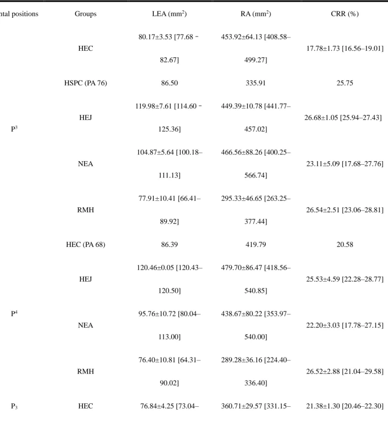
3.3 Crown-root surface area proportions
Nonparametric tests reveal significant differences in lateral enamel area (LEA) and root area (RA)
among groups, but no significant differences among groups in crown root ratio (CRR). Conover’s post hoc
tests demonstrate distinctions between fossil and modern taxa (Table S3), but it should be pointed out that
our sample size is small, statistical inferences should be addressed with caution.
For the P3, the lowest CRR values are found in the Chinese H. erectus specimens PA 67 (from
Zhoukoudian) and PA 832 (from Hexian) (Table 3 and Fig. 4E), suggesting a relatively large root surface
area, though these are not statistically significant. It overlaps with the lower range of Neanderthals, but not
with the values of Sangiran 4 and recent modern humans (Fig. 4E). The CRR of PA 76 (from Changyang) is
near the average for modern humans and overlaps the Neanderthal range (Fig. 4E). Modern humans have
significantly smaller absolute enamel and root surface area than Sangiran 4, early to mid-Middle Pleistocene
East Asian H. erectus and Neanderthals (Table S3), but do not differ from the estimates of the P3 PA 76 from
Changyang. For the P4, the Zhoukoudian H. erectus specimen PA 68 shows a similar signal to the P3s, with
(Table 3 and Fig. 4F). Modern humans have significantly smaller root area than all the fossil groups (Table
3 and Table S3), resulting in higher crown-root proportions (Fig. 4F). One late Middle Pleistocene Chinese
Homo sp. specimen (P3 PA 1578 from Panxian Dadong) shows a high CRR value, even exceeding the high
estimates of Neanderthals and modern humans (Fig. 4F). In contrast, the CRR ratio of the P4 PA 81 from
Changyang is considerably lower than the other groups, including the early to mid-Middle Pleistocene
Chinese H. erectus specimens (Table 3, Fig. 4F and Table S2).
3.4 Geometric morphometric analyses
GM analysis of root shape shows substantial morphological distinctions among groups (Fig. 5). There
is also an allometric signal observed along bgPC1 and/or bgPC2 for tooth positions P3, P4 and P
4 (Table S4),
mostly distinguishing the late Early/early Middle Pleistocene specimens from North Africa and Indonesia
from the Eurasian groups fossil and extant groups. H. erectus specimens from Sangiran and the late Early
Pleistocene North African Homo specimens from Tighenif are distinguished morphologically from modern
humans and Neanderthals along bgPC1, the former showing separated root branches while the latter mostly
exhibit fused roots/single root. Regarding P3s, recent modern humans are placed at the upper part of bgPC2
by showing separated root branches (Fig. 5A), while Neanderthals show incompletely separated roots. Two
of the East Asian specimens, the Zhoukoudian H. erectus specimen PA 67 and the late Middle Pleistocene
Homo sp. specimen PA 76 from Changyang site show similarities with Neanderthals, while the Hexian H.
erectus tooth PA 832 exhibits a primitive condition with three separated root branches (Fig. 5A). The Chinese
H. erectus specimen PA 68 falls between the ranges of of later the Homo groups (Fig. 5B), showing single
root with bifid apex. Fused root branches separate younger hominin groups from Sangiran H. erectus.
strongly expressed radical grooves at the distal or distolingual face of the root, and a tendency for separated
root tips at the apex. The H. erectus P3s from China (PA 110 Zhoukoudian, PA 526 Xichuan) are located at
the lower half of bgPC2, indicating that they tend to have stout root body and blunt apex (Fig. 5C), in
accordance with gross observation (Fig. 3E, F). In contrast, the root shape of Homo sp. specimen PA 1578
from Panxian Dadong falls within the modern human range and is quite distant from Neanderthals (Fig. 5C).
For the lower P4s, the Homo sp. specimen PA 81 from Changyang is closer to the Chinese H. erectus range,
showing an intermediate morphology between the more archaic figure represented by the Tighenif premolars
and the Neanderthal root shape (Fig. 5D).
4. Discussion
Premolar root anatomy in great apes and hominins has long been recognized to reflect taxonomy,
phylogeny, and function (e.g., Wood et al., 1988; Kupczik et al., 2003; Prado-Simón et al., 2012a; Moore et
al., 2016). Several studies have discovered a mosaic of primitive and derived dental traits among the
so-called archaic H. sapiens or post-erectus Homo from the East Asian mid- to late Middle Pleistocene (e.g.,
specimens from Panxian Dadong and Changyang) (Chia, 1957; Wu and Poirier, 1995; Liu et al., 2013), but
these traits are not diagnostic enough to link these specimens to any particular Homo species, and therefore
the taxonomic assignment of these specimens remains inconclusive (Liu et al., 2013).
Recent observations on the root-canal configuration (Pan and Zanolli, 2019) as well as the qualitative
and quantitative comparisons of external root morphology presented here reveal a modern-like trend of root
fusion and root number reduction for the East Asian mid- to late Middle Pleistocene Homo sp. specimens.
They differ from Chinese H. erectus in that the latter taxon generally shows stout premolar root(s) with thick
premolar roots of East Asian H. erectus often spread in different directions (including H. erectus from Yiyuan
site reported by Xing et al., 2016). These features of in East Asian H. erectus could be interpreted as a
functional response to withstand high biomechanical forces and better sustain high occlusal loads (Macho
and Spears, 1999; Kupczik et al., 2005, 2018; Xing et al., 2018). In any case, the stout, pillar-like morphology
of Chinese H. erectus premolars represent an autapomorphic feature characterizing this fossil human group
(Weidenreich, 1937; Liu et al., 2017; Xing et al., 2018). The analyses presented here of root and cementum
volume further confirms that larger roots and considerable cementum deposits are commonly found in
East Asian H. erectus, late Early Pleistocene Homo from Tighenif and those mid- to late Middle Pleistocene
hominins found in China.
4.1 Implications of root metrics and, crown-root tissue proportions
Compared with modern humans, Neanderthals have larger root and pulp volume in both the anterior
(Le Cabec et al., 2013) and molar dentition (Kupczik and Hublin, 2010). Our results show that Neanderthals
display larger premolar roots as well, although our sample size is small. Compared with modern humans,
East Asian Middle Pleistocene hominins show larger root, pulp chamber and cementum volume. An
increased root volume and pulp chamber volume may be due to a high frequency of occlusal loads. In addition,
a large pulp chamber could allow for deposition of tertiary dentine (Le Cabec et al., 2012, 2013).
In hominids, root surface area is linked to masticatory function, with larger root surface area suggested
to be adaptively associated with higher occlusal loads and higher mechanical resistance of food items
(Kupczik et al., 2003, 2005, 2018; Spencer, 2003; Kupczik and Dean, 2008). It has been suggested that the
larger molar root area in Neanderthals as compared to recent and Pleistocene H. sapiens could indicate that
on finite element analyses showed that the taurodont morphology of the Neanderthal post-canine dentition
did not improve the functional biomechanics of the tooth (Benazzi et al., 2014). Thus it is still uncertain if
the enlargement of premolar root area observed in Chinese H. erectus, together with their robust and stout
premolar roots (Xing et al., 2018), reflects an adaptation to mechanically resistant diet, or to other
evolutionary processes. Considering that their average premolar root area approximates that of Neanderthals,
we hypothesize that it more likely represents an adaptation to a high attrition diet or is a by-product of
pleiotropy or genetic drift (Benazzi et al., 2014). Future analyses based on finite element analyses will help
to resolve this question.
4.2 Taxonomic and phylogenetic value of external root shape
External root shape carries a taxonomic signal, but due to the lack of homologous landmarks, 3D
morphometric analysis is often limited to an broad approximation of the real root morphology (e.g., Emonet,
2009, 2012; Kupczik et al., 2018). (Semi)landmark-based GM is effective in depicting root morphology of
the anterior dentition, as the surface relief of incisors and canines is relatively simple with few bifurcations
or grooves (Le Cabec et al., 2013). In contrast to the above-mentioned studies, this analysis uses
deformation-based GM that for the first time compares the complete premolar root surface morphology in fossil and
modern humans, and explore the reliability of the radicular conformation for assessing taxonomy. Overall,
there is a clear distinction between the Early Pleistocene and Late Pleistocene to Holocene human groups.
The different root morphology of Neanderthals and modern humans are also clearly identified in these GM
analyses. However, Chinese early Middle Pleistocene H. erectus premolar roots (PA 67 P3 and PA 68 P4,
Zhoukoudian site, 800–770 Ka) tend to show a morphology that either approximates the Neanderthal
hominin from Changyang (PA 76 P3) is closely placed with one East Asian H. erectus specimen (PA 67), and
they both fall close to the range of Neanderthals. Whether this result indicates a phylogenetic link between
Chinese H. erectus and Neanderthals (or with the still underrepresented Denisovans which shows Tomes’
root in P3, a trait that is commonly seen in fossil hominins but is also seen in modern humans (Chen et al.,
2019)) or reflects homoplasy will need to be assessed in the future, integrating evidence from the crown
morphology and notably from the enamel-dentine junction, which is recognized as offering significant
phylogenetic information (e.g., Macchiarelli et al., 2006, 2013; Skinner et al., 2008a, b, 2009; Zanolli et al.,
2014; Zanolli, 2015; Pan et al., 2016, 2017).
Considering that the fossil and modern human groups included here can be clearly distinguished by
premolar morphology, we wanted to evaluate where the mid- to late Middle Pleistocene hominin specimens
from China would fit. As previously mentioned, the P3 specimen dated to the late Middle Pleistocene period
from Changyang falls next to the range of Neanderthals (Fig. 5A) and shows a close affinity with one early
Middle Pleistocene H. erectus (PA 67 from Zhoukoudian site); the mid-Middle Pleistocene P3 (PA 1578 from
Panxian Dadong), however, is clearly situated within the modern human distribution (Fig. 5C). The P4
specimen from Changyang (PA 81) shares a similar shape with older Chinese H. erectus and is distinguished
from the Early Pleistocene specimens from Tighenif, as well as from Neanderthals and modern humans (Fig.
5D). The root of PA 81 is quite robust, three-grooved and exhibits a thick cementum deposit (Pan and Zanolli,
2019). The absolutely large enamel and root area, large root and pulp chamber volume also point toward a
H. erectus-like signal. We thus interpret the root structural signature of this specimen as retaining primitive
features, compatible with the hypothesis that a H. erectus-like lineage survived in the late Middle Pleistocene.
H. erectus, it has smaller pulp chamber volume and relative root surface area within the range of modern
humans instead of H. erectus. Further analysis on other morphological aspects of this specimen is needed
before a taxonomic assessment can be made with any confidence. On the other hand, the late Middle
Pleistocene hominin PA 1578 from Panxian Dadong is clearly distinct from any of the Pleistocene samples
and falls within the variability of modern humans. In fact, previous study on crown morphology of the four
hominin teeth found at Panxian Dadong (I1, C1, P3, and P3) suggested that these teeth show a combination of
archaic and modern-like features that is in line with Middle and Upper Pleistocene populations from East and
West Asia (Liu et al., 2013). Our observations on the root morphology further recognize the primitive
morphology of Panxian Dadong P3 (e.g., a relatively stout root compared with modern humans, and
considerable cementum covering 2/3 part or the root, Fig. 3L). On the other hand, small root and pulp
chamber volume and a small relative root surface area place PA 1578 close to modern humans. We concur
that this specimen likely belonged to an ancient population that is close to modern humans. Based on crown
and root morphology, these “archaic H. sapiens” or “non-erectus” hominins like Panxian Dadong (Liu et al.,
2013), and Tongzi (Xing et al., 2019) from the mid- to late Middle Pleistocene and the Xujiayao hominin
from the early Late Pleistocene (Xing et al., 2015) express remarkable derived features that is typically found
in later Homo lineages. The dispersal of these “non-H. erectus” hominins might be further clarified following
a comparative study on the dentition of the Denisovan specimen from the Xiahe site (Chen et al., 2019).
Moreover, it is important to underscore that there is root shape variation in the East Asian H. erectus sample:
for instance, robust and fully separated root branches are found in PA 832 P3, a mid-Middle Pleistocene H.
erectus from Hexian site, while the older PA 67 P3 from Zhoukoudian shows incompletely separated double
with previous studies on crown morphology (Xing et al., 2014; Liu et al., 2017).
Previous investigations of hominin dental crown morphology using GM techniques (Martinón-Torres
et al., 2006; Gómez-Robles et al., 2007, 2008) have observed different evolutionary trends in different dental
positionswithin the same tooth class. For example, Neanderthals show a primitive P4 morphology but a
derived P3. Furthermore, morphogenesis of the upper and lower dentition are under the control of different
genetic programs (Thomas et al., 1997; McCollum and Sharpe, 2001). Given this, we emphasize that any
taxonomic interpretation of our results must be approached with caution and placed within a larger context—
especially with the incorporation of other dental morphological evidence. Nonetheless, our preliminary
results show that in contrast to non-metric configurations (e.g., root number and form (Abbott, 1984; Wood
et al., 1988; Moore et al., 2015; Moore et al., 2016)) and standard metrics (e.g., root length and width
(Kupczik and Hublin, 2010; Prado-Simón et al., 2012b; Le Cabec et al., 2013)), GM analysis of premolar
root surface is an useful indicator for taxonomy. On the other hand, we caution that other than root length
and some particular metrics that distinguish the roots of modern humans vs. Neanderthals (e.g., Le Cabec et
al., 2012, 2013), root shape and structure are highly phenotypically plastic and quite variable within groups.
Recent studies have demonstrated that there is considerable variation in premolar root number within a single
species, and even in the same individual (Kupczik et al., 2005; Shields, 2005; Moore et al., 2015, 2016).
However, as shown in our GM analyses, despite differences in root shape, it is still possible to quantify
differences between recent humans and Pleistocene human groups/taxa and separate them.
5. Conclusion
This study first provides a quantitative comparison of permanent maxillary and mandibular premolar
root and pulp chamber volume and a considerable amount of cementum deposit are commonly observed in
East Asian Middle Pleistocene H. erectus (Xing et al., 2018; this study). Together with previous
landmark-based GM studies on hominoid molars (Emonet et al., 2012), the present work suggests that external root
surface morphology is taxonomically informative. The 3D landmark-free approach to quantify dental root
surface morphology in fossil and modern humans, and used here is able to discriminate among several
hominin groups, and enabled evaluation of the taxonomic affinities of a number of East Asian Middle
Pleistocene hominin specimens. Integration of a larger sample and other dental evidence from more diverse
time periods localities should help to better characterize the spatial-temporal dispersal of multiple Homo
lineages in East Asia. In addition, as molar root form is more conservative in its phenotypic expression,
further study on molar root surface morphology may also provide useful information on the evolutionary
relationships in fossil hominins.
Crown-root metrics (lateral enamel area, root area and crown-root ratio) together with external root GM
analyses suggest that among the three Chinese mid- to late Middle Pleistocene Homo sp. specimens
investigated here, the two premolars from Changyang (the P3 PA 76 and the P4 PA 81) retain some primitive
features in their root structure and are possibly late representatives of a H. erectus-like lineage. Conversely,
the third specimen (P3 PA 1578 from Panxian Dadong) exhibits unambiguous modern-like features and may
represent evidence for an early presence of modern humans in Asia. In addition, recent study on the 240–172
Ka dental remains from Tongzi site, China hints at the existence of multiple paleodemes in East Asian during
mid- to late Middle Pleistocene period (Xing et al., 2019). Even if the available dating ranges of these
specimens do not fully overlap, the penecontemporaneity of the Changyang and Panxian Dadong hominins
human lineages in East Asia during the late Middle Pleistocene.
References
Abbott, S.A., 1984. A comparative study of tooth root morphology in the great apes, modern man and early hominids. Ph. D. Dissertation, University of London.
An, Z.S., Kun, H.C., 1989. New magnetostratigraphic dates of Lantian Homo erectus. Quaternary Research 32, 213–221.
Bailey, S.E., Liu, W., 2010. A comparative dental metrical and morphological analysis of a Middle Pleistocene hominin maxilla from Chaoxian (Chaohu), China. Quaternary International 211, 14–23. Beaudet, A., Dumoncel, J., Thackeray, F., Bruxelles, L., Duployer, B., Tenailleau, C., Bam, L., Hoffman, J., de Beer, F., Braga, J., 2016. Upper third molar internal structural organization and semicircular canal morphology in Plio-Pleistocene South African cercopithecoids. Journal of Human Evolution 95, 104–120. Benazzi, S., Nguyen, H.N., Kullmer, O., Hublin, J.-J., 2014. Exploring the biomechanics of taurodontism. Journal of Anatomy 226, 180–188.
Bookstein, F., 1991. Morphometric Tools for Landmark Data: Geometry and Biology. Cambridge University Press, Cambridge.
Bosshardt, D.D., Selvig, K.A., 1997. Dental cementum: the dynamic tissue covering of the root. Periodontology 2000 13, 41–75.
Braga, J., Zimmer, V., Dumoncel, J., Samir, C., de Beer, F., Zanolli, C., Pinto, D., Rohlf, F.J., Grine, F.E., 2019. Efficacy of diffeomorphic surface matching and 3D geometric morphometrics for taxonomic discrimination of Early Pleistocene hominin mandibular molars. Journal of Human Evolution 130, 21–35. Chen, F., Welker, F., Shen, C.-C., Bailey, S.E., Bergmann, I., Davis, S., Xia, H., Wang, H., Fischer, R., Freidline, S.E., 2019. A late Middle Pleistocene Denisovan mandible from the Tibetan Plateau. Nature 569,
409.
Chia, L., 1957. Notes on the human and some other mammalian remains from Changyang, Hupei. Vertabrata Palasiatica 1, 179–190.
Coleman, M.N., Colbert, M.W., 2007. CT thresholding protocols for taking measurements on three-dimensional models. American Journal of Physical Anthropology 133, 723–725.
Dou, L., Li, D., Xu, T., Tang, Y., Yang, D., 2017. Root anatomy and canal morphology of mandibular first premolars in a Chinese population. Scientific Reports 7, 750.
Dray, S., Dufour, A.-B., 2007. The ade4 package: implementing the duality diagram for ecologists. Journal of statistical software 22, 1–20.
Dumoncel, J., Durrleman, S., Braga, J., Jessel, J.-P., Subsol, G., 2014. Landmark-free 3D method for comparison of fossil hominins and hominids based on endocranium and EDJ shapes. American Journal of Physical Anthropology 153, suppl. 56, 110.
Durrleman, S., Pennec, X., Trouvé, A., Ayache, N., Braga, J., 2012a. Comparison of the endocranial ontogenies between chimpanzees and bonobos via temporal regression and spatiotemporal registration. Journal of Human Evolution 62, 74–88.
Durrleman, S., Prastawa, M., Korenberg, J.R., Joshi, S., Trouvé, A., Gerig, G., 2012b. Topology preserving atlas construction from shape data without correspondence using sparse parameters. In: Ayache, N., Delingette, H., Golland, P., Mori, K. (Eds.), Proceedings of Medical Image Computing and Computer Assisted Intervention. Nice, France, pp. 223–230
Durrleman, S., Prastawa, M., Charon, N., Korenberg, J.R., Joshi, S., Gerig, G., Trouvé, A., 2014. Morphometry of anatomical shape complexes with dense deformations and sparse parameters. NeuroImage 101, 35–49.
Emonet, E.-G., 2009. Khoratpithecus et la radiation des hominoïdes en Asie du Sud-Est au Miocène. Ph. D. Dissertation, Université de Poitiers.
Emonet, E.-G., Tafforeau, P., Chaimanee, Y., Guy, F., De Bonis, L., Koufos, G., Jaeger, J.-J., 2012. Three-dimensional analysis of mandibular dental root morphology in hominoids. Journal of Human Evolution 62, 146–154.
Emonet, E.G., Andossa, L., Taïsso Mackaye, H., Brunet, M., 2014. Subocclusal dental morphology of Sahelanthropus tchadensis and the evolution of teeth in hominins. American Journal of Physical Anthropology 153, 116–123.
Fajardo, R.J., Ryan, T., Kappelman, J., 2002. Assessing the accuracy of high-resolution X-ray computed tomography of primate trabecular bone by comparisons with histological sections. American Journal of Physical Anthropology 118, 1–10.
Glaunes, J.A., Joshi, S., 2006. Template estimation form unlabeled point set data and surfaces for computational anatomy. In: Pennec, X., Joshi, S. (Eds.), 1st MICCAI Workshop on Mathematical Foundations of Computational Anatomy: Geometrical, Statistical and Registration Methods for Modeling Biological Shape Variability. Copenhagen, Denmark, pp. 29–39
Gómez-Robles, A., Martinón-Torres, M., Bermúdez de Castro, J.M., Margvelashvili, A., Bastir, M., Arsuaga, J.L., Pérez-Pérez, A., Estebaranz, F., Martínez, L.M., 2007. A geometric morphometric analysis of hominin upper first molar shape. Journal of Human Evolution 53, 272–285.
Gómez-Robles, A., Martinón-Torres, M., Bermúdez de Castro, J.M., Prado, L., Sarmiento, S., Arsuaga, J.L., 2008. Geometric morphometric analysis of the crown morphology of the lower first premolar of hominins, with special attention to Pleistocene Homo. Journal of Human Evolution 55, 627–638.
Grün, R., Huang, P.-H., Wu, X., Stringer, C.B., Thorne, A.G., McCulloch, M., 1997. ESR analysis of teeth from the palaeoanthropological site of Zhoukoudian, China. Journal of Human Evolution 32, 83–91. Grün, R., Huang, P.-H., Huang, W., McDermott, F., Thorne, A., Stringer, C.B., Yan, G., 1998. ESR and U-series analyses of teeth from the palaeoanthropological site of Hexian, Anhui Province, China. Journal of Human Evolution 34, 555–564.
Huang, P.-H., Jin, S.-Z., Peng, Z.-C., Liang, R.-Y., Lu, Z.-J., Wang, Z.-R., Chen, J.-B., Yuan, Z.-X., 1993. ESR dating of tooth enamel: comparison with U-Series, FT and TL dating at the Peking Man Site. Applied Radiation and Isotopes 44, 239–242.
Huffman, O.F., 2001. Geologic context and age of the Perning/Mojokerto Homo erectus, East Java. Journal of Human Evolution 40, 353–362.
history of the Early Pleistocene hominids of Java: dentognathic evidence. American Journal of Physical Anthropology 128, 709–726.
Kaifu, Y., Kono, R.T., Sutikna, T., Saptomo, E.W., Jatmiko, Awe, R.D., Baba, H., 2015. Descriptions of the dental remains of Homo floresiensis. Anthropological Science 123, 129–145.
Karkanas, P., Schepartz, L.A., Miller-Antonio, S., Wang, W., Huang, W., 2008. Late Middle Pleistocene climate in southwestern China: inferences from the stratigraphic record of Panxian Dadong Cave, Guizhou. Quaternary Science Reviews 27, 1555–1570.
Kupczik, K., Spoor, F., Dean, M.C., 2003. Tooth root morphology and masticatory muscle force pattern in humans and nonhuman primates. American Journal of Physical Anthropolology Supplement 36, 134. Kupczik, K., Spoor, F., Pommert, A., Dean, M.C., 2005. Premolar root number variation in hominoids: genetic polymorphism vs. functional significance, In: Żądzińska E (Ed.), Current Trends in Dental Morphology Research. University of Lodz Press, Lodz, pp. 257–268.
Kupczik, K., Dean, M.C., 2008. Comparative observations on the tooth root morphology of Gigantopithecus blacki. Journal of Human Evolution 54, 196–204.
Kupczik, K., Olejniczak, A.J., Skinner, M.M., Hublin, J.-J., 2009. Molar crown and root size relationship in anthropoid primates. Frontiers in Oral Biology 13, 16–22.
Kupczik, K., Hublin, J.-J., 2010. Mandibular molar root morphology in Neanderthals and Late Pleistocene and recent Homo sapiens. Journal of Human Evolution 59, 525–541.
Kupczik, K., Toro-Ibacache, V., Macho, G.A., 2018. On the relationship between maxillary molar root shape and jaw kinematics in Australopithecus africanus and Paranthropus robustus. Royal Society Open Science 5, 180825.
Kupczik, K., Delezene, L.K., Skinner, M.M., 2019. Mandibular molar root and pulp cavity morphology in Homo naledi and other Plio-Pleistocene hominins. Journal of Human Evolution 130, 83–95.
Le Cabec, A., Kupczik, K., Gunz, P., Braga, J., Hublin, J.-J., 2012. Long anterior mandibular tooth roots in Neanderthals are not the result of their large jaws. Journal of Human Evolution 63, 667–681.
Le Cabec, A., Gunz, P., Kupczik, K., Braga, J., Hublin, J.-J., 2013. Anterior tooth root morphology and size in Neanderthals: Taxonomic and functional implications. Journal of Human Evolution 64, 169–193.
Liu, W., Schepartz, L.A., Xing, S., Miller-Antonio, S., Wu, X., Trinkaus, E., Martinón-Torres, M., 2013. Late Middle Pleistocene hominin teeth from Panxian Dadong, South China. Journal of Human Evolution 64, 337– 355.
Liu, W., Martinón-Torres, M., Kaifu, Y., Wu, X., Kono, R., Chang, C.-H., Wei, P., Xing, S., Huang, W., Bermúdez de Castro, J.M., 2017. A mandible from the Middle Pleistocene Hexian site and its significance in relation to the variability of Asian Homo erectus. American Journal of Physical Anthropology 162, 715–731. Loh, H., 1998. Root morphology of the maxillary first premolar in Singaporeans. Australian Dental Journal 43, 399–402.
Macchiarelli, R., Bondioli, L., Debénath, A., Mazurier, A., Tournepiche, J.-F., Birch, W., Dean, C., 2006. How Neanderthal molar teeth grew. Nature 444, 748–751.
Macchiarelli, R., Bayle, P., Bondioli, L., Mazurier, A., Zanolli, C., 2013. From outer to inner structural morphology in dental anthropology: integration of the third dimension in the visualization and quantitative analysis of fossil remains, In: Scott, G.R., Irish, J. (Eds.), Anthropological perspectives on tooth morphology: Genetics, evolution, variation. Cambridge University Press, Cambridge, pp. 250-277.
Macho, G.A., Spears, I.R., 1999. Effects of loading on the biochemical behavior of molars of Homo, Pan, and Pongo. American Journal of Physical Anthropology 109, 211–227.
Martinón-Torres, M., Bastir, M., De Castro, J.B., Gómez, A., Sarmiento, S., Muela, A., Arsuaga, J., 2006. Hominin lower second premolar morphology: evolutionary inferences through geometric morphometric analysis. Journal of Human Evolution 50, 523–533.
Martinón-Torres, M., Bermúdez de Castro, J.M., Gómez-Robles, A., Arsuaga, J.L., Carbonell, E., Lordkipanidze, D., Manzi, G., Margvelashvili, A., 2007. Dental evidence on the hominin dispersals during the Pleistocene. Proceedings of the National Academy of Sciences 104, 13279–13282.
Martinón-Torres, M., Xing, S., Liu, W., Bermúdez de Castro, J.M., 2018. A “source and sink” model for East Asia? Preliminary approach through the dental evidence. Comptes Rendus Palevol, 33–43.
McCollum, M.A., Sharpe, P.T., 2001. Developmental genetics and early hominid craniodental evolution. BioEssays 23, 481-493.
Plio-Pleistocene site of Drimolen, South Africa. Journal of Human Evolution 58, 374–405.
Molnar, S., 1971. Human tooth wear, tooth function and cultural variability. American Journal of Physical Anthropology 34, 175–189.
Moore, N.C., Skinner, M.M., Hublin, J.J., 2013. Premolar root morphology and metric variation in Pan troglodytes verus. American Journal of Physical Anthropology 150, 632–646.
Moore, N.C., Hublin, J.J., Skinner, M.M., 2015. Premolar root and canal variation in extant non-human hominoidea. American Journal of Physical Anthropology 158, 209–226.
Moore, N.C., Thackeray, J.F., Hublin, J.-J., Skinner, M.M., 2016. Premolar root and canal variation in South African Plio-Pleistocene specimens attributed to Australopithecus africanus and Paranthropus robustus. Journal of Human Evolution 93, 46–62.
NESPOS database, 2019, NEanderthal Studies Professional Online Service.
Pan, L., Dumoncel, J., de Beer, F., Hoffman, J., Thackeray, J.F., Duployer, B., Tenailleau, C., Braga, J., 2016. Further morphological evidence on South African earliest Homo lower postcanine dentition: enamel thickness and enamel dentine junction. Journal of Human Evolution 96, 82–96.
Pan, L., Thackeray, J.F., Dumoncel, J., Zanolli, C., Oettlé, A., De Beer, F., Hoffman, J., Duployer, B., Tenailleau, C., Braga, J., 2017. Intra-individual metameric variation expressed at the enamel-dentine junction of lower post-canine dentition of South African fossil hominins and modern humans. American Journal of Physical Anthropology 163, 806–815.
Pan, L., Zanolli, C., 2019. Comparative observations on the premolar root and pulp canal configurations of Middle Pleistocene Homo in China. American Journal of Physical Anthropology 168, 637–646.
Prado-Simón, L., Martinón-Torres, M., Baca, P., Olejniczak, A.J., Gómez‐Robles, A., Lapresa, M., Luis Arsuaga, J., Bermúdez de Castro, J.M., 2012a. Three-dimensional evaluation of root canal morphology in lower second premolars of early and middle pleistocene human populations from Atapuerca (Burgos, Spain). American Journal of Physical Anthropology 147, 452–461.
Prado-Simón, L., Martinón-Torres, M., Baca, P., Gómez-Robles, A., Lapresa, M., Carbonell, E., Bermúdez de Castro, J.M., 2012b. A morphological study of the tooth roots of the Sima del Elefante mandible (Atapuerca, Spain): a new classification of the teeth—biological and methodological considerations.
Anthropological Science 120, 61–72.
Radovčić, J., Smith, F.H., Trinkaus, E., Wolpoff, M.H., 1988. The Krapina Hominids: An Illustrated Catalog of Skeletal Collection. Mladost and the Croatian Natural History Museum, Zagreb.
Rink, W.J., Schwarcz, H.P., Smith, F.H., Radovčić, J., 1995. ESR dates for Krapina hominids. Nature 378, 24.
Sahnouni, M., Van der Made, J., 2009. The Oldowan in North Africa within a biochronological framework, In: Schick, K., Toth, N. (Eds.), New Approaches to the Archaeology of Human Origins. Stone Age Institute Press Bloomington, pp. 179–209.
Schlager, S., Profico, A., Di Vincenzo, F., Manzi, G., 2018. Retrodeformation of fossil specimens based on 3D bilateral semi-landmarks: Implementation in the R package “Morpho”. PLOS ONE 13, e0194073. Scott, G.R., Turner II, C.G., Townsend, G., Martinón-Torres, M., 2018. The Anthropology of Modern Human Teeth. Dental Morphology and its Variation in Recent and Fossil Homo sapiens. Cambridge University Press, Cambridge.
Selvig, K.A., 1965. The fine structure of human cementum. Acta Odontologica Scandinavica 23, 423–441. Sert, S., Bayirli, G.S., 2004. Evaluation of the root canal configurations of the mandibular and maxillary permanent teeth by gender in the Turkish population. Journal of Endodontics 30, 391–398.
Shen, G., Ku, T.-L., Cheng, H., Edwards, R.L., Yuan, Z., Wang, Q., 2001. High-precision U-series dating of Locality 1 at Zhoukoudian, China. Journal of Human Evolution 41, 679–688.
Shields, E.D., 2005. Mandibular premolar and second molar root morphological variation in modern humans: what root number can tell us about tooth morphogenesis. American Journal of Physical Anthropology 128, 299–311.
Skinner, M.M., Wood, B.A., Boesch, C., Olejniczak, A.J., Rosas, A., Smith, T.M., Hublin, J.-J., 2008a. Dental trait expression at the enamel-dentine junction of lower molars in extant and fossil hominoids. Journal of Human Evolution 54, 173–186.
Skinner, M.M., Gunz, P., Wood, B.A., Hublin, J.-J., 2008b. Enamel-dentine junction (EDJ) morphology distinguishes the lower molars of Australopithecus africanus and Paranthropus robustus. Journal of Human Evolution 55, 979–988.
Skinner, M.M., Gunz, P., Wood, B.A., Boesch, C., Hublin, J.J., 2009. Discrimination of extant Pan species and subspecies using the enamel–dentine junction morphology of lower molars. American Journal of Physical Anthropology 140, 234–243.
Spencer, M.A., 2003. Tooth-root form and function in platyrrhine seed-eaters. American Journal of Physical Anthropology 122, 325–335.
Sperber, G.H., 1974. Morphology of the cheek teeth of early South African hominids. Ph. D. Dissertation, University of the Witwatersrand.
Spoor, F., Hublin, J.-J., Braun, M., Zonneveld, F., 2003. The bony labyrinth of Neanderthals. Journal of Human Evolution 44, 141–165.
Thomas, B.L., Tucker, A.S., Qui, M., Ferguson, C.A., Hardcastle, Z., Rubenstein, J., Sharpe, P.T., 1997. Role of Dlx-1 and Dlx-2 genes in patterning of the murine dentition. Development 124, 4811–4818.
Tomes, C.S., 1923. A manual of dental anatomy: human and comparative. 8th ed. MacMillan Co., New York. Trinkaus, E., Maley, B., Buzhilova, A.P., 2008. Brief communication: Paleopathology of the Kiik‐Koba 1 Neandertal. American Journal of Physical Anthropology 137, 106–112.
Turner, C.G., 1981. Root number determination in maxillary first premolars for modern human populations. American Journal of Physical Anthropology 54, 59–62.
Turq, A., Jaubert, J., Maureille, B., Laville, D., 2008. Le cas des sépultures néandertaliennes du Sud-Ouest: et si on les vieillissait?, In: Vandermeersch, B., Cleyet-Merle, J.-J., Maureille, B., Turq, A. (Eds.), Première Humanité, Gestes Funéraires des Néandertaliens. Réunion des Musées Nationaux, Paris, pp. 40–41. Weidenreich, F., 1937. The dentition of Sinanthropus pekinensis: a comparative odontography of the hominids. Palaeontol. Sin. Series D I, 1–180.
White, T.D., Suwa, G., Simpson, S., Asfaw, B., 2000. Jaws and teeth of Australopithecus afarensis from Maka, Middle Awash, Ethiopia. American Journal of Physical Anthropology 111, 45–68.
Woo, J.-k., 1964. Mandible of Sinanthropus lantianensis. Current Anthropology 5, 98–101.
Wood, B., Engleman, C., 1988. Analysis of the dental morphology of Plio-Pleistocene hominids. V. Maxillary postcanine tooth morphology. Journal of Anatomy 161, 1–35.
hominids. IV. Mandibular postcanine root morphology. Journal of Anatomy 156, 107–139.
Wu, X., Wu, R., 1982. Human fossil teeth from Xichuan, Henan (In Chinese with English abstract). Vertabrata Palasiatica 20, 1–10.
Wu, X., Poirier, F.E., 1995. Human evolution in China: a metric description of the fossils and a review of the sites. Oxford University Press, New York.
Xing, S., 2012. Morphological variation of Zhoukoudian H. erectus teeth. Ph.D. Dissertation, Graduate University of Chinese Academy of Sciences.
Xing, S., Martinón-Torres, M., Bermúdez de Castro, J.M., Zhang, Y., Fan, X., Zheng, L., Huang, W., Liu, W., 2014. Middle Pleistocene hominin teeth from Longtan Cave, Hexian, China. PLOS ONE 9, e114265. Xing, S., Martinón‐Torres, M., Bermúdez de Castro, J.M., Wu, X., Liu, W., 2015. Hominin teeth from the early Late Pleistocene site of Xujiayao, Northern China. American Journal of Physical Anthropology 156, 224–240.
Xing, S., Sun, C., Martinón-Torres, M., Bermúdez de Castro, J.M., Han, F., Zhang, Y., Liu, W., 2016. Hominin teeth from the Middle Pleistocene site of Yiyuan, Eastern China. Journal of Human Evolution 95, 33–54.
Xing, S., Martinón-Torres, M., Bermúdez de Castro, J.M., 2018. The fossil teeth of the Peking Man. Scientific Reports 8, 1–11.
Xing, S., Martinón-Torres, M., Bermúdez de Castro, J.M., 2019. Late Middle Pleistocene hominin teeth from Tongzi, southern China. Journal of Human Evolution 130, 96–108.
Yamamoto, T., Hasegawa, T., Yamamoto, T., Hongo, H., Amizuka, N., 2016. Histology of human cementum: Its structure, function, and development. Japanese Dental Science Review 52, 63–74.
Yuan, S., Chen, T., Gao, S., 1986. Uranium series chronological sequence of some palaeolithic sites in South China (In Chinese with English abstract). Acta Anthropologica Sinica 2, 179–190.
Zanolli, C., Mazurier, A., 2013. Endostructural characterization of the H. heidelbergensis dental remains from the early Middle Pleistocene site of Tighenif, Algeria. Comptes Rendus Palevol 12, 293–304.
Zanolli, C., Bondioli, L., Coppa, A., Dean, C.M., Bayle, P., Candilio, F., Capuani, S., Dreossi, D., Fiore, I., Frayer, D.W., 2014. The late Early Pleistocene human dental remains from Uadi Aalad and Mulhuli-Amo
(Buia), Eritrean Danakil: macromorphology and microstructure. Journal of Human Evolution 74, 96–113. Zanolli, C., 2015. Brief communication: molar crown inner structural organization in Javanese Homo erectus. American Journal of Physical Anthropology 156, 148–157.
Zanolli, C., Pan, L., Dumoncel, J., Kullmer, O., Kundrát, M., Liu, W., Macchiarelli, R., Mancini, L., Schrenk, F., Tuniz, C., 2018. Inner tooth morphology of Homo erectus from Zhoukoudian. New evidence from an old collection housed at Uppsala University, Sweden. Journal of Human Evolution 116, 1–13.
Table 1. Composition of the study sample Specimen P3 P4 P3 P4 Provenance Chronological range a Citations b Middle Pleistocene H.
erectus from China
(HEC) PA 102 1 Chenjiawo 650–500 Ka (A) 1 PA 110 1 Layer 3 Zhoukoudian Loc. 1 320–282 Ka (B, C) 500 Ka (D) 2, 3
PMU M3887 c 1 Layer 5 550–500 Ka (E) 4, 5
PA 67 1 Layer 11 800–770 Ka (F) 2, 3 PA 68 1 PA 832 1 Hexian 437–387 Ka (E) 6, 7 PA 525 c 1 Xichuan Unknown 8 PA 526 c 1
Late Middle Pleistocene
Homo sp. from China
(HSPC) PA 76 1 Changyang 196–194 Ka (F) 9 PA 81 1 PA 1578 1 Panxian Dadong 296–233 Ka (G) 10 Early Pleistocene H. erectus (HEJ) Sangiran 4 2 2 Pucangan Fm. Sangiran 1.8–1.6 Ma (H) 5
Early Pleistocene Homo from North Africa (HNA)
Tighenif 1 2 2 Tighenif ~1.0 Ma (I) 11 Tighenif 2 1 1 Neanderthals (NEA) 3 7 4 5 Level 8 Krapina 140–120 Ka (J) 12, 13 1 2 1 Abri Bourgeois– Delaunay La Chaise 146–106 Ka (K) 13, 14 2 2 Regourdou 70 Ka (L) 13
Recent Modern Humans (RMH) 5 7 8 9 Asian/ European/ South African 15
a Age : (A) An and Kun (1989); (B) Grün et al. (1997); (C) Huang et al. (1993); (D) Shen et al. (2001); (E)
Grün et al. (1998); (F) Yuan et al. (1986); (G) Karkanas et al. (2008); (H) Huffman (2001); (I) Sahnouni and
Van der Made (2009); (J) Rink et al. (1995); (K) Macchiarelli et al. (2006) ; (L) Turq et al. (2008)
b Citations: (1) Woo (1964); (2) Xing (2012); (3) Xing et al. (2018); (4) Black et al. (1933); (5) Zanolli et al.
(2018); (6) Xing et al. (2014); (7) Liu et al. (2017); (8) Bailey and Liu (2010); (9) Chia (1957); (10) Liu et
al. (2013); (11) Zanolli and Mazurier (2013); (12) Radovčić et al. (1988); (13) NESPOS database (2019);
(14) Macchiarelli et al. (2006) ; (15) Pan et al. (2017)
Table 2. Definition of metrics taken for the study. Protocols proposed by Kupczik and Dean (2008);
Prado-Simón et al. (2012); Le Cabec et al. (2013).
Metrics Acronym Complete definition Dimension Descriptive statistics
Root length RL Along the long axis of the root, linear
distance between the root apex and the
center of the cervical plane, when more
than one root branches exist, the
maximum length was taken.
mm Table S1 and Figure
S2
Bucco-lingual root diameter D(b-l) At the cervical plane, the largest diameter
of the root along the bucco-lingual
direction.
mm Table S1
Mesio-distal root diameter D(m-d) At the cervical plane, the largest diameter
of the root along the mesio-distal
direction.
mm Table S1
Root area RA The total root surface area from the
cervical plane to the apex.
mm2 Table 3 and Table S2
Lateral enamel surface area LEA The outer enamel surface area between
the cervical plane and the most apical
plane (i.e., Plane B) showing the occlusal
enamel that is parallel to the cervical
plane.
Root volume RV Volume of the dentine, pulp chamber and
cementum in the root, between the
cervical plane and the root apex.
Figure 4 andTable S2
Root pulp cavity volume Vrpc The volume of the pulp chamber in the
root, between the cervical plane to the
pulp chamber apex.
mm3 Figure 4 and Table S2
Cementum volume Vce The total volume of the cementum. mm3 Table S2
D(b-l)/D(m-d) Ratio between bucco-lingual and
mesio-distal root diameters.
Dimensionless Table S1
The crown-root ratio CRR=
LEA/RA*100
Ratio between lateral enamel area and
root area.
Dimensionless Figure 4, Table 3 and
Table 3. Average values for the lateral enamel area (LEA), root area (RA) and crown-root ratio (CRR)
measured in the later Middle Pleistocene Homo sp. from China, together with comparative samples.
Dental positions Groups LEA (mm2) RA (mm2) CRR (%)
P3 HEC 80.17±3.53 [77.68– 82.67] 453.92±64.13 [408.58– 499.27] 17.78±1.73 [16.56–19.01] HSPC (PA 76) 86.50 335.91 25.75 HEJ 119.98±7.61 [114.60– 125.36] 449.39±10.78 [441.77– 457.02] 26.68±1.05 [25.94–27.43] NEA 104.87±5.64 [100.18– 111.13] 466.56±88.26 [400.25– 566.74] 23.11±5.09 [17.68–27.76] RMH 77.91±10.41 [66.41– 89.92] 295.33±46.65 [263.25– 377.44] 26.54±2.51 [23.06–28.81] P4 HEC (PA 68) 86.39 419.79 20.58 HEJ 120.46±0.05 [120.43– 120.50] 479.70±86.47 [418.56– 540.85] 25.53±4.59 [22.28–28.77] NEA 95.76±10.72 [80.04– 113.00] 438.67±80.22 [353.97– 540.00] 22.20±3.03 [17.78–27.15] RMH 76.40±10.81 [64.31– 90.02] 289.28±36.16 [224.40– 336.40] 26.52±2.88 [21.04–29.58] P3 HEC 76.84±4.25 [73.04– 360.71±29.57 [331.15– 21.38±1.30 [20.46–22.30]
79.85] 390.28] HSPC (PA 1578) 93.60 336.72 27.80 HNA 77.90±7.83 [68.94– 83.42] 391.95±31.16 [331.15– 390.28] 20.06±3.48 [16.21–22.99] NEA 75.37±15.60 [56.34– 103.82] 327.92±62.69 [260.64– 386.01] 23.02±3.75 [17.12–26.90] RMH 64.46±13.25 [49.16– 86.08] 261.65±64.19 [206.66– 335.04] 24.61±1.72 [21.24–26.87] P4 HEC 92.10±37.06 [65.89– 134.50] 353.65±49.76 [295.68– 395.20] 25.61±7.35 [20.51–26.51] HSPC (PA 81) 81.38 422.37 19.27 HNA 111.60±8.62 [103.30– 120.50] 456.60±10.60 [447.00– 468.20] 24.47±2.24 [22.06–26.51] NEA 86.16±18.50 [60.87– 110.60] 330.04±65.06 [261.08– 391.20] 26.16±4.86 [19.32–32.80] RMH 73.81±6.84 [60.46– 82.44] 267.71±38.39 [224.20– 300.98] 27.78±2.36 [25.24–33.21]
Abbreviations as follows: HEJ: Javanese H. erectus; HEC: Middle Pleistocene H. erectus from China; HSPC:
late Middle Pleistocene Homo sp. from China; HNA: Early Pleistocene Homo from North Africa; NEA:
Neanderthals; RMH: Recent modern humans. Data was reported in the form mean value ± SD [minimum–