HAL Id: tel-02410263
https://tel.archives-ouvertes.fr/tel-02410263
Submitted on 13 Dec 2019HAL is a multi-disciplinary open access
archive for the deposit and dissemination of sci-entific research documents, whether they are pub-lished or not. The documents may come from teaching and research institutions in France or abroad, or from public or private research centers.
L’archive ouverte pluridisciplinaire HAL, est destinée au dépôt et à la diffusion de documents scientifiques de niveau recherche, publiés ou non, émanant des établissements d’enseignement et de recherche français ou étrangers, des laboratoires publics ou privés.
Ciliogenesis Control Mechanisms in Cerebellar Neuron
Progenitors
Marco Zanini
To cite this version:
Marco Zanini. Ciliogenesis Control Mechanisms in Cerebellar Neuron Progenitors. Subcellular Pro-cesses [q-bio.SC]. Université Paris-Saclay, 2019. English. �NNT : 2019SACLS475�. �tel-02410263�
Ciliogenesis control
mechanisms in cerebellar
neuron progenitors
Thèse de doctorat de l'Université Paris-Saclay préparée à l’Université Paris Sud
École doctorale n°582 Cancérologie : biologie, médecine, santé Spécialité de doctorat : Aspects moléculaires et cellulaires de la biologie
Thèse présentée et soutenue à Orsay, le 5 Décembre 2019, par
Marco Zanini
Composition du Jury :
Simon Saule
PR1, Institut Curie, Orsay (CNRS UMR3347, Inserm U1021) Président
Julie Gavard
DR2, Centre de Recherche en Cancérologie et Immunologie
Nantes – Angers, Nantes (CNRS UMR6299, Inserm U892) Rapporteur
Frédéric Charron
Associate Professor, Department of Medicine, Institut de
Recherches Cliniques de Montréal, Montréal Rapporteur Lucia Di Marcotullio
PR, Department of Molecular Medicine, Rome Examinatrice
Olivier Ayrault
DR2, Institut Curie, Orsay (CNRS UMR3347, Inserm U1021) Directeur de thèse
NNT : 2 0 19 S A CL S 4 7 5
3
Acknowledgements
The first person I wish to acknowledge is my chief and PhD supervisor, Olivier. I wish to thank him for having hired me as a Master intern more than four years ago and for having renewed his trust one year later, when I became a PhD student. I am immensely grateful for his mentoring and teaching activities, which enabled me to learn a lot about different aspects of science, not only the technical-related ones. I am thankful because he has always granted me with trust, liberty and independence, hence allowing me to explore my strengths, but also my weaknesses. Many thanks also for having been patient, when for various personal reasons I was perhaps not performing well at work.
I am very grateful to Dr. Julie Gavard and Dr. Frédéric Charron for having accepted to be members of my PhD defense jury and review my manuscript. I am also really thankful to Pr. Lucia Di Marcotullio and Pr. Simon Saule for being part of my PhD committee.
I would like also to express my gratitude to Chia-Hsiang Chang and Pr. Jin-Wu Tsai for their great work and for the successful collaboration we set up and carried on together in the context of this project.
An enormous thanks goes to all the people of the lab, past and present members, who were directly involved in this study: Hamasseh Shirvani for having started and managed the project before my arrival; Hua Yu, for having trained me during my Master and for having being extremely kind and willing to help me in every situation, not only at work. Audrey Mercier, for her continuous support and for having been always available to hear me and advise me, whatever it was the matter, science or personal life; Antoine Forget, because even though he has never directly supervised me, he was a milestone for me in the lab and I learnt a lot from him; Sara Maria Cigna for her help and tips during my Master.
A big thanks also to the people of the technical support staff of the Institute Curie in Orsay: Claire Lovo for her great help with microscopy, Sophie Leboucher for her always punctual
4
work on the tissue IHC, Charlène Lasgi for the assistance with the FACS, and the whole team of the animal facilities, in particular Christophe Alberti, Elodie Belloir and Adlin Thadal.
I wish also to thank a lot other present members of the lab for their support throughout various phases of my PhD: Julie Talbot, for her extreme kindness and for having (maybe without even realizing it) pushed me to start to speak French; Emilie Indersie for all her advice and the amusing time spent all together in the lab; Gabriele Cancila for his enthusiasm that in part contributed to relight my passion for research in the last months.
Even without being involved the project, some can be equally (if not more) important for its success, just by providing love and support to who is in charge of managing it.
I am immensely grateful to my friend Benedetta, for all the company and the moral help she has always given me in any circumstances along these last four years. She was always there when I needed to stay or talk with someone, and I thank her a lot for this.
An enormous dzięki goes to Maria for having been so patient in the last months, when I could not be as present as both of us wished. Thank you for having always stayed on my side, provided massive support and love, shared good and bad moments and always understood my needs. I am also very grateful to Flavia, who was actually a colleague of mine at the time of the project, but I prefer to thank her here, in this section, because the firm friendship we developed is now a stronghold on which I can always count. Thank you for everything.
Similarly, I wish to thank Ludovica, as I really enjoyed the evenings spent all together in Paris this year.
A great thanks then goes to all the big group of Italian friends in Paris for the great moments shared together in these years and for making me feeling home also here, abroad.
Un grand merci également à Romain, Grégoire et Kanok du labo Ghysdael pour les bons moments passés ensemble.
Thanks to all the people of the Domain 1 for the good and relaxing time spent during the Happy Fridays and the ReSiPis.
E infine, anche se dovrei metterli per primi, il più grosso ringraziamento tra tutti va alla mia famiglia, in particolare ai miei genitori perché nella buona e nella cattiva sorte, loro ci sono sempre stati e hanno rappresentato un punto fermo su cui fare stabile affidamento per qualunque cosa.
5
Table of Contents
A
CKNOWLEDGEMENTS
... 3T
ABLE OF
C
ONTENTS
... 5A
BBREVIATIONS
... 9T
ABLE OF
F
IGURES
... 15INTRODUCTION ... 17
I.1
T
HE
C
EREBELLUM
... 19I.1.1 ANATOMY OF THE CEREBELLUM ... 19
I.1.2 MAIN FUNCTIONS OF THE CEREBELLUM ... 21
I.1.2.1 Functional compartmentalization of the cerebellum ... 21
I.1.3 MICROANATOMY OF THE CEREBELLAR CORTEX ... 23
I.1.3.1 Cells of the cerebellar cortex ... 23
I.1.3.2 Major cerebellar circuits... 24
I.2
D
EVELOPMENT OF
G
RANULE
N
EURONS
... 27I.2.1 EARLY SPECIFICATION AND PATTERNING OF THE CEREBELLUM ... 27
I.2.1.1 Regionalization of the neural tube ... 27
I.2.1.2 Specification of cerebellar territories in rhombomere 1 ... 29
I.2.2 EARLY HISTOGENESIS IN THE CEREBELLAR ANLAGE ... 30
I.2.2.1 Temporal origin of uRL derivatives... 32
I.2.3 GNPs: TANGENTIAL MIGRATION FROM THE uRL TO THE EGL ... 34
I.2.4 GNPs: FROM PROLIFERATION TO TERMINAL DIFFERENTIATION ... 36
I.2.4.1 Brief overview of the whole process ... 36
I.2.4.2 Proliferation and differentiation of GNPs at the histological level ... 36
I.2.4.3 Discovery of SHH as the main mitogen for GNPs ... 38
I.2.4.4 SHH boosts GNPs expansion by acting on the cell cycle machinery ... 39
I.2.4.4 Synergy and interplay with SHH during GNPs expansion ... 40
I.2.4.4.1 SDF-1α ... 40
I.2.4.4.2 Igf-II ... 41
I.2.4.4.3 Heparan sulfate proteoglycans ... 42
I.2.4.4.4 Laminin-integrin signaling ... 42
I.2.4.4.5 Notch2-Hes1 signaling ... 43
I.2.4.5 Mechanisms controlling cell cycle exit and differentiation of GNPs ... 44
I.2.4.5.1 Non canonical Wnt3 signaling ... 45
6
I.2.4.5.3 Mycn protein degradation at mitosis ... 45
I.2.4.5.4 Ubiquitin ligases ... 46
I.2.4.5.5 Vitronectin ... 47
I.2.4.6 Tangential migration and onset of parallel fibers formation ... 47
I.2.4.7 Terminal radial migration toward the IGL... 48
I.3
M
EDULLOBLASTOMA
... 51I.3.1 EPIDEMIOLOGY OF MEDULLOBLASTOMA ... 51
I.3.1.1 Risk factors associated to medulloblastoma ... 52
I.3.2 CLINICOPATHOLOGICAL FEATURES OF MEDULLOBLASTOMA ... 53
I.3.2.1 Histological characteristics ... 53
I.3.2.2 Metastases and recurrence of medulloblastoma ... 54
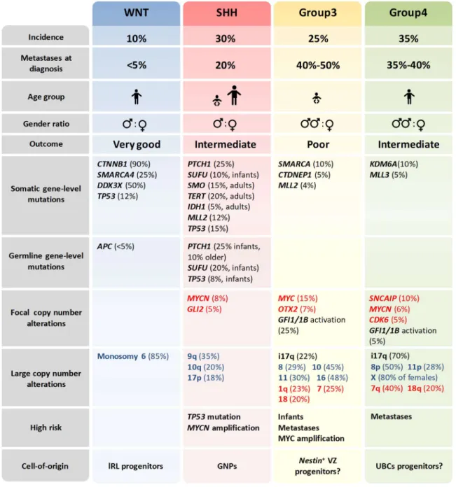
I.3.3 MOLECULAR CLASSIFICATION OF MEDULLOBLASTOMA ... 55
I.3.3.1 WNT-medulloblastoma ... 57
I.3.3.2 Group3-medulloblastoma ... 58
I.3.3.3 Group4-medulloblastoma ... 59
I.3.3.4 SHH-medulloblastoma ... 60
I.3.3.4.1 SHH-medulloblastoma intertumoral heterogeneity ... 61
I.3.3.4.2 Mouse models of SHH-medulloblastoma ... 63
I.3.3.4.3 Discovery of GNPs as the cell-of-origin of SHH-MB ... 64
I.3.4 STANDARD OF CARE, LIMITATIONS AND FUTURE DIRECTIONS ... 65
I.3.4.1 Targeted therapies in SHH-Medulloblastoma ... 67
I.4
T
HE
P
RIMARY
C
ILIUM
... 69I.4.1 DIFFERENT TYPES OF CILIA EXIST IN NATURE ... 69
I.4.2 STRUCTURE OF CILIA... 70
I.4.2.1 The ciliary shaft ... 70
I.4.2.2 The basal body ... 72
I.4.2.3 The transition zone ... 73
I.4.2.4 The ciliary pocket and the periciliary membrane ... 74
I.4.3 TRAFFICKING OF CILIARY PROTEINS ... 74
I.4.3.1 The intraflagellar transport ... 76
I.4.3.1.1 IFT motors ... 76
I.4.3.1.2 IFT particles ... 77
I.4.3.1.3 The BBsome ... 78
I.4.3.2 Ciliary import, transport and export of IFT cargoes ... 79
I.4.3.2.1 Transport of ciliary membrane proteins ... 80
I.4.4 CILIOGENESIS IS ENTANGLED TO CELL CYCLE PROGRESSION ... 81
I.4.4.1 Biogenesis of primary cilia ... 82
I.4.4.2 Disassembly of primary cilia ... 83
I.4.5 PRIMARY CILIA ARE SIGNALING CENTERS IN VERTEBRATES ... 84
I.4.5.1 Overview of the Hedgehog signaling ... 85
I.4.5.2 Major components of the Hedgehog signaling ... 86
I.4.5.2.1 Hedgehog proteins ... 86
I.4.5.2.2 Patched is the HH receptor ... 88
I.4.5.2.3 Smoothened is repressed by Patched ... 88
I.4.5.2.4 The Gli transcription factors ... 90
7
I.4.5.3.1 HH pathway OFF ... 92
I.4.5.3.2 HH pathway ON ... 94
I.4.5.4 Roles of primary cilia in normal GNs development ... 96
I.4.5.5 Roles of primary cilia in SHH-MB tumorigenesis ... 97
I.5
C
ENTRIOLAR
S
ATELLITES
... 99I.5.1 COMPOSITION OF CENTRIOLAR SATELLITES ... 99
I.5.1.1 Pericentriolar material 1 is the major component of CS ... 99
I.5.1.2 CS are mainly composed by centrosomal proteins ... 100
I.5.2 CENTRIOLAR SATELLITES INTEGRITY AND LOCALIZATION ... 101
I.5.2.1 The trafficking of CS depends on microtubles ... 102
I.5.2.2 CS integrity and localization depends on many integral components ... 102
I.5.2.3 Post-translational modifications contribute at shaping CS ... 103
I.5.2.4 Cell cycle dependent regulation of CS localization and integrity ... 104
I.5.3 FUNCTIONS OF CENTRIOLAR SATELLITES ... 105
I.5.3.1 CS are required for numerous centrosome functions ... 105
I.5.3.2 CS are regulators of ciliogenesis ... 106
I.6
A
TOH
1
... 111I.6.1 ROLES OF bHLH TRANSCRIPTION FACTORS IN NEUROGENESIS ... 111
I.6.2 STRUCTURE AND EXPRESSION OF ATOH1 ... 113
I.6.2.1 Structure of Atonal/Atoh1 proteins ... 113
I.6.2.2 Atoh1 is expressed in many tissues, besides the cerebellum ... 114
I.6.2.2.1 Atoh1 in the hindbrain ... 114
I.6.2.2.2 Atoh1 in the dorsal neural tube ... 115
I.6.2.2.3 Atoh1 in the inner ear ... 115
I.6.2.2.5 Atoh1 in the intestine ... 116
I.6.2.2.6 Atoh1 in Merkel cells ... 116
I.6.2.2.7 Concluding remarks ... 117
I.6.2.3 Atoh1 expression is highly regulated in the GN lineage ... 117
I.6.2.3.1 Transcriptional control of Atoh1 gene in the cerebellum ... 117
I.6.2.3.2 Post-translational control of Atoh1protein expression in the cerebellum... 120
I.6.3 FUNCTIONS OF ATOH1 IN THE GRANULE NEURON LINEAGE ... 121
I.6.3.1 Functions of Atoh1 during normal GNs development ... 121
I.6.3.2 Functions of Atoh1 during SHH-MB formation and expansion ... 122
I.7
O
BJECTIVES OF THE
S
TUDY
... 125RESULTS ... 127
A
RTICLE AND
M
AIN
F
IGURES
... 1298
DISCUSSION ... 197
D.1 Highlights of our findings and take-home messages ... 198
D.2 Atoh1 may control cell cycle and timing of neurogenesis, by regulating ciliogenesis ... 199
D.3 Possible consequences of loss or gain of Cep131 on centrosome duplication ... 201
D.4 Role of CS in neurogenesis and consequences of their absence in post mitotic GNs ... 202
D.5 Contribution of Atoh1 to SHH signaling ... 203
D.6 Atoh1 collaborates with the SHH pathway at multiple steps. ... 205
D.7 Dual and opposite roles of Atoh1 in GNs development ... 207
BIBLIOGRAPHY ... 211
ANNEX ... 253
9
Abbreviations
ACVR2A/B: Activin receptor 2A/B
APC/C: Anaphase promoting complex/cyclosome APC: Adenomatous polyposis cancer
Arf4: ADP-ribosylation factor 4
Arl13b: ADP-ribosylation factor-like protein 13b Ascl1: Achaete-scute homolog 1
AtEAM: Atoh1 E-box associated motif Atoh1: Atonal homolog 1
ATP: Adenosine triphosphate Aurka: Aurora A kinase
Barhl1: BarH-like 1 homeobox protein BBIP10: BBSome Interacting Protein 10 BBS: Bardet-Biedl syndrome
BCC: Basal cell carcinoma bHLH: Basic helix-loop-helix
Bmi1: B cell-specific Moloney murine leukemia virus integration site 1 BMP2/4/7: Bone morphogenetic protein 2/4/7
BNDF: Brain-derived neurotrophic factor Boc: Brother of Cdo
BRCA2: Breast cancer 2
BrdU: 5-bromo-2'-deoxyuridine
Ccdc14: Coiled-coil domain containing protein 14 Ccnd1/2: Cyclin D1/2
Cdc25b: Cell division cycle 25B
CDK1/4/6: Cyclin-dependent kinase 1/4/6 Cdkn1b: Cyclin-dependent kinase inhibitor 1b Cdkn2a/c: Cyclin-dependent kinase inhibitor 2a/c
Cdo: Cell adhesion molecule-related/down-regulated by oncogenes
Cep72/83/97/110/131/152/164/290: Centrosomal protein of 72/83/97/110/131/152/164/290 kDa
ChIP-seq: Chromatin-immuniprecipitation sequencing Ci: Cubitus interruptus
CKIδ: Casein kinase I δ cKO: Conditional knockout CLS: Cilium-localization signals CMP: Ciliary membrane protein CNS: Central nevous system
10 COPI/II: Coat protein I/II
CP110: Centriolar colied-coil protein of 110kDa Crcx4: C-X-C chemokine receptor type 4 CRD: Cystein-rich domain
CREB: Cyclic AMP-responsive element-binding protein CS: Centriolar satellites
CSF: Cerebrospinal fluid CSI: Craniospinal irradiation CT: Chemotherapy
Cyld: Cylindromatosis D/N: Desmoplastic/nodular
DCI: Dorsal commissural interneuron DCN: Deep cerebellar nuclei
DDX3X: DEAD-box helicase 3 X-linked Dhh: Desert Hedgehog
DRG: Dynein regulatory complex E: Embryonic day
ECM: Extracellular matrix EdU: 5-ethynyl-2'-deoxyuridine Egf: Epidermal growth factor En1/2: Engrailed 1/2
EphB2: Ephrin type-B receptor 2
ERBB4: Erb-b2 receptor tyrosine kinase 4 Evc: Ellis van Creveld protein
EZH2: Enhancer of zeste homolog 2 Fgf8: Fibroblast growth factor 8 FOP: FGFR1 oncogene partner For20: FOP-related protein of 20 kDa FoxM1: Forkhead box M1
GABA: Gamma-Aminobutyric acid Gas1: Growth arrest specific1
Gbx2: Gastrulation brain homeobox 2 GEF: Guanine nucleotide exchange factor Gfap: Glial fibrillary acidic protein
GFI1: Growth factor independent 1 GFP: Green fluorescent protein
Gli1/2/3: Glioma-associated oncogene 1/2/3 GliA/FL/R: Gli activator/full-length/repressor form
GN: Granule neuron
GNP: Granule neuron progenitor GPCR: G-protein coupled receptor
11 Gpr161: G-protein-coupled receptor 161
Gprk2: G-protein-coupled receptor kinase 2 GSK-3β: Glycogen synthase kinase-3β GTP: Guanosine triphosphate
HC: Hair cell
HDAC1/6: Histons deacetylase 1/6 Hes1/5: Enhancer of split 1/5 HH: Hedgehog
Hhip1: Hedgehog-interacting protein 1 Hic1: Hypermethylated in cancer 1 Hoxa2: Homeobox A2
Hrs: hours
HSPGs: Heparan sulfate proteoglycans
Huwe1: HECT, UBA and WWE domain containing E3 ubiquitin protein ligase 1 Id2: Inhibitor of DNA-binding/differentiation 2
IDH1: Isocitrate Dehydrogenase 1 iEGL: Inner external granular layer IFT: Intraflagellar transport
Ift20/88/172: Intraflagellar transport protein 20/88/172 Igf2: Insulin-like growth factor 2
IGL: Internal granular layer Ihh: Indian Hedgeohog ILK: Integrin linked kinase IsO: Isthmic organizer Itch: Itchy homolog JBTS: Jubert syndromes JNK: c-Jun N-terminal kinase Kap3: Kinesin-associated protein 3
KBTBD4: Kelch repeat and BTB domain containing 4 KDM6A: Lysine demethylase 6A
Kif3a/3b/3c/7/17/24: Kinesin family member 3a/3b/3c/7/17/24 KMT2C: Lysine methyltransferase 2C
KO: Knockout
Lh2A/B: LIM/homeobox protein A/B
Lmx1a/b: LIM homeobox transcription factor 1, alpha/beta LOH: Loss of heterozigosis
lRL: Lower rhombic lip
MAPK: Mitogen-activated protein kinases MB: Medulloblastoma
MBEN: Extensive nodularity MDM4: Murine double minute 4
12 MEP: Multipotent epithelial progenitor
MHB: Midbrain-hindbrain boundary Mib1: Mindbomb 1
MKS: Meckel syndrome ML: Molecular layer
MRI: Magnetic resonance imaging MTOC: Microtuble organizing center mTOR: Mammalian target-of-rapamycin MYCL1: L-myc-1 proto-oncogene Mycn: N-myc proto-oncogene
Nek2: Never in mitosis related Kinase 2 NeuN: Neuronal nuclei antigen
NeuroD1: Neurogenic differentiation factor 1 Neurog1/2: Neurogenin-1/2
NICD: Notch intracellular domain NMDA: N-Methyl-D-aspartic acid
NPC: Neural progenitor cell NPHP: Nephronophthisis NTZ: Nuclear transitory zone Odf2: Outer dense fiber protein 2 oEGL: Outer external granular layer Ofd1: Orofaciodigital Syndrome 1
Olig2: Oligodendrocyte transcription factor 2 Otx2: Orthodenticle homeobox 2
P: Post-natal day
PALB2: Partner and localizer of BRCA2 Par6α: Partitioning defective 6 homolog alpha Pax2/5/6: Paired box 2/5/6
PC: Purkinje cell
PCL: Purkinje cell layer Pcm1: Pericentriolar material 1 PDD: Processing determinant domain PDGF: Platelet-derived growth factor PDXs: Patient-derived xenograft PI3K: Phosphatidylinositol 3-kinases
PIK3C2B/G: Phosphatidylinositol-4-phosphate 3-kinase catalytic subunit type 2 beta/gamma PKA/C: Protein kinase A/C
Plk1/4: Polo-like kinase 1/4
PPM1D: Protein Phosphatase, Mg++/Mn++ dependent 1D,
PRC1: Polycomb repressive Complex 1 PRC2: Polycomb repressive complex 2
13 PRDM6: PR/SET domain 6
Ptch1/2: Patched homolog 1/2
PTEN: Phosphatase and TENsin homolog Ptf1a: Pancreas associated transcription factor 1a PTM: Post-translational modification
r1 - r8: Rhombomere 1 - Rhombomere 8 Rb1: Retinoblastoma 1
Rbpj: Recombination signal binding protein for immunoglobulin kappa J region RL: Rhombic lip
RLS: Rostral rhombic lip migratory stream RND: Resistance-nodulation-division Robo1/2: Roundabout guidance receptor 1/2 RT: Radiotherapy
SC: Supporting cell SCF: Skip-Cullin-F-box
SDF-1α: Stromal cell-derived factor 1 alpha SEM: Standard error of the mean
SHH: Sonic hedgehog
siRNA: small interfering RNA Slit1/2: Slit Guidance Ligand 1
Smad4: Small mothers against decapentaplegic
SMARCA4: SWI/SNF related, matrix associated, actin dependent regulator of chromatin, subfamily A, member 4
Smo: Smoothened
SNCAIP: Synuclein alpha interacting protein Spop: Speckle-type POZ protein
SSX2IP: SSX family member 2 interacting protein TERT: Telomerase reverse transcriptase
TGF-β: Transforming growt factor-beta Tieg1: TGFbeta inducible early gene-1 TP53: Tumor protein 53
Tuj1: Neuron-specific class III beta-tubulin UBC: Unipolar brush cell
Unc5c: Uncoordinated-5C uRL: Upper rhombic lip
USP9X: Ubiquitin specific peptidase 9 X-linked UV: Ultraviolet
VZ: Venticular zone
WHO: World Health Organization
Wnt1/3: Wingless-type MMTV integration site family member 1/3 YAP1: Yes associated protein 1
14 Zic1: Zinc finger of the cerebellum family member 1 β-TrCP: Beta-transducin repeats-containing protein γ-TURC: Gamma-tubulin ring complexes
15
Table of Figures
Figure I. Anatomy of the cerebellum.
Figure II. Functional classification of cerebellar regions.
Figure III. Cortical cerebellar neurons and main neuronal circuitries. Figure IV. Early embryonic patterning of the vertebrate brain. Figure V. Germinal zones in the early cerebellar anlage.
Figure VI. Sequential phases of embryonic and post-natal cerebellar development. Figure VII. Developmental phases of GNPs in post-natal times.
Figure VIII. Pro-proliferative pathways active in GNPs in the EGL.
Figure IX. Anti-proliferative pathways triggering terminal differentiation of GNPs. Figure X. Molecular subgroups of MB.
Figure XI. Heterogeneity in SHH-MB. Figure XII. Structure of a cilium.
Figure XIII. Mechanisms of protein trafficking through the cilium. Figure XIV. Cilium-centrosome behaviour during the cell cycle.
Figure XV. Synthesis steps of the active HH protein (HH-N) from the precursor. Figure XVI. Relevant domains and sites in mouse Gli2 and Gli3 proteins. Figure XVII. HH signaling in vertebrates.
Figure XVIII. Centriolar satellites.
Figure XIX. Mechanisms controlled by CS during ciliogenesis in normal conditions. Figure XX. Major networks regulating Atoh1 expression in GNPs across embryonic and post-natal developmental stages.
Figure XXI. Expression of SHH target genes upon Atoh1 manipulation in GNPs.
19
I.
1
The Cerebellum
By housing more than half of the neurons of the whole brain, the cerebellum (Latin for "little brain") is one of the most architecturally complex region of the vertebrate central nervous system (CNS). Since the end of the XIX century, for over 80 years, the cerebellum has been considered the area of the CNS exclusively dedicated to motor control and coordination. However, this view has progressively expanded during the last three decades, as numerous studies based on novel functional imaging, neural tracing and clinical data analysis have highlighted new and unexpected roles in higher cognitive activities such as learning, attention, language and emotions.
In this first introductive chapter, the anatomy and functional compartmentalization of the cerebellum will be initially illustrated, before delving into the description of the histological architecture of the cerebellar cortex.
I.1.1
A
NATOMY OF THEC
EREBELLUMResiding within the posterior fossa of the skull, the cerebellum represents the anteriormost region of the hindbrain, locating beneath the cerebral hemispheres and posterior to the brain stem1 and the IVth ventricle.
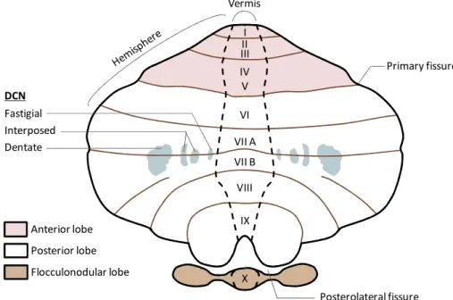
At a first glance, the cerebellum can be clearly divided in three major parts consisting in two large lateral hemispheres separated by a narrow midline region called vermis (Latin for "worm"). In addition, the surface of the cerebellum is crossed by two deep transversal (medio-lateral) grooves called primary and posterolateral fissures, which delineate three major cerebellar lobes, namely the anterior, posterior and flocculonodular lobes (Roostaei et al., 2014) (Figure I). The anterior and the posterior lobes are further shaped by shallow transversal fissures which overall subdivide the entire cerebellum in a total of ten lobules, indicated with Roman numbers. In particular, the anterior lobe contains the first five lobules (I-V), the posterior lobe the following four (from VI to IX, with the lobule VII split in VIIA and VIIB),
20
while the flocculonodular lobe coincides with the lobule X (Roostaei et al., 2014) (Figure I). Next, the surface of each lobules is also shaped by a series of tiny parallel transversal gyrus, referred as folia, which adding up to the lobular organization, further increases the total surface of the cerebellum, eventually enabling the packaging of a large area of neural tissue in a relatively small volume (Roostaei et al., 2014).
The internal macro-organization of the cerebellum is relatively simple. It consists into an external cortex of grey matter, mainly composed by cell bodies, surrounding a large body of white matter, made primarily by myelinated neuronal fibers. Embedded within the white matter lie three pairs of "deep cerebellar nuclei" (DCN), composed of grey matter (Stilling, 1864). From medial to lateral, these nuclei are respectively called fastigial, interposed (which is further divided into globose and emboliform nuclei) and dentate (Figure I). With only few exceptions, the majority of the cerebellar outputs are projected from the DCN.
Morphologically, the cerebellum is connected to the brainstem via three pair of peduncles which, according to their relative position are named superior, middle and inferior. The totality of the afferent and efferent neuronal fibers directed toward and leaving the cerebellum pass through these peduncles, which therefore represent also the sole functional connection of the cerebellum with the other regions of the CNS (Roostaei et al., 2014).
Figure I. Anatomy of the cerebellum. Schematic dorsal view of an unfolded human cerebellar cortex. The vermis in medial in the cerebellum and delimited from the lateral hemispheres by black gapped lines. The major fissures, including those dividing the three lobes (indicated with different colours) are represented by transversal brown lines. The position and name of the lobules is indicated with Roman numbers at the level of the vermis. The position of the deep cerebellar nuclei (DCN) inside the cerebellum is projected in the drawing by light blue areas.
VI VII A VII B VIII X IX I II III IV V Vermis Anterior lobe Posterior lobe Flocculonodular lobe Primary fissure Posterolateral fissure Fastigial Interposed Dentate DCN
21
I.1.2
M
AINF
UNCTIONS OF THEC
EREBELLUMThe major role of the cerebellum consists in controlling, coordinating and if needed correcting the body voluntary movements. Such complex activities occur in relation to memorized motor schemes and environmental or internal stimuli.
The cerebellum is believed to receive two major types of input information (Ito, 2013; Ohyama et al., 2003; Ramnani, 2012). The first information is motor type: it principally comes from the motor cortex, which contains the pre-motor and primary motor areas dedicated to the planning and execution of movement. This way the cerebellum is informed about the type of movement that is intended to perform. The second information is sensory type: it reports details about the actual status of the body and the surrounding environment at the onset of movement. Once received, these information are integrated and processed within the cerebellar circuitries which are supposed to store memories of "correct" motor schemes learnt by trials and errors during the individual's life. By basing on these schemes and by evaluating motor and sensory inputs, the cerebellum produces an output which is eventually directed to the motor cortex via a relay through the thalamus. Such output may correct the activity of cortical motor neurons, thus tuning their output to accomplish the best execution of the movement (Ito, 2013; Ohyama et al., 2003; Ramnani, 2012). Nevertheless, if the movement is not precisely or correctly executed, then an error signal is generated, conceivably at the level of the inferior olive of the medulla, and delivered to the cerebellum. Such error input is believed to re-wire the cerebellar circuitries in order to modify the stored motor memory and allow the cerebellum to correct such error in the future if similar conditions will be met again (Ito, 2013; Ohyama et al., 2003; Ramnani, 2012).
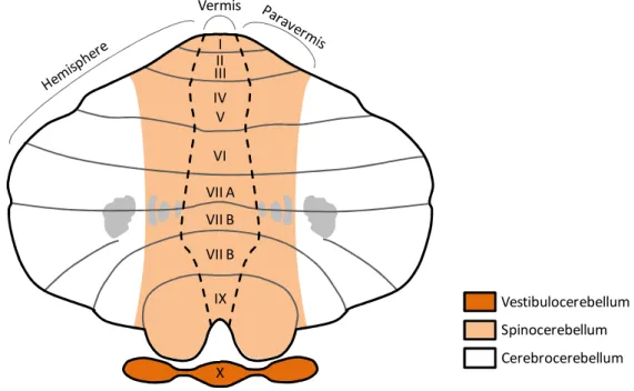
I.1.2.1 Functional compartmentalization of the cerebellum
Depending on the nature of the afferencies received, the efferencies projected and thus on the type of body activities controlled, the cerebellum can be divided into three main parts, namely vestibulocerebellum, spinocerebellum and cerebrocerebellum (Figure II). The vestibulocerebellum, is the posteriormost region of the cerebellum, it corresponds to the flocculonodular lobe and it is the most evolutionary ancient part of this organ. By receiving inputs from the vestibular nuclei on the brainstem and from the visual cortex, the vestibulocerebellum is dedicated to the regulation of the equilibrium, the posture and the coordination of eyes positioning upon head movements.
22
The spinocerebellum locates in the cerebellum midline and consists into the vermis and the medial part of the cerebellar hemispheres (also known as paravermis), the fastigial and interposed nuclei. It receives auditory, visual and somatosensory information from all the body thanks to its connections with the spinal cord and various brainstem nuclei (Roostaei et al., 2014). The somatosensory inputs coming from different parts of the body are processed in dedicated areas of the spinocerebellum, so that a somatotopic map of the body is created on its cortex (Manni and Petrosini, 2004). Also the spinocerebellum participates in the regulation of posture and equilibrium, but it is also involved in the coordination of regular movements of the limbs, like those required for walking.
Finally, the cerebrocerebellum corresponds to the lateral part of the cerebellar hemispheres and includes the dentate nuclei. It represents the most evolutionary recent functional part of the cerebellum and it has tremendously expanded with the appearance of the primates lineage (Herculano-Houzel, 2010). In humans it accounts for around 90% of the volume of the whole cerebellum. The cerebrocerebellum is primarily responsible for the functions described in the previous paragraph given its high connectivity with the cerebral cortex. Hence, the cerebrocerebellar is ultimately required for the planning and the proper timing of complex movements, as well as high cognitive activities, including language (Buckner, 2013).
Figure II. Functional classification of cerebellar regions. The drawing depicts an equivalent representation of the cerebellum as shown in Figure I. The three functional regions of the cerebellum, namely the vestibulocerebellum, the spinocerebellum and the cerebrocerebellum are indicated with different colors.
VI VII A VII B VII B X IX I II III IV V Vermis Vestibulocerebellum Spinocerebellum Cerebrocerebellum
23
I.1.3
M
ICROANATOMY OF THEC
EREBELLARC
ORTEXI.1.3.1 Cells of the cerebellar cortex
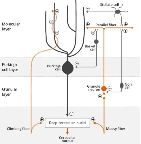
Despite the functional compartmentalization described above, the cytoarchitecture of the cerebellar cortex seems to be essentially identical and repeated along its whole extension (Ito, 2006; Roostaei et al., 2014; Voogd and Glickstein, 1998) (Figure III).
Depending on the cell type composition, the cerebellar cortex can be subdivided into three layers. The deepest layer is also the thickest and it is called granular layer or internal granular layer (IGL), to distinguish it from its "external" counterpart that transitorily appears during cerebellar development (as it will be described later). The IGL gets its name after the granule neurons (GNs), small, glutamatergic (excitatory) neurons that tightly packed reside in this layer. Interestingly, GNs account for the most abundant neuronal population of the whole adult brain. In addition to the GNs, the IGL hosts also some less abundant populations of interneurons as the GABAergic (inhibitory) Golgi and Lugaro cells and the glutamatergic unipolar brush cells (UBCs), the latter found only in the vestibulocerebellum.
Above the IGL there is the Purkinje cell layer (PCL), which consists of one single layer of cell bodies (or somata) of the Purkinje cells (PCs), a GABAergic neuron type responsible for transmitting the output of the cerebellar cortex (as described shortly). Alongside to the somata of PCs, the PCL also contains small interneurons called candelabrum cells, and the Bergmann glia cells, a particular type of astrocyte with roles in modulating PCs activity (Lainé and Axelrad, 1994).
The uppermost layer of the cortex is called molecular layer (ML) and it is composed mostly by the large and expanded dendritic tree of the PCs and by the axons of the GNs. Indeed, each GN in the IGL extends its axon toward the ML, where it bifurcates generating two characteristic T-shaped branches that extend parallel to the cerebellar surface, up to 4-6mm in humans (Ito, 2006). These specialized axons of the GNs are called parallel fibers and they transmit excitatory signal to the Purkinje cells by forming synapses with their dendritic trees. It is estimated that each GN can synapse with thousands of Purkinje cells. In addition to parallel fibers and PCs dendritic trees, the molecular layer also houses a set of inhibitory interneurons like the basket cells and the stellate cells.
24
I.1.3.2 Major cerebellar circuits
Similarly to its neuronal composition, the neuronal circuitries of the cerebellar cortex seem highly repeated and stereotyped (Ito, 2006; Roostaei et al., 2014; Voogd and Glickstein, 1998) (Figure III). Indeed, such neuronal networks can be ultimately reduced to a single functional module that is reproduced multiple times across the cerebellar cortex, with only few regional little diversifications.
Fundamentally, the cerebellum receives two major types of excitatory inputs relayed by two distinct afferent pathways, namely the mossy fibers (MFs) and the climbing fibers (CFs). The MFs are projections generated by a multitude of sources, including pre-cerebellar brainstem nuclei (as some pontine nuclei relaying the cerebral cortex inputs) and the spinal cord. They basically transport the motor and sensory type information required by the cerebellum for generating its output. The CFs instead originate from a unique source, the inferior olive, and are believed to relay the error inputs to the cerebellum.
Figure III. Cortical cerebellar neurons and main neuronal circuitries. Major synaptic interaction between neurons of the cerebellar cortex. The (+) and (-) symbols at the synapses indicate excitatory and inhibitory synapses respectively. The neurotransmitter phenotype of neurons is color-coded: glutamatergic neurons are in orange and GABAergic in grey.
+
-+ +
+
Deep cerebellar nuclei + -+ + -+ + + Stellate cell Purkinje cell Granule neuron Parallel fiber Mossy fiber Climbing fiber Cerebellar output Molecular layer Purkinje cell layer Granular layer Basket cell Golgi cell
25
Once entered the cerebellum through the cerebellar peduncles, collaterals of both MFs and CFs are sent toward the cerebellar cortex and the DCN. In this way, the same excitatory input is received by the two neuronal substrates of the cerebellum.
At the level of the cortex, CFs are directed to the molecular layer where they literally "climb" the branches of the dendritic trees of PCs forming multiple synaptic connection with them. The number of synapses here generated is so high that a single action potential travelling along a CF is generally sufficient to activate a PC.
On the contrary, MFs are directed to the IGL where they extensively branch and contact the dendrites of GNs forming specialized synapses called "cerebellar glomeruli". Cerebellar glomeruli are morphologically unique synapses in the brain, displaying a typical globular shape where a MF terminal enlarges to engage up to fifty GN dendrites (Jakab and Hámori, 1988). In addition, cerebellar glomeruli contain the axon terminal of various Golgi cells, which by releasing GABA modulate the activation of GNs.
As GNs downstream signal to PCs through their parallel fibers, the cortical circuits are organized in such a way that either directly or indirectly, the excitatory signals of CFs and MFs are eventually received by PCs. Upon activation of PCs, their inhibitory output is sent toward the DCN via their long axons, which extends through the cerebellar white matter.
In the DCN, integration of the excitatory inputs from CFs and MFs and inhibitory inputs from PCs occurs, and, if generated, excitatory signals are sent out the cerebellum to various downstream targets (e.g. to the cerebral cortex via a thalamic relay from the dentate nuclei). Besides the above described neuronal network, other elements add up to these circuitries to further refine and modulate the information processing. Cortical interneurons indeed participate in the signal propagation by regulating the activation of both GNs and PCs via local feedback and feedforward circuitries. As anticipated, Golgi cells in the IGL counteract the activation of GNs by the MFs at the level of the cerebellar glomeruli. As Golgi cells are themselves upstream activated by parallel fibers, this represents an example of a feedback inhibition on GNs (Figure III). Stellate and basket cells in the ML instead receive synaptic inputs from parallel fibers and form downstream inhibitory synapses with PCs. Therefore, these interneurons mediate a feedforward inhibition on PCs in the context of the GNs-PCs pathway (Figure III).
27
I.
2
Development of Granule Neurons
Cerebellar GNs represent the most abundant neuronal pool of the whole adult brain. Their neurogenesis requires precise temporally regulated steps of specification, migration, proliferation and maturation happening during both embryonic development and early post-natal times in both human and mouse.
This second introductive chapter describes how the cerebellum is formed during embryogenesis and how histogenesis of GNs is achieved, highlighting the molecular modules implicated in such developmental process.
I.2.1
E
ARLYS
PECIFICATION ANDP
ATTERNING OF THEC
EREBELLUMI.2.1.1 Regionalization of the neural tube
The entire vertebrate CNS originates from the ectoderm, one of the three germ layers of the early embryo, which also contributes to the epidermis. During gastrulation, signals from the primitive node regionalize the ectoderm specifying the prospective CNS in the so-called "neural plate", a relatively uniform sheet of cells located at the dorsal midline of the embryo. Subsequently, a combination of migratory events, changes in cell shape, and mechanical pressure due to the expanding lateral epidermis will cause the borders of the neural plate to thick-up, bend over the midline and eventually fuse dorsally giving rise to the neural tube. The process that starts with formation of the neural plate and terminates with closure of the neural tube is denominated neurulation (Darnell and Gilbert, 2017; Stiles and Jernigan, 2010).
During neurulation, inductive signals from the anterior endoderm and the notochord initiate the spatial patterning of the neural plate both along the antero-posterior and the dorso-ventral axis. Even much before the complete closure of the neural tube, such regionalization becomes visible by the appearance of three swellings in the anterior neural tube, denominated neuromeres or brain vesicles. The neuromeres will give rise to the various brain structures and they are named, from anterior to posterior, forebrain (or prosencephalon), midbrain (or mesencephalon) and hindbrain (or rhombencephalon) (Figure IV). Conversely, the remaining posterior part of the
28
neural tube maintains its cylindrical shape and will become the spinal cord (Darnell and Gilbert, 2017)
Later on, the regionalized expression of Hox genes further transiently patterns the hindbrain into eight segmental units, called rhombomeres (r1 to r8) leading to initial neuronal identity diversification (Keynes and Krumlauf, 1994) (Figure IV).
Sometimes, the anatomical boundary between the midbrain and the hindbrain (midbrain-hindbrain boundary, MHB), also called isthmus or isthmic constriction (as the neural tube is here "squeezed" between these two neuromeres), is referred as the rhombomere 0.
Early fate mapping studies utilizing the quail-chick transplants system led to the nowadays accepted knowledge that all the cerebellar neurons arise from the dorsal (alar) r1. By embryonic day (E) 7.5 in mouse, the r1 territory is specified in the anteriormost part of the hindbrain, just posterior to the MHB, thanks to the regionalized expression of a group of homeobox transcription factors (Figure IV).
Figure IV. Early embryonic patterning of the vertebrate brain. (Left drawing) Schematic view of a sagittal section of the embryonic mouse brain at around E9. The anteriormost brain vesicle is the forebrain (Fb) followed by the midbrain (Mb) and by the hindbrain (Hb), the latter further divided into segmental units, the rhombomeres (r1 to r8). At the dorsal midline of the hindbrain, the roofplate (Rp) appears as a thin layer covering the IVth ventricle (IVth v). (Middle drawing) Schematic dorsal view of the boundary between midbrain and hindbrain at the same developmental stage. The isthmic organizer (IsO) localizes at the boundary between midbrain and hindbrain. The medio-lateral expansion of the roofplate at the level of the anterior hindbrain can be appreciated from this view. (Right scheme) Expression domains of the major genes implicated in anterior hindbrain patterning in relation to the middle drawing. In addition, the fundamental regulatory networks between these genes are shown, based on the reviews of Martinez et al. (2013), Wurst and Bally-Cuif, (2001b) and Sillitoe and Joyner (2007). Such networks establish, reinforce and maintain the represented gene expression pattern. The r1 is specified in a region devoid of Otx2 and Hoxa2 expression. Lmx1b may initiate the IsO gene expression program. Fgf8 and Wnt1 are expressed at the IsO, but into two distinct posterior and anterior domains respectively. Pax2 and En1/2 are expressed into broader territories encompassing the IsO and the r1.
IsO (Hb) r1 (Hb) r2-r8 Mb Gbx2 Hoxa2 Fgf8 Wnt1 Lmx1b Otx2 Pax2 En1/2 Rp A P R L Rp Fb IVthv
29
In particular, the MHB coincides with the boundary between the expression domain of Orthodenticle homeobox 2 (Otx2, expressed in the midbrain and in the forebrain) and Gastrulation brain homeobox 2 (Gbx2, expressed from r1 to r3) (Simeone et al., 1992; Wassarman et al., 1997). Importantly, the expression of Otx2 and Gbx2 is mutually exclusive as both can cross-repress each other’s expression. Notably, such regulation is functional at maintaining a neat separation between the midbrain and the hindbrain territories (Inoue et al., 2012). The posterior edge of the r1 is instead defined by the anteriormost outreach of Homeobox A2 (Hoxa2) expression, which extends in the r2 and r3.
Therefore, the cerebellum ultimately originates in a territory of the hindbrain marked by the expression of Gbx2, and by the absence of Otx2 and Hoxa2 (Figure IV). This conclusion is supported by a number of genetic studies demonstrating that perturbing the expression of these genes results into either loss or ectopic specification of prospective cerebellar territories (Eddison et al., 2004; Gavalas et al., 1997; Wassarman et al., 1997).
I.2.1.2 Specification of cerebellar territories in rhombomere 1
Once it is formed, the r1 undergoes spatial patterning in order to regionally specify the identity of its derivatives, including the cerebellar neurons and glia. During embryogenesis, tissue patterning is generally controlled by signaling centers, also known as organizers, which by releasing morphogens or other signaling molecules play instructive roles on the fate of adjacent tissues. Patterning of the anterior hindbrain is principally orchestrated by a transversal ring of cells localizing at the MHB, which altogether constitute the so called isthmic organizer (IsO) (Marin and Puelles, 1994; Martinez et al., 1991; Martínez et al., 1995) (Figure IV). The IsO is established around E8.5 and its onset is marked by the expression of morphogens like Fibroblast growth factor 8 (Fgf8) and Wingless-type MMTV integration site family member 1 (Wnt1), whose secretion and diffusion mediate the instructive functions of the IsO in the surrounding territories (Crossley and Martin, 1995; Martinez et al., 2013; McMahon and Bradley, 1990). Although seemingly dispensable for IsO formation, Otx2 and Gbx2 are instrumental for determining its positioning at the MHB. Indeed, genetic manipulations causing anterior or posterior shift of Otx2 and Gbx2 expression domains eventually reposition the IsO at the new Otx2-Gbx2 interface (Simeone, 2000; Wurst and Bally-Cuif, 2001a).
Conversely, converging evidence from studies in mouse and chicken embryos, pinpointed the LIM homeobox transcription factor Lmx1b as the main responsible for IsO induction (Adams et al., 2000; Guo et al., 2007). Lmx1b is expressed in the posteriormost midbrain, where it
30
activates the expression of Wnt1 at the MHB. Here, Wnt1 signaling downstream activates Fgf8 expression, thereby establishing the IsO (Guo et al., 2007; Matsunaga et al., 2002). In addition, also the Paired box 2 (Pax2) transcription factor is a known regulator of Fgf8 expression, thus it may contribute to IsO formation as well (Ye et al., 2001).
Among the two principal IsO morphogens, Fgf8 seems to be the main mediator of IsO patterning activity, while Wnt1 may just have a limited role consisting into a mitogenic stimulus for neuroepithelial cells (Chi et al., 2003; Liu et al., 1999; Martinez et al., 1999). Hence, regionalization of the r1, which also includes specification and maintenance of the cerebellar identity in the alar r1, mainly requires the morphogenetic activity of Fgf8. However, by E9, also the transcription factors Lmx1b and Pax2, Engrailed 1 (En1), Engrailed 2 (En2) and Paired box 5 (Pax5) add up to Fgf8 signaling to regulate r1 patterning (Asano and Gruss, 1992; Davis and Joyner, 1988; Rowitch and McMahon, 1995; Sillitoe and Joyner, 2007; Wurst and Bally-Cuif, 2001a). Some of the known genetic interactions required for anterior hindbrain specification and patterning are represented in Figure IV.
I.2.2
E
ARLYH
ISTOGENESIS IN THEC
EREBELLARA
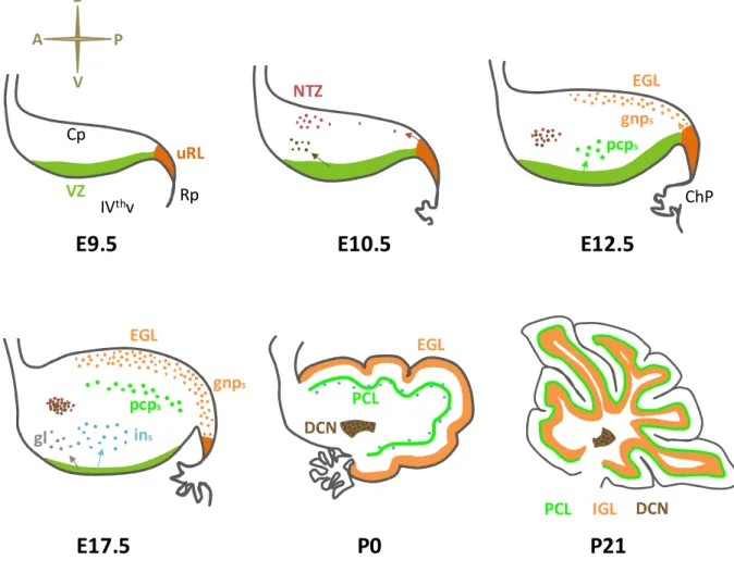
NLAGEOnce the presumptive cerebellar territory is specified, cerebellar cells hystogenesis initiates. As mentioned above, it is nowadays accepted that all the cerebellar neurons derive from the alar plate of r1. At E9.5 in mice, two molecularly and spatially distinct germinal regions can be there identified: the upper rhombic lip (uRL) and the ventricular zone (VZ) (Hatten and Heintz, 1995) (Figure V). The rhombic lip (RL) localizes at dorsal margin of the hindbrain neuroepithelium, lining adjacent to the roofplate of the IVth ventricle. The anterior segment of the RL, which is contained in the r1, is called "upper" RL (uRL), in contrast to the "lower" RL (lRL) which extends posteriorly in the remaining rhombomeres (Figure V). Importantly, the uRL is the source of all the cerebellar glutamatergic neurons, including the GNs, the UBCs and the large excitatory neurons of the DCN (Alder et al., 1996; Machold and Fishell, 2005; Wang et al., 2005; Wingate and Hatten, 1999) (Figure VI and VI). In addition, the uRL also contributes to some pre-cerebellar pontine nuclei part of the vestibular, auditory and proprioceptive sensory systems (Wang et al., 2005). Conversely, the lRL mainly gives rise to neurons populating different sets of precerebellar pontine and medullary nuclei (Rodriguez and Dymecki, 2000; Wang et al., 2005; Wingate, 2001).
31
The VZ is instead placed along the lining of the dorsal aspect of the IVth ventricle and produces all the cerebellar GABAergic neurons, including PCs, Golgi, basket, stellate cells and the small inhibitory neurons of the DCN (Hoshino et al., 2005; Leto et al., 2006; Maricich and Herrup, 1999; Sudarov et al., 2011) (Figure V and VI). In addition, VZ progenitors also contribute to great part of the cerebellar glial population (Sudarov et al., 2011).
Two genes encoding for class II-basic helix-loop-helix (bHLH) transcription factors, namely Atonal homolog 1 (Atoh1) and Pancreas transcription factor 1a (Ptf1a), are required for the fate commitment of RL and VZ progenitors respectively and they are also used as bona fide markers for these two germinal zones (Akazawa et al., 1995; Ben-Arie et al., 1997; Hoshino et al., 2005; Machold and Fishell, 2005; Wang et al., 2005; Yamada et al., 2014) (Figure V). Interestingly, once the expression of Atoh1 and Ptf1a is induced in the cerebellar anlage (around E9.5), these two transcription factors are able to repress each other's expression, hence robustly separating uRL and VZ (Yamada et al., 2014).
Therefore, similarly to what happens for forebrain cortical neurogenesis, generation of glutamatergic and GABAergic neurons is spatially compartmentalized in the cerebellum (Schuurmans et al., 2004) (Figure V).
Figure V. Germinal zones in the early cerebellar anlage. (Left drawing) Dorso-lateral view of a mouse embryo's head at E9.5. (Middle drawing) Dorsal view of the hindbrain (Hb) at the same stage. The rhombic lip (RL, orange/brown) appears at the dorsal margins of the Hb, adjacent to the roofplate (Rp). The upper RL (uRL, orange) is specified anteriorly, within the rhombomere 1 (r1). In contrast the lower RL (lRL, brown) borders the remaining posterior part of the Hb. (Top-right drawing) Sagittal section view of the dorsal (alar) r1, which will give rise to the cerebellum. The uRL is visible posteriorly, next to the Rp, while the ventricular zone (VZ, green) is more anteriorly located and lines above the IVth ventricle (IVthv). The dorsal surface or the alar r1 is covered by the meninges (pia). Atoh1 is expressed in both the uRL and the lRL, while Ptf1a expression is observed only in the VZ. The uRL generates cerebellar glutamatergic neurons, while the VZ generates GABAergic neurons and glia. r1 Hb Mb IsO uRL lRL IVthv Rp A P R L D P A IsO uRL VZ Rp V pia Atoh1 Ptf1a eye Mb
32
I.2.2.1 Temporal origin of uRL derivatives
As stated above, starting from E9.5 the uRL (but also the lRL) territory is marked by the expression of Atoh1. Microscopy studies have shown that at later stages, the uRL becomes progressively spatially compartmentalized (Yeung et al., 2014). Indeed, by E13, Atoh1 expression becomes slightly reduced at the interior face of the uRL, where neuron progenitors initiate to express the multipass transmembrane protein Wintless. The exterior face of the uRL instead continues to produce high levels of Atoh1 and its transcriptional target Paired box 6 (Pax6). By E15, the separation between these two faces of the uRL becomes even more defined, when a thin layer of Lmx1a-expressing cells appears between them. However, the functional role of this regionalization is still poorly known, although it is likely that different uRL zones may give rise to different populations of uRL derivatives (Chizhikov et al., 2010; Yeung et al., 2014, 2016).
Conversely, much better characterized is the temporal pattern of neuronal subtype specification in the uRL from E9.5 onwards (Figure VI). Here, the use of various mouse models, including those expressing the inducible CreER recombinase under the control of Atoh1 regulative regions, were useful for transiently or permanently tracing the fate of all Atoh1+ uRL precursors
(Machold and Fishell, 2005; Wang et al., 2005). From these studies, it was found that the first subset of cerebellar glutamatergic neurons generated in the uRL are those eventually populating the DCN (together with the previously mentioned neurons of the pre-cerebellar pontine nuclei). Upon specification, these neuron precursors rapidly downregulate Atoh1 and leave the uRL moving anteriorly and laterally along the cerebellar surface producing the so-called rostral RL migratory stream (RLS). Around E12.5, most of these migrating neurons cluster in a region at the anterior margin of the cerebellum named nuclear transitory zone (NTZ), which represents a primordium of the DCN (Figure VI).
Only subsequently, from E12.5 to E17, a second, distinct wave of fate-specification generates GN-committed cells. The GN progenitors (GNPs) here specified also take part to the RLS (sometimes referred as a "late" RLS), but differently from the "early" RLS precursors they maintain Atoh1 expression and transitorily crowd on the cerebellar surface forming a mitotically active region denominated external granular layer (EGL). Here, GNPs first massively expand clonally and only after birth they gradually exit the cell cycle and migrate radially toward the IGL, where they complete their differentiation to GNs (Figure VI).
The last glutamatergic neuronal type generated within the uRL are the UBCs progenitors, which appear in the uRL concomitantly with GNPs, between E15.5 and E17.5 (Englund et al., 2006;
33
Sekerková et al., 2004). By birth, differentiating UBCs delaminate from the uRL, migrate through the immature cerebellar white matter and reach their final localization in the developing IGL of the prospective posteriormost cerebellar lobes.
Therefore, at least two, main, temporally distinct phases of neurogenesis take place in the uRL, with E12.5 roughly representing the temporal border between them.
Figure VI. Sequential phases of embryonic and post-natal cerebellar development. Sagittal section of the dorsal r1 at progressive time-points during mouse development. By E9.5, the prospective cerebellum (Cp) is specified and the two germinal zones, namely the uRL and the VZ, appear. By E10.5, the uRL generates precursors of the glutamatergic DCN that crowd anteriorly in the cerebellum in the NTZ (red cells); simultaneously, the VZ produces the small GABAergic interneurons of the DCN (dark green cells). At E12.5, the uRL ceases to generate DCN neurons, and switches to GNPs (gnps in the picture) that initiate to populate the EGL; the VZ is instead actively forming PC precursors (pcps) since already 24 hours; the roofplate (Rp) is differentiating into the choroid plexus (ChP). At E17.5, the PC precursors initiate to arrange in the PCL, while the VZ produces cortical interneurons (ins) and glial cells (gl); the GNPs initiate to massively proliferate in the EGL. By birth (P0), the PCL is formed and the NTZ has differentiated to DCN. The GNPs in the EGL are actively expanding and as a result the cerebellar lobes begin to be shaped; in the following days GNPs will progressively terminally differentiate and migrate inward to populate the IGL. By three weeks after birth (P21), the cerebellar development is completed; all GNPs have differentiated to GNs and have reached the IGL below the PCL. (IVthv: IVth ventricle).
uRL VZ Rp IVthv Cp vv v vvv ChP vvv v NTZ EGL gnps pcps pcps gnps EGL ins gl vvvvvv EGL PCL IGL PCL
E9.5
E10.5
E12.5
E17.5
P0
P21
D V
P A
34
Interestingly, lineage tracing studies performed by Machold and Fishell (2005) demonstrated that all the Atoh1+ precursors present in the uRL at E10.5 had completely left this compartment
via the RLS by E12.5. Nevertheless, Atoh1+ cells are still observed in the uRL at E12.5. This
fact suggests that Atoh1 expression is continuously de novo induced in the naïve neuron progenitors appearing or arriving in the uRL after departure of the earlier specified precursors. Interestingly, this hypothesis is consistent with the evidence that diffusible signals as Bone Morphogenetic Protein 7 (BMP7), secreted by the roof plate and its derivative, the choroid plexus, seem required for continuous Atoh1 induction in the uRL (Alder et al., 1999; Chizhikov et al., 2006; Fernandes et al., 2012; Machold et al., 2007; Qin et al., 2006; Tong and Kwan, 2013).
Finally, how intrinsic and extrinsic cues temporally orchestrate lineage commitment of the uRL precursors remains puzzling. A potential role may be played by the BMP signaling itself, as variable temporal gradients of BMPs ligands could differentially instruct uRL cells about their fate (Tong et al., 2015). In this context, a fine modulation of such developmental program may be offered by the Notch1 signaling, which has been shown to contrast the activation of some BMPs responsive genes in the cerebellar anlage (Machold et al., 2007).
I.2.3
GNP
S:T
ANGENTIALM
IGRATION FROM THE URL
TO THEEGL
After specification, GNPs leave the uRL via the late RLS by moving tangentially and anteriorly over the dorsal surface of the cerebellar anlage, below the meninges, to form a new, transient germinal region, the EGL (Hatten and Heintz, 1995; Wingate and Hatten, 1999). At this stage GNPs are marked by expression of transcription factors as Atoh1, Pax6, the zinc finger protein Zic1 and BarH-like 1 homeobox protein (Barhl1) (Rahimi-Balaei et al., 2018). Pioneer studies using chick embryos clearly showed that the anterior migratory route followed by GNPs is absent of any significant shift or deviation along the medio-lateral axis. Because of that, the medio-lateral position occupied by GNPs in the EGL corresponds to their original medio-lateral location in the uRL (Ryder and Cepko, 1994). In addition, also the timing of formation affects GNPs terminal position in the EGL: early specified GNPs move more rostrally compared to late ones, hence prospectively populating the anteriormost cerebellar lobules (Ryder and Cepko, 1994).35
As it is typical of migrating neuron progenitors, also GNPs adopt a unipolar shape during their migration, extending a short cytoplasmic protrusion, called the leading process, toward the direction of movement. The leading process is believed to explore the territory, responding to attractive or repellent signals in the environment. Interestingly, while migration of neurons in the developing CNS is normally guided by glial fibers or neuron axons, the rostral migration of GNPs away from the uRL seems to be independent of these substrates. Rather, GNPs seem to form homotypic interactions between them resulting in the appearance of chain-like trails when leaving the uRL (Rieger et al., 2009). Another peculiarity of GNPs rostral migration is its saltatory pattern, which is characterized by relatively long pauses alternated to rapid forward twitches (Gilthorpe et al., 2002).
A number of extracellular signals and cell autonomous mechanisms regulate the tangential migration of GNPs at this stage.
Among them, the cell adhesion transmembrane protein N-Cadherin is required for polarizing GNPs during their path toward the EGL and for enabling the formation of their homotypic interactions (Rieger et al., 2009).
The secreted proteins Slit Guidance Ligand 1 and 2 (Slit1/2) produced in the uRL instead work as repellent cues for GNPs, inducing their departure from the uRL via binding to the Roundabout guidance receptors 1 and 2 (Robo1/2) (Gilthorpe et al., 2002; Marillat et al., 2002; Yuan et al., 1999).
On the contrary, the chemokine Stromal cell-derived factor 1α (SDF-1α) secreted by the dorsal meninges represents the major attractive signal for GNPs to the EGL (Zhu et al., 2002). Finally, Netrin-1 is a secreted ligand generally implicated in migration and axon guidance, acting either as an attractive or a repellent signal depending on the surface receptor expressed by the responding cells (Lai Wing Sun et al., 2011). While Netrin-1 secreted by the hindbrain's floorplate is known to ventrally attract the prospective pontine and medullar neurons leaving the lRL (Gilthorpe et al., 2002; Serafini et al., 1996; Yung et al., 2018), its role in GNPs is less clear. Mutant mice for the Netrin-1 receptor Unc5c, which mediates a repellent activity of Netrin-1, display expanded migration of GNPs over the cerebellar surface with invasion of midbrain territories (Przyborski et al., 1998). This led to the initial hypothesis that Netrin-1 could exclude GNPs from migrating to undesired territories, as those outside the EGL. Nevertheless, no major cerebellar defects are observed in Netrin-1 knockout (KO) mice by birth and direct administration of Netrin-1 does not influence the migration direction of uRL GNPs in vitro or in vivo (Alcantara et al., 2000; Bloch-Gallego et al., 1999; Gilthorpe et al., 2002).
36
Conversely, Netrin-1 expression is upregulated in the post-natal EGL and according to in vitro studies at this stage it would stimulate GNPs final radial migration to the IGL (Alcantara et al., 2000). Therefore, the influence of Netrin-1 on GNPs migration appears to be age dependent.
I.2.4
GNP
S: FROMP
ROLIFERATION TO TERMINALD
IFFERENTIATIONI.2.4.1 Brief overview of the whole process
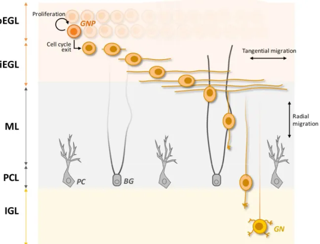
Once reached the EGL, GNPs undergo a phase of massive proliferation which accounts for the enormous number of mature GNs in the adult cerebellum. In mice, this clonal expansion initiates as soon as the first GNPs get to the EGL (around E13) and peaks around post-natal day (P) 7, the time at which the EGL thickens the most (Pons et al., 2001). However, after birth this exponential growth becomes increasingly counteracted by a progressive wave of cell cycle exit that will eventually convert all the dividing GNPs into post-mitotic cells by the end of the third post-natal week (Hanaway, 1967; Miale and Sidman, 1961). Once post-mitotic, GNPs slide below the still dividing progenitors, hence splitting the EGL into two sub-layers: the outer EGL (oEGL) containing the still mitotically active GNPs and the inner EGL (iEGL) constituted by their differentiating derivatives. Within the iEGL, post-mitotic GNPs undergo tangential migration that results in the extension of two long processes at the opposite poles of their somata. These processes will later maturate into their axons, the parallel fibers in the ML. Finally, the somata of differentiating GNs leaves the iEGL descending radially through the immature ML and the PCL to reach their final localization, the IGL. While descending, GNs produce a new leading process oriented inward in the cerebellum. This new process extends perpendicular to the two previously generated, therefore forming the characteristic "T-shaped" parallel fiber. Once located in the IGL, GNs complete their maturation forming the synaptic connections with MFs terminals. The whole process is illustrated in Figure VII.
I.2.3.2 Proliferation and differentiation of GNPs at the histological level
During the last 25 years several lineage tracing experiments helped to dissect the fate of GNPs in the EGL.
One of the first fundamental and not obvious observation was that GNPs seem to be unipotent progenitors, hence irreversibly committed to become GNs (Zhang and Goldman, 1996a, 1996b).
37
Although this observation has always been confirmed in vivo, it was shown that treatment of primary GNPs with BMP2 in vitro can twist their fate toward the astrocyte lineage (Okano-Uchida et al., 2004). Whether this property is the result of artifacts of the culturing system or it is biologically relevant also in vivo under certain circumstances, remains unknown.
Another important property of GNPs is that they only undergo symmetrical divisions in the EGL. In other words, a single GNP undergoing mitosis generates two identical cells which will either both re-enter the cell cycle, or both differentiate (Espinosa and Luo, 2008; Nakashima et al., 2015). Thanks to this peculiarity, GNPs pool expansion follows an exponential curve, with each single GNP generating a progeny with a median of 250 cells from E17.5 to P21 (Espinosa and Luo, 2008).
Figure VII. Developmental phases of GNPs in post-natal times. After the last division in the oEGL, GNPs exit the cell cycle and slide below forming the iEGL. Within the iEGL, GNPs migrate tangentially extending a leading and a trailing process that ultimately differentiate into the parallel fibers composing the ML (at this stage still immature). Subsequently, a third, perpendicular process is elongated inward in the cerebellum and the somata initiate to move along it. The fibers of the Bergmann Glia cells (BG) are utilized as substrate along which the radial migration proceeds. Migrating GNs reach the PCL, which hosts the PCs and BG cell bodies. From here, they will move to the IGL where they will terminate their differentiation to mature GNs. oEGL: outer external granule layer, iEGL: inner external granule layer, ML: molecular layer, PCL: Purkinje cells layer, IGL: internal granule layer, PC: Purkinje cell.
38
Interestingly, clonal pools of GNPs tend to exit the cell cycle synchronously and will also project their parallel fibers within the same sub-layer of the ML (Espinosa and Luo, 2008; Zong et al., 2005). In addition, while early born GNPs generally send their parallel fibers in deeper sub-layers of the ML, late born ones do it in upper sub-layers (Espinosa and Luo, 2008). This uneven spatio-temporal pattern of GN neurogenesis is likely to reflect precise requirements for proper wiring of cerebellar circuitries at later developmental stages. However, the actual nature of these requirements is still puzzling.
I.2.4.3 Discovery of SHH as the main mitogen for GNPs
During CNS development, various cell autonomous and non-cell autonomous mechanisms temporally and spatially control the expansion of neuron progenitors.
Aimed at dissecting these actors for GNPs proliferation in the EGL, the earliest works focused on a series of growth factors that were previously shown to mitotically activate other neuron progenitors in the developing brain. Thanks to these initial studies, it was shown that GNPs proliferation was elicited by Epidermal growth factor (Egf), Fibroblast growth factors (Fgf) and Insulin-like growth factors (Igf), all molecules produced in the developing cerebellum (Gao et al., 1991; Lin and Bulleit, 1997; Tao et al., 1996; Ye et al., 1996).
However, only from the end of the '90s it became clear that the most powerful signal driving GNPs divisions is Sonic Hedgehog (SHH), a secreted ligand produced by the PCs, which are arranging in the PCL at the time of EGL formation.
The first evidence of this regulation came from the fact that mutations in SHH pathway components were systematically found in certain types of medulloblastomas (MBs) malignant pediatric tumors arising from the developing cerebellum2 (Goodrich et al., 1997; Pietsch et al.,
1997; Raffel et al., 1997; Vorechovský et al., 1997). This suggested that SHH could act as a mitogen for some population of cerebellar neurons. Second, in situ hybridization revealed that a source of SHH in the cerebellum are the PCs (Wechsler-Reya and Scott, 1999). Very interestingly, it was previously shown that the selective ablation of PCs lineage, dramatically impaired GNPs pool expansion in the EGL, indicating that somehow PCs were required for GNPs proliferation (Smeyne et al., 1995). Therefore, the effects of the putative mitogen SHH were tested in primary GNPs cultures and, strikingly, it was found that it was capable of potently stimulating GNPs divisions (Dahmane and Ruiz-i-Altaba, 1999; Lewis et al., 2004;
2 Nowadays, these types of MBs are referred as SHH-MBs, and GNPs are known to be their cell of origin. MB
39
Reya and Scott, 1999). Finally, when all these observations were integrated with the confirmation that GNPs in the EGL express various transcriptional targets of SHH and that the blockade of SHH activity by anti-SHH antibodies impaired GNPs proliferation in cerebellar slices, it became clear that SHH functions as the main mitogen for GNPs (Dahmane and Ruiz-i-Altaba, 1999; Wallace, 1999; Wechsler-Reya and Scott, 1999).
Interestingly, SHH is expressed by PCs with a precise spatio-temporal pattern (Lewis et al., 2004). It is only after E17.5 that Shh is significantly up-regulated by PCs, and this is associated with concomitant activation of SHH targets in the overlying GNPs. In addition, SHH seems to be higher expressed by PCs occupying the anterior lobes compare to those in the posterior ones. A direct consequence is the uneven intensity of SHH signaling experienced by GNPs in the EGL along the anteroposterior axis. Such effect may be important for regulating the complexity of cortical foliation, which is in large part dependent on the extent of GNPs expansion (Corrales et al., 2004, 2006).
I.2.4.4 SHH boosts GNPs expansion by acting on the cell cycle machinery
The SHH signaling will be thoroughly described in Chapter I.4, while here only a brief overview will be presented.
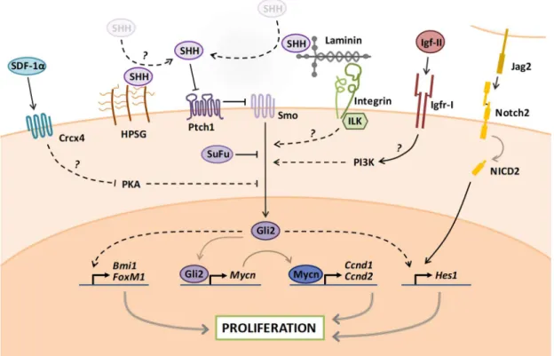
The SHH pathway in GNPs is activated when SHH binds and inactivates the plasma membrane receptor Patched homolog 1 (Ptch1), thereby relieving its inhibition on the G-protein coupled receptor (GPCR) Smoothened (Smo). This event initiates a signaling cascade which primarily culminates with the activation of the zinc-finger protein Glioma-associated oncogene 2 (Gli2), the main transcriptional effector of SHH in GNPs. Activation of Gli2 is counteracted by a Gli2-binding partner, called Suppressor of Fused (SuFu). In addition, Gli2 activates the expression of its paralog Gli1, which further amplifies the SHH transcriptional response in GNPs (Corrales et al., 2004) (Figure VIII).
The SHH-driven proliferation of GNPs is achieved through various mechanisms, among which the regulation of the cell cycle G1/S checkpoint is crucial. Indeed, activation of SHH signaling in GNPs potently induces the upregulation of N-myc proto-oncogene (Mycn) protein which in turn activates the transcription of Cyclin D1 and Cyclin D2 (Ciemerych et al., 2002; Huard et al., 1999; Kenney and Rowitch, 2000; Kenney et al., 2003; Knoepfler et al., 2002; Ma et al., 2015). Accumulation of D-type Cyclins in the G1 phase is critical for cells to cross the G1/S restriction point. Indeed, D-type Cyclins bind and activate Cyclin dependent kinases as CDK4/6 which initiate the phosporylation-dependent inhibition of Retinoblastoma-associated protein 1