RESEARCH OUTPUTS / RÉSULTATS DE RECHERCHE
Author(s) - Auteur(s) :
Publication date - Date de publication :
Permanent link - Permalien :
Rights / License - Licence de droit d’auteur :
Bibliothèque Universitaire Moretus Plantin
Institutional Repository - Research Portal
Dépôt Institutionnel - Portail de la Recherche
researchportal.unamur.be
University of Namur
Mitochondrial respiratory states and rates
Gnaiger, Erich; Arnould, Thierry; Detraux, Damien; STORDER, Julie
Published in:MitoFit Preprint Arch (2019) DOI:
doi.org/10.26124/mitofit:190001.v5 Publication date:
2019
Link to publication
Citation for pulished version (HARVARD):
Gnaiger, E, Arnould, T, Detraux, D & STORDER, J 2019, 'Mitochondrial respiratory states and rates', MitoFit Preprint Arch (2019) , no. 1, MitoFit Preprint Arch (2019) doi:10.26124/mitofit:190001.v2, pp. 1-40.
https://doi.org/doi.org/10.26124/mitofit:190001.v5
General rights
Copyright and moral rights for the publications made accessible in the public portal are retained by the authors and/or other copyright owners and it is a condition of accessing publications that users recognise and abide by the legal requirements associated with these rights. • Users may download and print one copy of any publication from the public portal for the purpose of private study or research. • You may not further distribute the material or use it for any profit-making activity or commercial gain
• You may freely distribute the URL identifying the publication in the public portal ? Take down policy
If you believe that this document breaches copyright please contact us providing details, and we will remove access to the work immediately and investigate your claim.
Mitochondrial respiratory states and rates
Gnaiger E, Aasander Frostner E, Abdul Karim N, Abdel-Rahman EA, Abumrad NA, Acuna-Castroviejo D, Adiele RC, Ahn B, Ali SS, Almeida A, Alton L, Alves MG, Amati F, Amoedo ND, Amorim R, Andreadou I, Antunes D, Aragó M, Aral C, Arandarčikaitė O, Arias Reyes C, Armand AS, Arnould T, Avram VF, Bach de Courtade SM, Bailey DM, Bairam A, Bajpeyi S, Bajzikova M, Bakker BM, Bardal T, Barlow J, Bastos Sant'Anna Silva AC, Batterson P, Battino M, Bazil J, Beard DA, Bednarczyk P, Beleza J, Bello F, Ben-Shachar D, Bento G, Bergdahl A, Berge RK, Bergmeister L, Bernardi P, Berridge MV, Bettinazzi S, Bishop D, Blier PU, Blindheim DF, Boardman NT, Boetker HE, Borchard S, Boros M, Børsheim E, Borutaite V, Botella J, Bouillaud F, Bouitbir J, Boushel RC, Bovard J, Bravo-Sagua R, Breton S, Brown DA, Brown GC, Brown RA, Brozinick JT, Buettner GR, Burtscher J, Calabria E, Calbet JA, Calzia E, Cannon DT, Cano Sanchez M, Canto AC, Cardoso LHD, Carvalho E, Casado Pinna M, Cassar S, Cassina AM, Castelo MP, Cavalcanti-de-Albuquerque JP, Celen MC, Cervinkova Z, Chabi B, Chakrabarti L, Chakrabarti S, Chaurasia B, Chen Q, Chicco AJ, Chinopoulos C, Chowdhury SK, Cizmarova B, Clementi E, Coen PM, Cohen BH, Coker RH, Collin A, Coxito P, Crisóstomo L, Crispim M, Crossland H, Dahdah N, Dalgaard LT, Dambrova M, Danhelovska T, Darveau CA, Das AM, Dash RK, Davidova E, Davis MS, De Goede P, De Palma C, Dembinska-Kiec A, Detraux D, Devaux Y, Di Marcello M, Di Paola FJ, Dias C, Dias TR, Diederich M, Distefano G, Djafarzadeh S, Doermann N, Doerrier C, Dong L, Donnelly C, Drahota Z, Duarte FV, Dubouchaud H, Duchen MR, Dumas JF, Durham WJ, Dymkowska D, Dyrstad SE, Dyson A, Dzialowski EM, Eaton S, Ehinger J, Elmer E, Endlicher R, Engin AB, Escames G, Evinova A, Ezrova Z, Falk MJ, Fell DA, Ferdinandy P, Ferko M, Ferreira JCB, Ferreira R, Ferri A, Fessel JP, Filipovska A, Fisar Z, Fischer C, Fischer M, Fisher G, Fisher JJ, Ford E, Fornaro M, Fuertes Agudo M, Fulton M, Galina A, Galkin A, Gallee L, Galli GL, Gama Pérez P, Gan Z, Ganetzky R, Gao Y, Garcia G, Garcia-Rivas G, Garcia-Roves PM, Garcia-Souza LF, Garipi E, Garlid KD, Garrabou G, Garten A, Gastaldelli A, Gayen J, Genders AJ, Genova ML, Giovarelli M, Glatz JFC, Goikoetxea Usandizaga N, Goncalo Teixeira da Silva R, Goncalves DF, Gonzalez-Armenta JL, Gonzalez-Franquesa A, Gonzalez-Freire M, Gonzalo H, Goodpaster BH, Gorr TA, Gourlay CW, Grams B, Granata C, Grefte S, Grilo L, Guarch ME, Gueguen N, Gumeni S, Haas CB, Haavik J, Hachmo Y, Haendeler J, Haider M, Hajrulahovic A, Hamann A, Han J, Han WH, Hancock CR, Hand SC, Handl J, Hargreaves IP, Harper ME, Harrison DK, Hassan H, Hatokova Z, Hausenloy DJ, Heales SJR, Hecker M, Heiestad C, Hellgren KT, Henrique A, Hepple RT, Hernansanz-Agustin P, Hewakapuge S, Hickey AJ, Ho DH, Hoehn KL, Hoel F, Holland OJ, Holloway GP, Holzner L, Hoppel CL, Hoppel F, Houstek J, Huete-Ortega M, Hyrossova P, Iglesias-Gonzalez J, Irving BA, Isola R, Iyer S, Jackson CB, Jadiya P, Jana PF, Jang DH, Jang YC, Janowska J, Jansen K, Jansen-Dürr P, Jansone B, Jarmuszkiewicz W, Jaskiewicz A, Jaspers RT, Jedlicka J, Jespersen NR, Jha RK, Jones JG, Joseph V, Jurczak MJ, Jurk D, Jusic A, Kaambre T, Kaczor JJ, Kainulainen H, Kampa RP, Kandel SM, Kane DA, Kapferer W, Kappler L, Karabatsiakis A, Karavaeva I, Karkucinska-Wieckowska A, Kaur S, Keijer J, Keller MA, Keppner G, Khamoui AV, Kidere D, Kilbaugh T, Kim HK, Kim JKS, Kimoloi S, Klepinin A, Klepinina L, Klingenspor M, Klocker H, Komlodi T, Koopman WJH, Kopitar-Jerala N, Kowaltowski AJ, Kozlov AV, Krajcova A, Krako Jakovljevic N, Kristal BS, Krycer JR, Kuang J, Kucera O, Kuka J, Kwak HB, Kwast K, Kwon OS, Laasmaa M, Labieniec-Watala M, Lagarrigue S, Lai N, Land JM, Lane N, Laner V, Lanza IR, Laouafa S, Laranjinha J, Larsen TS, Lavery GG, Lazou A, Ledo AM, Lee HK, Leeuwenburgh C, Leffler M, Lehti M, Lemieux H, Lenaz G, Lerfall J, Li PA, Li Puma L, Liang L, Liepins E, Liu J, López LC, Lucchinetti E, Ma T, Macedo MP, Machado IF, Maciej S, MacMillan-Crow LA, Magalhaes J, Magri A, Majtnerova P, Makarova E, Makrecka-Kuka M, Malik AN, Marcouiller F, Markova M, Martin DS, Martins AD, Martins JD, Maseko TE, Maull F, Mazat JP, McKenna HT, McKenzie M, Menze MA, Merz T, Meszaros AT, Methner A, Michalak S, Mila Guasch M, Minuzzi LM, Moellering DR, Moisoi N, Molina AJA, Montaigne D, Moore AL, Moore C, Moreau K, Moreno-Sánchez R, Moreira BP, Mracek T, Muccini AM, Munro D, Muntane J, Muntean DM,
Murray AJ, Musiol E, Nabben M, Nair KS, Nehlin JO, Nemec M, Neufer PD, Neuzil J, Neviere R, Newsom SA, Norman J, Nozickova K, Nunes S, O'Brien K, O'Brien KA, O'Gorman D, Olgar Y, Oliveira B, Oliveira J, Oliveira MF, Oliveira MT, Oliveira PF, Oliveira PJ, Olsen RE, Orynbayeva Z, Osiewacz HD, Paez H, Pak YK, Pallotta ML, Palmeira CM, Parajuli N, Passos JF, Passrugger M, Patel HH, Pavlova N, Pecina P, Pedersen TM, Perales JC, Pereira da Silva Grilo da Silva F, Pereira SP, Perez Valencia JA, Perks KL, Pesta D, Petit PX, Pettersen IKN, Pichaud N, Pichler I, Piel S, Pietka TA, Pinho SA, Pino MF, Pirkmajer S, Plangger M, Porter C, Porter RK, Preguica I, Prigione A, Procaccio V, Prochownik EV, Prola A, Pulinilkunnil T, Puskarich MA, Puurand M, Radenkovic F, Ramzan R, Rattan SIS, Reano S, Reboredo P, Rees BB, Renner-Sattler K, Rial E, Robinson MM, Roden M, Rodrigues AS, Rodriguez E, Rodriguez-Enriquez S, Rohlena J, Rolo AP, Ropelle ER, Roshanravan B, Røsland GV, Rossignol R, Rossiter HB, Rubelj I, Rybacka-Mossakowska J, Saada A, Safaei Z, Salin K, Salvadego D, Sandi C, Saner N, Santos D, Sanz A, Sardao V, Sarlak S, Sazanov LA, Scaife P, Scatena R, Schartner M, Scheibye-Knudsen M, Schilling JM, Schlattner U, Schneider Gasser EM, Schönfeld P, Schots PC, Schulz R, Schwarzer C, Scott GR, Selman C, Sendon PM, Shabalina IG, Sharma P, Sharma V, Shevchuk I, Shirazi R, Shiroma JG, Siewiera K, Silber AM, Silva AM, Sims CA, Singer D, Singh BK, Skolik R, Smenes BT, Smith J, Soares FAA, Sobotka O, Sokolova I, Soliz J, Sommer N, Sonkar VK, Sova M, Sowton AP, Sparagna GC, Sparks LM, Spinazzi M, Stankova P, Starr J, Stary C, Stelfa G, Stepto NK, Stevanovic J, Stiban J, Stier A, Stocker R, Storder J, Sumbalova Z, Suomalainen A, Suravajhala P, Svalbe B, Swerdlow RH, Swiniuch D, Szabo I, Szewczyk A, Szibor M, Tanaka M, Tandler B, Tarnopolsky MA, Tausan D, Tavernarakis N, Teodoro J, Tepp K, Thakkar H, Thapa M, Thyfault JP, Tomar D, Ton R, Torp MK, Towheed A, Tretter L, Trewin AJ, Trifunovic A, Trivigno C, Tronstad KJ, Trougakos IP, Truu L, Tuncay E, Turan B, Tyrrell DJ, Urban T, Valentine JM, Van Bergen NJ, Van der Ende M, Van Hove J, Varricchio F, Vella J, Vendelin M, Vercesi AE, Victor VM, Vieira Ligo Teixeira C, Vidimce J, Viel C, Vieyra A, Vilks K, Villena JA, Vincent V, Vinogradov AD, Viscomi C, Vitorino RMP, Vlachaki Walker J, Vogt S, Volani C, Volska K, Votion DM, Vujacic-Mirski K, Wagner BA, Ward ML, Warnsmann V, Wasserman DH, Watala C, Wei YH, Weissig V, Whitfield J, Wickert A, Wieckowski MR, Wiesner RJ, Williams CM, Winwood-Smith H, Wohlgemuth SE, Wohlwend M, Wolff JN, Wrutniak-Cabello C, Wüst RCI, Yokota T, Zablocki K, Zanon A, Zanou N, Zaugg K, Zaugg M, Zdrazilova L, Zhang Y, Zhang YZ, Zíková A, Zischka H, Zorzano A, Zujovic T, Zvejniece L
Corresponding author: Gnaiger E
Chair COST Action CA15203 MitoEAGLE – http://www.mitoeagle.org
Department of Visceral, Transplant and Thoracic Surgery, D. Swarovski Research Laboratory, Medical University of Innsbruck, Innrain 66/4, A-6020 Innsbruck, Austria
Email: mitoeagle@i-med.ac.at; Tel: +43 512 566796, Fax: +43 512 566796 20
612 coauthors
MitoEAGLE Task Group, COST Action CA15203 MitoEAGLE
http://www.mitofit.org/index.php/Gnaiger_2019_MitoFit_Preprint_Arch
Copyright: © 2019 Gnaiger et al. This is an Open Access preprint (not
peer-reviewed) distributed under the terms of the Creative Commons Attribution License, which permits unrestricted use, distribution, and reproduction in any medium, provided the original authors and source are credited. © remains with the authors, who have granted MitoFit an Open Access preprint licence in perpetuity.
Editor MitoFit Preprint Archives: Gnaiger E
More information:
www.mitoeagle.org
Funded by the Horizon 2020 Framework Programme of the European Union
Table of contents
AbstractExecutive summary
1. Introduction – Box 1: In brief: Mitochondria and Bioblasts 2. Coupling states and rates in mitochondrial preparations
2.1. Cellular and mitochondrial respiration
2.1.1. Aerobic and anaerobic catabolism and ATP turnover 2.1.2. Specification of biochemical dose
2.2. Mitochondrial preparations 2.3. Electron transfer pathways 2.4. Respiratory coupling control
2.4.1. Coupling
2.4.2. Phosphorylation, P», and P»/O2 ratio
2.4.3. Uncoupling
2.5. Coupling states and respiratory rates
2.5.1. LEAK-state
2.5.2. OXPHOS-state
2.5.3. Electron transfer-state 2.5.4. ROX state and Rox 2.5.5. Quantitative relations 2.5.6. The steady-state
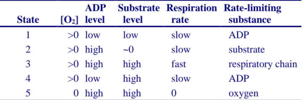
2.6. Classical terminology for isolated mitochondria
2.6.1. State 1 2.6.2. State 2 2.6.3. State 3 2.6.4. State 4 2.6.5. State 5
2.7. Control and regulation
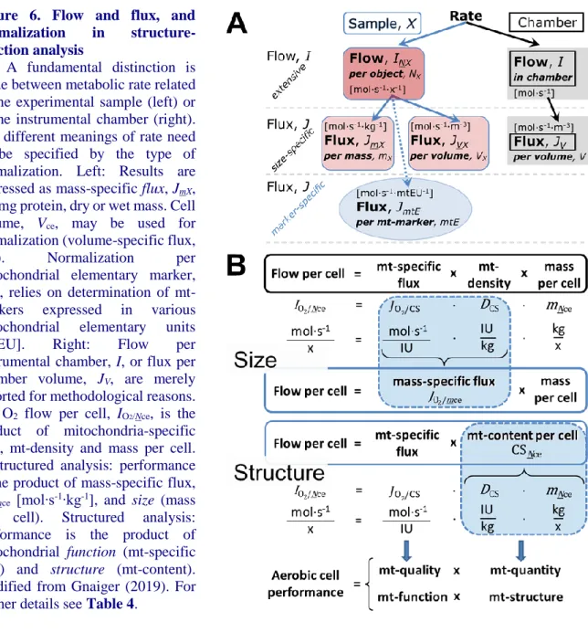
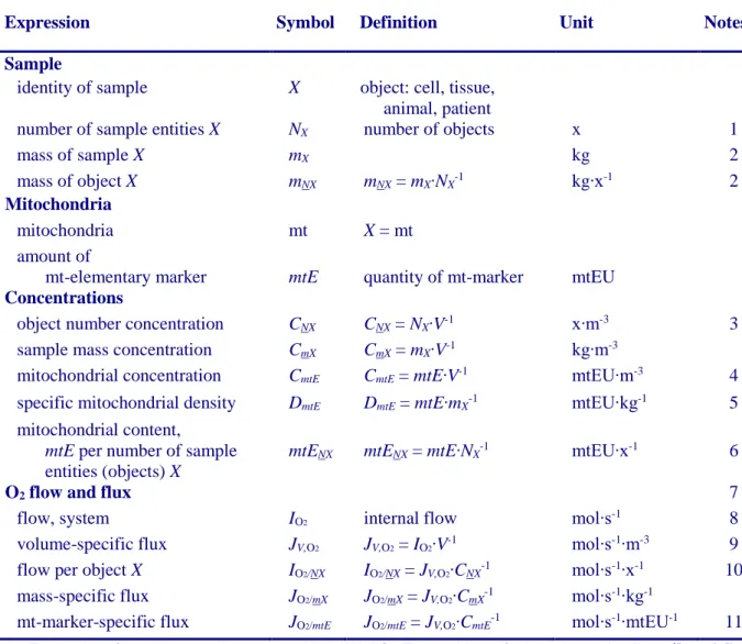
3. What is a rate? – Box 2: Metabolic flows and fluxes: vectoral, vectorial, and scalar 4. Normalization of rate per sample
4.1. Flow: per object
4.1.1. Number concentration 4.1.2. Flow per object
4.2. Size-specific flux: per sample size
4.2.1. Sample concentration 4.2.2. Size-specific flux
4.3. Marker-specific flux: per mitochondrial content
4.3.1. Mitochondrial concentration and mitochondrial markers 4.3.2. mt-Marker-specific flux
5. Normalization of rate per system
5.1. Flow: per chamber 5.2. Flux: per chamber volume
5.2.1. System-specific flux 5.2.2. Advancement per volume
6. Conversion of units
7. Conclusions – Box 3: Recommendations for studies with mitochondrial preparations Acknowledgements
Author contributions
Competing financial interests References
Supplement
S1. Manuscript phases and versions - an open-access approach S2. Joining COST Actions
Abstract As the knowledge base and importance of mitochondrial physiology to human health expands,
the necessity for harmonizing the terminology concerning mitochondrial respiratory states and rates has become increasingly apparent. The chemiosmotic theory establishes the mechanism of energy transformation and coupling in oxidative phosphorylation. The unifying concept of the protonmotive force provides the framework for developing a consistent theoretical foundation of mitochondrial physiology and bioenergetics. We follow guidelines of the International Union of Pure and Applied Chemistry (IUPAC) on terminology in physical chemistry, extended by considerations of open systems and thermodynamics of irreversible processes. The concept-driven constructive terminology incorporates the meaning of each quantity and aligns concepts and symbols with the nomenclature of classical bioenergetics. We endeavour to provide a balanced view of mitochondrial respiratory control and a critical discussion on reporting data of mitochondrial respiration in terms of metabolic flows and fluxes. Uniform standards for evaluation of respiratory states and rates will ultimately contribute to reproducibility between laboratories and thus support the development of databases of mitochondrial respiratory function in species, tissues, and cells. Clarity of concept and consistency of nomenclature facilitate effective transdisciplinary communication, education, and ultimately further discovery.
Keywords: Mitochondrial respiratory control, coupling control, mitochondrial preparations,
protonmotive force, uncoupling, oxidative phosphorylation: OXPHOS, efficiency, electron transfer: ET, electron transfer system: ETS, proton leak, ion leak and slip compensatory state: LEAK, residual oxygen consumption: ROX, State 2, State 3, State 4, normalization, flow, flux, oxygen: O2
Executive summary
In view of the broad implications for health care, mitochondrial researchers face an increasing responsibility to disseminate their fundamental knowledge and novel discoveries to a wide range of stakeholders and scientists beyond the group of specialists. This requires implementation of a commonly accepted terminology within the discipline and standardization in the translational context. Authors, reviewers, journal editors, and lecturers are challenged to collaborate with the aim to harmonize the nomenclature in the growing field of mitochondrial physiology and bioenergetics, from evolutionary biology and comparative physiology to mitochondrial medicine. In the present communication we focus on the following concepts in mitochondrial physiology:
1. Aerobic respiration is the O2 flux in catabolic reactions coupled to phosphorylation of ADP to
ATP, and O2 flux in a variety of O2 consuming reactions apart from oxidative phosphorylation
(OXPHOS). Coupling in OXPHOS is mediated by the translocation of protons across the mitochondrial inner membrane (mtIM) through proton pumps generating or utilizing the protonmotive force that is maintained between the mitochondrial matrix and intermembrane compartment or outer mitochondrial space. Compartmental coupling depends on ion translocation across a semipermeable membrane, which is defined as vectorial metabolism and distinguishes OXPHOS from cytosolic fermentation as counterparts of cellular core energy metabolism (Figure 1). Cell respiration is thus distinguished from fermentation: (1) Electron acceptors are supplied by external respiration for the maintenance of redox balance, whereas fermentation is characterized by an internal electron acceptor produced in intermediary metabolism. In aerobic cell respiration, redox balance is maintained by O2 as the electron
acceptor. (2) Compartmental coupling in vectorial OXPHOS contrasts to exclusively scalar substrate-level phosphorylation in fermentation.
2. When measuring mitochondrial metabolism, the contribution of fermentation and other cytosolic interactions must be excluded from analysis by disrupting the barrier function of the plasma membrane. Selective removal or permeabilization of the plasma membrane yields mitochondrial preparations—including isolated mitochondria, tissue and cellular preparations—with structural and functional integrity. Subsequently, extramitochondrial concentrations of fuel substrates, ADP, ATP, inorganic phosphate, and cations including H+
can be controlled to determine mitochondrial function under a set of conditions defined as coupling control states. We strive to incorporate an easily recognized and understood concept-driven terminology of bioenergetics with explicit terms and symbols that define the nature of respiratory states.
3. Mitochondrial coupling states are defined according to the control of respiratory oxygen flux by the protonmotive force, pmF, in an interaction of the electron transfer system generating the pmF and the phosphorylation system utilizing the pmF. Capacities of OXPHOS and electron transfer are measured at kinetically-saturating concentrations of fuel substrates, ADP and inorganic phosphate, and O2, or at optimal uncoupler concentrations, respectively, in the
absence of Complex IV inhibitors such as NO, CO, or H2S. Respiratory capacity is a measure
of the upper boundary of the rate of respiration; it depends on the substrate type undergoing oxidation in a mitochondrial pathway, and provides reference values for the diagnosis of health and disease. Evaluation of the impact of evolutionary background, age, gender and sex, lifestyle and environment represents a major challenge for mitochondrial respiratory physiology and pathology.
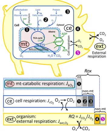
Figure 1. Internal and external respiration
(mt) Mitochondrial catabolic respiration, JkO2,
is the O2 consumption in the oxidation of fuel substrates (electron donors) and reduction of O2 catalysed by the electron transfer system, ETS, which drives the protonmotive force,
pmF. JkO2 excludes mitochondrial residual
oxygen consumption, mt-Rox ().
(ce) Cell respiration or internal cellular O2 consumption, JrO2, takes into account all
chemical reactions, r, that consume O2 in the cells. Catabolic cell respiration is the O2 consumption associated with catabolic pathways in the cell, including (mt) mitochondrial catabolism; mt-Rox (); non-mt O2 consumption by catabolic reactions, particularly peroxisomal oxidases and microsomal cytochrome P450 systems (); and non-mt Rox by reactions unrelated to catabolism ().
(ext) External respiration balances internal
respiration at steady-state, including extracellular Rox () and aerobic respiration by the microbiome (). O2 is transported
from the environment across the respiratory cascade, i.e., circulation between tissues and diffusion across cell membranes, to the intracellular compartment. The respiratory quotient, RQ, is the molar CO2/O2 exchange ratio; when combined with the respiratory nitrogen quotient, N/O2 (mol N given off per mol O2 consumed), the RQ reflects the proportion of carbohydrate, lipid and protein utilized in cell respiration during aerobically balanced steady-states. Bicarbonate and CO2 are transported in reverse to the extracellular milieu and the organismic environment. Hemoglobin provides the molecular paradigm for the combination of O2 and CO2 exchange, as do lungs and gills on the morphological level. Consult Table 8 for a list of terms and symbols.
4. Incomplete tightness of coupling, i.e., some degree of uncoupling relative to the mitochondrial pathway-dependent coupling stoichiometry, is a characteristic of energy-transformations across membranes. Uncoupling is caused by a variety of physiological, pathological, toxicological, pharmacological and environmental conditions that exert an influence not only on the proton leak and cation cycling, but also on proton slip within the proton pumps and the structural integrity of the mitochondria. A more loosely coupled state is induced by stimulation of mitochondrial superoxide formation and the bypass of proton pumps. In addition, the use of protonophores represents an experimental uncoupling intervention to assess the transition from a well-coupled to a noncoupled state of mitochondrial respiration.
5. Respiratory oxygen consumption rates have to be carefully normalized to enable meta-analytic studies beyond the question of a particular experiment. Therefore, all raw data on rates and
variables for normalization should be published in an open access data repository. Normalization of rates for: (1) the number of objects (cells, organisms); (2) the volume or mass of the experimental sample; and (3) the concentration of mitochondrial markers in the instrumental chamber are sample-specific normalizations, which are distinguished from system-specific normalization for the volume of the instrumental chamber (the measuring system).
6. The consistent use of terms and symbols facilitates transdisciplinary communication and will support the further development of a collaborative database on bioenergetics and mitochondrial physiology.
Box 1: In brief – Mitochondria and Bioblasts
‘For the physiologist, mitochondria afforded the first opportunity for an experimental approach to structure-function relationships, in particular those involved in active transport, vectorial metabolism, and metabolic control mechanisms on a subcellular level’ (Ernster and Schatz 1981).
Mitochondria are oxygen-consuming electrochemical generators that evolved from the endosymbiotic alphaproteobacteria which became integrated into a host cell related to Asgard Archaea (Margulis 1970; Lane 2005; Roger et al. 2017). They were described by Richard Altmann (1894) as ‘bioblasts’, which include not only the mitochondria as presently defined, but also symbiotic and free-living bacteria. The word ‘mitochondria’ (Greek mitos: thread; chondros: granule) was introduced by Carl Benda (1898). Mitochondrion is singular and mitochondria is plural. Abbreviation: mt, as generally used in mtDNA.
Contrary to current textbook dogma, which describes mitochondria as individual organelles, mitochondria form dynamic networks within eukaryotic cells. Mitochondrial movement is supported by microtubules. Mitochondrial size and number can change in response to energy requirements of the cell via processes known as fusion and fission; these interactions allow mitochondria to communicate within a network (Chan 2006). Mitochondria can even traverse cell boundaries in a process known as horizontal mitochondrial transfer (Torralba et al. 2016). Another defining morphological characteristic of mitochondria is the double membrane. The mitochondrial inner membrane (mtIM) forms dynamic tubular to disk-shaped cristae that separate the mitochondrial matrix, i.e., the negatively charged internal mitochondrial compartment, from the intermembrane space; the latter being enclosed by the mitochondrial outer membrane (mtOM) and positively charged with respect to the matrix.
Intracellular stress factors may cause shrinking or swelling of the mitochondrial matrix that can ultimately result in permeability transition (mtPT; Lemasters et al. 1998). The mtIM contains the non-bilayer phospholipid cardiolipin, which is also involved in the mtOM (Gebert et al. 2009) but is not present in any other eukaryotic cellular membrane. Cardiolipin has many regulatory functions (Oemer
et al. 2018); it promotes and stabilizes the formation of supercomplexes (SCInIIInIVn) based on dynamic
interactions between specific respiratory complexes (McKenzie et al. 2006; Greggio et al. 2017; Lenaz et al. 2017), and it supports proton transfer on the mtIM from the electron transfer system to F-ATPase (ATP synthase; Yoshinaga et al. 2016). The mtIM is plastic and exerts an influence on the functional properties of incorporated proteins (Waczulikova et al. 2007).
Mitochondria constitute the structural and functional elementary components of cell respiration. Mitochondrial respiration is the reduction of molecular oxygen by electron transfer coupled to electrochemical proton translocation across the mtIM. In the process of OXPHOS, the catabolic reaction of oxygen consumption is electrochemically coupled to the transformation of energy in the phosphorylation of ADP to adenosine triphosphate (ATP; Mitchell 1961, 2011). Mitochondria are the powerhouses of the cell that contain the machinery of the OXPHOS-pathways, including transmembrane respiratory complexes (proton pumps with FMN, Fe-S and cytochrome b, c, aa3 redox systems);
alternative dehydrogenases and oxidases; the coenzyme ubiquinone (Q); F-ATPase or ATP synthase; the enzymes of the tricarboxylic acid cycle (TCA), fatty acid and amino acid oxidation; transporters of ions, metabolites and co-factors; iron/sulphur cluster synthesis; and mitochondrial kinases related to catabolic pathways. TCA cycle intermediates are vital precursors for macromolecule biosynthesis (Diebold et al. 2019). The mitochondrial proteome comprises over 1,200 proteins (Calvo et al. 2015; 2017), mostly encoded by nuclear DNA (nDNA), with a variety of functions, many of which are relatively well known, e.g., proteins regulating mitochondrial biogenesis or apoptosis, while others are still under investigation, or need to be identified, e.g., mtPT pore and alanine transporter. The
mammalian mitochondrial proteome can be used to discover and characterize the genetic basis of mitochondrial diseases (Williams et al. 2016; Palmfeldt and Bross 2017).
Numerous cellular processes are orchestrated by a constant crosstalk between mitochondria and other cellular components. For example, the crosstalk between mitochondria and the endoplasmic reticulum is involved in the regulation of calcium homeostasis, cell division, autophagy, differentiation, and anti-viral signaling (Murley and Nunnari 2016). Mitochondria contribute to the formation of peroxisomes, which are hybrids of mitochondrial and ER-derived precursors (Sugiura et al. 2017). Cellular mitochondrial homeostasis (mitostasis) is maintained through regulation at transcriptional, post-translational and epigenetic levels (Ling and Rönn 2018; Lisowski et al. 2018), resulting in dynamic regulation of mitochondrial turnover by biogenesis of new mitochondria and removal of damaged mitochondria by fusion, fission and mitophagy (Singh et al. 2018). Cell signalling modules contribute to homeostatic regulation throughout the cell cycle or even cell death by activating proteostatic modules, e.g., the ubiquitin-proteasome and autophagy-lysosome/vacuole pathways; specific proteases like LON, and genome stability modules in response to varying energy demands and stress cues (Quiros et al. 2016). In addition, several post-translational modifications, including acetylation and nitrosylation, are capable of influencing the bioenergetic response, with clinically significant implications for health and disease (Carrico et al. 2018).
Mitochondria of higher eukaryotes typically maintain several copies of their own circular genome known as mitochondrial DNA (mtDNA; hundred to thousands per cell; Cummins 1998), which is maternally inherited in many species. However, biparental mitochondrial inheritance is documented in some exceptional cases in humans (Luo et al. 2018), is widespread in birds, fish, reptiles and invertebrate groups, and is even the norm in some bivalve taxonomic groups (Breton et al. 2007; White et al. 2008). The mitochondrial genome of the angiosperm Amborella contains a record of six mitochondrial genome equivalents acquired by horizontal transfer of entire genomes, two from angiosperms, three from algae and one from mosses (Rice et al. 2016). In unicellular organisms, i.e., protists, the structural organization of mitochondrial genomes is highly variable and includes circular and linear DNA (Zikova et al. 2016). While some of the free-living flagellates exhibit the largest known gene coding capacity, e.g., jakobid Andalucia godoyi mtDNA codes for 106 genes (Burger et al. 2013), some protist groups, e.g., alveolates, possess mitochondrial genomes with only three protein-coding genes and two rRNAs (Feagin et al. 2012). The complete loss of mitochondrial genome is observed in the highly reduced mitochondria of Cryptosporidium species (Liu et al. 2016). Reaching the final extreme, the microbial eukaryote, oxymonad Monocercomonoides, has no mitochondrion whatsoever and lacks all typical nuclear-encoded mitochondrial proteins, showing that while in 99% of organisms mitochondria play a vital role, this organelle is not indispensable (Karnkowska et al. 2016).
In vertebrates, but not all invertebrates, mtDNA is compact (16.5 kB in humans) and encodes 13 protein subunits of the transmembrane respiratory Complexes CI, CIII, CIV and ATP synthase (F-ATPase), 22 tRNAs, and two ribosomal RNAs. Additional gene content has been suggested to include microRNAs, piRNA, smithRNAs, repeat associated RNA, long noncoding RNAs, and even additional proteins or peptides (Rackham et al. 2011; Duarte et al. 2014; Lee et al. 2015; Cobb et al. 2016). The mitochondrial genome requires nuclear-encoded mitochondrially targeted proteins, e.g., TFAM, for its maintenance and expression (Rackham et al. 2012). The nuclear and the mitochondrial genomes encode peptides of the membrane spanning redox pumps (CI, CIII and CIV) and F-ATPase, leading to strong constraints in the coevolution of both genomes (Blier et al. 2001).
Given the multiple roles of mitochondria, it is perhaps not surprising that mitochondrial dysfunction is associated with a wide variety of genetic and degenerative diseases. Robust mitochondrial function is supported by physical exercise and caloric balance, and is central for sustained metabolic health throughout life. Therefore, a more consistent set of definitions for mitochondrial physiology will increase our understanding of the etiology of disease and improve the diagnostic repertoire of mitochondrial medicine with a focus on protective medicine, lifestyle and healthy aging.
1. Introduction
Mitochondria are the powerhouses of the cell with numerous morphological, physiological, molecular, and genetic functions (Box 1). Every study of mitochondrial health and disease faces
Evolution, Age, Gender and sex, Lifestyle, and Environment (MitoEAGLE) as essential background
conditions intrinsic to the individual person or cohort, species, tissue and to some extent even cell line. As a large and coordinated group of laboratories and researchers, the mission of the global MitoEAGLE Network is to generate the necessary scale, type, and quality of consistent data sets and conditions to address this intrinsic complexity. Harmonization of experimental protocols and implementation of a quality control and data management system are required to interrelate results gathered across a spectrum of studies and to generate a rigorously monitored database focused on mitochondrial respiratory function. In this way, researchers from a variety of disciplines can compare their findings using clearly defined and accepted international standards.
With an emphasis on quality of research, published data can be useful far beyond the specific question of a particular experiment. For example, collaborative data sets support the development of open-access databases such as those for National Institutes of Health sponsored research in genetics, proteomics, and metabolomics. Indeed, enabling meta-analysis is the most economic way of providing robust answers to biological questions (Cooper et al. 2009). However, the reproducibility of quantitative results and databases depend on accurate measurements under strictly-defined conditions. Likewise, meaningful interpretation and comparability of experimental outcomes requires harmonization of protocols between research groups at different institutes. In addition to quality control, a conceptual framework is also required to standardise and harmonise terminology and methodology. Vague or ambiguous jargon can lead to confusion and may convert valuable signals to wasteful noise. For this reason, measured values must be expressed in standard units for each parameter used to define mitochondrial respiratory function. A consensus on fundamental nomenclature and conceptual coherence, however, are missing in the expanding field of mitochondrial physiology. To fill this gap, the present communication provides an in-depth review on harmonization of nomenclature and definition of technical terms, which are essential to improve the awareness of the intricate meaning of current and past scientific vocabulary. This is important for documentation and integration into databases in general, and quantitative modelling in particular (Beard 2005).
In this review, we focus on coupling states and fluxes through metabolic pathways of aerobic energy transformation in mitochondrial preparations in the attempt to establish a conceptually-oriented nomenclature in bioenergetics and mitochondrial physiology in a series of communications, prepared in the frame of the EU COST Action MitoEAGLE open to global bottom-up input.
2. Coupling states and rates in mitochondrial preparations
‘Every professional group develops its own technical jargon for talking about matters of critical
concern ... People who know a word can share that idea with other members of their group, and a shared vocabulary is part of the glue that holds people together and allows them to create a shared culture’ (Miller 1991).
2.1. Cellular and mitochondrial respiration
2.1.1. Aerobic and anaerobic catabolism and ATP turnover: In respiration, electron transfer
is coupled to the phosphorylation of ADP to ATP, with energy transformation mediated by the protonmotive force, pmF (Figure 2). Anabolic reactions are coupled to catabolism, both by ATP as the intermediary energy currency and by small organic precursor molecules as building blocks for biosynthesis (Diebold et al. 2019). Glycolysis involves substrate-level phosphorylation of ADP to ATP in fermentation without utilization of O2, studied mainly in living cells and organisms. Many cellular
fuel substrates are catabolized to acetyl-CoA or to glutamate, and further electron transfer reduces nicotinamide adenine dinucleotide to NADH or flavin adenine dinucleotide to FADH2. Subsequent
mitochondrial electron transfer to O2 is coupled to proton translocation for the control of the
protonmotive force and phosphorylation of ADP (Figure 2B and 2C). In contrast, extramitochondrial oxidation of fatty acids and amino acids proceeds partially in peroxisomes without coupling to ATP production: acyl-CoA oxidase catalyzes the oxidation of FADH2 with electron transfer to O2; amino
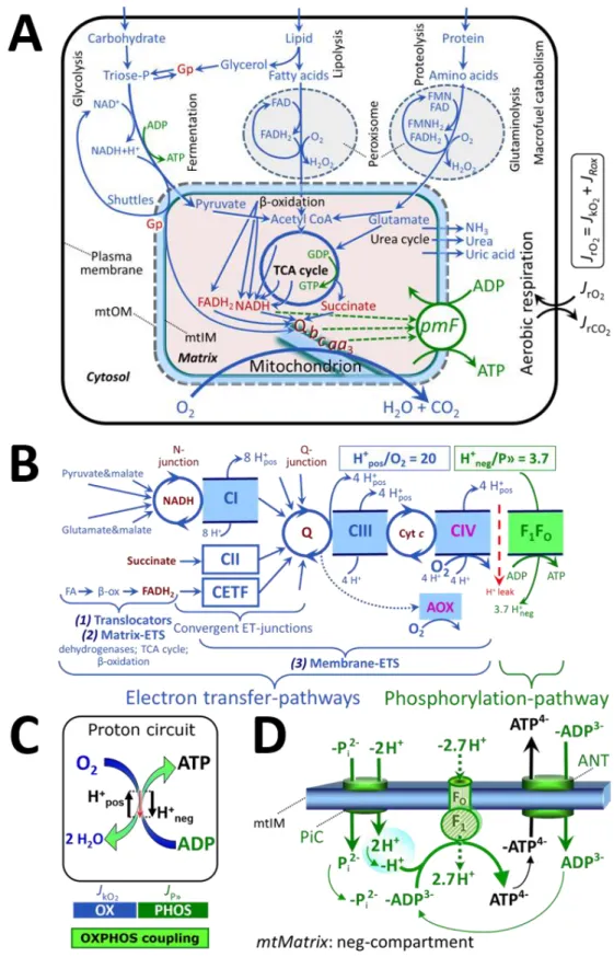
Figure 2. Cell respiration and oxidative phosphorylation (OXPHOS)
Mitochondrial respiration is the oxidation of fuel substrates (electron donors) with electron transfer to O2 as the electron acceptor. For explanation of symbols see also Figure 1.
(A) Respiration of living cells: Extramitochondrial catabolism of macrofuels and uptake of
small molecules by the cell provide the mitochondrial fuel substrates. Dashed arrows indicate the connection between the redox proton pumps (respiratory Complexes CI, CIII and CIV) and the transmembrane protonmotive force, pmF. Coenzyme Q (Q) and the cytochromes b, c, and aa3 are redox systems of the mitochondrial inner membrane, mtIM. Glycerol-3-phosphate, Gp.
(B) Respiration in mitochondrial preparations: The mitochondrial electron transfer system
(ETS) is (1) fuelled by diffusion and transport of substrates across the mtOM and mtIM, and in addition consists of the (2) matrix-ETS, and (3) membrane-ETS. Electron transfer converges at the N-junction, and from CI, CII and electron transferring flavoprotein complex (CETF) at the Q-junction. Unlabeled arrows converging at the Q-junction indicate additional ETS-sections with electron entry into Q through glycerophosphate dehydrogenase, dihydroorotate dehydrogenase, proline dehydrogenase, choline dehydrogenase, and sulfide-ubiquinone oxidoreductase. The dotted arrow indicates the branched pathway of oxygen consumption by alternative quinol oxidase (AOX). ET-pathways are coupled to the phosphorylation-pathway. The H+pos/O2 ratio is the outward proton flux from the matrix space to the positively (pos) charged vesicular compartment, divided by catabolic O2 flux in the NADH-pathway. The H+neg/P» ratio is the inward proton flux from the inter-membrane space to the negatively (neg) charged matrix space, divided by the flux of phosphorylation of ADP to ATP. These stoichiometries are not fixed because of ion leaks and proton slip. Modified from Lemieux et al. (2017) and Rich (2013).
(C) OXPHOS-coupling:
The H
+circuit couples O
2
flux through the catabolic
ET-pathway, J
kO2, to flux through the phosphorylation-pathway of ADP to ATP, J
P».
(D) Phosphorylation-pathway catalyzed by the proton pump F1FO-ATPase (F-ATPase, ATP synthase), adenine nucleotide translocase (ANT), and inorganic phosphate carrier (PiC). The H+neg/P» stoichiometry is the sum of the coupling stoichiometry in the F-ATPase reaction (-2.7 H+pos from the positive intermembrane space, 2.7 H+neg to the matrix, i.e., the negative compartment) and the proton balance in the translocation of ADP3-, ATP4- and Pi 2-(negative for substrates). Modified from Gnaiger (2019).
The plasma membrane separates the intracellular compartment including the cytosol, nucleus, and organelles from the extracellular environment. Cell membranes include the plasma membrane and organellar membranes. The plasma membrane consists of a lipid bilayer with embedded proteins and attached organic molecules that collectively control the selective permeability of ions, organic molecules, and particles across the cell boundary. The intact plasma membrane prevents the passage of many water-soluble mitochondrial substrates and inorganic ions—such as succinate, adenosine diphosphate (ADP) and inorganic phosphate (Pi) that must be precisely controlled at
kinetically-saturating concentrations for the analysis of mitochondrial respiratory capacities. Respiratory capacities delineate, comparable to channel capacity in information theory (Schneider 2006), the upper boundary of the rate of O2 consumption measured in defined respiratory states. The intact plasma membrane limits
the scope of investigations into mitochondrial respiratory function in living cells, despite the activity of solute carriers, e.g., the dependent dicarboxylate transporter SLC13A3 and the sodium-dependent phosphate transporter SLC20A2, which transport specific metabolites across the plasma membrane of various cell types, and the availability of plasma membrane-permeable succinate (Ehinger
et al. 2016). These limitations are overcome by the use of mitochondrial preparations.
2.1.2. Specification of biochemical dose: Substrates, uncouplers, inhibitors, and other chemical
reagents are titrated to analyse cellular and mitochondrial function. Nominal concentrations of these substances are usually reported as initial amount of substance concentration [mol∙L-1] in the incubation
medium. Kinetically-saturating conditions are evaluated by substrate kinetics to obtain the maximum reaction velocity or maximum pathway flux, in contrast to solubility-saturated conditions. When aiming at the measurement of kinetically-saturated processes—such as OXPHOS-capacities—the concentrations for substrates can be chosen according to half-saturating substrate concentrations, c50,
for metabolic pathways, or the Michaelis constant, Km, for enzyme kinetics. In the case of hyperbolic
kinetics, only 80% of maximum respiratory capacity is obtained at a substrate concentration of four times the c50, whereas substrate concentrations of 5, 9, 19 and 49 times the c50 are theoretically required
for reaching 83%, 90%, 95% or 98% of the maximal rate (Gnaiger 2001). Other reagents are chosen to inhibit or alter a particular process. The amount of these chemicals in an experimental incubation is selected to maximize effect, avoiding unacceptable off-target consequences that would adversely affect the data being sought. Specifying the amount of substance in an incubation as nominal concentration in the aqueous incubation medium can be ambiguous (Doskey et al. 2015), particularly for cations (TPP+;
fluorescent dyes such as safranin, TMRM; Chowdhury et al. 2015) and lipophilic substances (oligomycin, uncouplers, permeabilization agents; Doerrier et al. 2018), which accumulate in the
mitochondrial matrix or in biological membranes, respectively. Generally, dose/exposure can be specified per unit of biological sample, i.e., (nominal moles of xenobiotic)/(number of cells) [mol∙cell-1]
or, as appropriate, per mass of biological sample [mol∙kg-1]. This approach to specification of
dose/exposure provides a scalable parameter that can be used to design experiments, help interpret a wide variety of experimental results, and provide absolute information that allows researchers worldwide to make the most use of published data (Doskey et al. 2015).
2.2. Mitochondrial preparations
Mitochondrial preparations are defined as either isolated mitochondria or tissue and cellular preparations in which the barrier function of the plasma membrane is disrupted. Since this entails the loss of cell viability, mitochondrial preparations are not studied in vivo. In contrast to isolated mitochondria and tissue homogenate preparations, mitochondria in permeabilized tissues and cells are
in situ relative to the plasma membrane. When studying mitochondrial preparations,
substrate-uncoupler-inhibitor-titration (SUIT) protocols are used to establish respiratory coupling control states (CCS) and pathway control states (PCS) that provide reference values for various output variables (Table 1). Physiological conditions in vivo deviate from these experimentally obtained states; this is because kinetically-saturating concentrations, e.g., of ADP, oxygen (O2; dioxygen) or fuel substrates,
may not apply to physiological intracellular conditions. Further information is obtained in studies of kinetic responses to variations in fuel substrate concentrations, [ADP], or [O2] in the range between
kinetically-saturating concentrations and anoxia (Gnaiger 2001).
The cholesterol content of the plasma membrane is high compared to mitochondrial membranes (Korn 1969). Therefore, mild detergents—such as digitonin and saponin—can be applied to selectively permeabilize the plasma membrane via interaction with cholesterol; this allows free exchange of organic molecules and inorganic ions between the cytosol and the immediate cell environment, while maintaining the integrity and localization of organelles, cytoskeleton, and the nucleus. Application of permeabilization agents (mild detergents or toxins) leads to washout of cytosolic marker enzymes— such as lactate dehydrogenase—and results in the complete loss of cell viability (tested by nuclear staining using plasma membrane-impermeable dyes), while mitochondrial function remains intact (tested by cytochrome c stimulation of respiration). Digitonin concentrations have to be optimized according to cell type, particularly since mitochondria from cancer cells contain significantly higher contents of cholesterol in both membranes (Baggetto and Testa-Perussini, 1990). For example, a dose of digitonin of 8 fmol∙cell-1 (10 pg∙cell-1; 10 µg∙10-6 cells) is optimal for permeabilization of endothelial
cells, and the concentration in the incubation medium has to be adjusted according to the cell concentration (Doerrier et al. 2018). Respiration of isolated mitochondria remains unaltered after the addition of low concentrations of digitonin or saponin. In addition to mechanical cell disruption during homogenization of tissue, permeabilization agents may be applied to ensure permeabilization of all cells in tissue homogenates.
Suspensions of cells permeabilized in the respiration chamber and crude tissue homogenates contain all components of the cell at highly dilute concentrations. All mitochondria are retained in chemically-permeabilized mitochondrial preparations and crude tissue homogenates. In the preparation of isolated mitochondria, however, the mitochondria are separated from other cell fractions and purified by differential centrifugation, entailing the loss of mitochondria at typical recoveries ranging from 30% to 80% of total mitochondrial content (Lai et al. 2018). Using Percoll or sucrose density gradients to maximize the purity of isolated mitochondria may compromise the mitochondrial yield or structural and functional integrity. Therefore, mitochondrial isolation protocols need to be optimized according to each study. The term mitochondrial preparation neither includes living cells, nor submitochondrial particles and further fractionated mitochondrial components.
2.3. Electron transfer pathways
Mitochondrial electron transfer (ET) pathways are fuelled by diffusion and transport of substrates across the mtOM and mtIM. In addition, the mitochondrial electron transfer system (ETS) consists of the matrix-ETS and membrane-ETS (Figure 2B). Upstream sections of ET-pathways converge at the NADH-junction (N-junction). NADH is mainly generated in the tricarboxylic acid (TCA) cycle and is oxidized by Complex I (CI), with further electron entry into the coenzyme Q-junction (Q-junction).
Similarly, succinate is formed in the TCA cycle and oxidized by CII to fumarate. CII is part of both the TCA cycle and the ETS, and reduces FAD to FADH2 with further reduction of ubiquinone to ubiquinol
downstream of the TCA cycle in the Q-junction. Thus FADH2 is not a substrate but is the product of
CII, in contrast to erroneous metabolic maps shown in many publications. β-oxidation of fatty acids (FA) supplies reducing equivalents via (1) FADH2 as the substrate of electron transferring flavoprotein
complex (CETF); (2) acetyl-CoA generated by chain shortening; and (3) NADH generated via 3-hydroxyacyl-CoA dehydrogenases. The ATP yield depends on whether acetyl-CoA enters the TCA cycle, or is for example used in ketogenesis.
Selected mitochondrial catabolic pathways, k, of electron transfer from the oxidation of fuel substrates to the reduction of O2 are stimulated by addition of fuel substrates to the mitochondrial
respiration medium after depletion of endogenous substrates (Figure 2B). Substrate combinations and specific inhibitors of ET-pathway enzymes are used to obtain defined pathway control states in mitochondrial preparations (Gnaiger 2019).
2.4. Respiratory coupling control
2.4.1. Coupling: In mitochondrial electron transfer, vectorial transmembrane proton flux is
coupled through the redox proton pumps CI, CIII and CIV to the catabolic flux of scalar reactions, collectively measured as O2 flux, JkO2 (Figure 2). Thus mitochondria are elementary components of
energy transformation. Energy is a conserved quantity and cannot be lost or produced in any internal process (First Law of Thermodynamics). Open and closed systems can gain or lose energy only by external fluxes—by exchange with the environment. Therefore, energy can neither be produced by mitochondria, nor is there any internal process without energy conservation. Exergy or Gibbs energy (‘free energy’) is the part of energy that can potentially be transformed into work under conditions of constant temperature and pressure. Coupling is the interaction of an exergonic process (spontaneous, negative exergy change) with an endergonic process (positive exergy change) in energy transformations which conserve part of the exergy change. Exergy is not completely conserved, however, except at the limit of 100% efficiency of energy transformation in a coupled process. The exergy or Gibbs energy change that is not conserved by copling is irreversibly lost or dissipated, and is accounted for as the entropy change of the surroundings and the system, multiplied by the temperature of the irreversible process.
Pathway control states (PCS) and coupling control states (CCS) are complementary, since mitochondrial preparations depend on (1) an exogenous supply of pathway-specific fuel substrates and oxygen, and (2) exogenous control of phosphorylation (Figure 2).
2.4.2. Phosphorylation, P», and P»/O2 ratio: Phosphorylation in the context of OXPHOS is
defined as phosphorylation of ADP by Pi to form ATP. On the other hand, the term phosphorylation is
used generally in many contexts, e.g., protein phosphorylation. This provides the argument for introducing a symbol more discriminating and specific than P as used in the P/O ratio (phosphate to atomic oxygen ratio), where P indicates phosphorylation of ADP to ATP or GDP to GTP (Figure 2): The symbol P» indicates the endergonic (uphill) direction of phosphorylation ADP→ATP, and likewise P« the corresponding exergonic (downhill) hydrolysis ATP→ADP. P» refers mainly to electrontransfer phosphorylation but may also involve substrate-level phosphorylation as part of the TCA cycle (succinyl-CoA ligase, phosphoglycerate kinase) and phosphorylation of ADP catalyzed by pyruvate kinase, and of GDP phosphorylated by phosphoenolpyruvate carboxykinase. Transphosphorylation is performed by adenylate kinase, creatine kinase (mtCK), hexokinase and nucleoside diphosphate kinase. In isolated mammalian mitochondria, ATP production catalyzed by adenylate kinase (2 ADP ↔ ATP + AMP) proceeds without fuel substrates in the presence of ADP (Komlódi and Tretter 2017). Kinase cycles are involved in intracellular energy transfer and signal transduction for regulation of energy flux. The P»/O2 ratio (P»/4 e-) is two times the ‘P/O’ ratio (P»/2 e-). P»/O2 is a generalized symbol, not
specific for reporting Pi consumption (Pi/O2 flux ratio), ADP depletion (ADP/O2 flux ratio), or ATP
production (ATP/O2 flux ratio). The mechanistic P»/O2 ratio—or P»/O2 stoichiometry—is calculated
from the proton–to–O2 and proton–to–phosphorylation coupling stoichiometries (Figure 2B):
P»/O2 = Hpos+ /O2
Hneg+ /P» (1)
The H+
pos/O2 coupling stoichiometry (referring to the full four electron reduction of O2) depends on the
catabolic ET-pathway from reduced fuel substrates (electron donors) to the reduction of O2 (electron
acceptor). This varies with: (1) a bypass of CI by single or multiple electron input into the Q-junction; and (2) a bypass of CIV by involvement of alternative oxidases, AOX. AOX are expressed in all plants, some fungi, many protists, and several animal phyla, but are not expressed in vertebrate mitochondria (McDonald et al. 2009).
The H+
pos/O2 coupling stoichiometry equals 12 in the ET-pathways involving CIII and CIV as
proton pumps, increasing to 20 for the NADH-pathway through CI (Figure 2B). A general consensus on H+
pos/O2 stoichiometries, however, remains to be reached (Hinkle 2005; Wikström and Hummer
2012; Sazanov 2015). The H+
neg/P» coupling stoichiometry (3.7; Figure 2B) is the sum of 2.7 H+neg
required by the F-ATPase of vertebrate and most invertebrate species (Watt et al. 2010) and the proton balance in the translocation of ADP, ATP and Pi (Figure 2C). Taken together, the mechanistic P»/O2
ratio is calculated at 5.4 and 3.3 for the N- and S-pathway, respectively (Eq. 1). The corresponding classical P»/O ratios (referring to the 2 electron reduction of 0.5 O2) are 2.7 and 1.6 (Watt et al. 2010),
in agreement with the measured P»/O ratio for succinate of 1.58 ± 0.02 (Gnaiger et al. 2000).
2.4.3. Uncoupling: The effective P»/O2 flux ratio (YP»/O2 = JP»/JkO2) is diminished relative to the
mechanistic P»/O2 ratio by intrinsic and extrinsic uncoupling or dyscoupling (Figure 3). This is distinct
from switching between mitochondrial pathways that involve fewer than three proton pumps (‘coupling sites’: Complexes CI, CIII and CIV), bypassing CI through multiple electron entries into the Q-junction, or bypassing CIII and CIV through AOX (Figure 2B). Reprogramming of mitochondrial pathways leading to different types of substrates being oxidized may be considered as a switch of gears (changing the stoichiometry by altering the substrate that is oxidized) rather than uncoupling (loosening the tightness of coupling relative to a fixed stoichiometry). In addition, YP»/O2 depends on several
experimental conditions of flux control, increasing as a hyperbolic function of [ADP] to a maximum value (Gnaiger 2001).
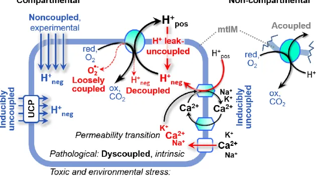
Figure 3. Mechanisms of respiratory uncoupling
An intact mitochondrial inner membrane, mtIM, is required for vectorial, compartmental coupling. Inducible uncoupling, e.g., by activation of UCP1, increases LEAK-respiration; experimentally noncoupled respiration provides an estimate of ET-capacity obtained by titration of protonophores stimulating respiration to maximum O2 flux. H+ leak-uncoupled, decoupled, and loosely coupled respiration are components of intrinsic uncoupling (Table 2). Pathological dysfunction may affect all types of uncoupling, including permeability transition (mtPT), causing intrinsically dyscoupled respiration. Similarly, toxicological and environmental stress factors can cause extrinsically dyscoupled respiration. ‘Acoupled’ respiration is the consequence of structural disruption with catalytic activity of non-compartmental mitochondrial fragments. Reduced fuel substrates, red; oxidized products, ox.
Uncoupling of mitochondrial respiration is a general term comprising diverse mechanisms (Figure 3):
1. Proton leak across the mtIM from the positive to the negative compartment (H+ leak-uncoupled)
2. Cycling of other cations, strongly stimulated by mtPT; comparable to the use of protonophores, cation cycling is experimentally induced by valinomycin in the presence of K+
3. Decoupling by proton slip in the redox proton pumps (CI, CIII and CIV) when protons are effectively not pumped in the ETS, or are not driving phosphorylation (F-ATPase)
4. Loss of vesicular (compartmental) integrity when electron transfer is acoupled
5. Electron leak in the loosely coupled univalent reduction of O2 to superoxide (O2•–; superoxide
anion radical)
Differences of terms—uncoupled vs. noncoupled—are easily overlooked, although they relate to different meanings of uncoupling (Figure 3 and Table 2).
2.5. Coupling states and respiratory rates
To extend the classical nomenclature on mitochondrial coupling states (Section 2.6) by a concept-driven terminology that explicitly incorporates information on the meaning of respiratory states, the terminology must be general and not restricted to any particular experimental protocol or mitochondrial preparation (Gnaiger 2009). Diagnostically meaningful and reproducible conditions are defined for measuring mitochondrial function and respiratory capacities of core energy metabolism. Standard respiratory coupling states are obtained while maintaining a defined ET-pathway state with constant fuel substrates and inhibitors of specific branches of the ET-pathway. Concept-driven nomenclature aims at mapping the meaning and concept behind the words and acronyms onto the forms of words and acronyms (Miller 1991). The focus of concept-driven nomenclature is primarily the conceptual why, along with clarification of the experimental how. (Table 1).
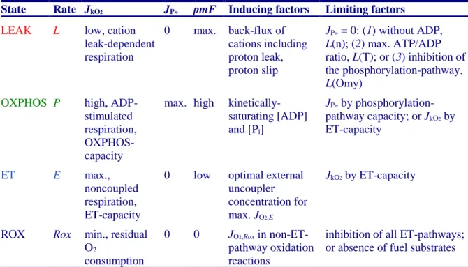
Table 1. Coupling states and rates, and residual oxygen consumption in mitochondrial preparations. Respiration- and phosphorylation-flux, JkO2and JP», are rates, characteristic of a state in conjunction with the protonmotive force, pmF. Coupling states are established at kinetically-saturating concentrations of fuel substrates and O2.
State Rate JkO2 JP» pmF Inducing factors Limiting factors
LEAK L low, cation leak-dependent respiration 0 max. back-flux of cations including proton leak, proton slip JP» = 0: (1) without ADP, L(n); (2) max. ATP/ADP ratio, L(T); or (3) inhibition of the phosphorylation-pathway, L(Omy)
OXPHOS P high, ADP-stimulated respiration, OXPHOS-capacity
max. high kinetically-saturating [ADP] and [Pi]
JP» by phosphorylation-pathway capacity; or JkO2 by
ET-capacity
ET E max.,
noncoupled respiration, ET-capacity
0 low optimal external uncoupler concentration for max. JO2,E
JkO2 by ET-capacity
ROX Rox min., residual O2
consumption
0 0 JO2,Rox in
non-ET-pathway oxidation reactions
inhibition of all ET-pathways; or absence of fuel substrates
LEAK: The contribution of intrinsically uncoupled O2 consumption is studied by preventing the
stimulation of phosphorylation either in the absence of ADP or by inhibition of the phosphorylation-pathway. The corresponding states are collectively classified as LEAK-states when O2 consumption compensates mainly for ion leaks, including the proton leak. The chelator
OXPHOS: The ET- and phosphorylation-pathways comprise coupled segments of the OXPHOS-system and provide reference values of respiratory capacities. The OXPHOS-capacity is measured at kinetically-saturating concentrations of ADP and Pi.
ET: Compared to capacity, the oxidative ET-capacity reveals the limitation of OXPHOS-capacity mediated by the phosphorylation-pathway. By application of external uncouplers, ET-capacity is measured as noncoupled respiration.
The three coupling states, LEAK, OXPHOS, and ET are shown schematically with the corresponding respiratory rates, abbreviated as L, P, and E, respectively (Figure 4). We distinguish metabolic pathways from metabolic states and the corresponding metabolic rates; for example: ET-pathways, ET-states, and ET-capacities, E, respectively (Table 1). The protonmotive force, pmF, is
maximum in the LEAK-state of coupled mitochondria, driven by LEAK-respiration at a minimum
back-flux of cations to the matrix side, high in the OXPHOS-state when it drives phosphorylation, and very
low in the ET-state when uncouplers short-circuit the proton cycle (Table 1).
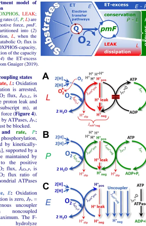
Figure 4. Four-compartment model of oxidative phosphorylation
Respiratory states (ET, OXPHOS, LEAK;
Table 1) and corresponding rates (E, P, L) are connected by the protonmotive force, pmF. (1) ET-capacity, E, is partitioned into (2) dissipative LEAK-respiration, L, when the Gibbs energy change of catabolic O2 flux is irreversibly lost, (3) net OXPHOS-capacity, P-L, with partial conservation of the capacity to perform work, and (4) the ET-excess capacity, E-P. Modified from Gnaiger (2019).
Figure 5. Respiratory coupling states
(A)
LEAK-state
and
rate, L
: Oxidation
only, since phosphorylation is arrested,
J
P»= 0, and catabolic O
2flux, J
kO2,L, is
controlled mainly by the proton leak and
slip, J
mH+neg(motive, subscript m), at
maximum protonmotive force (Figure 4).
ATP may be hydrolyzed by ATPases, J
P«;
then phosphorylation must be blocked.
(B)
OXPHOS-state
and
rate,
P:
Oxidation coupled to phosphorylation,
J
P», which is stimulated by
kinetically-saturating [ADP] and [P
i], supported by a
high protonmotive force maintained by
pumping of protons to the positive
compartment, J
mH+pos. O
2flux, J
kO2,P, is
well-coupled at a P»/O
2flux ratio of
J
P»,P∙J
O2,P-1. Extramitochondrial ATPases
may recycle ATP, J
P«.
(C)
ET-state and
rate, E: Oxidation
only, since phosphorylation is zero, J
P»=
0, at optimum exogenous uncoupler
concentration
when
noncoupled
respiration, J
kO2,E, is maximum. The
F-ATPase
may
hydrolyze
2.5.1. LEAK-state (Figure 5A): The LEAK-state is defined as a state of mitochondrial respiration when O2 flux mainly compensates for ion leaks in the absence of ATP synthesis, at
kinetically-saturating concentrations of O2, respiratory fuel substrates and Pi. LEAK-respiration is
measured to obtain an estimate of intrinsic uncoupling without addition of an experimental uncoupler: (1) in the absence of adenylates, i.e., AMP, ADP and ATP; (2) after depletion of ADP at a maximum ATP/ADP ratio; or (3) after inhibition of the phosphorylation-pathway by inhibitors of F-ATPase (oligomycin), or adenine nucleotide translocase (carboxyatractyloside). Adjustment of the nominal concentration of these inhibitors to the concentration of biological sample applied can minimize or avoid inhibitory side-effects exerted on ET-capacity or even some dyscoupling.
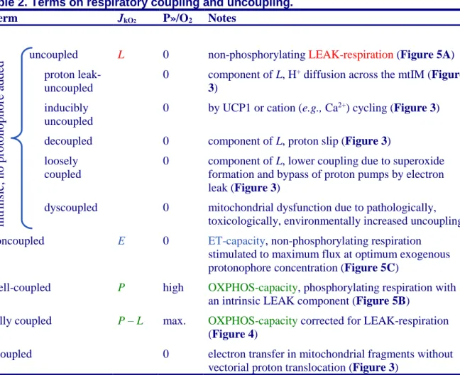
Table 2. Terms on respiratory coupling and uncoupling.
Term JkO2 P»/O2 Notes
uncoupled L 0 non-phosphorylating LEAK-respiration (Figure 5A) proton leak-
uncoupled
0 component of L, H+ diffusion across the mtIM (Figure
3)
inducibly uncoupled
0 by UCP1 or cation (e.g., Ca2+) cycling (Figure 3) decoupled 0 component of L, proton slip (Figure 3)
loosely coupled
0 component of L, lower coupling due to superoxide formation and bypass of proton pumps by electron leak (Figure 3)
dyscoupled 0 mitochondrial dysfunction due to pathologically, toxicologically, environmentally increased uncoupling noncoupled E 0 ET-capacity, non-phosphorylating respiration
stimulated to maximum flux at optimum exogenous protonophore concentration (Figure 5C)
well-coupled P high OXPHOS-capacity, phosphorylating respiration with an intrinsic LEAK component (Figure 5B)
fully coupled P – L max. OXPHOS-capacity corrected for LEAK-respiration (Figure 4)
acoupled 0 electron transfer in mitochondrial fragments without vectorial proton translocation (Figure 3)
• Proton leak and uncoupled respiration: The intrinsic proton leak is the uncoupled leak current of protons in which protons diffuse across the mtIM in the dissipative direction of the downhill protonmotive force without coupling to phosphorylation (Figure 5A). The proton leak flux depends non-linearly on the protonmotive force (Garlid et al. 1989; Divakaruni and Brand 2011), which is a temperature-dependent property of the mtIM and may be enhanced due to possible contamination by free fatty acids. Inducible uncoupling mediated by uncoupling protein 1 (UCP1) is physiologically controlled, e.g., in brown adipose tissue. UCP1 is a member of the mitochondrial carrier family that is involved in the translocation of protons across the mtIM (Jezek et al. 2018). Consequently, this short-circuit lowers the pmF and stimulates electron transfer, respiration, and heat dissipation in the absence of phosphorylation of ADP.
• Cation cycling: There can be other cation contributors to leak current including calcium and probably magnesium. Calcium influx is balanced by mitochondrial Na+/Ca2+ or H+/Ca2+
exchange, which is balanced by Na+/H+ or K+/H+ exchanges. This is another effective uncoupling
mechanism different from proton leak (Table 2).
• Proton slip and decoupled respiration: Proton slip is the decoupled process in which protons are only partially translocated by a redox proton pump of the ET-pathways and slip back to the original vesicular compartment. The proton leak is the dominant contributor to the overall leak
int
rinsic
, no
protonophor
e
adde
d
current in mammalian mitochondria incubated under physiological conditions at 37 °C, whereas proton slip increases at lower experimental temperature (Canton et al. 1995). Proton slip can also happen in association with the F-ATPase, in which the proton slips downhill across the pump to the matrix without contributing to ATP synthesis. In each case, proton slip is a property of the proton pump and increases with the pump turnover rate.
• Electron leak and loosely coupled respiration: Superoxide production by the ETS leads to a bypass of redox proton pumps and correspondingly lower P»/O2 ratio. This depends on the actual
site of electron leak and the scavenging of hydrogen peroxide by cytochrome c, whereby electrons may re-enter the ETS with proton translocation by CIV.
• Dyscoupled respiration: Mitochondrial injuries may lead to dyscoupling as a pathological or toxicological cause of uncoupled respiration. Dyscoupling may involve any type of uncoupling mechanism, e.g., opening the mtPT pore. Dyscoupled respiration is distinguished from experimentally induced noncoupled respiration in the ET-state (Table 2).
• Protonophore titration and noncoupled respiration: Protonophores are uncouplers which are titrated to obtain maximum noncoupled respiration as a measure of ET-capacity.
• Loss of compartmental integrity and acoupled respiration: Electron transfer and catabolic O2
flux proceed without compartmental proton translocation in disrupted mitochondrial fragments. Such fragments are an artefact of mitochondrial isolation, and may not fully fuse to re-establish structurally intact mitochondria. Loss of mtIM integrity, therefore, is the cause of acoupled respiration, which is a nonvectorial dissipative process without control by the protonmotive force.
2.5.2. OXPHOS-state (Figure 5B): The OXPHOS-state is defined as the respiratory state with kinetically-saturating concentrations of O2, respiratory and phosphorylation substrates, and absence of
exogenous uncoupler, which provides an estimate of the maximal respiratory capacity in the OXPHOS-state for any given ET-pathway OXPHOS-state. Respiratory capacities at kinetically-saturating substrate concentrations provide reference values or upper limits of performance, aiming at the generation of data sets for comparative purposes. Physiological activities and effects of substrate kinetics can be evaluated relative to the OXPHOS-capacity.
As discussed previously, 0.2 mM ADP does not kinetically-saturate flux in isolated mitochondria (Gnaiger 2001; Puchowicz et al. 2004); greater [ADP] is required, particularly in permeabilized muscle fibers and cardiomyocytes, to overcome limitations by intracellular diffusion and by the reduced conductance of the mtOM (Jepihhina et al. 2011; Illaste et al. 2012; Simson et al. 2016), either through interaction with tubulin (Rostovtseva et al. 2008) or other intracellular structures (Birkedal et al. 2014). In addition, kinetically-saturating ADP concentrations need to be evaluated under different experimental conditions such as temperature (Lemieux et al. 2017) and with different animal models (Blier and Guderley 1993). In permeabilized muscle fiber bundles of high respiratory capacity, the apparent Km for
ADP increases up to 0.5 mM (Saks et al. 1998), consistent with experimental evidence that >90% kinetic saturation is reached only at >5 mM ADP (Pesta and Gnaiger 2012). Similar ADP concentrations are also required for accurate determination of OXPHOS-capacity in human clinical cancer samples and permeabilized cells (Klepinin et al. 2016; Koit et al. 2017). 2.5 to 5 mM ADP is sufficient to obtain the actual OXPHOS-capacity in many types of permeabilized tissue and cell preparations, but experimental validation is required in each specific case.
2.5.3. Electron transfer-state (Figure 5C): O2 flux determined in the ET-state yields an estimate
of ET-capacity. The ET-state is defined as the noncoupled state with kinetically-saturating concentrations of O2, respiratory substrate and optimum exogenous uncoupler concentration for
maximum O2 flux. Uncouplers are weak lipid-soluble acids which function as protonophores. These
disrupt the barrier function of the mtIM and thus short-circuit the protonmotive system, functioning like a clutch in a mechanical system. As a consequence of the nearly collapsed protonmotive force, the driving force is insufficient for phosphorylation, and JP» = 0. The most frequently used uncouplers are
carbonyl cyanide m-chloro phenyl hydrazone (CCCP), carbonyl cyanide
p-trifluoromethoxyphenylhydrazone (FCCP), or dinitrophenol (DNP). Stepwise titration of uncouplers stimulates respiration up to or above the level of O2 consumption rates in the OXPHOS-state; respiration
is inhibited, however, above optimum uncoupler concentrations (Mitchell 2011). Data obtained with a single dose of uncoupler must be evaluated with caution, particularly when a fixed uncoupler concentration is used in studies exploring a treatment or disease that may alter the mitochondrial content or mitochondrial sensitivity to inhibition by uncouplers. There is a need for new protonophoric