HAL Id: tel-02303046
https://tel.archives-ouvertes.fr/tel-02303046
Submitted on 2 Oct 2019HAL is a multi-disciplinary open access archive for the deposit and dissemination of sci-entific research documents, whether they are pub-lished or not. The documents may come from teaching and research institutions in France or abroad, or from public or private research centers.
L’archive ouverte pluridisciplinaire HAL, est destinée au dépôt et à la diffusion de documents scientifiques de niveau recherche, publiés ou non, émanant des établissements d’enseignement et de recherche français ou étrangers, des laboratoires publics ou privés.
The application of metabolomics by high field nuclear
magnetic resonance to study thyroid signalisation
pathologies in mice
Houda Boumaza
To cite this version:
Houda Boumaza. The application of metabolomics by high field nuclear magnetic resonance to study thyroid signalisation pathologies in mice. Human health and pathology. Université de Lyon, 2019. English. �NNT : 2019LYSEN003�. �tel-02303046�
Numéro National de Thèse : 2019LYSEN003
THESE de DOCTORAT DE L’UNIVERSITE DE LYON
Opérée parL’Ecole Normale Supérieure de Lyon
Ecole Doctorale N° 340
Biologie Moléculaire, Intégrative et Cellulaire (BMIC) Spécialité de doctorat : Biologie
Soutenue publiquement le 08/03/2019, par:
Houda BOUMAZA
Etude métabolomique par résonance
magnétique nucléaire de pathologies
associées à la signalisation
thyroïdienne chez la souris
Devant le jury composé de :
SAVARIN, Philippe Professeur des universités, Université de Paris 13 - Rapporteur DAMBLON, Christian Professeur des universités, Université de Liège - Rapporteur SHINTU, Laetitia Maître de conférence, Aix-Marseille Université - Examinatrice RODIEN, Patrice Professeur des universités - praticien hospitalier, CHU-Angers - Examinateur
FLAMANT, Frédéric Directeur de recherche, INRA, ENS de Lyon - Directeur de thèse ELENA-HERRMANN, Bénédicte Chargée de recherche, CNRS, Université Grenoble Alpes - Co-directrice de thèse
Acknowledgements
Je souhaiterais remercier le Professeur Philippe Savarin et le Professeur Christian Damblon pour avoir accepté d’évaluer ce travail de thèse, ainsi que le Docteur Laetitia Shintu et le Docteur Patrice Rodien pour avoir accepté d’être membres du jury.
Ce manuscrit de thèse est l’aboutissement de trois années de travail et de nombreux défis. Je souhaiterais donc remercier les personnes talentueuses avec lesquelles j’ai pu interagir avant et tout au long de ma thèse.
Je remercie mes directeurs de thèses : Docteur Frédéric Flamant et Docteur Bénédicte Elena-Herrmann de m’avoir fait confiance et de m’avoir donné l’opportunité d’effectuer cette thèse dans des conditions favorables. Je les remercie pour leur encadrement et leur disponibilité permanente. Merci de m’avoir permis de développer une autonomie et une indépendance dans ce travail.
Je remercie le Docteur Elodie Jobard de m’avoir formée en métabolomique, d’avoir été toujours présente pour répondre à des questions techniques, d’avoir partagé avec moi ses expériences et son parcours.
Je remercie le Docteur Gilles Rautureau d’avoir été toujours disponible pour répondre à mes questions sur le projet et sur mon parcours professionnel.
Je remercie Suzy Markossian de m’avoir formée et beaucoup aidée pour effectuer les expériences biologiques à l’IGFL. Merci pour sa disponibilité et sa bonne humeur contagieuse.
Je remercie le Docteur Karine Gauthier pour son aide précieuse dans ce projet de thèse.
J’ai eu la chance d’effectuer ma thèse en même temps que mon amie rencontrée au laboratoire Manhal Mili. Je la remercie sincèrement pour ses encouragements, sa bienveillance et sa bonne humeur.
Je remercie les anciens membres de l’équipe de métabolomique avec qui il a été très agréable de travailler et d’échanger : Docteur Tony Palama, Docteur Aurélien Scalabre et Sylvie Guibert.
Je remercie également tous les membres du CRMN anciens et nouveaux arrivants pour l’ambiance chaleureuse : Anne, Guido, Diane, Olivier, James, Tanguy, Pierrick, Cécile, Dorothea, Audrey, David, Baptiste et tous les autres.
Je remercie aussi les membres de l’IGFL de m’avoir accueillie. Merci à Romain, Sabine et Denise.
Je remercie l’Agence Nationale de Recherche pour le financement de cette thèse.
Pour finir, je remercie mes proches qui ont également contribué par leur présence et leur soutien à l’accomplissement de ce doctorat.
Merci à ma mère Samia, mon frère Bader et mes sœurs Rym et Maroua, ma belle-sœur Marine, mon petit neveu Taqqyeddine, mon beau-frère Driss et mon beau-père Kamel, pour leur soutien constant, et les agréables moments passés en famille toujours ressourçant. Un grand merci à ma maman à qui je dois tant. Merci à ma grand-mère, qui nous a quittés aujourd’hui, de m’avoir toujours encouragée à faire des études et aller le plus loin possible.
Merci à ma deuxième famille, mes amis. Merci à Inès, à Sophiana, à Anaïs, à Linda, à Coralie, à Sabrina, à Myriam, à Mehdi et bien d’autres. Merci à tous ceux que j’ai rencontrés dans le milieu associatif qui ont su enrichir mon âme.
Enfin, je remercie toutes les personnes qui ont contribué de près ou de loin à l’aboutissement de ce travail qui est ci-important à mes yeux.
Résumé
La métabolomique par résonance magnétique nucléaire (RMN) permet d’étudier la
réponse métabolique globale d’un système biologique à un stimulus ou un événement
physiopathologique (maladie, manipulation génétique, etc.). Cette discipline connaît un essor
important dans la recherche clinique et biologique, et constitue ainsi un outil à fort potentiel
pour la découverte de biomarqueurs de maladies, et l’étude de la fonction des gènes.
Cette thèse est dédiée à l’application de la métabolomique par RMN à hauts champs
pour l’étude des pathologies associées à la signalisation thyroïdienne chez la souris. L’objectif
global est d’identifier des biomarqueurs spécifiques liés aux différentes maladies
hormonales : l’hypothyroïdie et la maladie génétique émergente résistance à l’hormone
thyroïdienne due à une mutation au niveau du récepteur TRα1 (RTHα). Cette dernière est
particulièrement difficile à diagnostiquer à cause du manque de marqueurs biochimiques et de
symptômes spécifiques à cette maladie. De plus, elle présente des similitudes avec
l’hypothyroïdie au niveau symptomatique. Des modèles murins de RTHα et de
l’hypothyroïdie ont été analysés, et l’investigation a été menée sur l’urine et le plasma
sanguin dans le but de différencier métaboliquement ces maladies et d’identifier des
biomarqueurs spécifiques à RTHα. Des signatures métaboliques liées à chaque maladie ont
été identifiées dans l’urine et le plasma sanguin. Cinq métabolites qui varient de façon
significative ont été identifiés dans l’urine comme étant liés à la maladie RTHα :
trimethylamine, dimethylamine, isovalerylglycine, N-acetylglucosamine et la choline. Dans le
sang, ce sont les lipides insaturés qui varient de façon significative chez les souris mimant la
L’impact des hormones thyroïdiennes (HT) et le récepteur TRβ sur le métabolisme
hépatique a été également étudié dans ce présent manuscrit. Un modèle murin présentant une
inactivation du récepteur TRβ au niveau des hépatocytes (LTRβ-KO) a été généré pour
étudier cette question. Pour comprendre la fonction des HT médiée par le récepteur TRβ, les
réponses métaboliques hépatiques à HT, obtenues sur des extraits hépatiques aqueux et tissus
hépatiques intacts, des souris TRβ-KO et des souris sauvages ont été comparées. Les résultats
obtenus suggèrent la présence d’un effet direct (par le récepteur TRβ) et un effet indirect des
hormones thyroïdiennes sur le métabolisme hépatique.
Abstract
Metabolomics by nuclear magnetic resonance (NMR) allows studying the metabolic
response of a global biological system to a stimuli or a physiopathological even (diseases,
genetic modifications, etc.). This discipline is growing especially in the clinical and biological
fields, and represents a strong potential tool to identify biomarkers related to diseases, and
study the function of genes.
This thesis is dedicated to the application of metabolomics by high field NMR to study
thyroid signalisation pathologies in mice. The main goal is to identify biomarkers related to
the emerging genetic disease called resistance to thyroid hormone due to a mutation in thyroid
hormone receptor TRα1 (RTHα). This disease is particularly difficult to diagnose because of
the lack of biochemical markers and specific symptoms. In addition, it presents common
features with hypothyroidism in term of symptoms. Mice models of RTHα and
hypothyroidism were analysed, and the investigation were driven on urine and blood plasma
in order to differentiate metabolically theses diseases and identify biomarkers related to
RTHα. Metabolic fingerprints related to each disease were identified in both urine and blood
plasma. Five metabolites vary significantly in the urine of RTHα mice: trimethylamine,
dimethylamine, isovalerylglycine, N-acetylglucosamine and choline. Unsaturated lipids vary
significantly in the blood plasma of RTHα mice.
The impact of thyroid hormones (TH) and the thyroid hormone receptor TRβ on the
liver metabolism were also studied in the present manuscript through NMR-based
(LTRβ-KO), were used to study this question. To understand the function of TH mediated by TRβ,
the liver metabolic response to TH, obtained from liver aqueous extracts and intact liver
tissues, TRβKO and wild-type mice were compared. The results suggest the presence of
direct and indirect effects of thyroid hormones on the liver metabolism.
Table of contents
Acknowledgements Résumé Abstract Table of contents Table of abbreviations IntroductionChapter 1 – Metabolomics: from fundamentals to clinical applications
1.1 Metabolomics approach
1.1.1 Metabolomics and metabonomics 1.1.2 From genomics to metabolomics 1.1.3 Platforms for metabolomics 1.2 Nuclear magnetic resonance
1.2.1 The principle of NMR 1.2.2 NMR parameters 1.2.3 NMR methods
1.3 NMR-based metabolomics for the investigation of biological samples 1.3.1 Biological samples explored by metabolomics
1.3.2 Targeted and untargeted analyses 1.4 Metabolomics and biomarkers
1.4.1 Metabolomics to study pathologies 1.4.2 Metabolomics and genetic diseases 1.5 Conclusion
Chapter 2 - Thyroid hormones system and alterations
2.1 Thyroid hormone system
2.1.1 Thyroid hormones function 2.1.2 Thyroid hormones receptors 2.1.3 Mechanism of actions 2.1.4 Regulation
2.1.5 Alterations in thyroid hormone system 2.2 Resistance to thyroid hormone α
2.2.1 The first cases
2.2.2 RTHα is different from RTHβ
2.2.3 A high variability in clinical features 2.2.4 Molecular aspect
2.2.5 Relation between genotype and phenotype? 2.2.6 Mouse models to study RTHα
Chapter 3 – Materials and methods
3.1 Introduction
3.2 Biological samples preparation
2 4 6 8 11 13 17 17 17 18 19 20 20 22 25 27 27 28 28 28 29 29 30 30 30 31 33 34 34 35 36 36 37 38 39 40 43 43 45
3.2.1 Collection and storage 3.2.2 Preparation and handling 3.3 NMR-based metabolomics
3.3.1 Data acquisition 3.3.2 Data processing
3.3.3 Multivariate analysis of metabolic profiles 3.3.4 Metabolite identification
Chapter 4: 1H NMR-based metabolomics for the study of the impact of the excess and the privation of thyroid hormones on murine metabolism
4.1 Introduction 4.2 Study design 4.3 Results
4.3.1 Quality of 1H NMR data
4.3.2 Discrimination according to mice phenotype in urine 4.3.3 Impact of thyroid hormones on the metabolism
4.3.4 Determination of a metabolomic signature associated with hypothyroidism
4.3.5 Determination of a metabolic signature associated with TH treatment 4.4 Conclusion
Chapter 5: NMR as a putative diagnostic tool for the presence of thyroid hormone receptor alpha 1 mutations
5.1 Introduction 5.2 Study design 5.3 results
5.3.1 Quality of 1H NMR data
5.3.2 Discrimination between group of mutants and wild-type group 5.3.3 Discrimination between each mutant group and wild-type group 5.3.4 Metabolomic analysis segregates two types of frameshift mutations 5.3.5 Discrimination between ThraS/+ mice with human-like mutations and wild-type mice
5.3.6 Determination of metabolic signature of Thra mutation in urine and blood plasma
5.3.7 RTHα signature is different from hypothyroidism signature
5.4 Discussion
Chapter 6: Functional genomics by metabonomics to study thyroid hormone receptor β
6.1 Introduction 6.2 Study design 6.3 Results
6.3.1 Untargeted NMR Metabolomics analysis of the liver response to thyroid hormone
6.3.2 Impact of TH nuclear receptor on liver metabolism 6.3.3 Impact of TRβ on liver metabolism
45 46 47 47 49 52 55 56 56 58 60 60 61 61 64 65 69 70 70 72 74 74 74 75 77 79 81 83 84 87 88 89 89 89 91 92
6.3.4 Summary 6.4 Conclusion
Conclusion and perspectives References
Appendices
Appendix 1: Clinical features of RTHα patients
Appendix 2: Metabolites identified from 1D and 2D NMR profiles of mice urine Appendix 3: Metabolites identified from 1D and 2D NMR profiles of mice plasma Appendix 4: Metabolites identified from 1D and 2D NMR profiles of mice liver aqueous extracts 94 96 98 101 111 111 117 120 122
Table of abbreviations
ANOVA: Analysis Of Variance CPMG: Carr-Purcell-Meiboom-Gill DA: Discriminant Analysis
DBD: DNA-Binding Domain
FHCTD: Thyroid Hormone Cell Transport Defects FID: Free Induction Decays
FT4: Free T4
GC: Gas Chromatography
HCA: Hierarchical Clustering Analysis HDL: High Density Lipoproteins HMDB: Human Metabolome Database
HPT axis: Hypothalamus/Pituitary/Thyroid axis HP: High Pressure
HR-MAS: High Resolution-Magic Angle Spinning HSQC: Heteronuclear Single Quantum Correlation KI: Knocking In
KO: Knocking Out
LBD: Ligand-Binding Domain LC: Liquid Chromatography
MCT: Monocarboxylate Transporter
MMMDB: Mouse Multiple tissue Metabolome Database MS: Mass Spectrometry
NMR: Nuclear Magnetic Resonance NOESY: Nuclear Overhauser Effect OPLS: Orthogonal Partial Least Squares PCA: Principal Component Analysis PLS: Projection to Latent Structures PQN: Probabilistic Quotient Normalization PTU: Propylthiouracil
RTH: Resistance to Thyroid Hormone
RTHα: Resistance to Thyroid Hormone due a mutation in thyroid hormone receptor α RTHβ: Resistance to Thyroid Hormone due a mutation in thyroid hormone receptor β SRV: Statistical Recoupling of Variables
TH: Thyroid Hormone
THCTD: Thyroid Hormone Cell Transport Defects THMD: Thyroid Hormone Metabolism Defect TOCSY: TOtal Correlation SpectroscopY TSP: 3-Trimethylsilylpropionic acid TRα: Thyroid hormone Receptor α TRβ: Thyroid hormone Receptor β TRH: Thyrotropin-Releasing Hormone TRs: Thyroid Receptors TSH: Thyroid-Stimulating Hormone T3: 3, 5, 3’-triiodothyronine T4: Thyroxine RXR: Retinoid X Receptor UP: Ultra Performance UV: Unit Variance
Introduction
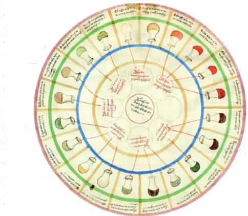
The relation between the variation in the composition and the aspect of biofluids or tissues and pathologies was identified hundreds of years back1. This concept exists from the Middle age, in a “urine charts”, the colors, smells and tastes were related to specific medical conditions (Figure 1). These variations come, of course, from the metabolism, which is influenced by certain conditions. Nowadays, modern-day metabolomics uses the same principle to analyze biological samples, but with modern techniques.
Figure 1: “Urine charts”.
Ulrich Pinder published this illustration in 1506, in his book “Epiphanie Medicorum”. This chart connects colors, smells and tastes of urine to diseases.
The big challenge in biological and clinical studies is to discover biological markers, which lead to diagnose easily and at early stage of a disease to improve human health. The goal is to take care of patients as soon as possible to curb the disease. Biomarkers are
“biological characteristics that are measured and evaluated as indicators of normal or pathological processes, and pharmacologic responses to a therapeutic intervention”2. A good biomarker must be easily measurable, and help for the diagnosis. High throughput “-omics” methods (genomics, transcriptomics, proteomics, metabolomics, etc.) lead to a new diagnostic approach and to targeted therapies.
In practical terms, metabolomics consists in the measurement of the small molecules, named metabolites, involved in biochemical processes present in a biological system (tissue, cells, biofluids, etc.). Metabolites are the final products of interactions between genes, proteins and the impact of environment. Thus, each individual has his own metabolic state at a specific time, which is also defined by the metabotype3. Analyzing metabolites provides information that cannot be obtained only from the genome or the proteome. With this approach, the overall response of a biological system to a physiological or pathological event (disease, genetic manipulation, drugs, nutrition, lifestyle, etc.) can be defined.
Metabolomics was applied to different biological and clinical conditions like cancers, cardiovascular diseases, diabetes, inborn errors of metabolism, etc4-12. Metabolomics studies in this field aim to understand the impact of diseases on the organism, and to discover biomarkers that predict disease incidence, severity, and progression and detect abnormalities. In addition to its application in the discovery of biomarkers, metabolomics is often used to understand biological processes of diseases and genetic mutations3,10,13-15. Metabolomics is now developing as an important functional genomics tool to evaluate the impact of gene change16.
This thesis is dedicated to the application of NMR-based metabolomics for the investigation of thyroid signalisation pathologies in mice. We prospectively explored urine and blood plasma of murine models to obtain metabolic fingerprints of hypothyroidism, excess of thyroid hormone and the emerging genetic disease called resistance to thyroid hormone due to a mutation in thyroid hormone receptor TRα1 (RTHα). RTHα is particularly difficult to diagnose because of the lack of biochemical markers and specific symptoms. Nowadays, only few researchers were carried out to understand this disease, and try to identify biomarkers to facilitate its diagnosis. RTHα patients present common features with hypothyroid patients with near normal thyroid function tests. The study of hypothyroidism here serves as a base to understand RTHα. We investigate in the end the impact of thyroid
hormones and the role of thyroid hormone nuclear receptor β in the liver, by using liver samples.
The first chapter presents the metabolomics approach and its application as a diagnostic tool in the medical field. We set then in the second chapter the background of the present study. We describe thyroid hormone functions, molecular actions, their receptors and the different hormonal diseases. Materials and methods are detailed in the chapter 3. The different steps of our metabolomics studies are described.
The fourth chapter is dedicated to the investigation by using non-targeted 1 H-NMR-based metabolomics of two biofluids (urine and blood plasma) issue from mice to identify non-hormonal biomarkers related to hypothyroidism. It was crucial for us to study this disease at first to understand then the specific metabolic changes caused by RTHα. Here, murine models for hypothyroidism were compared to a control group to obtain metabolic fingerprints related to each disease in both urine and plasma. Clear discriminations between murine model for hypothyroidism and the control group were observed and specific metabolic signatures of the hypothyroid condition were identified.
The fifth chapter concerns the study of the new emerging genetic disease called resistance to thyroid hormone due to a mutation in thyroid hormone receptor TRα (RTHα), which its symptoms are close to those of hypothyroidism. Patients present a high variability in clinical features (skeletal dysplasia, growth retardation, intellectual disability, etc.), and the absence of reliable biochemical markers make the diagnosis of this disease difficult. The main goal of the study is to find, by the use of NMR based-metabolomics approach, a metabolic signature related to RTHα in mice, identify specific biomarkers and differentiate it from hypothyroidism. Five mice models carrying a mutation in Thra gene were studied, and the investigation were driven on urine and blood. Five metabolites vary significantly in the urine of RTHα mice: trimethylamine, dimethylamine, isovalerylglycine, N-acetylglucosamine and choline. Unsaturated lipids vary significantly in RTHα mice blood plasma.
In the sixth chapter, the impact of thyroid hormones (TH) and the thyroid hormone receptor TRβ on the liver metabolism were studied through NMR-based metabolomics. A mouse model, with a specific knockout of TRβ gene in hepatocytes (LTRβ-KO), was generated to study this question. To understand the function of TH mediated by TRβ, the liver
metabolic response to TH, obtained from liver aqueous extracts and intact liver tissues, LTRβ-KO and wild-type mice were compared. The results suggest the presence of direct and indirect effects of thyroid hormones on the liver metabolism.
1. Metabolomics: from fundamentals to
clinical applications
1.1. Metabolomics approach
1.1.1. Definitions
Metabolites are low molecular weights molecules (less than 1.5kDa) that are involved in metabolic pathways. They are the reactants, intermediates and end products of complex multiple cascade, which is initiated by the genome followed by the transcriptome and proteome. They can be endogenous (from the organism) or exogenous (i.e. nutrition, medication, microbiota, etc.). They include polysaccharides, lipids, steroids, organic acids, amino acids, ketones and peptides, or else. They fulfill diverse functions, ranging from signal transduction to energy fuels17,18. Each type of biological sample (fluid, tissue, cells, organism) or each subtype (type of cell, type of fluid, etc.) has his own characteristic set of metabolites3.
The Metabolome represents the entire set of metabolites present in a biological system19. The human metabolome is estimated to contain about 150 000 metabolites (endogenous and exogenous) or more20. Environmental factors (diet, gut microbial activity, disease, medication, stress level, physical activity, etc.) have an effect on the metabolism, which lead to changes in the concentrations of metabolites, and thus in the metabolome.
Metabolomics19,21 corresponds to the quantification and identification as many as possible of the metabolites present in a biological system (biological fluid, cell, tissue, organism, etc.). Analyzing metabolites in a biological system provides information on the metabolic phenotype or metabotype of individuals or populations3. Thanks to technological advances in the identification, quantification and analysis of metabolites, metabolomics has grown significantly in different fields1,22. Metabolomic analyses are often built as case-control studies, which aim to compare two groups of samples coming from two different
physio-pathological states. By the use of bioinformatics and biostatistics tools, the difference between the two groups in term of molecules can be identified23.
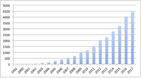
The application of metabolomics in different fields has increased strongly over the past years (Figure 1.1.1).
Figure 1.1.1: Number of metabolomics publications per year.
The research was performed in the Web of science (WOS) database, using metabolomics or metabonomics as keywords. The data was collected up to 2017.
1.1.2. From genomics to metabolomics
Omics strategies represent an important step in sciences area, and they are widely used nowadays. The principle of these approaches is to identify and quantify the entire set of biomolecules (genes, transcripts, proteins, metabolites, etc.) present in a given biological matrix (tissues, cells, organisms, biofluids), and to determine the relationship between their variations and specific pathophysiological states or external perturbations. Studying only genes, transcripts or proteins is insufficient to understand the complete phenotypic changes induced by these factors24. The number of metabolites is smaller than the estimated number of genes (25.000), transcripts (100.000) and proteins (1.000.000) found in humans10. Metabolites represent a more sensitive level25 than the other biomolecules, and they are the result of the interaction between theses biomolecules and the environment. In addition to the interactions
between metabolites and the other biomolecules, metabolites are also the building blocks of proteins (amino-acids), genes and transcripts (nucleotides). Metabolic pathways reflect the genome and proteome states with up to 10.000-fold increase in metabolite concentration resulting from a single amino acid change in a protein or base change in a gene22,26. Metabolic changes are detectable within minutes of a biological event and thus provide an almost ‘real-time’ feedback22.
Now, metabolomics is considered as a strong phenotyping tool, which provides molecular information that complement data obtained from genomics, transcriptomics and proteomics (Figure 1.1.2).
Figure 1.1.2: Correlation between omics sciences.
Omics strategies (Genomics, Transcriptomics, Proteomics and Metabolomics) aim at identifying all the biomolecules present in biological system (genes, RNA, proteins, metabolites).
1.1.3. Platforms for metabolomics
The two main technologies that are amply employed in metabolomics studies are NMR spectroscopy and mass spectrometry24,27,28. NMR spectroscopy is used in metabolomics studies to detect hydrogen atoms in metabolites, and identify compounds with identical masses8. Each metabolite, with concentrations above the detection limit, gives a 1H NMR spectrum. Thus, a 1H NMR spectrum of a complex mixture (biofluids, cells/tissue extracts) corresponds to the superposition of the spectra of all the present metabolites, which means
GENOMICS TRANSCRIPTOMICS PROTEOMICS METABOLOMICS
DNA RNA Proteins
Traduction Transcription Metabolites Genotype Phenotype Enzymatic reactions Amino acids Cabohydrates Nucleic acids Lipids
that NMR provides a direct fingerprint of all observable metabolites in a short time. NMR has several advantages like a minimal sample preparation, low cost per sample, excellent data acquisition and reproducibility29,30. However, NMR has a low sensitivity and limited detection range (micromolar) compared to MS (nanomolar). NMR is not destructive; samples can be reused for other analyses, and has also the ability to analyze intact tissues like biopsies.
In the other hand, MS31 is highly sensitive and has the capability to detect a broader range of metabolites. But samples are usually separated before analysis using chromatography (gas chromatography (GC), liquid chromatography (LC), ultraperformance (UP), high pressure (HP)) or electrophoresis (capillary electrophoresis/electrospray ionization).
NMR and MS are thus highly complementary17,18, and their use is often necessary for full molecular characterization32.
Whatever the technique used, the result of the detection and quantification of metabolites is called a spectrum.
1.2 Nuclear magnetic resonance
NMR was first described in 1938, and become rapidly a powerful and interdisciplinary method33. Demonstration of the use of protons (1H) NMR and the development of Fourier Transformation (FT) were developed in the followed few years32. The first use of 1H NMR for metabolic studies was described in 197734, where some metabolites were identified in a suspension of red blood cells. From here, 1H NMR was recognized to have a promising role in the investigation of diseases, especially since the development of modern spectrometers and software.
Atom is composed of a nucleus and electrons. The nucleus, itself, is composed of protons and neutrons. All these particles possess an intrinsic property called spin. NMR is a nuclei specific spectroscopy, which detects only nuclei with a non-zero nuclear spin because they act as elementary magnet contrary to nuclei with zero nuclear spin. The isotopes of particular interest and use to organic chemists are: 1H, 13C, 15N, 19F and 31P. 1H NMR remain the best described and the most used in metabolomics, because approximately all the metabolites present in a biological sample contain hydrogens26,27.
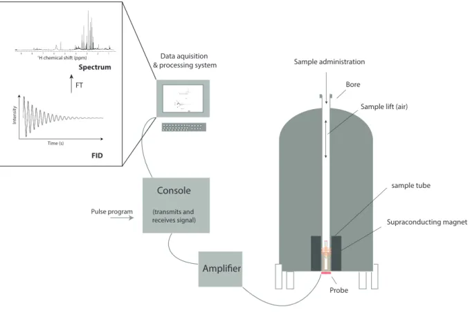
In practical terms, when the sample is placed in a strong magnetic field, an electromagnetic radiation (radiofrequency pulses) is used to excite the hydrogens. After their excitation, the protons returns to equilibrium, and the energy is recorded as an oscillating electromagnetic signal, named the free induction decay (FID). A mathematical algorithm, Fourier transformation (FT), is applied to the FID to produce 1H NMR spectra of intensity versus chemical shift (δ) using the computer. The magnitude or intensity of NMR resonance signals is displayed along the vertical axis of a spectrum, and is proportional to the molar concentration of the different metabolites present in the sample. NMR can calculate metabolites concentration by using an internal or external standard with a known concentration.
Figure 1.2.1: Diagram of NMR spectrometer.
A supraconducting magnet is present in the heart of the spectrometer. 5 mm glass tube is oriented between the poles of a powerful magnet. Radio frequency (RF) radiation of appropriate energy is broadcast into the sample from an antenna coil. A receiver coil surrounds the sample tube, and dedicated electronic devices and a computer monitor emission of absorbed RF energy. A free induction decay (FID) is acquired and then converted to NMR spectrum by Fourier transformation (FT) algorithm.
1.2.2 NMR Parameters
Each signal in a 1H NMR spectrum may be split into one or more peaks, which is named signal multiplicity (1 peak = singlet, 2 peaks = doublet, 3 peaks = triplet, 4 peaks = quartet, and several peaks = multiplet). The multiplicity tells us how many neighbouring hydrogen atoms are present around the hydrogens producing a specific peak. The chemical shift δ (expressed in ppm) and the multiplicity are central to provide information about the structure of the molecule, and then facilitate its identification. Coupling constant J (expressed
Bore sample tube Probe Amplifier Console (transmits and receives signal) Pulse program Supraconducting magnet Sample administration Data aquisition & processing system
FID In tensit y Time (s) FT 1 2 3 4 5 6 7 8 9 1H chemical shift (ppm)
Sample lift (air)
FID Intensit y Time (s) FT 1 2 3 4 5 6 7 8 91H chemical shift (ppm) Spectrum
in Hz) corresponds to the measure of the interaction between a pair of protons is also often used in the identification of metabolites.
In order to standardize the NMR scale it is necessary so set a 0 reference point to which all protons can then be compared. This association with the reference signal is called the chemical shift. This shift is measured in parts per million (ppm).
Figure 1.2.2: Schematic representation of NMR parameters.
The spectrum of a complex mixture like biological samples, which contain hundreds of metabolites, corresponds to the sum of individual metabolites spectra (Figure 1.2.2).
δ 3 δ2 δ1 Intensity 1H Chemical shift (ppm) Doublet Singulet Triplet Coupling constant Coupling constant J J J
Figure 1.2.3: Example of a 600 MHz 1H-NMR urine spectrum. 1 2 3 4 5 6 7 8 9 1H chemical shift (ppm)
1.2.3 NMR methods
Biofluids and tissue or cell extracts are considered as homogenous liquids. All the molecules present inside are in a homogeneous chemical environment. They are thus analyzed by liquid state NMR experiments in metabolomics. The obtained spectra are well resolved and the resonances are well defined. A minimal sample preparation by the addition of specific buffer to the samples is needed here.
Other biological samples like cells, biopsies or small organisms (Caenorhabditis elegans, Daphnia, etc.) are used to understand the cellular metabolisms of physiological and pathological processes. This kind of sample is considered as semi-solid samples. They are heterogeneous, and a different NMR technique is used in this case. The use of liquid NMR state experiments here gives spectra with very poor resolution.
The development of a technique called high-resolution 1H magic angle spinning (MAS) NMR spectroscopy has enabled to acquire high-resolution NMR data on small pieces of intact tissues with no pretreatment35-38. The sample heterogeneity causes line broadening. This method is based on the fact that the sample is spinning at an angle (so-called magic angle) of 54.7°, which reduces the loss of information caused by line broadening effect39.
Figure 1.2.4: Typical NMR spectra.
(A) Typical spectrum of liver extract acquired at 700MHz. (B) Typical spectrum of intact liver tissue acquired at 700MHz with HR-MAS technique.
The limited of the 1D spectra is the presence of overlapped signals; a given pic can hide two different metabolites. To facilitate metabolite identification, two-dimensional (2D) 1H NMR spectra are highly recommended to provide additional information about metabolite content. Total Correlation SpectroscopY (TOCSY)40 and J-resolved41 experiments reveal molecular connectivities and the multiplicity of resonances, respectively. 1H-13C HSQC (Heteronuclear Single Quantum Correlation), which investigate heteronuclear coupling, is also used. 2D NMR spectroscopy is useful for increasing signal dispersion and to clarify the connectivities between signals. They help to identify metabolites.
1.0 1.5 2.0 2.5 3.0 3.5 4.0 4.5 5.0 5.5 0 6 6.5 7 7.5 8 8.5 9 1H chemical shift A B
1.3 NMR-based metabolomics for the investigation of biological
samples
1.3.1 Biological samples explored by metabolomics
Different sample types can be used in metabolomics studies: biofluids (whole blood, plasma, serum, urine, saliva, amniotic fluid, faecal water, etc.), intact cells and tissues (cardiac, liver, kidney, etc.). To answer to a specific biological or clinical issue, it is primordial to choose the more appropriate type of sample. In clinical studies, the priority is given to biofluids because there is a real need to non-invasive methods to understand and diagnose diseases. For biological studies, tissues and cells in addition to biofluids are used to understand mechanisms in a specific organ. In this case, animal models are investigated because it is ethically non acceptable to take biopsies from healthy individuals.
Biofluids are easily obtained, and non-invasive compared to biopsies17,18,24. Urine, one of the most widely studied biofluids in metabolomics, reflects a more short-term state of the organism. Urine contains endogenous and exogenous (like drugs, food) metabolites, which made it very interesting in disease biomarker discovery2 and toxicology29.
About 3000 urine metabolites were identified18. For metabolomics studies, it is important to collect the first morning voids, which are following by an overnight fast to reduce the impact of food or mediation42. Fasting plasma and serum changes reflect more chronic and long-term snapshots of the system43. However, it is very important to consider external factors related to ethnicity, diet, diurnal rhythm, etc., for human samples before drawing conclusions. Analyzing cerebrospinal fluid (CSF) offers great understanding of neurological disorders. Saliva is a good source of biomarkers related to pathologies44. Sweat and breast milk is used in metabolomics to improve the infants nutrition.
Metabolomic study on the whole organism does not provide information about a specific cell type or organ. Investigate specific cells and organs is more appropriate to understand the local mechanisms. These two approaches can be combined to provide complementary information.
Standardization of protocols has enabled to work in the same conditions and make the data comparable45,46.
1.3.2 Targeted and untargeted analyses
A targeted metabolomics consists in looking for specific metabolites in a biological system. In practical terms, it is the quantification (concentrations are determined) or semi-quantification (intensities are determined) of a set of metabolites that might be associated to the common classes, to study specific metabolic pathways for example. Lipidomics9, a subtype of metabolomics, which aim to study only lipids, can be considered as targeted metabolomics. This discipline can be also considered as untargeted metabolomics because there are different types of lipids. An extraction step for wanted metabolites is often needed in this approach.
In the other hand, untargeted metabolomics is the quantification or semi-quantification of all the detectable metabolites present in a biological sample. Sample preparation is usually minimal.
1.4 Metabolomics and biomarkers
1.4.1 Metabolomics to study pathologies
Clinical metabolomics is nowadays an area of intense investigation. This use was born from the need to diagnose diseases, understand their mechanisms, identify new drugs targets, and monitor therapeutic outcomes. Specific investigations are carried to identify potential biomarkers related to pathologies10,13,47. Metabolic changes as a disease symptom have already been recognized in ancient medicine1. The challenge of biological and clinical researches is to develop fast and reliable methods for diagnosing the disease non-invasively. Since the start of metabolomics, the number of studies in biological and clinical researches has grown quickly. The study design consists often in the comparison between control (healthy) and case subjects, and the same thing in terms of cells, tissues and biofluids, in human and/or animal models. We can find also epidemiologic studies, which aim to determine the causes of diseases outcomes.
Several pathologies were highly investigated through metabolomics especially cancer including lung, colorectal, breast, prostate, bladder, gastric and thyroid28,38,48-55. It was discovered that blood acetate is associated with biliary tract cancer54, urine taurine are associated to bladder cancer48. Investigation of cerebrospinal fluid revels a deregulation in cholesterol and phospholipids metabolisms in brain cancer56.
For Cardiovascular diseases, amino-acid metabolism were found altered, and an increase of methylated arginine species and lipids (specially fatty acids) were noticed6,7,57.. In neurological field, Alzheimer and depression are highly studied20,58. Other diseases like infectious diseases59,60 and diabetes12 and others were also investigated.
1.4.2 Metabolomics and genetic diseases
The association of genetic variation with metabolite levels is well documented61,62. A number of studies have highlighted the influence of genetics on the metabolites levels 63,64. It was discovered more than 150 genetic loci that associate with blood levels of more than 300 distinct metabolites65,66. For example, metabolomics approach was used to explore several genetic diseases like Inborn errors metabolism, which is actually implemented in clinical routines67. Mitochondrial diseases, a group of disorders that can result from abnormalities in the mitochondrial and nuclear genomes, could be also recognized by metabolomics68.
1.5 Conclusion
The application of metabolomic approach in medical research is a dynamic field, and has a high potential in disease diagnosis. These discoveries make metabolomics a promising diagnostic tool, with has the advantage to be non-invasive and highly reproducible. Metabolomic technologies offer a sensitive means to search human biofluids for metabolite profiles potentially usable as biomarkers for diseases. In this thesis, we will use a 1H NMR based metabolomics to investigate murine models for thyroid hormone pathologies.
2 Thyroid hormone system and diseases
2.1 Thyroid hormone system
2.1.1 Thyroid hormone functions
Thyroid hormones (TH), thyroxine (T4) and 3,5,3’-triiodothyronine (T3), the main secretion products of the thyroid gland, are essential for normal growth, development and metabolism regulation69-71. T4 is more abundant in the blood, T3 is considered as the major active hormone due to its high affinity for nuclear receptors and intracellularly generated from T4.
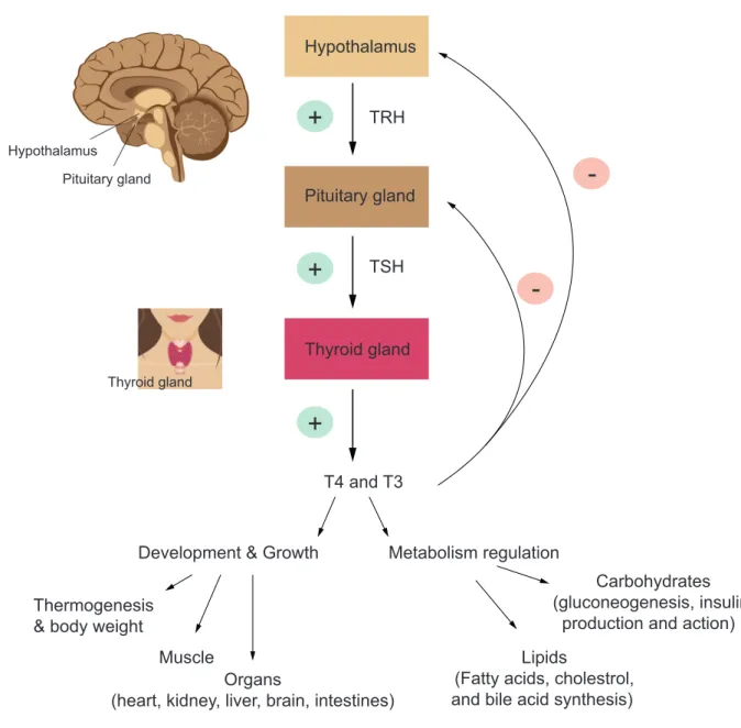
TH play significant roles during embryogenesis and childhood. They are involved in nearly all tissues, with major effects on the basal metabolic rate and oxygen consumption72,73. TH are known to maintain heart rate, myocardial contractility and vascular function74. They are also key regulators of thermogenesis, which allows the maintenance of body internal temperature75,76. They are involved in body weight regulation, in the development and maintenance of adult bone mass and strength77. It is well established that changes in TH level, compared to the reference range, is associated with body weight change in both men and women78. Skeletal muscle is an important TH target for contractile function and regeneration79. TH regulate lipids metabolism such as: cholesterol synthesis and efflux, bile acid synthesis, fatty acid metabolism. Carbohydrates are also regulated by thyroid hormones (Figure 2.1.1).
Figure 2.1.1: The hypothalamus-pituitary-thyroid (HPT) axis and thyroid hormones (TH) actions.
HPT axis regulates and maintains thyroid hormones homeostasis. Hypothalamus secretes TRH in response to low concentration of circulating T4/T3. This secretion stimulates the pituitary gland, which secretes TSH in turn. This leads to T4/T3 release in the blood. T3 negative feedback on TRH and TSH levels keeps the T3/T4 ration constant in circulation. Thyroid hormones have a central role in development, growth and metabolism regulation.
2.1.2 Thyroid hormones receptors Thyroid gland Hypothalamus Pituitary gland Thyroid gland TRH TSH T4 and T3
Development & Growth Metabolism regulation
Carbohydrates (gluconeogenesis, insulin
production and action) Lipids
(Fatty acids, cholestrol, and bile acid synthesis) Thermogenesis
& body weight
+
+
+
-Muscle Organs(heart, kidney, liver, brain, intestines)
Hypothalamus Pituitary gland
Thyroid hormones exert their effects via thyroid hormone receptors (TRs), member of the superfamily of hormone-responsive nuclear transcription factors. These receptors share a similar structure and mechanism of action80. THRA and THRB encode the two major TR isoforms: TRα1 and TRβ (β1 and β2), which are distributed differentially in tissues. TRα1 is predominantly expressed in brain (central nervous system), heart (myocardium), skeletal muscle, adipose tissue and gastrointestinal tract. TRα2 and TRα3 are variants of TRs, which are non TH-binding proteins, and their function is not understood81. TRβ is expressed in the sensory tissue (the inner ear and retina), the kidney, the liver and cardiac ventricles82.
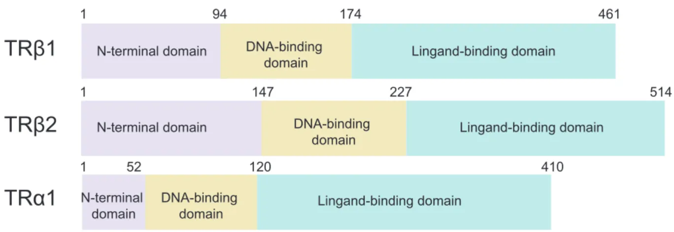
As described in figure 2.1.2, all these receptors contain three conserved domains: a N-terminal domain, a DNA-binding domain (DBD) and a ligand-binding domain (LBD). The α and β receptors have a similar DBD and LBD, but differ in their N-terminal domain. The three receptors (TRα1, TRβ1, TRβ2) bind the same ligand due to these structural homologies69.
TRs are considered as transcription factors because they regulate target gene expression directly through DNA response element (TRE)83.
Non-genomic actions of thyroid hormone were also reported and are little studied 84. They have extranuclear actions, which are not TRE-mediated.
Figure 2.1.2: Schematic alignment of thyroid hormone receptors: TRβ1, TRβ2 and TRα1.
TH receptors are composed of three domains: N-terminal domain (activation function), DNA-binding domain (DBD) and Ligand-binding domain (LBD). DBD and LBD are highly conserved domains between the three TH receptors, contrary to the N-terminal domain.
TRβ1
TRβ2
TRα1
1 94 174 461 1 147 227 514 1 52 120 410 N-terminal domain N-terminal domain N-terminal domain DNA-binding domain DNA-binding domain DNA-binding domain Lingand-binding domain Lingand-binding domain Lingand-binding domain2.1.3 Mechanisms of action
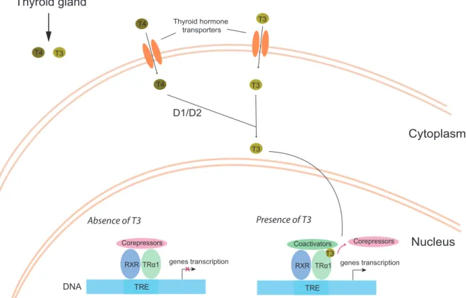
The thyroid gland secretes T4 and T3 hormones, which are transported into the cells by membrane transporters like the monocarboxylate transporter (MCT) family85, and more precisely MCT8. T3 is secreted by the thyroid gland, but also converted from T4 under the action of type 1 or type 2 of 5’ deiodinase, which are tissue-dependent. Unliganded TR in the nucleus forms a heterodimer with the retinoid X receptor (RXR), which then binds the thyroid hormone response element (TRE) in the regulatory sequences of TH-responsive gene. This results in the activation or repression of TH target genes transcription.
In the absence of T3, corepressor proteins are associated to the TR-RXR complex and prevent genes transcription86. However, in the presence of T3, corepressor proteins leave the complex, and coactivator proteins are recruited, which lead to target genes transcription87 (Figure 2.1.3).
Figure 2.1.3: Mechanisms of action of thyroid hormones. TRE TRα1 RXR Corepressors genes transcription DNA Absence of T3 TRE TRα1 RXR Coactivators T3 genes transcription Presence of T3 Corepressors Thyroid gland T4 T3 T4 Thyroid hormone T3 transporters T4 T3 Cytoplasm Nucleus T3 D1/D2
Membrane transporters transport T4 and T3, after their secretion by thyroid gland, into the cell. A part of T4 is converted to T3 under the action of type 1 and 2 5’ deiodinase (D1 and D2). Unliganded TR heterodimerizes with RXR, and then the complex recognizes TRE. In the absence of T3, corepressor proteins bind the complex to repress target gene expression. In the presence of T3, corepressor proteins are dropped and coactivator proteins are recruited, which lead to target gene transcription.
2.1.4 Regulation
TH regulates a large amount of genes, which are involved in growth, metabolic balance and thermogenesis88. TH synthesis and secretion are regulated by a negative-feedback system that involves the hypothalamus, pituitary, and thyroid gland (hypothalamic/pituitary/thyroid (HPT) axis)89. Hypothalamus secretes TRH. Thyrotropes, endocrine cells from pituitary, secrete then Thyroid-Stimulating hormone (TSH). TSH acts on the thyroid gland to induce thyroxine (T4) and 3,5,3’-triiodothronine (T3) hormones production, which then act on body growth and development and metabolism regulation. High levels of T4 and T3 in turn feed back lead to TSH level secretion diminution in the pituitary, which then regulate T4 and T3 levels.
2.1.5 Alterations in thyroid hormone system
TH action requires (i) the availability of TH (production/secretion, conversion of T4 to T3) (ii) intact and adapted membrane transport (iii) cytosolic and nuclear processing (iv) association with intact receptors (v) interaction with regulators (activator or co-repressor proteins). Alteration in one of these steps can lead to a heavy metabolic disorder90.
Associated diseases are the most common endocrine disorders worldwide91. In addition to clinical symptoms, biochemical tests like determination of TSH and free T4 levels in blood are used to suspect thyroid disorder. Hypothyroidism and hyperthyroidism lead to alterations in metabolism (lipids, carbohydrates), growth and energy homeostasis92. Hypothyroidism,
results from low levels of TH, presents a low metabolic rate, and leads to cardiovascular diseases, whereas hyperthyroidism, which is a catabolic syndrome, is associated with a high metabolic rate, and leads to tachycardia and loss of body mass. These diseases are related to the secretion and production of the thyroid hormones by the organism.
Other diseases are known to lead to hypothyroid or hyperthyroid phenotypes, without alteration in TH production, but caused by genetic mutations. Thyroid hormone cell transport defects (THCTD) caused by a mutation in MCT8 gene lead to production of defective cell-transport proteins, which reduce hormone cell-transport and causes reduced levels of intracellular TH90. Thyroid hormone metabolism defect (THMD) is caused by a mutation in selenocysteine-binding protein 2, which interferes with conversion of T4 to T3, resulting in a low T3 and high T493.
Other diseases known under the name of resistance to thyroid hormone (RTH)94 were discovered. The first described cause was a mutation in TRβ gene in 1989, which is characterized by high serum concentration of free T4 and T3, and normal or slightly elevated TSH concentration. This disease is called RTHβ. Twenty-four years later, the first mutation in TRα gene was discovered, and the disease has been named RTHα.
2.2 Resistance to thyroid hormones α
Resistance to thyroid hormone (RTH) was first described as a clinical entity in 196795. Patients are hyposensitive to TH and they present reduced clinical and biochemical manifestations of TH action relative to the circulating hormone levels. The molecular explication of this syndrome was determined in 1989 when the first case of RTH caused by a mutation in the THRB gene was discovered. This disorder was named resistance to thyroid
hormone due to a mutation in the thyroid hormone receptor β, abbreviated by RTHβ96,97.
RTHβ is characterized by the impairment of the HPT axis: elevated levels of thyroid
hormone, normal or elevated levels of TSH. Patients present with different degree of goiter, hearing abnormalities, tachycardia, mental retardation, attention-deficit, and delayed bone growth and maturation94,97,98. They present features of hypothyroidism and hyperthyroidism,
which is explained by variable resistance in different tissues. The incidence of RTHβ is
actually estimated to be 1 in 40.000 with 160 different mutations approximately99,100. Patients
with RTHβ are currently found in more than 400 families101. This syndrome can be suspected in patients with these features and can easily be recognized by physicians.
2.2.1 The first cases
Patients with mutations in THRA were not identified until 2012. The first case of RTHα
was discovered, after a whole-exome sequencing, in a little 6-year-old girl, which presents classical features of hypothyroidism (e.g. growth retardation, developmental retardation, skeletal dysplasia, low heart rate, and severe constipation) and nearly normal thyroid function biochemical tests102.
Other cases, female and male of different ages, were discovered with different mutations in TRα1100,103-112. The phenotype of the identified patients consists of varying degrees of growth impairment, mental and motor development, delayed bone, constipation, and
near-normal thyroid function tests. Some cases have specific health issues like autism106, chronic
anaemia104,110,113, epilepsy114, etc. (Appendix 1).
2.2.2 RTHα is different from RTHβ
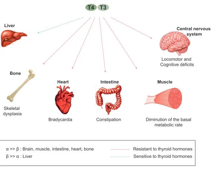
RTHα is clinically distinct from RTHβ. TRα and TRβ, are differentially expressed during development and are differentially distributed in adult tissues, which explains the difference between the two diseases. TRβ has an important role in inner ear, cerebellar, and retinal development, TSH regulation, and mediating the metabolic actions of T3 in the liver71. TRα has specific roles in the heart, brain, intestine and in mediating adaptive thermogenesis in brown adipose tissue (BAT)71,115. RTHα is thus characterized by a tissue-selective hypothyroidism (e.g. skeleton, gastrointestinal tract, myocardium, etc.) and near-normal thyroid function biochemical tests. In figure 3.2.1, organs that present a dominance of TRα (bones, heart, intestine, brain and particularly the central nervous system, and muscles) are resistant to TH. While, liver that presents more than 80% of TRβ is resistant to TH. The pituitary-thyroid axis in RTHα is not dysregulated as noticed in RTHβ patients.
Figure 2.2.1: The sensitivity of organs to thyroid hormones depends on the type of thyroid receptor present.
2.2.3 A high variability in the clinical features
The high variability of clinical features and the absence of specific traits and reliable biochemical markers make the diagnosis of this disease difficult. In addition, identified RTHα patients present some common features with hypothyroid patients, which may delay the diagnosis in some cases. Since its first description, 45 cases of RTHα have been reported worldwide, corresponding to 25 different mutations of TRα1. In 2015, the sequence of 60 000 anonymous exomes were released by the Exac database (http://exac.broadinstitute.org), revealing the existence of 68 THRA missense or frameshift, which almost certainly alter TRα1 function. It is thus thought that the incidence of RTHα may be similar to RTHβ incidence.
Heart Intestine Muscle
Central nervous system
Locomotor and Cognitive déficits
Diminution of the basal metabolic rate Constipation Bradycardia Bone Skeletal dysplasia T4 T3 Liver
Resistant to thyroid hormones Sensitive to thyroid hormones α >> β : Brain, muscle, intestine, heart, bone
The inability to quickly recognize patients with RTHα is regrettable because the developmental consequences of the disease can be greatly reduced by an early therapeutic intervention. Motor skills and body growth have been restored in a young patient treated with an excess of thyroxine to overcome tissue resistance108,109. Two adult patients with normal adult stature received thyroxine treatment during childhood by chance105. Authors suggest here that the neurocognitive abnormalities are may be less severe because of this treatment105. Another study on a murine model (Arg384cys) showed also that increased concentrations of thyroid hormones can reverse neurological abnormalities116.
It is thus primordial to find a reliable way to identify these patients, and to manage them at early stage to minimize the consequences of this disease.
2.2.4 Molecular aspect
All the identified patients are heterozygous for THRA mutation, which generate a mutant receptor that inhibits wild-type receptor function in a dominant negative manner106,108,112. All the known mutations are present in the carboxyterminus, the ligand-binding domain (LBD) of the receptor (Figure 3.2.2), to which the helix 12 belongs. These mutations lead to the destabilization of this helix102.
Two types of mutations were identified in RTHα patients: frameshift 103,104,107,114 and
missense mutations102,105-108.
Amino acid substitutions (D211G, H361Q, R384H, A263V) reduce the affinity of TRα1 for T3. While, c-terminal substitutions (P398R and E403K), truncations (C392X and E403X), and frameshift mutations (C380fs387X, A382PfsX7, F397fs406X, and F401S) alter ligand binding, by modifying or eliminating the helix 12, and prevent thus coactivator recruitment117.
Figure 2.2.2: THRA mutations consequences.
Thyroid hormones regulate target genes transcription via nuclear receptors TRα and TRβ. These receptors are composed of 3 distinct regions: N-terminal domain, DNA binding domain (DBD) and ligand binding domain (LBD). All the known RTHα mutations are located in the LBD (A). Mutations in TRα1 disturb target gene transcriptional regulation. Two functional alterations were observed in the presence of T3: the dissociation of corepressor proteins is prevented (B), or affinity between TRα1 and T3 is reduced (C).
2.2.5 Relation between genotype and phenotype?
There are thus different mutations with different degrees of severity of the disease. Two patients (6-year-old and 15-year-old girls), which were found with the same mutation (E403X)102,118 present similar features. Two other patients were found with TRα1 mutation in the same position (A263V and A263S), but, as noticed, the original amino acid alanine was substituted by valine in the first case and by serine in the other case. The functional analysis of the A263V mutant indicates a greater defect in transcriptional activity of TRα1 than that induced by the A263S mutation. It was also noticed that a more up-stream mutation lead to a more severe phenotype, like the mutation C392X118.
DBD AF1 LBD N-term C-term 1 52 120 410 TRα1 E403X/K P398R F397fs406X C392X R384C/H A382PfsX7 C380fs387X N359Y A263V/S D21 1G TRE TRα1 RXR Corepressors genes transcription DNA T3 Coactivators TRE TRα1 RXR Coactivators T3 genes transcription A B C Corepressors
All these observations suggest that the variability in the symptoms is maybe related to the type of the mutation and its position. The relationship between genotype and phenotype need to be further discerned. To understand this relationship and to study the impact of RTHα
disease on the whole organism, different murine models were developed.
2.2.6 Mouse models to study RTHα
Mouse Thra and human THRA genes present sequence similarities. The two TRα1 amino acid sequences differ at three positions only (AA34, 37, and 170), which makes mouse lines with Thra mutations highly relevant animal models for RTHα disease. Prior to the identification of human RTHα cases, several researchers were done using TRα knockout and knockin mouse models to predict the phenotype of human disorder99,115,119-124. The first mouse model of RTHα was a knockout of Thra gene, in which TRα1 and TRα2 isoforms were inactivated124. Mice failed to survive 5 weeks after birth, and features of hypothyroidism and serious delayed maturation in the small intestine and bones were observed in these mice124. It was reported also in another study that a mutation in TRα1 exhibit several distinct neurological abnormalities: extreme anxiety, reduced recognition memory, and locomotor dysfunction116. Another model was developed before its discovery in a human patient107,121. Other researchers showed the significant implication of TRα1 on metabolic homeostasis119. The phenotype of mice models is often close to the clinical features of RTHα patients.
These models allowed the good understanding of the function of THRA gene and the TRα receptor, and facilitate the comprehension of RTHα disease and its impact on the organism.
For the metabolomic study presented in this thesis, we have used mouse models that were previously generated by Markossian et al117 to understand the relationship between specific THRA mutations and phenotype. Five novel germline mutations (four frameshift: E395fs401X, E395fs485X, E395fs406X, and K389fs479X; and one missense N359Y), which are closely modeling the mutations found in RTHα patients, were introduced in mouse Thra gene. Markossian et al have investigated skeleton, blood, heart, cerebellum and intestinal epithelium. They concluded that like human patients, mutant mice displayed a
hypothyroid-like phenotype, with altered development. Phenotype severity varied between the different mouse models117.
From a global literature screening, about a hundred of research studies have been conducted to study the thyroid system by metabolomics approaches. About 50% of these studies address about thyroid cancer, which is explained by its increased incidence worldwide. The main medical treatment in the case of thyroid cancer is the total thyroidectomy (removal of the thyroid), even if the tumour is benign. A number of studies, with different methods, were performed to distinguish between cancer patients and healthy individuals, and between benign and malignant thyroid nodules55,125 to improve thyroid cancer diagnosis. Several toxicological studies, which highlight the harmful impact of certain chemicals (mainly glyphosphate-based herbicide and decabromodiphenyl) on the thyroid hormone system126,127. A few studies, also aimed providing deeper understanding of thyroid disease mechanism, such Graves’ disease14, hypothyroidism128, hyperthyroidism129, depression, low T3 syndrome, type 2 diabetes12 and selenium deficiency. Others correspond to population-based studies seeking for linear relationship between either TSH or free T4 (FT4) and metabolite levels55,129. Other works focused on the direct effect of thyroid hormones status on the brain130 or hepatic lipids, to provide deeper understanding of the mechanism behind the biological phenomenon.
At this point, metabolomics investigations of genetic diseases that disturb thyroid system such as RTH (α and β) or THCTD or THMD, have never been reported in the literature.
We notice overall that the thyroid system has little been explored by metabolomics approaches, and more precisely genetic diseases. A number of studies have highlighted the influence of genetics on the metabolites levels 63,64. The association of genetic variation with metabolite levels is well documented61. For example, metabolomics approach was used to explore several genetic diseases like Inborn errors metabolism, which is actually implemented in clinical routines67. Mitochondrial diseases, a group of disorders that can result from abnormalities in the mitochondrial and nuclear genomes, could be also recognized by metabolomics68.
In the work described in the following chapters of this thesis, we study different pathologies related to the thyroid hormone system. We exploit the metabolic fingerprint of hypothyroidism. We investigate the same series of mouse models of RTHα used by Markossian et al. 117 to assess the capacity of 1H NMR analysis of body fluids to recognize the presence of Thra mutations. And in the end, we study the impact of thyroid hormone on the hepatic metabolism and the specific role of the thyroid hormone receptor β in this mediation.
3 Materials, methods and analytical
workflow
3.1 Introduction
The NMR metabolomic global workflow contains several steps: sample preparation, NMR data acquisition, data processing, statistical analysis, metabolite identification, and interpretation. Two different approaches can be used: untargeted approach (with no a priori hypothesis) and targeted approach (applied on a predefined set of metabolites). The different steps are detailed in the following section (Figure 3.1).
3.2 Biological samples preparation
3.2.1 Collection and storage
NMR liquid samples preparation is straightforward and the protocols46 are standardized and highly reproducible. This step can be automated by using a robotic liquid handling technology. It is very important to collect the biofluids with the same manner, and to handle properly the samples. Because, in some cases, errors related to samples handling (time of collection, contaminations) can introduce variations in signal intensity that are not from metabolism.
• Urine
Time of collection for urine samples can make a quantitative and qualitative difference in the urine metabolome. The first morning void is the preferred type, because it is collected after several hours of fasting, which minimizes the impact of food or medication on the urine metabolome. It is important to respect the same time of collection between subjects, and between time points in the case of longitudinal studies. For human subjects, urine collection is easy contrary to animals. Animal houses need to be clean before their first urine, which are around 7 am for the majority. In addition, in some cases, we find urine and feces in the same place, which can alter considerably the composition of urine. The volume of mouse urine needed for classical metabolomics studies is 200µl. This quantity is sometimes not reached, and we need to wait for a second sample, in fasting condition also, to obtain the needed volume. Urine is then quickly stored at -80°C after collection, before NMR analysis.
• Blood
The main difference between blood serum and plasma is the presence or absence of clotting. For plasma, the whole blood is collected into tubes with anti-coagulant and then a centrifugation step lead to separate the liquid state from blood cells. For serum, whole blood is collected into tubes and is allowed to clot for a specified time and temperature before centrifugation to pellet the clot and cells. Different studies showed that there is not a difference between these two types in term of metabolite composition. In our study, we