HAL Id: ensl-00182491
https://hal-ens-lyon.archives-ouvertes.fr/ensl-00182491
Submitted on 26 Oct 2007
HAL is a multi-disciplinary open access
archive for the deposit and dissemination of
sci-entific research documents, whether they are
pub-lished or not. The documents may come from
teaching and research institutions in France or
abroad, or from public or private research centers.
L’archive ouverte pluridisciplinaire HAL, est
destinée au dépôt et à la diffusion de documents
scientifiques de niveau recherche, publiés ou non,
émanant des établissements d’enseignement et de
recherche français ou étrangers, des laboratoires
publics ou privés.
zinc finger protein form a complex implicated in vulval
development
Vincent Coustham, Cécile Bedet, Monier Karine, Sonia Schott, Karali
Marianthi, Francesca Palladino
To cite this version:
Vincent Coustham, Cécile Bedet, Monier Karine, Sonia Schott, Karali Marianthi, et al.. The C.
elegans HP1 homologue HPL-2 and the LIN-13 zinc finger protein form a complex implicated in
vulval development. Developmental Biology, Elsevier, 2006, pp.308-322. �ensl-00182491�
The C. elegans HP1 homologue HPL-2 and the LIN-13 zinc finger protein
form a complex implicated in vulval development
Vincent Coustham, Cécile Bedet, Karine Monier, Sonia Schott,
Marianthi Karali, Francesca Palladino
⁎
Laboratoire de Biologie Moleculaire de la Cellule, Ecole Normale Supérieure de Lyon, 46 allée d’Italie, 69007 Lyon, France Received for publication 21 December 2005; revised 10 April 2006; accepted 11 April 2006
Available online 23 May 2006
Abstract
HP1 proteins are essential components of heterochromatin and contribute to the transcriptional repression of euchromatic genes via the
recruitment to specific promoters by corepressor proteins including TIF1 and Rb. The Caenorhabditis elegans HP1 homologue HPL-2 acts in the
“synMuv” (synthetic multivulval) pathway, which defines redundant negative regulators of a Ras signaling cascade required for vulval induction.
Several synMuv genes encode for chromatin-associated proteins involved in transcriptional regulation, including Rb and components of the Mi-2/
NuRD and TIP60/NuA4 chromatin remodeling complexes. Here, we show that HPL-2 physically interacts in vitro and in vivo with the multiple
zinc finger protein LIN-13, another member of the synMuv pathway. A variant of the conserved PXVXL motif found in many HP1-interacting
proteins mediates LIN-13 binding to the CSD of HPL-2. We further show by in vivo localization studies that LIN-13 is required for HPL-2
recruitment in nuclear foci. Our data suggest that the LIN-13/HPL-2 complex may physically link a subset of the Rb related synMuv proteins to
chromatin.
© 2006 Elsevier Inc. All rights reserved.
Keywords: C. elegans; HP1; Chromatin; LIN-13; Vulva
Introduction
HP1 proteins are a highly conserved family of proteins
which directly contribute to the higher order packaging of
chromatin by binding to modified histones (
Li et al., 2002
).
All HP1 proteins are structurally related and characterized
by the presence of two conserved domains, an N-terminal
chromo domain (CD) separated by a variable hinge region
from a C-terminal-related domain termed the chromo
shadow domain (CSD). The CD is responsible for binding
to methylated histone H3-K9, while the CSD is required for
dimerization and most protein–protein interactions within the
nucleus (
Eissenberg, 2001
). Many of these interactions play
an important role in directing heterochromatin formation
and/or gene silencing. A large number of the CSD interaction
partners contain a PXVXL pentapeptide motif, including the
transcription intermediary factor TIF1, the TBP-associated
factor TAFII130, and the large subunit of chromatin assembly
factor 1 (CAF-1) (
Le Douarin et al., 1996; Murzina et al.,
1999; Vassallo and Tanese, 2002
). These results suggest that
HP1 proteins function as adapters, bringing together different
proteins in multiprotein complexes via protein–protein
interactions with the CD and CSD. The multiple interactions
of the CSD underscore the complexity of HP1 dynamics.
However, it remains to be established how many partners
HP1 has under a given set of conditions and how important
these interactions are in different cellular contexts. The
Caenorhabditis elegans vulva is a simple, well-described
system particularly suited to study how chromatin regulation
can function in a specific developmental pathway. Previously,
we showed by RNAi that the C. elegans HP1-like protein
HPL-2 contributes to vulval development by acting in the Rb
related synMuvB pathway antagonistic to the Ras-mediated
signal transduction pathway responsible for vulval cell fate
determination (
Couteau et al., 2002
). In addition to
LIN-35Rb, synMuvB genes encode homologues of the class I
⁎ Corresponding author. Fax: +33 4 72728080.E-mail address:francesca.palladino@ens-lyon.fr(F. Palladino). 0012-1606/$ - see front matter © 2006 Elsevier Inc. All rights reserved. doi:10.1016/j.ydbio.2006.04.474
histone deacetylase HDA-1, EFL-1/E2F, DPL-1/DP (
Ceol and
Horvitz, 2001; Lu and Horvitz, 1998
) and counterparts of the
NuRD nucleosome remodeling and histone deacetylase
com-plex (
Solari and Ahringer, 2000; von Zelewsky et al., 2000
).
Based on the identity of the cloned class B synMuv genes, it has
been proposed that they remodel chromatin and repress
transcription of genes important for vulval cell fate
specifica-tion. More recently, a TIP60/NuA4-like complex was also
implicated in the negative regulation of Ras signaling during
vulval cell fate specification (
Ceol and Horvitz, 2004
). These
results strongly suggest that different chromatin remodeling and
modification complexes may cooperate in the same
develop-mental pathway. To gain insight into the specific role played by
HPL-2 in vulval cell fate determination, we carried out a
two-hybrid screen to identify potential partners. One of the proteins
identified is the LIN-13 zinc finger protein, another member of
the synMuvB pathway (
Melendez and Greenwald, 2000;
Ferguson and Horvitz, 1989; Thomas et al., 2003
). The
interaction between the two proteins was confirmed in vivo
and depends on the CSD of HPL-2 and a conserved HP1
binding motif in the N-terminus of LIN-13. Finally, we show
that the localization of HPL-2 in nuclear foci is completely
dependent on 13 but not other synMuvB genes such as
lin-35 and lin-9. Altogether, these results suggest the existence of a
hierarchy in the recruitment of the Rb-related synMuvB
complex to chromatin.
Materials and methods
Yeast strains and media
Strains used are the following: CG1945 (MATa gal4-542 gal80-538 ade2-101 his3-200 leu2-3,-112 trp1-901 ura3-52 lys2-801 [URA3∷GAL4 17mers (X3)-CyC1TATA-lacZ] LYS2∷GAL1UAS-GAL1TATA-HIS3 CYHR) (gift from L. Segalat); Y187 (MATα gal4Δ gal480Δ ade2-101 his3 leu2-3,112 trp1-901 ura3-52 URA3∷UASGAL1-lacZ met−); and PJ69-4a/α (MATA/α trp-1-901 leu2-3,112 ura3-52 his3-200 gal4Δ gal480ΔLYS2∷GAL1-HIS3 GAL2-ADE2 met2∷GAL7-lacZ. Basic methodology for yeast was as described by Fink (1991).
C. elegans strains and genetics
Strains were maintained according to the standard protocol (Brenner, 1974). The following mutant alleles were used: LGI lin-35(n745); LGIII hpl-2(ok916), hpl-2(ok917), hpl-2(tm1489), lin-13(n388), unc-32(e189), unc-49 (e407), dpy-17(e164); X lin-15(n767). ok916, ok917 and tm1489 alleles were backcrossed four times prior to analysis and the presence of the deletion allele confirmed by PCR analysis. The hpl-2 wild-type sequence fully rescued synMuv and synthetic lethal phenotypes and partially rescued the sterility of all three alleles. For expression studies lin-39∷GFP (gift from A. Hajnal) and qIs56[lag-2∷GFP] were used. To construct the lin-13(n388)dpy-17(e164)hpl-2(tm1489)unc-49(e407) strain, lin-13(n388)/unc-32(e189) worms were crossed with dpy-17(e164);lin15(n767) males. Recombinant lin-13 (n388)dpy-17(e164)/+;lin15(n767) were selected based on the Muv pheno-type in their progeny at 20°C and then crossed with unc-49(e407)hpl-2 (tm1489) males. lin-13(n388)dpy-17(e164)hpl-2(tm1489)unc-49(e407)/+ recombinants were selected as Dpy Unc animals segregating in their progeny at 20°C. Loss of the lin15(n767) allele was confirmed by duplex PCR (M. Koelle laboratory protocol) using the following primers: for 5 ′-GGGAAGGTTACGTCGAACAC-3′; rev1 5′-GATTCGTCGATGGAGG-CACC-3′; rev2 5′-GCCTCGTCGGCAATTGAATG-3′. The presence of the tm1489 deletion in lin-13 hpl-2 mutants was confirmed by duplex PCR using the
following primers: for1 (outside deletion) 5 ′-TACATGCAACAAACCGCGCT-3′; for2 (inside deletion) GACAGTATAAGTTCCCCGAC-3′; rev 5′-GTTTACCAGCTTTTCCGTGTG-3′, followed by sequencing using both outside primers described below. The presence of the lin-13(n388) C to T substitution was confirmed by sequencing.
Two-hybrid screen
The full-length hpl-2a cDNA was subcloned into plasmid pAS2.1 (Clontech) to create pCV1. The two-hybrid screen was performed by mating, as described (Fromont-Racine et al., 1997) using yeast strains CG1945 and Y187 and the C. elegans cDNA libraryλACT-RB2 (Durfee et al., 1993; Elledge et al., 1991). Positive interactions were selected using the lacZ and HIS3 reporter genes on HIS selection plates supplemented with 10 mM 3-AT. Interacting partners were confirmed by re-transformation in 4a, mating with a PJ69-4α carrying either pAS2.1 or pCV1 vectors, and selection for expression of ADE2 and HIS3 reporters. For directed two-hybrid assays, baits were transformed in PJ69-4α strain and mated with strain PJ69-4a carrying preys cloned in the pGADT7 vector (Clontech).
PCR of hpl-2 fragments
For domain mapping experiments, specific fragments of hpl-2a cDNA sequence were amplified by PCR with oligonucleotides designed at following positions: CD, for 5′-ATGTCGAGCAAATCAACAAA-3′, rev 5′-CTCAAACTCGTCCAACATCTC-3′; CSD, for 5′-GAAACGAAT-CAAAATGACAAATTC-3′, rev 5′-TTAAAGCTCGTCGGCTTTTGG-3′; Hinge, for 5′-AGGGAATTTTCAAAGAGAGAG-3′, rev 5′-CTTCTTTTCATCTT-TATCCTCTTC-3′;Hinge+CSD,for5′-AGGGAATTTTCAAAGAGAGAG-3′,rev 5′-TTAAAGCTCGTCGGCTTTTGG-3′. For HPL-2Δ, primers used were for 5′-ATGTCGAGCAAATCAACAAA-3′, rev 5′-ACTGTTCACCTCCTTCGCTGG-3′. For this last construct, a stop codon was added at nucleotide position 453, corresponding to the 5′ breakpoint of ok916 and ok917 deletion alleles. For LIN-13, point mutations were introduced resulting in a change at position 442 (V to D) and position 444 (V to E) using the Quickchange Site-directed Mutagenesis kit (Stratagene) on the cDNA isolated from two-hybrid experiment. Primers used were 5 ′-GAACCATTCTTGTCAACCGTTGGATCCG-GAGGTTGCTTACTTCCCAAAACCG-3′ and 5′-CGGTTTTGGGAAGTAAG-CAACCTCCGGATCCAACGGTTGACAAGAATGGTTC-3′. The presence of the point mutations was confirmed by sequencing.
Deletion mapping
Total genomic DNA was extracted from mixed-stage populations of ok916 and ok917 mutant worms. Oligonucleotides were designed as described by the C. elegans Knockout Consortium. Nested PCR was performed using BIO-X-ACT polymerase (Bioline). The tm1489 deletion predicted by the National BioResource Project was confirmed by sequencing using primers described below.
Reverse transcriptase - polymerase chain reaction (RT-PCR) and
sequencing
Total RNA was isolated from mixed-stage populations of homozygous ok916, ok917 and N2 worms using Trizol reagent (Sigma). First-strand synthesis and RT-PCR were performed in a single reaction using the Access RT-PCR Kit (Promega). Oligonucleotides were designed at the following positions: 4799– 4819 (for exon 2, 5′-AGGACAACGTGTTCATGGTGG-3′); 7011–7030 (rev exon 5-2B, 5′-CTTCAGTCATCTCAACGTCC-3′); 7617–7638 (rev exon 6-2B, 5′-ATCATCGTCTGGTACAGTGTCG-3′).
RNAi
RNAi injection and feeding experiments were carried out as described (Fire et al., 1998; Kamath and Ahringer, 2003). When necessary, to produce a weaker RNAi effect, the concentration of IPTG in feeding plates was reduced to 0.1 mM IPTG. Induction was 12 h at room temperature. Most RNAi feeding clones were obtained from Geneservice. For the lin-13
feeding clone, the cDNA sequence of lin-13 obtained in the two-hybrid screen was subcloned into the L4440 feeding vector and transformed into HT115. RNAi of lin-13 by feeding resulted in the same range of highly penetrant temperature-sensitive phenotypes previously reported by injection (Melendez and Greenwald (2000), including sterility, evl (everted vulva), and Muv in combination with class A synMuv mutations at 15–20°C, and larval arrest at 25°C (data not shown). lin-15A-specific RNA for injection was prepared as previously described (von Zelewsky et al., 2000). For hpl-2∷GFP localization studies, embryos were dissected from gravid mothers derived either from L1 larvae grown on RNAi feeding plates, or from injected L4 larvae. For lin-9 and lin-35 feeding, the efficacy of RNAi was measured by placing lin-15A mutant animals on these plates and looking for 100% Muv animals. For lin-13, mys-1, trr-1, hda-1 and let-418, RNAi feeding yielded the phenotypes previously described (Kamath and Ahringer, 2003).
GST pulldown assay
GST-HPL-2 fusion protein expressed in E. coli using the full-length hpl-2a cDNA cloned in pGEX-KG (Pharmacia) was immobilized on gluthatione-agarose beads (Sigma). Proteins subcloned in pGADT7 vector were produced and labeled with [35S]methionine using the TNT T7-coupled reticulocyte
lysate system (Promega). Beads were incubated with in vitro produced proteins 2 h at 4°C in PBS with 0.05–0.2% Triton, washed and analyzed on SDS-PAGE.
Preparation of embryo extracts
Hermaphrodites carrying a rescuing LIN-13∷GFP transgene were grown in liquid as described (Mains and McGhee, 1999). Embryos recovered following alkaline hypochlorite treatment of gravid adults were recovered by centrifugation, resuspended in 5 volumes of cold TNT buffer (Tris–HCl 50 mM pH = 8; NaCl 150 mM; Triton X-100 1% + protease inhibitor cocktail (Complete EDTA, Roche Applied Science, plus 1 mM PMSF) and sonicated on ice. Lysates were spun at high speed for 10 min at 4°C, and supernatants were collected and assayed for protein concentration using Bradford reagent (Bio-Rad Laboratories). Protein concentration of the LIN-13∷GFP embryonic lysate was≈5 mg/ml.
Immunoprecipitation and Western blot
3 μl of rabbit polyclonal affinity purified anti-HPL-2 antibody or preimmune serum, or 4μg of anti-GFP (Clones 7.1 and 13.1, Roche Applied Science) or anti-FLAG (M2 monoclonal, Sigma) antibodies were added to 250 μl of LIN-13∷GFP embryonic lysate diluted twice in TNT buffer. Samples were incubated at 4°C for 1 h, 30 and 50μl of a 1:1 slurry of protein A–sepharose (Amersham Bioscience) or protein G–sepharose (Sigma) beads were added. Samples were incubated another 2 h at 4°C. The beads were washed two times in 1 ml of buffer A (10 mM Tris–HCl pH = 8; 150 mM NaCl; 0,5% T X-100), two times in 1 ml of buffer B (10 mM Tris–HCl pH = 8; 500 mM NaCl; 0,5% T X-100), and one time in 1 ml of buffer C (10 mM Tris–HCl pH = 8). Proteins were resolved by SDS-PAGE followed by Western blot analysis. Primary antibodies were rabbit affinity purified anti-HPL-2 developed in the lab and diluted at 1:2000 or anti-GFP (Roche) diluted at 1:1000. Secondary antibodies were ImmunoPure Recomb Protein A– Peroxidase Conjugated (1:15,000, Pierce) or an anti-mouse IgG (from sheep) conjugated to peroxidase (1:10,000; Amersham Bioscience). Proteins were visualized using a chemiluminescent reagent kit (Super Signal, Pierce).
Construction of hpl-2
∷
RFP
hpl-2∷RFP was constructed by replacing the GFP insertion in pFG2 (Couteau et al., 2002) with a monomeric RFP fragment from pRSET B (Invitrogen). Transgenic worms were generated as described previously (Mello et al., 1991). For colocalization experiments, hpl-2∷RFP and a rescuing lin-13∷GFP construct (gift of I. Greenwald) were coinjected with pRF4 to generate transgenic worms.
hpl-2
∷
GFP acquisition, restoration and analysis
Number of nuclear HPL-2-GFP foci were counted in nuclei of live embryos fed with RNAi against different synMuv genes. To compare number of foci in equivalent conditions, analyses were restricted to HPL-2∷GFP embryos contain-ing between 60 and 80 cells (due to GFP intensity variation in embryos). Parameters of image acquisition, restoration, and analysis were kept constant for embryos issued of various genetic backgrounds. Image stacks of 50 sections of whole HPL-2-GFP embryos were acquired every 0.3 μm, using the 63× objective lens (NA = 1.4) of a motorized Zeiss Axioplan2 fluorescence microscope, equipped with a Coolsnap HQ camera, driven by Metamorph (v. 6.3). The 3D point spread function of our system was measured to restore image stacks using a 3D deconvolution procedure (set up with default parameters), available as a plugin in Metamorph. Maximum intensity projections of deconvolved sections were performed and observed with the same dynamic display to count nuclear foci. Superimposed nuclei as well as nuclei which could not be clearly visualized were not included in the analysis. In practice, approximately one-third of the nuclei revealed to be suitable for counting nuclear foci in the majority of embryos. The same individual counted the number of nuclear HPL-2-GFP foci in 160 to 641 nuclei for each genetic background issued from 7 to 27 representative embryos. Two other individuals independently confirmed the results. To take into account any variability of RNAi between embryos, the average number of foci per nucleus for each embryo was determined for each RNAi experiment. One-way analysis of variance, followed by Tukey Honest Significant Differences, was performed on the average number of foci per nucleus in each embryo. This design assigns any contribution of the variable RNAi effects between embryos to the residual error and therefore is unbiased with respect to the number of nuclei observed in each embryo. Differences between mutant genotypes and wild type were considered significant when zero was excluded from their 99,9% or 99.99% confidence interval. Computation was performed using R (http://www.r-project.org/).
Immunofluorescence experiments
Embryos were freeze cracked and fixed with MetOH followed by 3% paraformaldehyde at RT. Incubation was with anti-GFP mouse monoclonal antibody (Roche) and anti-H3K9Me3 rabbit polyclonal antibody (Upstate), both diluted at 1/500. Images were acquired using a Zeiss LSM510 confocal microscope and LSM510 v3.2 software.
Reporter gene expression analysis
lin-39∷GFP and lag-2∷GFP strains were crossed with unc49(e407)hpl-2 (ok917), unc49(e407)hpl-2(tm1489) mutants, or placed on lin-13(RNAi) feeding plates.
Real-time quantitative RT-PCR
Gene expression was evaluated by real-time quantitative RT-PCR (LightCycler, Roche) using the LightCycler FastStart DNA Master SYBR Green 1 RT-PCR kit (Roche) on total reverse transcribed cDNA oligo-dT primed (First strand cDNA Synthesis Kit, Fermentas) obtained from RNA extracted from mixed stage wild-type or hpl-2(tm1489) worm populations grown at 25°C on 14-cm plates supplemented to 2.5% agar and 1% peptone. The measures were normalized to actin (act-4) RNA levels. The sequence of the primers were as follows: act-4 for 5 ′-AGG-TCATCACCGTTGGAAAC-3′, rev 5′-TTCCTGGGTACATGGTGGTT-3′; lag-2 for 5′-TGCGAAAACTTTTGTGATGC-3′, rev 5′-TCGATGTTTGATTTGGC-TGA-3′; lin-39 for 5′-TGGGAGGTCCTCAATATCCA-3′, rev 5′-CACCACT-GATGCCTTTCTTG-3′.
Results
HPL-2 interacts with the LIN-13 zinc finger protein in vitro
and in vivo
The hpl-2 gene gives rise to two alternatively spliced
transcripts, of which only one, hpl-2a, contains the
conserved full-length CD and CSD domains (
Figs. 3
A,B;
Couteau et al., 2002
). In all studies described here, hpl-2
refers to the hpl-2a transcript only. We have previously
shown that hpl-2 encodes a ubiquitously expressed protein
required for germline differentiation and vulval development
(
Couteau et al., 2002
). Given that HP1 family proteins have
been shown to interact with a number of nuclear factors
(
Eissenberg and Elgin, 2000; Li et al., 2002
), the specificity
of action of HPL-2 in germline and somatic development is
likely to be due to interaction with specific proteins. To
identify potential partners of HPL-2, a yeast two-hybrid
screen was carried out. The bait used was composed of the
full-length HPL-2 protein fused to the GAL-4 DNA binding
domain (DB-HPL-2). Dimerization via the CSD has been
shown to be required for HP1 to interact with at least a
subset of proteins (
Brasher et al., 2000; Thiru et al., 2004
).
To ask whether HPL-2 can dimerize, DB-HPL-2 and
AD-HPL-2 fusion proteins were expressed in 4a and
PJ69-4
α yeast strains respectively, and interaction was tested by
mating. In the presence of both fusion proteins, HIS3 and
ADE2 reporter genes were activated (
Fig. 1
A). This result
was confirmed in an in vitro GST pulldown assay, using a
purified recombinant N-terminally epitope tagged
GST-HPL-2 and an in vitro translated
35S-HPL-2 (
Fig. 1
B). We
conclude that the self-dimerization property of HP1 family
proteins is conserved in C. elegans. The DB-HPL-2 fusion
protein was then used as a bait to screen a random primed C.
elegans cDNA bank (kind gift from R. Barstead). Out of
4.8 × 10
7transformants screened, we isolated one clone
encoding a fragment of lin-13, a member of the LIN-35Rb
class of genes involved in vulval development (
Ferguson and
Horvitz, 1989; Thomas et al., 2003
). The LIN-13 protein is
characterized by 24 zinc fingers of the C2-H2 class and a
LXCXE Rb binding motif (
Melendez and Greenwald, 2000
).
The clone isolated, in frame with the GAL-4-AD domain,
contains residues 173 to 909 of the protein, including the
LXCXE motif and the first five zinc fingers (
Fig. 1
C).
Interaction was validated by re-transformation in strains
PJ69-4a and PJ69-4α, mating and testing reporter gene
activity (
Fig. 1
A). To confirm the interaction, we performed
binding studies using a GST-tagged HPL-2 protein and in
vitro translated
35S-LIN-13 protein in a GST-pulldown assay
(
Fig. 1
B and supplementary material).
35S-LIN-13 was
retained on GST-HPL-2 but not GST-only resin, confirming
the specificity of the two-hybrid interaction. We next asked if
HPL-2 and LIN-13 also associate in vivo in C. elegans. To
this end, we performed immunoprecipitation (IP)
experi-ments from extracts expressing a functional LIN-13
∷
GFP
fusion protein using either GFP or HPL-2 specific
anti-bodies, followed by Western blot analysis of the resulting
immunocomplexes. As shown in
Fig. 1
D, we found that
HPL-2 antibodies efficiently coimmunoprecipitated
LIN-13
∷
GFP from these extracts. Conversely, GFP antibodies
directed against LIN-13
∷
GFP were able to
coimmunopre-cipitate HPL-2. Neither HPL-2 nor LIN-13
∷
GFP could be
precipitated by non-specific antibodies. Therefore, the
HPL-2/LIN-13 interaction observed in vitro is also found in vivo.
The CSD and Hinge region of HPL-2 are required for
interaction with LIN-13 via a PLVPV HP1 consensus motif
For most known HP1-interacting proteins, association with
HP1 involves the CSD (
Lechner et al., 2000, 2005; Linder et
al., 2001; Murzina et al., 1999; Nakayama et al., 2001;
Vassallo and Tanese, 2002
). To define the region of HPL-2
required for interaction with LIN-13, we performed binding
studies with in vitro translated proteins deleted for the
different domains of HPL-2. We tested CD alone, hinge (H)
alone, H + CSD, and CSD alone (
Figs. 2
A and B). While the
CSD alone was sufficient for interaction with LIN-13, this
interaction was approximately 10-fold stronger when the
hinge region was also included, as quantified by ImageQuant.
Neither the hinge region nor the CD alone was able to
interact with LIN-13. These results indicate that the CSD is
essential for the interaction with LIN-13, and that amino acids
present in the variable hinge region may contribute to this
interaction. Since the CSD of HP1 proteins often interacts
with a conserved pentapeptide motif PXVXL, we tested
whether a PLVPV motif present in the N-terminal portion of
LIN-13 included in the two-hybrid clone (
Fig. 1
C) is
responsible for the interaction with HPL-2. We used site
directed mutagenesis to change the conserved amino acids
Val-442 and Val-444 to Asp (D) and Glu (E), respectively, to
create AD-LIN-13
PLDPE. Substitution of these amino acids
significantly reduced the interaction between HP1 and
TAF
II130 (
Vassallo and Tanese, 2002
). After verifying the
expression of the LIN-13
PLDPEmutant protein by Western
blot analysis (data not shown), we tested its interaction with
HPL-2 in the yeast two-hybrid system. As shown in
Fig. 2
C,
the interaction of LIN-13 with HPL-2 was lost when the
PLVPV sequence was mutated. Altogether, these results show
that HPL-2 shares interaction properties with its homologues
in other species.
Deletion of conserved residues within the CSD abolishes
HPL-2 homodimerization but not interaction with LIN-13
Three hpl-2 deletion alleles, ok916, ok917, and tm1489,
have been independently isolated. tm1489 comes from the
Japanese National Bioresource Project, while ok916 and
ok917 come from the C. elegans Knockout Consortium. We
mapped the extent of the deletion in these alleles by
sequencing the entire hpl-2 coding sequence from
homozy-gous mutant animals (see Supplementary material for
experimental details). ok916 is a 1739-bp deletion. RT-PCR
analysis on total RNA from ok916 mutant animals showed
that this deletion results in the splicing of exon 3 to a cryptic
splice donor site within exon 6, creating an open reading
frame of 167 amino acids (aa) (
Fig. 3
A). ok917 is a smaller
deletion of 779 bp which joins exon 3 to exon 5 of hpl-2b
and results in a frameshift. Sequence analysis showed that this
mutation results in an open reading frame of 171 aa. The
region deleted in ok916 and ok917 alleles includes conserved
residues within the CSD domain (
Figs. 3
B, C). To study the
potential effect of the loss of this region on the ability of
HPL-2 to dimerize and interact with LIN-13, we inserted a
stop codon after nucleotide position 453 within the hpl-2
sequence, corresponding to the 5′ breakpoint of the ok916
and ok917 deletion alleles. This resulted in DB- and
AD-tagged HPL-2 proteins with a partially deleted CSD.
Following Western blot analysis to confirm expression of
these truncated proteins in yeast (data not shown), two-hybrid
assays were performed to test their ability to homodimerize
and interact with LIN-13. Surprisingly, we found that
although the truncated protein was unable to homodimerize,
it maintained the ability to interact with LIN-13 when assayed
on SC minus histidine plus 1 mM 3-AT (
Fig. 4
) but not on
higher concentrations of 3-AT or on SC minus adenine,
suggesting that the interaction is nonetheless weaker
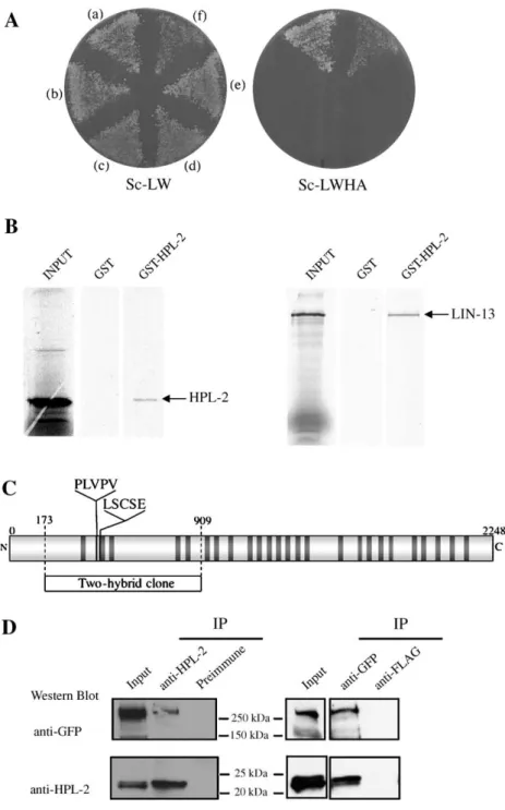
Fig. 1. HPL-2 self-dimerizes and interacts with LIN-13 in yeast two-hybrid and GST pulldown assays. (A) Two-hybrid assay. Left, selective media lacking leucine and tryptophan. Right, selective media lacking leucine, trpyptophane, adenine and histidine supplemented with 1 mM 3-aminotriazole (3-AT). (a) DB-HPL-2 × AD-LIN-13; (b) DB × AD-LIN-AD-LIN-13; (c) DB-HPL-2 × AD; (d) DB × AD; (e) DB × AD-HPL-2; (f) DB-HPL-2 × AD-HPL-2. (B) In vitro translated and labeled35S-HPL-2 (left panel) and35S-LIN-13 (right panel) were incubated with immobilized control GST or GST-HPL-2 (seeFig. 2). Bound interactors were eluted, resolved on SDS-PAGE and visualized by autoradiography. (C) Schematic representation of the LIN-13 protein. Solid bars represent zinc fingers. Position of both LSCSE Rb binding and PLVPV HP1 binding consensus motifs are indicated. The fragment corresponding to the cDNA clone recovered in the two-hybrid screen is indicated below the full-length sequence. (D) HPL-2 coimmunoprecipitates with LIN-13. Worm extracts derived from LIN-13∷GFP transgenic animals were immunoprecipitated with anti-HPL-2 (left) or anti-GFP antibody (right) as indicated (IP), and the lysates were analyzed by Western blot. Input represents 1/50 of starting material. The third lane in each panel shows control IPs using either HPL-2 pre-immune sera, or anti-FLAG antibodies.compared to full-length HPL-2. These results suggest that
while the CSD residues missing in the ok916 and ok917
deletion alleles are essential for dimerization, they may not be
absolutely required for the interaction of HPL-2 with the
PLVPV motif of LIN-13. Sequencing of the tm1489 allele
confirmed that it is a deletion which starts 58 nucleotides
upstream of the ATG and takes out the entire first and second
exons, including sequences coding for the CD (
Fig. 3
A). This
deletion is a clear molecular null allele, consistent with the
observed phenotype, as described below.
hpl-2 and lin-13 genetically interact with the synMuv pathway
of vulval development
All three hpl-2 mutant alleles show phenotypes similar to
those we previously reported for hpl-2 (RNAi) animals,
including sterility, slow growth and an everted vulva (evl,
Table 1
A;
Couteau et al., 2002
). However, the penetrance of
these phenotypes appeared to be higher for the ok917 and
tm1489 alleles, which at 25°C were also thin and scrawny and
showed additional defects in vulva development. These data are
consistent with the molecular analysis suggesting that ok916
and perhaps ok917 are not null alleles. The difference in the
severity of phenotypes between ok916 and ok917 is likely to be
due to differences in the residues added to the carboxyl terminus
of the respective proteins.
In C. elegans, the vulva is derived from three of the six
equivalent vulva precursor cells (VPCs), P(5
–7).p (
Sulston and
Horvitz, 1977
). The three other VPCs, P3.p, P4.p and P8.p
normally adopt a cell fate giving rise to non-vulval cells. A
conserved Ras-mediated signaling cascade is responsible for
inducing the P(5–7).p cells to adopt a vulval fate (
Aroian et al.,
1990; Beitel et al., 1990
) by overcoming inhibitory signals from
two functionally redundant sets of genes, known as synMuvA
Fig. 2. Mapping of HPL-2 and LIN-13 interaction domains. (A) Schematic representation of full-length and truncated HPL-2 proteins tested by GST pulldown. CD, chromo domain; CSD, chromo shadow domain; H, hinge region. (B) GST-pulldown assays were performed with in vitro translated35S-LIN-13 incubated in a batchassay with immobilized GST or truncated proteins containing only the chromo domain (CD), hinge region (H), chromo shadow domain (CSD), or both chromo shadow and hinge regions (CSD + H). (C) The PLVPV motif present in LIN-13 is required for interaction with HPL-2. The full-length two-hybrid clone AD-LIN-13 and mutated AD-LIN-13PLDPEwere tested for interaction with DB-HPL-2 in a two-hybrid assay. Left, selective media lacking leu and trp. Right, selective media lacking
Fig. 4. HPL-2Δ does not homodimerize but is able to interact with LIN-13. Left, selective media lacking leu and trp. Right, selective media lacking leu, trp, his with 1 mM 3-AT. (A) DB-HPL-2Δ × AD-LIN-13, (B) DB-HPL-2Δ × AD, (C) DB × AD-HPL-2Δ, (D) DB-HPL-2Δ × AD-HPL-2Δ, (E) DB-HPL-2Δ × AD-HPL-2, (F) DB-HPL-2 × AD-HPL-2.
Fig. 3. Deletion mapping of the ok916, ok917 and tm1489 mutants. (A) Genomic structure of the C. elegans hpl-2a and hpl-2b gene products showing the region deleted in the three mutants (stippled lines). Deletion breakpoints are given with reference to the ATG start codon and bases of positional ambiguity are in the parentheses. (B) Schematic representation of the HPL-2A and HPL-2B proteins, showing the chromo domain (CD), chromo shadow domain (CSD), and hinge region (H). Dotted lines designate the highly conserved CSD region of HPL-2A aligned against other HP1 proteins in panel C. (C) Alignment of the amino acid sequence of the C-terminal end of the CSD from C. elegans HPL-2A (CeHPL-2A) and HPL-2B (CeHPL-2B), D. melanogaster HP1 isoforms (DmHP1a,-b,-c) and H. sapiens HP1 isoforms (HsHP1α,-β,-γ). Black and gray shaded residues denote identical and conserved amino acids respectively, and amino acids essential for interaction are indicated by triangles.
and B (
Ferguson and Horvitz, 1989
). When any two redundant
signaling systems are disabled, P3.p, P4.p and P8.p cells adopt
vulval fates and produce a Muv phenotype. Given that RNAi
experiments suggested a role for hpl-2 in the synMuvB pathway
(
Couteau et al., 2002
), we tested the hpl-2 mutant alleles for a
Muv phenotype in either synMuvA or B mutant backgrounds.
We found that for all three hpl-2 deletion alleles, RNAi
inactivation of class A, but not class B synMuv genes results in
a highly penetrant Muv phenotype at 20°C (
Table 1
B and data
not shown). This phenotype is dependent on a functional RTK/
Ras/Map kinase pathway (
Couteau et al., 2002
and data not
shown). At 25°C, however, ok917 and tm1489 mutations alone
result in a Muv phenotype (
Table 1
A). Furthermore, at 25°C
inactivation of synMuvA or B genes in an hpl-2 mutant context
results in severe developmental defects, including larval
lethality. Altogether, these results suggest that although at
20°C hpl-2 behaves as synMuvB genes, at 25°C it has
additional functions redundant with both synMuvA and B
genes.
Like hpl-2, lin-13 strictly behaves as a synMuvB gene at
15°C and 20°C but shows a Muv phenotype at 25°C (
Table 1
C;
Ferguson and Horvitz, 1989; Melendez and Greenwald, 2000;
Thomas et al., 2003
). Surprisingly, we found that at 20°C, hpl-2
lin-13 double mutant animals without maternal contribution
also showed a Muv phenotype (38%, n = 211, see
Supplemen-tary material for strain construction). In addition, in the presence
of maternal contribution of both gene products, these animals
also showed a synergistic increase in sterility. Since both the
lin-13 and hpl-2 alleles used in these experiments are nulls, these
results suggest that although both genes participate in vulval
development by acting in the same complex with at least some
of the synMuvB genes, they may have additional, redundant
functions in both vulval and germline development.
The fact that at 25°C hpl-2 mutants show an interaction with
both synMuvA and B mutants raised the possibility that hpl-2
may belong to the recently described synMuvC group of genes
(
Ceol and Horvitz, 2004
). However, while synMuvC mutants
alone or in combination with synMuvB mutants mostly affect
the specification of P8.p, the Muv phenotype associated with
hpl-2 single mutants at 25°C concerns P3.p, P4.p and P8.p cells,
which were induced respectively in 44%, 78% and 56% of
animals showing a Muv phenotype (n = 23). Furthermore, we
found that inactivation of the synMuvC gene trr-1/TRRAP in an
hpl-2 mutant background resulted in synthetic lethality. While
trr-1(RNAi) feeding on wild-type worms did not cause any
embryonic lethality at either 20°C or 25°C (n = 66 and n = 191,
Table 1Phenotypic analysis of hpl-2 deletion mutants and interaction with lin-13 A
Genotype of progeny Temperature (°C) % Muv(a) % Sterility % evl n
Wild type 25 0 0 0 >2000 hpl-2(RNAi) 25 <1(b) 24–53(b) 1–5(b) 1852(b) hpl-2(ok916) 20 0 <1 0 995 hpl-2(ok916) 25 <1 98 1–10 376 hpl-2(ok917) 20 0 1–16 0 1770 hpl-2(ok917) 25 23 >99 45 669 hpl-2(tm1489) 20 0 14 0 341 hpl-2(tm1489) 25 34 100 72 1614 B
synMuv allele combined with hpl-2(tm1489) Class % Muv at 20°C % Muv at 25°C
+ − 0 (n = 341) 34 (n = 1614) lin-15A(RNAi)(a) A 100 (n = 72) NA lin-9(RNAi)(b) B 0 (n = 209) 77,2(c)(n = 217) lin-35(RNAi)(b) B 0 (n = 174) 73,4(c)(n = 163) lin-53(RNAi)(b) B 0(d)(n = 124) 0(d)(n = 98) C
Genotype of progeny Genotype of mother Temperature (°C) % Muv(a) % Sterility n
lin-13(n388) lin-13(n388)/+ 20 0 3–10 895 lin-13(n388) lin-13(n388)/+ 25 90 100 119 hpl-2(tm1489) hpl-2(tm1489)/+ 25 0 10 212 lin-13(n388) hpl-2(tm1489) lin-13(n388) hpl-2(tm1489)/+ 20 0 95 128 lin-13(n388) hpl-2(tm1489) lin-13(n388) hpl-2(tm1489)/+ 25 96 100 127 lin-13(n388) lin-13(n388) 20 0 100 >500 lin-13(n388) hpl-2(tm1489) lin-13(n388) hpl-2(tm1489) 20 38 100 211(b)
(A) hpl-2 mutant phenotypes. Animals were derived from homozygous mothers grown at 20°C, and when indicated shifted at 25°C as early L4. Ranges indicate variations between experiments.(a)Animals were scored as Muv if they showed at least one ectopic pseudovulva under a dissecting microscope.(b)Taken fromCouteau et al. (2002). (B) Genetic interaction between hpl-2(tm1489) mutants and synMuv genes. synMuvA or B genes were inactivated in hpl-2(tm1489) animals by RNAi by either injection (a) or feeding (b). Animals were scored as Muv as in (A). When specified, Muv phenotypes were scored among escapers of the larval arrest phenotype (c), or among escapers of the embryonic lethality (d) in early broods.
(C) Genetic interaction between hpl-2 and lin-13. Heterozygous parents were grown at 20°C or 25°C as indicated.(a)Animals were scored as Muv as in (A).(b)hpl-2
respectively), trr-1(RNAi) feeding on hpl-2(tm1489) worms
resulted in 30% (n = 212) embryonic lethality at 20°C and 60%
(n = 202) embryonic lethality at 25°C. In both cases, 100% of
the remaining progeny arrested development as early larvae.
trr-1(RNAi) was also found to result in embryonic lethality in
combination with several other synMuvB genes tested (
Ceol
and Horvitz, 2004
). Although we considered the possibility that
the synergistic effect, we observe might be due to the enhanced
response to dsRNA recently reported for several synMuvB
mutants (
Wang et al., 2005
), we think this unlikely as the
synMuvB mutations found to synergize with trr-1(RNAi) by
Ceol and Horvitz (2004)
include ones shown to have an
enhanced RNAi response (lin-35, dpl-1), as well as ones that do
not (lin-36, lin-37). Altogether, these results suggest that hpl-2
and the synMuvC genes act in distinct pathways in vulval
development, while for viability trr-1 may function redundantly
with hpl-2.
HPL-2 localization in nuclear foci depends on LIN-13
We previously showed that an HPL-2
∷
GFP fusion protein is
ubiquitously expressed in most, if not all nuclei throughout
embryonic and larval development and into adulthood, with a
signal concentrated in a limited number of nuclear foci in early
embryos (
Couteau et al., 2002
). Interestingly, a LIN-13
∷
GFP
fusion protein shows very similar nuclear foci in embryos
(
Melendez and Greenwald, 2000
), suggesting that both HPL-2
and LIN-13 may be enriched in particular nuclear subdomains.
To test whether the two proteins colocalize in at least some of
these nuclear foci, we constructed a transgenic strain
coexpres-sing both HPL-2
∷
RFP and LIN-13
∷
GFP fusion proteins. In
early stage live embryos, we observed an overlap of most of the
larger LIN-13 and HPL-2 nuclear foci (
Figs. 5
A–C). These
results are consistent with HPL-2 and LIN-13 interacting at a
subset of specific chromosomal sites. We next asked whether
there is interdependence in the localization of HPL-2 and
LIN-13. To this end, we performed lin-13(RNAi) on HPL-2
∷
GFP
strain and introduced the LIN-13
∷
GFP construct in the hpl-2
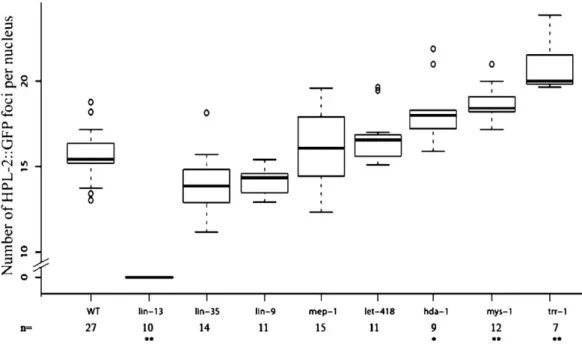
(tm1489) mutant strain. In wild-type embryos, hpl-2
∷
GFP is
concentrated in a median number of 15.4 foci per nucleus (
Fig.
6
). Strikingly, these HPL-2 foci were completely lost in lin-13
(RNAi) animals (
Figs. 5D–G, 6
). By contrast, LIN-13 nuclear
foci were unaltered in the absence of hpl-2 (
Figs. 5
L–M).
We next tested whether RNAi of other synMuvB genes,
including lin-9, lin-35, hda-1, let-418 and mep-1 might also
effect HPL-2 localization (
Figs. 5I–K and Fig. 6
). While lin-9,
lin-35, let-418 and mep-1 had no significant effect on the number
of HPL-2 nuclear foci (one-way ANOVA test, followed by
Tukey Honest Significant Differences; P < 0.001), in hda-1
(RNAi) embryos the median number of foci per nucleus was
found to increase to 18.0 (
Fig. 6
). We also tested the synMuvA
gene lin-15A but were unable to obtain a reliable count of the
number of foci per nucleus due to defects in nuclear morphology
in a significant fraction of the embryos observed. Interestingly,
inactivation of the synMuvC genes trr-1 and mys-1 had a
pronounced effect on the median number of HPL-2 foci per
nucleus, which was found to increase to 20.0 and 18.4
respectively (
Fig. 5H
and
Fig. 6
). Altogether, these results
suggest that while LIN-13 alone may be sufficient for recruiting
HPL-2 in nuclear foci, other synMuv genes, including the class
B gene hda-1, and the class C genes trr-1 and mys-1, may also
either directly or indirectly influence this recruitment to a subset
of chromosomal sites.
HP1 specifically recognizes the H3 N-terminal tails when
di-and tri-methylated on lysine 9 (MeK9H3) (
Bannister et al.,
2001; Jacobs et al., 2001; Lachner et al., 2001; Nakayama et al.,
2001; Nielsen et al., 2002
). To ask whether H3-K9 methylation
might also play a role in the recruitment of HPL-2, we stained
fixed samples with tri-methyl K9 antibodies in combination
with anti-GFP antibodies to detect the HPL-2
∷
GFP fusion
protein. As shown in
Fig. 7
, although a partial overlap between
the two signals can be detected in some cases, the majority of
the large HPL-2 foci do not colocalize with tri-methyl K9
staining. Similar results were obtained with di-methyl K9
antibodies (data not shown). These results suggest that the large
HPL-2 foci that we observe do not correspond to chromosomal
regions enriched in H3-K9 methylation, although at other
chromosomal sites HPL-2 and H3-K9 methylation may overlap.
lin-13 and hpl-2 regulate the expression of specific genes
To gain insight into the molecular mechanism responsible for
the hpl-2 and lin-13 phenotypes, we sought to identify potential
targets of these two genes using various cell type-specific GFP
reporter transgenes. Several synMuv genes have been shown to
regulate the lin-39Hox gene in VPCs (
Chen and Han, 2001
). To
test whether lin-13 and hpl-2 have a similar function, we used
an integrated lin-39
∷
GFP reporter transgene normally weakly
expressed in a subset of VPCs and more strongly in nerve cord
neurons (
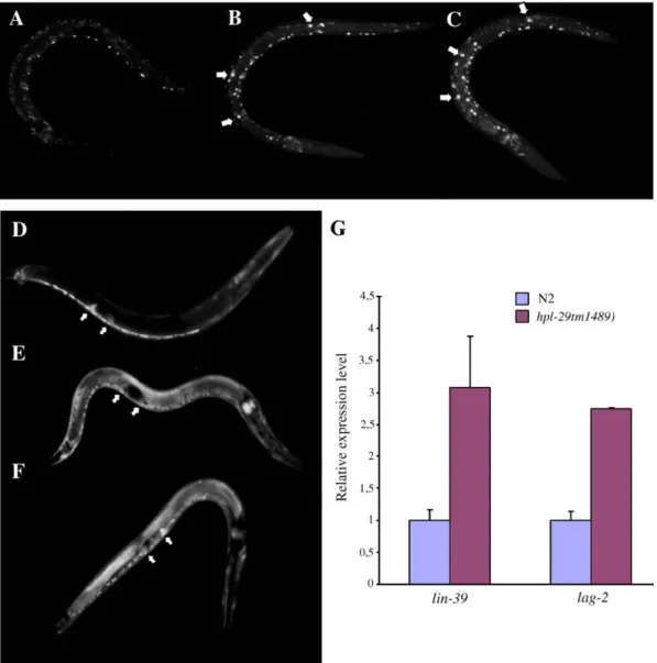
Fig. 8
A). In hpl-2 and lin-13 mutants, we observed an
increased expression of the lin-39 reporter in neuronal cells as
well as additional unidentified cells in the mid-body region
(
Figs. 8
B, C). Although we were unable to specifically quantify
GFP expression in VPCs, the relevant targets for vulval cell fate
specification, these results suggest that hpl-2 and lin-13 may
play a more general role in the regulation of lin-39 expression.
Fig. 5. HPL-2 localization in nuclear foci depends on LIN-13. (A–C) Localization of HPL-2 and LIN-13 in a live 64-cell stage embryo. (A) LIN-13∷GFP (B) HPL-2∷RFP (C) Merge. Images correspond to one optical section, not submitted to deconvolution. Scale bar represents 10μm. Inserts in panel C show an enlargement of the boxed nucleus. Scale bar represents 2.5μm. (D–G) HPL-2∷GFP foci disappear in lin-13(RNAi)-treated animals. (D) HPL-2∷GFP expression in a live wild-type embryo (74-cell stage). (E) HPL-2∷GFP expression in a live lin-13(RNAi) embryo (72-cell stage). Scale bar represents 10μm. (F–G) Images show an enlargement of boxed nuclei visible in panels D and E, respectively. (H–K) Enlargements of live embryos expressing hpl-2∷GFP in different RNAi backgrounds, trr-1 (H), lin-35 (I), lin-15B (J), lin-9 (K). (L–M) LIN-13∷GFP foci are not affected in hpl-2(tm1489) animals. LIN-13∷GFP expression is shown in a live wild-type embryo (L) or in a live hpl-2(tm1489) mutant embryo (M). Images shown in panels D–K correspond to maximum intensity projection of deconvolved sections and are display with the same dynamic for comparison purposes, except for lin-13 images (E, G), whose dynamics were increased by a factor of 4. Images shown in panels L–M correspond to maximum intensity projection of deconvolved sections and are display with the same dynamic for comparison purposes Scale bar represents 2.5μm (F–K).
We also found that a lag-2
∷
GFP reporter was widely
derepressed in both mutant backgrounds, as previously shown
for HDA-1 and more recently other synMuv genes (
Dufourcq et
al., 2002; Poulin et al., 2005
). As shown in
Fig. 8
D, in wild-type
animals expression of lag-2
∷
GFP is restricted to the distal tip
cell of the gonad, the ventral nerve cord and a few cells of the
vulva. In hpl-2 and lin-13 animals, lag-2
∷
GFP expression was
widely derepressed, notably in cells of the pharynx, hypodermis
and intestine (
Figs. 8
E, F). The effect on lag-2 and lin-39
expression is unlikely to be due to a general effect of the hpl-2
and lin-13 mutations on transgene expression as we did not
notice any effect on the expression of several other transgenes
in the soma, either in an extrachromosomal or integrated
context (data not show). Furthermore, quantitative real-time
PCR analysis using RNA derived from a mixed stage
population of worms revealed a threefold increase in lin-39
expression and a two and a half-fold increase in lag-2
expression in hpl-2 animals compared to wild-type (
Fig. 8
G).
Altogether, these results are consistent with hpl-2 and lin-13
playing a role in either directly or indirectly repressing the
expression of specific genes.
Discussion
HPL-2 interaction with LIN-13 relies on conserved domains
HP1 family proteins are essential components of constitutive
heterochromatin in both yeast and Drosophila. More recent
evidence from mammals suggests a broader role for HP1 family
proteins in the epigenetic regulation of gene expression. Several
examples of gene-specific recruitment of HP1 via its association
with transcriptional regulators that bind specific DNA
sequences have been reported (
Ayyanathan et al., 2003; Nielsen
et al., 2001; Ogawa et al., 2002
). In vitro, the association of
TIF1 with HP1 has been shown to be required for progression
through differentiation (
Cammas et al., 2004
), while Rb has
been found to be associated with the recruitment of HP1 to the
cyclin E promoter (
Nielsen et al., 2001
). An interaction with the
Fig. 6. Quantitation of HPL-2∷GFP foci in wild-type and mutant conditions. A box plot representation of the number of HPL-2∷GFP foci in different synMuv RNAi backgrounds is shown. The x axis indicates genotypes; the y axis indicates the number of HPL-2∷GFP foci observed per nucleus. The center horizontal line of each box indicates the median value; the box to and bottom indicate the first and third quartile values; the line above and below the boxes extend to the entire range of measurements. n Represents the number of embryos analyzed for each genotype. *P < 0.001; **P < 0.0001, one-way analysis of variance and Turkey multiple comparison of mean differences compared to WT.Fig. 7. HPL-2∷GFP foci are not enriched in H3K9Me3. (A) HPL-2∷GFP (B) H3K9Me3 (C) merge. Insert shows an enlargement of the boxed in nucleus. Scale bar represents 10μm.
E2F-6 complex has also been shown to mediate recruitment of
HP1 to the promoter of E2F and myc-responsive genes (
Ogawa
et al., 2002
). Nonetheless, the role played by HP1 proteins in
specific developmental context remains poorly understood. In
C. elegans, the synMuvB pathway of vulval development
includes homologues of the recently described RB containing
complexes Myb-MuvB or dREAM (
Korenjak et al., 2004;
Lewis et al., 2004
). Here, we have shown that HPL-2 physically
interacts with the LIN-13 zinc finger protein, another
component of the Rb-related synMuvB pathway, both in vitro
and in vivo, and that this interaction is required for the
recruitment of HPL-2 in nuclear foci. Our results suggest
that HPL-2 shares interaction properties with its
homo-logues in other species. Most notably, interaction with
LIN-13 involves both CSD and hinge regions, and is
mediated by a PLVPV motif in the N-terminus of LIN-13.
Unique and redundant functions of hpl-2 and lin-13 in
development
We find that hpl-2 and lin-13 share several properties that
distinguish them from classical synMuvB genes. By definition,
synMuvB mutants only result in a Muv phenotype in
combination with mutations in the synMuvA pathway, and do
not genetically interact (
Ferguson and Horvitz, 1989
). By
contrast, at 25°C mutations in hpl-2 and lin-13 alone give rise to
a significant percentage of Muv animals. In addition, at 20°C
hpl-2 and lin-13 double mutants show a Muv phenotype and a
synergistic increase in sterility. Altogether, these results
suggests that although HPL-2 and LIN-13 may participate in
vulval development by acting in the same complex with at least
some of the synMuvB proteins, they are likely to have
additional, redundant functions in both vulval and germline
Fig. 8. HPL-2 and LIN-13 influence lin-39∷GFP and lag-2∷GFP gene expression. (A–C) Expression of lin-39∷GFP in late L2 larvae of strain qIs56 at 25°C is shown in green. Anterior end is up, posterior down. White arrows point to examples of cells in which ectopic GFP expression is observed. (a) lin-39∷GFP expression in a wild-type animal; (b) lin-39∷GFP expression in a hpl-2(tm1489) animal; (c) lin-39∷GFP in a lin-13(RNAi) animal. (D–F) Expression of lag-2∷GFP in L2 larvae at 25°C is shown in green. White arrows indicate distal tip cells (DTC). (d) lag-2∷GFP expression in a wild-type animal; (e) lag-2∷GFP expression in a hpl-2 (tm1489) mutant background; (f) lag-2∷GFP expression in a lin-13(RNAi) animal. (G) Endogenous lag-2 and lin-39 RNA levels were quantified in wild-type and hpl-2(tm1489) worms by real-time quantitative RT-PCR. lag-2 and lin-39 expression levels were arbitrarily set to 1 in the wild-type background. Error bars, SEM.development. The exquisite temperature dependence of the
phenotypes observed for both lin-13 and hpl-2 has already been
noted for other synMuv genes (
Ferguson and Horvitz, 1989;
Thomas et al., 2003
). Elevated temperature conditions have
been shown to be associated with defects in heterochromatin
assembly in different systems (
Allshire et al., 1995; Ayoub et
al., 1999; Spofford, 1976
), consistent with the idea that at least a
subset of synMuv genes including hpl-2 and lin-13 act in
chromatin-related pathways. The Muv phenotype observed at
25°C could be attributed to the participation of hpl-2 and lin-13
to both synMuvA and B pathways, or to yet another pathway at
this temperature. Ceol and Horvitz recently described a third
class of synMuv genes, called synMuvC, that encode
homo-logues of the Tip60/NuA4 histone acetyltransferase complex
and act redundantly with both class A and B genes (
Ceol and
Horvitz, 2001
). Our data suggest that in vulval cell fate
specification, hpl-2 acts independently of synMuvC genes,
consistent with multiple chromatin modifying proteins being
implicated in this specific developmental pathway.
The observation that depletion of both maternal and zygotic
activity at 25°C results in larval arrest for lin-13, and adult
sterility for hpl-2, is consistent with LIN-13 and HPL-2 having
additional, independent functions in larval development.
Consistently, HP1 family proteins are known to interact
specifically with a number of different nuclear proteins and in
addition to LIN-13 we have isolated a number of other potential
HPL-2 partners (M. Karali and F. Palladino, unpublished). In
the context of the dynamic structure of chromatin, only a subset
of HP1 interactions are likely to take place in a given cell type at
a specific stage of development.
lin-13 and hpl-2 may regulate the expression of specific genes
Our data suggest that lin-13 and hpl-2 may play a general
role in regulating the expression of at least two genes, the
lin-39Hox and the lag-2 Notch ligand genes. Both lag-2
∷
GFP and
lin-39
∷
GFP reporters were widely derepressed in both hpl-2
and lin-13 mutant backgrounds. Interestingly, the C. elegans
Polycomb group proteins MES-2, MES-3 and MES-6, involved
in the chromatin based control of gene expression in the
germline, have also been shown to repress Hox expression in
the soma (
Ross and Zarkower, 2003
), while lag-2 expression
has previously been shown to be negatively regulated by the
histone deacetylase hda-1 (
Dufourcq et al., 2002
) and more
recently other synMuvB and C genes (
Poulin et al., 2005
).
During vulval development, lag-2 acts in a LIN-12/Notch
signaling pathway which prevents certain VPCs from adopting
the primary vulval cell fate (
Chen and Greenwald, 2004;
Sternberg, 1988
). The global ectopic expression of the
lag-2
∷
GFP reporter we observe in hpl-2 and lin-13 mutant animals
suggests that lag-2 expression may be under control of lin-13
and hpl-2. lin-12 gain of function alleles (
Greenwald et al.,
1983
) and ectopically expressed mutant forms of LAG-2
(
Henderson et al., 1997
) result in multivulval phenotypes. In
principle, derepression of LAG-2 could therefore result in the
same phenotype and contribute to the Muv phenotype observed
in single hda-1, lin-13 and hpl-2 mutant strains (
Dufourcq et al.,
2002
, our data). We note however that, in contrast to lin-12 gain
of function alleles which cause a Muv phenotype because all six
Pn.p cells adopt 2° vulval cell fates (
Greenwald and Seydoux,
1990
), in hpl-2 and lin-13 single mutants ectopically induced
VPCs adopt 1° and 2° cell fate, as shown for other synMuvs
(data not shown and
Sternberg, 1988
). Regardless of the precise
mechanism, our results are consistent with HPL-2 and LIN-13,
together with other synMuv proteins, regulating gene
expres-sion either directly or indirectly in multiple developmental
pathways.
A function for LIN-13 in recruiting HPL-2 to chromatin
Consistent with an interaction between HPL-2 and
LIN-13, we have shown that the foci observed in embryos with
HPL-2
∷
RFP and LIN-13
∷
GFP fusion proteins overlap.
Furthermore, HPL-2 localization in these foci is strictly
dependent on LIN-13, while LIN-13 distribution does not
seem to be affected in the absence of HPL-2. Altogether,
these results strongly suggest that LIN-13 directly recruits
HPL-2 to a limited number of target loci. Nonetheless, our
localization studies show that other synMuv genes may also
influence HPL-2 localization. Most notably, we found that
inactivation of synMuvC genes and to a lesser extent hda-1/
HDAC, resulted in a significant increase in the number of
HPL-2 nuclear foci. The synMuvC genes encode
homo-logues of the TIP60/NuA4 histone acetylase complex, which
could act either in transcriptional repression or activation
(
Ceol and Horvitz, 2004
), while class I histone deacetylase
such as HDA-1 are associated with repression. One
interpre-tation of the effect of inactivation of trr-1, mys-1, and to a lesser
extent hda-1, on HPL-2 distribution is that the dynamic
acetylation/deacetylation equilibrium of specific chromosomal
regions is perturbed, leading to recruitment of HPL-2 to ectopic
sites. One possible consequence of this ectopic recruitment may
be to reduce the amount of HPL-2 available for repression at
target genes. We note that while LIN-13 distribution was not
affected by inactivation of most of the synMuvB genes tested in
this study (data not shown), inactivation of trr-1 had an effect on
LIN-13 distribution similar to that observed for HPL-2,
reinforcing the idea that chromatin architecturation may be
perturbed in a synMuvC mutant context.
Although HP1 proteins have been shown to bind chromatin
by specifically recognizing histone H3 N-terminal tails
methyl-ated on lysine 9 (MeK9H3), several recent reports suggest that
HP1 can also be found on chromatin independently of MeK9H3
(
Cowell et al., 2002; Greil et al., 2003; Li et al., 2003
), indicating
the existence of more than one mechanism of HP1 recruitment.
Our data suggest that LIN-13 may provide one such mechanism.
Nonetheless, MeK9H3 may target HPL-2 to other chromosomal
regions, as we were able to observe some overlap in HPL-2 and
MeK9H3 localization, and HPL-2 does show an in vitro binding
preference for peptides methylated on lysine 9 (data not shown).
Furthermore, LIN-13 cannot be the sole mechanism for targeting
HPL-2 because while in larva and adults hpl-2 expression
persists in most if not all cell types, including VPCs and
hypodermal cells (
Couteau et al., 2002
), LIN-13 expression is
very low and limited to a few cell types including hypodermal
cells but not VPCs (
Melendez and Greenwald, 2000
).
Interest-ingly, recent data suggest that for LIN-35Rb, the relevant
transcription targets for vulval cell fate specification are likely
to be in the hypodermal syncytium (
Myers and Greenwald, 2005
).
If HPL-2 and LIN-13 interact as a single complex in vulval cell
fate specification, they are also likely to act in hypodermal cells,
where both proteins are expressed. Altogether, our data suggest
that LIN-13, HPL-2 and other synMuv proteins including HDA-1,
LIN-35Rb, and LIN-9 may be found either in the same or in
distinct repressor complexes depending on the specific
develop-mental pathway. In Drosophila, the presence of at least two
distinct RB complexes, termed dREAM and myb-MuvB, has
recently been described (
Korenjak et al., 2004; Lewis et al., 2004
).
Interestingly, while both complexes were found to contain
homologues of synMuvB gene products, they differed in the
presence of individual subunits. In a more general way, we
suggest that in C. elegans the LIN-13/HPL-2 complex may act as
a chromatin scaffold that, in turn, coordinates the activities of
large macromolecular complexes that modify chromatin structure
to silence gene expression, including the synMuvB complex. In
this context, LIN-13 may fulfill a function similar to the
mam-malian KAP1-KRAB repressor system, which directs binding and
deposition of HP1 to silence gene expression (
Abrink et al., 2001;
Lechner et al., 2000; Peng et al., 2000; Schultz et al., 2002
).
It has been recently shown that members of the C. elegans
Rb pathway, including the synMuvB genes lin-35Rb, dpl-1DP
and lin-9, negatively regulate RNAi, presumably through the
repression of a subset of RNAi genes in the soma (
Wang et al.,
2005
). Of particular interest, hpl-2 and lin-13, but not other
synMuv genes, were found to have a similar function. However,
despite the fact that both HPL-2 and LIN-13 proteins contain a
putative Rb binding motif, so far we have been unable to detect
a direct interaction between LIN-35Rb and either HPL-2 or
LIN-13. Further analysis is required to gain better
understand-ing of the link between LIN-35Rb, LIN-13, HPL-2 and other
synMuv class genes in vulval and other developmental
pathways. These studies should contribute to an understanding
of how HP1 and other chromatin-associated proteins may be
adapted to specific pathways of development in higher
eukaryotes.
Acknowledgments
We are grateful to M. Labouesse for critical reading of the
manuscript, J.B. Sibarita for providing expertise with
deconvo-lution, G. Yvert for providing expertise with statistical analyses,
and the Microscopy platform of the IFR128. Thanks to Y.
Kohara for cDNA clones, K. Giesler and E. Goillot for yeast
strains, the C. elegans Knockout Consortium and the National
BioResource Project for hpl-2 alleles. Some C. elegans strains
were obtained from the Caenorhabditis Genetic Center, which
is supported by the National Center for Research. This work
was supported by the CNRS and the Association pour la
Recherche sur le Cancer (ARC). V. Coustham was supported by
the Ministere de la Recherche and the ARC. K. Monier was
supported by the Federation pour la Recherche Medicale.
References
Abrink, M., Ortiz, J.A., Mark, C., Sanchez, C., Looman, C., Hellman, L., Chambon, P., Losson, R., 2001. Conserved interaction between distinct Kruppel-associated box domains and the transcriptional intermediary factor 1 beta. Proc. Natl. Acad. Sci. U. S. A. 98, 1422–1426.
Allshire, R.C., Nimmo, E.R., Ekwall, K., Javerzat, J.P., Cranston, G., 1995. Mutations derepressing silent centromeric domains in fission yeast disrupt chromosome segregation. Genes Dev. 9, 218–233.
Aroian, R.V., Koga, M., Mendel, J.E., Ohshima, Y., Sternberg, P.W., 1990. The let-23 gene necessary for Caenorhabditis elegans vulval induction encodes a tyrosine kinase of the EGF receptor subfamily. Nature 348, 693–699. Ayoub, N., Goldshmidt, I., Cohen, A., 1999. Position effect variegation at the
mating-type locus of fission yeast: a cis-acting element inhibits covariegated expression of genes in the silent and expressed domains. Genetics 152, 495–508.
Ayyanathan, K., Lechner, M.S., Bell, P., Maul, G.G., Schultz, D.C., Yamada, Y., Tanaka, K., Torigoe, K., Rauscher III, F.J., 2003. Regulated recruitment of HP1 to a euchromatic gene induces mitotically heritable, epigenetic gene silencing: a mammalian cell culture model of gene variegation. Genes Dev. 17, 1855–1869. Bannister, A.J., Zegerman, P., Partridge, J.F., Miska, E.A., Thomas, J.O., Allshire, R.C., Kouzarides, T., 2001. Selective recognition of methylated lysine 9 on histone H3 by the HP1 chromo domain. Nature 410, 120–124. Beitel, G.J., Clark, S.G., Horvitz, H.R., 1990. Caenorhabditis elegans ras gene let-60 acts as a switch in the pathway of vulval induction. Nature 348, 503–509. Brasher, S.V., Smith, B.O., Fogh, R.H., Nietlispach, D., Thiru, A., Nielsen, P.R., Broadhurst, R.W., Ball, L.J., Murzina, N.V., Laue, E.D., 2000. The structure of mouse HP1 suggests a unique mode of single peptide recognition by the shadow chromo domain dimer. EMBO J. 19, 1587–1597.
Brenner, S., 1974. The genetics of Caenorhabditis elegans. Genetics 77, 71–94. Cammas, F., Herzog, M., Lerouge, T., Chambon, P., Losson, R., 2004. Association of the transcriptional corepressor TIF1beta with heterochroma-tin protein 1 (HP1): an essential role for progression through differentiation. Genes Dev. 18, 2147–2160.
Ceol, C.J., Horvitz, R.H., 2001. dpl-1 DP and efl-1 E2F Act with lin-35 Rb to antagonise Ras signaling in C. elegans vulval development. Mol. Cell 7, 461–473.
Ceol, C.J., Horvitz, H.R., 2004. A new class of C. elegans synMuv genes implicates a Tip60/NuA4-like HAT complex as a negative regulator of Ras signaling. Dev. Cell 6, 563–576.
Chen, N., Greenwald, I., 2004. The lateral signal for LIN-12/Notch in C. elegans vulval development comprises redundant secreted and transmembrane DSL proteins. Dev. Cell 6, 183–192.
Chen, Z., Han, M., 2001. C. elegans Rb, NuRD, and Ras regulate lin-39-mediated cell fusion during vulval fate specification. Curr. Biol. 11, 1874–1879. Couteau, F., Guerry, F., Muller, F., Palladino, F., 2002. A heterochromatin
protein 1 homologue in Caenorhabditis elegans acts in germline and vulval development. EMBO Rep. 3, 235–241.
Cowell, I.G., Aucott, R., Mahadevaiah, S.K., Burgoyne, P.S., Huskisson, N., Bongiorni, S., Prantera, G., Fanti, L., Pimpinelli, S., Wu, R., Gilbert, D.M., Shi, W., Fundele, R., Morrison, H., Jeppesen, P., Singh, P.B., 2002. Heterochromatin, HP1 and methylation at lysine 9 of histone H3 in animals. Chromosoma 111, 22–36.
Dufourcq, P., Victor, M., Gay, F., Calvo, D., Hodgkin, J., Shi, Y., 2002. Functional requirement for histone deacetylase 1 in Caenorhabditis elegans gonadogenesis. Mol. Cell. Biol. 22, 3024–3034.
Durfee, T., Becherer, K., Chen, P.L., Yeh, S.H., Yang, Y., Kilburn, A.E., Lee, W.H., Elledge, S.J., 1993. The retinoblastoma protein associates with the protein phosphatase type 1 catalytic subunit. Genes Dev. 7, 555–569. Eissenberg, J.C., 2001. Molecular biology of the chromo domain: an ancient
chromatin module comes of age. Gene 275, 19–29.
Eissenberg, J.C., Elgin, S.C., 2000. The HP1 protein family: getting a grip on chromatin. Curr. Opin. Genet. Dev. 10, 204–210.
Elledge, S.J., Mulligan, J.T., Ramer, S.W., Spottswood, M., Davis, R.W., 1991. Lambda YES: a multifunctional cDNA expression vector for the isolation of genes by complementation of yeast and Escherichia coli mutations. Proc. Natl. Acad. Sci. U. S. A. 88, 1731–1735.