HAL Id: tel-02488600
https://tel.archives-ouvertes.fr/tel-02488600v2
Submitted on 27 Feb 2020HAL is a multi-disciplinary open access archive for the deposit and dissemination of sci-entific research documents, whether they are pub-lished or not. The documents may come from teaching and research institutions in France or
L’archive ouverte pluridisciplinaire HAL, est destinée au dépôt et à la diffusion de documents scientifiques de niveau recherche, publiés ou non, émanant des établissements d’enseignement et de recherche français ou étrangers, des laboratoires
Cell-type specific CB1 receptor modulation of
hippocampal synaptic plasticity and memory
Jose Fernando Oliveira da Cruz
To cite this version:
Jose Fernando Oliveira da Cruz. Cell-type specific CB1 receptor modulation of hippocampal synaptic plasticity and memory. Neurons and Cognition [q-bio.NC]. Université de Bordeaux; Università degli studi (Catane, Italie), 2017. English. �NNT : 2017BORD0905�. �tel-02488600v2�
U
NIVERSITY OF
B
ORDEAUX
,
F
RANCE
D
OCTORAL
S
CHOOL OF
L
IFE AND
H
EALTH
S
CIENCES
-
S
PECIALTY
N
EUROSCIENCE
AND
U
NIVERSITY OF
C
ATANIA
,
I
TALY
I
NTERNATIONAL
P
H
D
P
ROGRAM IN
N
EUROSCIENCE
–
XXIX
C
YCLE
Cell-type specific CB1 receptor
modulation of hippocampal synaptic
plasticity and memory
José Fernando OLIVEIRA DA CRUZ
2017
Presented and defended publicly on 18-12-2017
Thesis Supervisor:
Thesis Co-Supervisor:
Members of the Jury:
Dr. Giovanni Marsicano (Bordeaux, France)
Prof. Filippo Drago (Catania, Italy)
Prof. Carla CANNIZZARO – University of Palermo – Examiner Prof. Luigia TRABACE – University of Foggia – Examiner
Prof. Sabatino MAIONE – University of Campania – President of the Jury Prof. Cesare MANCUSO – Università Cattolica del Sacro Cuore – Rapporteur
DOCTORAL DISSERTATION SUBMITTED IN PARTIAL FULFILLMENT OF THE
THÈSE EN COTUTELLE PRÉSENTÉE POUR OBTENIR LE GRADE DE
DOCTEUR DE
L’UNIVERSITÉ DE BORDEAUX
École Doctorale Sciences de la Vie et de la Santé - SPÉCIALITÉ NEUROSCIENCES
ET DE
L’UNIVERSITÉ DE CATANE
International PhD Program in Neuroscience – XXIX Cycle
Par
José Fernando OLIVEIRA DA CRUZ
Contrôles distincts de la plasticité synaptique de l'hippocampe et de
la mémoire par différentes populations de récepteurs CB1
Sous la direction de Giovanni MARSICANO et de Filippo DRAGO
Soutenue le 18 Décembre 2017
Membres du jury:
Prof. Carla CANNIZZARO – Université de Palerme – Examinateur Prof. Luigia TRABACE – Université de Foggia – Examinateur
This work was supported by a fellowship from the International PhD Program in Neuroscience – XXIX CYCLE of the University of Catania (Italy) and by a doctoral extension grant provided by the French “Fondation pour la Recherche Médicale” (FDT20160435664).
The work in this thesis has been carried out as a joint-PhD program in the laboratories of:
Dr. Giovanni Marsicano
Prof. Filippo Drago
Neurocentre Magendie – INSERM U1215 University of Bordeaux
146 Rue Leo Saignat 33077 Bordeaux cedex
France
Department of Biomedical and Biotechnological Sciences School of Medicine University of Catania Via S. Sofia 64 95125 Catania Italy
TABLE OF CONTENTS
TABLE OF CONTENTS ... 6
ACKNOWLEDGMENTS ... 8
LIST OF PUBLICATIONS ... 10
Articles published or in process of publication in peer reviewed scientific journals ... 10
LIST OF COMMUNICATIONS ... 11 Poster communications ... 11 Oral communications ... 12 ABSTRACT ... 13 RÉSUMÉ ... 14 LONG RÉSUMÉ ... 16 LIST OF ABREVIATIONS ... 21 LIST OF FIGURES ... 24
Section I – GENERAL INTRODUCTION ... 25
Part 1 – The endocannabinoid system in the brain ... 26
I – Introduction ... 26
II – Cannabinoid receptors in the brain ... 28
III – Distribution of CB1 receptors in the brain ... 30
IV – Metabolism of Endocannabinoids ... 33
V – Methodologies to dissect the function of CB1 receptors ... 38
Part 2 – Synaptic plasticity ... 43
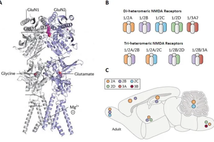
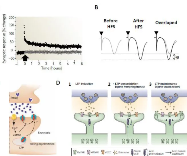
I – NMDA receptors... 43
II – NMDA receptor-dependent LTP ... 46
III – Mechanisms of LTP ... 48
IV – CB1 receptor modulation of synaptic transmission and plasticity ... 51
I – Astrocytes: morphology, distribution, physiology and function ... 62
II – The tripartite synapse and the neuroglial interactions ... 66
III – Network properties of astrocytes ... 67
IV – Gliotransmission ... 69
V – Astrocytes, CB1 receptors and gliotransmission ... 71
Part 4 – Memory ... 74
I – Plasticity and memory: cellular and molecular mechanisms of memory formation... 76
II – Novel object recognition memory ... 78
III – Memory and CB1 Receptors ... 80
IV – Memory and astrocytes ... 83
Part 5 – Dopaminergic System and the ECS ... 86
I – The dopaminergic system ... 87
II – Dopamine type-1 (D1) receptors ... 88
III – The role of D1 receptors in LTP ... 90
IV – D1 and CB1 receptors – A potential crosslink to modulate memory functions ... 93
Section II – RESEARCH OBJECTIVES ... 95
Section III – RESULTS ... 99
Part 1 – Astroglial CB1 receptors determine synaptic D-serine availability to enable recognition memory ... 100
Part 2 – Deletion of CB1 receptors in hippocampal D1-positive cells impairs object recognition memory and associated synaptic plasticity ... 142
Section IV – GENERAL DISCUSSION ... 156
Part 1 – Astroglial CB1 receptors determine synaptic D-serine availability to enable recognition memory ... 157
Part 2 – Deletion of CB1 receptors in hippocampal D1-positive cells impairs object recognition memory and associated synaptic plasticity ... 167
Section V – REFERENCES ... 170
Section VI – ANNEX ... 194
ACKNOWLEDGMENTS
The scientific work is a collaborative rather than solitary experience in which many people take part. During the last 4 years, I was not alone and so I would like to thank those people that contributed in several domains for this expedition that is due to be achieved.
Em primeiro lugar gostava de agraceder à minha familia pela ajuda, apoio e confiança que recebi durante todo este percursso. Se hoje entrego esta tese, em parte foi porque durante a minha vida, a minha familia me ajudou nos momentos mais dificeis. Dedico uma palavra especial à minha avó que sempre me motivou na luta pelos meus objectivos e que sempre disse que gostaria de me ver doutorar. Infelizmente, ela partiu durante este percurso e não terá a oportunidadede estar presente. No entanto, fica aqui perpetuado o meu eterno agradecimento a toda a sua ajuda.
Then, I would like to specially thank Giovanni. I would like to thank him for the opportunity that he gave me to belong to his team and to develop my PhD project under his supervision. The way you manage the team reflects a great deal of sobriety, professionalism, curiosity for science, drive for excellence and good internal atmosphere between members. I will surely miss such a place upon my departure but I have hopefully learnt important scientific and personal lessons that will help me throughout my career.
Also, I would like to thank Edgar Soria-Gomez and Arnau Busquets-Garcia. During these last 4 years, I have learned a lot from you and in key moments of my PhD, when doubts about the path that I chose persisted, your insights and advice where key to keep me on track. Likewise, I would like to thank Michelangelo Colavita and Carolina Muguruza for the good times well spent in scientific discussions and musical rehearsals. Also, to thank Robert Rozeske for the afternoons spend in collaboration with Michele and Carol, preparing this musical project of the “Next Week”. Although this project never sky-rocked to fame, it nevertheless contributed to some well spent musical afternoons.
Another special mention to the office I4 and all the people that passed there. Indeed, the endless discussions provided some good brainstorming that showed the immense curiosity that drives these passion people. Besides Arnau, Edgar and Carolina, I also would like to thank Bastien Redon and more recently to Christina Ioannidou.
Also, I would like to thank the remaining Marsicano team members for all the support and thoughtful discussions that we had (scientific, political, philosophical or just simple casual
Part of this PhD project was developed as part of a scientific collaboration. Thus, I would like also to thank Valentin Langlais, Aude Panatier and Stephane Oliet for the discussions and knowledge shared about this interesting field of the glial cells. Also, I would like to thank Laurie Robin and her work as it was an important part of the project in which my thesis is included.
A special mention must go to François Georges and Federico Massa. To Francois Georges, I would like to thank the patience to teach me electrophysiology that is currently the main technical expertise that I have acquired during my PhD. To Federico Massa because in times of technical difficulties, his help was very important to solve these demoniac bugs that haunt scientists.
I would like to thank Filippo Drago, as my institutional supervisor at the University of Catania, for the opportunity to engage in this interesting PhD degree. I would like also to thank professor Salomone for the help and availability during the whole period of my PhD training.
Because living in a foreign country is not easy and we often experience: “Saudades de casa”, I cannot forget the “usual suspects” (AKA a grande familia portuguesa de Bordéus). Having a good group of friends and colleagues to which I could speak in the language of Camões was like an “oxygen balloon” during this period. All the dinners, discussions, perspectives of life shared or just simply that “beer at the end of the day”, these were important moments of sobriety that were crucial to keep me functional. Obrigado malta!
Se existe formação da qual não me posso esquecer, é a Estudantina Universitária de Coimbra. Todos as lições de camaradagem, preserverança e paciência que aprendi neste grupo nos meus anos na academia de Coimbra permanecem comingo e ainda hoje sinto que devo a esse grupo parte do cientista e pessoa que hoje sou.
Then, even if we just met in the final stages of my PhD I would like to thank Marta Collu. Your support during these final months (plus the Swiss chocolate that you gave me) as well as the moments that we shared were an important part. Obrigado Marta!
I cannot forget to thank another important that a part of this PhD that happened (almost) every Monday and Thursday at 19h at the DOJO Bordeaux. During these days master Raymond Mallia taught us the art of taekwondo, which was key for my mental stability during these last years.
I am sure that in the moment I will print this thesis, I will realize that many other important people deserve to be in this section. For those, my apologies and a sincere “thank you”.
LIST
OF
PUBLICATIONS
Articles published or in process of publication in peer
reviewed scientific journals
PUBLISHED
OLIVEIRA DA CRUZ, J. F., ROBIN, L. M., DRAGO, F., MARSICANO, G. & METNA-LAURENT, M. 2016. Astroglial type-1 cannabinoid receptor (CB1): A new player in the tripartite synapse. Neuroscience, 323, 35-42.
SUBMITTED
(1) Astroglial CB1 receptors determine synaptic D-serine availability to enable recognition memory
Laurie M. Robin*, Jose F. Oliveira da Cruz*, Valentin C. Langlais*, Mario Martin-Fernandez, Mathilde Metna-Laurent, Arnau Busquets-Garcia, Luigi Bellocchio, Edgar Soria-Gomez, Thomas Papouin, Ilaria Belluomo, Isabel Matias, Barbara Bosier, Filippo Drago, Ann Van Eeckhaut, Ilse Smolders, Francois Georges, Alfonso Araque, Aude Panatier, Stéphane H.R. Oliet# and Giovanni
Marsicano#
*: equal contribution, #: equal supervision
IN PREPARATION
(2) Deletion of CB1 receptors in hippocampal D1-positive cells impairs object recognition memory and associated synaptic plasticity
Jose F. Oliveira da Cruz*, Arnau Busquets-Garcia*, Luigi Bellocchio, Zhe Zhao, Filippo Drago,
Marjorie Varilh, Giovanni Marsicano#, Edgar Soria-Gomez#
LIST
OF
COMMUNICATIONS
Poster communications
2017 – NEUROFRANCE 2017 (BORDEAUX, FRANCE)
Deletion of CB1 receptors in D1-positive cells impairs object recognition memory
Jose F. Oliveira da Cruz, Arnau Busquets-Garcia, Luigi Bellocchio, Zhe Zhao, Marjorie Varilh, Giovanni Marsicano, Edgar Soria-Gomez
2016 – INTERNATIONAL NEUROSCIENCE CONFERENCE 2016 (MARSEILLE, FRANCE)
Astroglial CB1 receptors control memory via D-serine
Jose F. Oliveira da Cruz, Laurie M. Robin, Valentin C. Langlais, Mathilde Metna-Laurent, Arnau Busquets-Garcia, Luigi Bellocchio, Edgar Soria-Gomez1,2, Thomas Papouin, Barbara Bosier,
Filippo Drago, Ann Van Eeckhaut, Ilse Smolders, Francois Georges, Aude Panatier, Stephane H.R. Oliet, Giovanni Marsicano
2015 – CANNABINOID CONFERENCE (SESTRI LEVANTE
, ITALY)
Astroglial CB1 receptors are required for in vivo hippocampal long-term potentiation of synaptic transmission
José F. Oliveira da Cruz, Laurie Robin, Valentin C. Langlais, Arnau Busquets-Garcia, Edgar Soria-Gomez, Filippo Drago, Aude Panatier, François Georges, Mathilde Metna-Laurent, Stéphane Oliet, Giovanni Marsicano
2015 – 13
TH SUMMER SCHOOL OF NEUROSCIENCE (CATANIA, ITALY)Astroglial type-1 cannabinoid receptors (CB1) are required for memory formation
José F. Oliveira da Cruz, Laurie Robin, Valentin C. Langlais, Arnau Busquets-Garcia, Edgar Soria-Gomez, Filippo Drago, Aude Panatier, François Georges, Mathilde Metna-Laurent, Stéphane Oliet, Giovanni Marsicano
2015 – NEURIZONS 2015 (GOTTINGEN
, GERMANY)
Astroglial type-1 cannabinoid receptors (CB1) are required for memory formation
José F. Oliveira da Cruz, Laurie Robin, Valentin C. Langlais, Arnau Busquets-Garcia, Edgar Soria-Gomez, Filippo Drago, Aude Panatier, François Georges, Mathilde Metna-Laurent, Stéphane Oliet, Giovanni Marsicano
2015 – ANNUAL MEETING OF THE BORDEAUX DOCTORAL SCHOOL (BORDEAUX, FRANCE)
Cannabinoid receptor 1 in hippocampal astrocytes is crucial for memory formation
José F. Oliveira da Cruz, Laurie Robin, Valentin C. Langlais, Arnau Busquets-Garcia, Edgar Soria-Gomez, Filippo Drago, Aude Panatier, François Georges, Mathilde Metna-Laurent, Stéphane Oliet, Giovanni Marsicano
Oral communications
2015 – NEURIZONS 2015 (GOTTINGEN
, GERMANY)
Astroglial type-1 cannabinoid receptors (CB1) are required for memory formation
José F. Oliveira da Cruz, Laurie Robin, Valentin C. Langlais, Arnau Busquets-Garcia, Edgar Soria-Gomez, Filippo Drago, Aude Panatier, François Georges, Mathilde Metna-Laurent, Stéphane Oliet, Giovanni Marsicano
2013 – INTERNATIONAL ASTROCYTE SCHOOL (BERTINORO, ITALY)
The role of Astroglial Type-1 Cannabinoid Receptor in Memory Functions
Jose F. Oliveira da Cruz, Mathilde Metna-Laurent, Laurie Robin, Valentin C. Langlais, Arnau Busquets-Garcia, Thierry Leste-Lasserre, Thomas Papouin, Stéphane Oliet, François Georges, Giovanni Marsicano
ABSTRACT
The endocannabinoid system is a major brain modulatory system that controls memory and learning mainly via the cannabinoid receptor type 1 (CB1)-dependent regulation of neuronal and glial activity. In the hippocampus, bidirectional communication between neurons and astrocytes shapes synaptic plasticity and behavior. CB1 receptors have been shown to be present in the astrocytes and to mediate the disruptive effects of cannabinoids in synaptic plasticity and working memory. Yet, it is not currently known the role of this receptor in the physiological modulation of memory processes. Also, previous studies have shown that CB1 receptors expressed in dopamine D1 receptor-expressing cells are involved in the modulation of hippocampal-dependent aversive memories. However, their involvement in the modulation of non-aversive long-term memory formation and synaptic plasticity is presently unknown. In this thesis, I aimed at identifying the cellular and molecular mechanisms by which specific CB1 receptors in distinct brain neuronal and glial populations contribute to the physiological modulation of synaptic plasticity and learning and memory. For this aim we used conditional genetic mutant mice lacking CB1 receptors specifically in astrocytes or in D1-positive cells. By coupling these genetic mouse models with behavioral, pharmacological, and in vitro and in vivo electrophysiological approaches, we dissected the role of these CB1 receptors in the formation of memory. First, we show that astroglial CB1 receptors in the hippocampus control long-term potentiation (LTP) of CA3-CA1 synaptic transmission and long-term recognition memory. By allowing physiological availability of D-serine at NMDA receptors via gliotransmission, astrocytes are important elements controlling glia-neuron interactions that underlie synaptic plasticity and memory functions. The data show that astroglial CB1 receptors control plasticity and memory by regulating the synaptic availability of D-serine at NMDA receptors. Second, we show that CB1 receptors D1-positive cells control the consolidation, but not acquisition, of new memories and the enhancement of LTP induced by learning, showing that specific subpopulations CB1 receptor-expressing cells differentially modulate these processes.
Overall, by showing that the endocannabinoid system in astrocytes is an important modulator of learning and memory and by suggesting that CB1 receptors in D1-positive cells are important for specific components of memory formation, we provide functional evidence for the complex cell type-dependent regulation of long-term recognition memory by the CB1 receptors.
RÉSUMÉ
Le système endocannabinoïde est un système neuromodulateur majeur du cerveau. Ainsi, il contrôle la mémoire et l’apprentissage, et ce, principalement par l'intermédiaire des récepteurs aux cannabinoïdes de type 1 (CB1) qui régulent de manière fine les activités neuronales et gliales. Dans l’hippocampe, une communication bidirectionnelle entre neurones et astrocytes modèle la plasticité synaptique et le comportement. Il a été rapporté que les effets disruptifs des cannabinoïdes sur la plasticité synaptique et la mémoire de travail sont dépendants de récepteurs CB1 présents dans les astrocytes. Cependant, le rôle de ce récepteur dans la modulation physiologique des processus mnésiques n’est pas encore connu. De précédentes études ont également montré que les récepteurs CB1 exprimés dans les cellules hébergeant le récepteur dopaminergique D1 sont impliqués dans la modulation hippocampique de la mémoire associée aux évènements aversifs. Toutefois, leur implication dans la modulation de la formation de la mémoire associée à des évènements non aversifs ainsi que dans la plasticité synaptique sous-jacente reste encore inconnue. Dans cette thèse, mon objectif était d’identifier les mécanismes cellulaires et moléculaires par lesquels des populations distinctes de récepteurs CB1 dans des populations gliales et des régions cérébrales bien définies contribuent à la modulation physiologique de la plasticité synaptique, de l’apprentissage et de la mémoire. Pour ce faire, nous avons utilisé des souris mutantes conditionnelles dans lesquelles le récepteur CB1 a été rendu silencieux sélectivement dans les astrocytes ou dans les cellules exprimant le récepteur D1. En couplant ces modèles génétiques murins avec des approches comportementales, pharmacologiques et électrophysiologiques in vitro et in vivo, nous avons disséqué le rôle de ces populations de récepteurs CB1 dans la formation de la mémoire. Tout d’abord, nous avons montré que les récepteurs CB1 astrogliaux dans l’hippocampe contrôlaient la potentialisation à long terme (PLT) de la transmission synaptique CA3-CA1 et la mémoire de reconnaissance à long terme. En contrôlant, via la gliotransmission, la disponibilité effective de D-sérine aux récepteurs NMDA, les astrocytes sont des éléments importants contrôlant les interactions glie-neurones qui sous-tendent la plasticité synaptique et les fonctions mnésiques. Les données obtenues montrent que les récepteurs CB1 astrogliaux contrôlent la plasticité et la mémoire en régulant la disponibilité synaptique de la D-sérine aux récepteurs NMDA. Deuxièmement, nous avons montré que les récepteurs CB1 dans les cellules exprimant le récepteur D1 contrôlaient la consolidation, mais pas l’acquisition, de nouveau souvenirs et l’augmentation de la PLT induite par l’apprentissage. Ces résultats indiquent que des populations spécifiques de cellules exprimant le récepteur CB1 modulent ces processus de manière différentielle.
En conclusion, ces travaux démontrent que le système endocannabinoïde dans les astrocytes est un important modulateur de l’apprentissage et de la mémoire alors que les récepteurs CB1 dans les cellules exprimant le récepteur D1 semblent importants pour des composantes spécifiques de la formation de la mémoire. Prise dans son ensemble, cette thèse apporte des preuves fonctionnelles quant à la régulation complexe de la mémoire de reconnaissance à long-terme par des populations distinctes de récepteurs CB1.
LONG
RÉSUMÉ
Le système endocannabinoïde est un modulateur majeur du système nerveux central et des tissus périphériques contrôlant et régulant d’important processus physiologiques. Les cannabinoïdes exogènes et endogènes agissent principalement via le récepteur cannabinoïde de type 1 (CB1) qui régule des fonctions cérébrales clés telles que la consommation alimentaire, le métabolisme énergétique, les réponses immunitaires, les réponses de stress, les performances motrices, la perception de la douleur et la mémoire. Dans l’hippocampe, une région cérébrale importante pour l’apprentissage et la mémoire, la présence des récepteurs CB1 (principalement présynaptiques) sur les neurones GABAergiques et glutamatergiques modulent l’activité neuronale en diminuant la libération de neurotransmetteurs. En combinant des approches génétiques, de mutagénèses conditionnelles, et pharmacologiques, il a été montré que des récepteurs CB1 de populations neuronales bien distinctes sont responsables des altérations des processus mnésiques induites par les cannabinoïdes. Toutefois, il reste de nombreux points à explorer concernant le rôle physiologique des récepteurs CB1 dans ces fonctions. De plus, les récepteurs CB1 peuvent être présents dans d’autres types cellulaires que les neurones, e.g. les astrocytes, et le rôle physiologique de ces récepteurs dans l’apprentissage et la mémoire reste inconnu.
Il a été montré que les astrocytes de l'hippocampe expriment des récepteurs CB1 fonctionnels. Les astrocytes constituent le type de cellules gliales le plus abondant dans le soutien au réseau neuronal en fournissant les substrats métaboliques nécessaires au fonctionnement optimal du cerveau. Toutefois, il a été démontré au cours des dernières décennies que les astrocytes étaient impliqués dans d’autres fonctions importantes telles que la modulation directe de l’activité et la plasticité synaptique via une communication bidirectionnelle avec les structures synaptiques neuronales (les terminaisons présynaptiques et les épines postsynaptiques). Ce concept, communément désigné sous les termes de "synapse tripartite", définit ces synapses comme étant composées de trois éléments : les terminaisons pré- et post-synaptiques ainsi que les processus fins astrocytiques les entourant. Cette localisation permet aux astrocytes de percevoir les signaux neuronaux et de libérer des molécules modulant leur activité. Les modifications fonctionnelles et/ou structurelles à court- et long-terme de la transmission synaptique, qui constituent la plasticité synaptique, ont été proposées comme étant des mécanismes cellulaires clés sous-jacents à la formation de nouveaux souvenirs. Plus précisément, l’induction de la potentialisation à long terme (PLT) dépendante des récepteurs NMDA a été montrée au niveau des synapses de l’hippocampe
sérine, le co-agoniste principal du récepteur NMDA, considéré comme un gliotransmetteur, est nécessaire à l’induction de la PLT dans cette région cérébrale particulière. De plus, les récepteurs CB1 astrogliaux sont les médiateurs des effets perturbateurs des cannabinoïdes synthétiques et naturels sur la plasticité synaptique et la mémoire de travail. Actuellement, le rôle physiologique des récepteurs CB1 astrogliaux dans la modulation des fonctions mnésiques et synaptiques n’est pas connu.
Comme mentionné précédemment, le ciblage d’un gène spécifique couplé à des méthodes technologiques avancées ont permis l’identification des récepteurs CB1 (i) dans des localisations intracellulaires jusqu’alors inconnues (mitochondrie) et (ii) au sein de nouvelles populations cellulaires cérébrales. Ainsi, les récepteurs CB1 ont été récemment localisés au sein de cellules exprimant le récepteur dopaminergique de type 1 (D1). Néanmoins, le nombre de cellules exprimant D1 dans l'hippocampe étant relativement faible, il est difficile de les identifier anatomiquement. Bien que la nature de ces cellules demeure insaisissable (neuronale ou gliale), des preuves fonctionnelles pointent vers l’existence de ces cellules au sein des structures de l’hippocampe avec un impact probable sur les fonctions médiées par celui-ci. Bien que ces cellules représentent une petite fraction du nombre total de cellules exprimant CB1, elles représentent une sous-population neuronale ou gliale exprimant le récepteur CB1. Ainsi, il est nécessaire d’étudier le rôle de ces nouvelles sous-populations de cellules exprimant le récepteur CB1 et d’identifier le mécanisme moléculaire et les conséquences comportementales qui sont liées à leur activité.
Le principal objectif de ma thèse a été d’identifier les mécanismes moléculaires et cellulaires par lesquels les récepteurs CB1 au sein de populations cellulaires spécifiques, qu'elles soient neuronales (par exemple les cellules dopaminergiques) ou gliales (par exemple les astrocytes) contribuent à la modulation physiologique de l’apprentissage et de la mémoire. Ceci est important non seulement pour la compréhension des fonctions cérébrales mais également pour l'appréhension des mécanismes par lesquels certaines dérégulations peuvent conduire à des états pathologiques.
Afin d’atteindre l’objectif principal de cette thèse, nous avons utilisé une combinaison d’outils génétiques (mutagénèse constitutive et conditionnelle du récepteur CB1 chez la souris) et pharmacologiques (agonistes et antagonistes du récepteur CB1) couplés à des paradigmes comportementaux précédemment conçus et validés pour étudier la formation de la mémoire chez la souris. De plus, en combinant ces précédentes approches à de l’électrophysiologie in
vitro et in vivo, l'objectif était de disséquer les mécanismes impliqués dans la modulation de la
Spécifiquement, le premier but était d’étudier les mécanismes cellulaires impliqués dans la modulation physiologique de la mémoire à long-terme par les récepteurs CB1 astrogliaux. Au cours de ces travaux, nous nous sommes intéressés à plusieurs questions concernant (i) le rôle de ces récepteurs dans la modulation de la formation des mémoires à court- et long-terme, (ii) le rôle des récepteurs CB1 astrogliaux dans la modulation de la PLT, (iii) les mécanismes par lesquels les récepteurs CB1 contrôlent les fonctions astrogliales afin de permettre la gliotransmission et iv) la caractérisation du mécanisme sous-tendant le contrôle des fonctions mnésiques par le récepteur CB1.
La délétion spécifique des récepteurs CB1 astrogliaux chez la souris (GFAP-CB1-KO) altère la tâche de mémoire de reconnaissance d’un nouvel objet, montrant que ces récepteurs sont nécessaires pour la formation de la mémoire d’objet nouveau. De plus, nous avons montré que la transmission du récepteur NMDA dans l’hippocampe était nécessaire à la formation de ce type de mémoire. Ensuite, par l’enregistrement in vivo du potentiel de champ postsynaptique excitateur (fEPSP) dans les voies CA3-CA1 de l’hippocampe chez des souris sauvages ou mutantes anesthésiées, nous avons montré que les souris GFAP-CB1-KO avait une altération in
vitro et in vivo de la PLT dépendante du récepteur NMDA dans l’hippocampe. Pour étudier le
mécanisme cellulaire impliqué dans le phénotype présenté par les souris GFAP-CB1-KO, nous avons exploré la relation entre les astrocytes et leurs homologues neuronaux au sein des synapses tripartites de l’hippocampe. En montrant qu’un agoniste du récepteur CB1 pouvait induire une augmentation des niveaux intracellulaires de calcium au sein des astrocytes des souris sauvages mais pas des souris GFAP-CB1-KO, nous avons révélé que les récepteurs CB1 astrogliaux pouvaient contrôler les niveaux de calcium intracellulaire. Considérant que les niveaux de calcium intracellulaires sont les mécanismes astrocytiques potentiels impliqués dans la sécrétion de gliotransmetteurs, nous avons étudié si plusieurs gliotransmetteurs supposément libérés par les astrocytes d’une manière dépendante du calcium étaient également affectés par la modulation du récepteur CB1. Nous avons observé que la D-sérine, un important co-agoniste du récepteur NMDA, était modulée par le récepteur CB1, fournissant un bon candidat pour le phénotype observé chez les souris GFAP-CB1-KO. De plus, nous avons montré que les récepteurs CB1 astrogliaux étaient nécessaires au maintien de concentrations appropriées de D-sérine au sein de la fente synaptique. Ces concentrations assuraient un niveau adapté d’occupation du site de liaison du co-agoniste sur le récepteur NMDA. Ensuite, par analyse du rôle potentiel de la transmission de D-sérine chez les souris GFAP-CB1-KO, nous avons montré que les récepteurs CB1 astrogliaux régulaient les niveaux synaptiques de D-sérine, un élément nécessaire pour la PLT dépendante du récepteur NMDA aussi bien in vitro qu'in vivo. Nous avons également montré par la modulation des niveaux de D-sérine (réalisée par administration exogène de D-sérine ou par augmentation endogène de cette dernière via
performances de mémoire dans le test de reconnaissance de nouvel objet. Ce contrôle s'effectue par l'intermédiaire de la signalisation de la D-sérine pendant les phases initiales de la consolidation de la mémoire. De manière générale, les résultats présentés dans la première partie de cette Thèse montrent que les récepteurs CB1 astrogliaux sont nécessaires pour la formation de la mémoire de reconnaissance d’objet et pour l’induction de la PLT hippocampique via la modulation du gliotransmetteur D-sérine, illustrant un mécanisme physiologique inattendu sous-tendant la plasticité synaptique et la formation de la mémoire.
Dans une seconde partie de la Thèse, nous avions pour objectif d’explorer le rôle des récepteurs CB1 dans les cellules D1-positives dans la modulation des fonctions de mémoire de reconnaissance d’objet à court- et long-terme, et ce afin de comprendre quelle région cérébrale était responsable de ce phénotype afin d’étudier le rôle de ces récepteurs dans la modulation de la PLT.
Bien qu’il n’y ait actuellement aucune preuve anatomique de la présence des récepteurs CB1 sur les cellules D1-positives, des preuves fonctionnelles ont suggéré qu’ils pourraient être présents dans l'hippocampe. La délétion spécifique des récepteurs CB1 des cellules D1-positives chez la souris (D1-CB1-KO) altère spécifiquement la formation de la mémoire de reconnaissance de nouvel objet à long-terme mais pas à court-terme indiquant que les récepteurs CB1 dans ce type de cellules en particulier étaient nécessaires à la formation de la mémoire à long-terme. Il est intéressant de noter que l’expression dépendante de la CRE recombinase du récepteur CB1 dans le striatum des souris D1-CB1-KO n’a pas permis de restaurer les performances de mémoire des souris mutantes alors que la même manipulation dans l’hippocampe a permis de rétablir totalement les performances de mémoire des souris D1-CB1-KO. Ceci indique donc que les récepteurs CB1 de l’hippocampe dans les neurones D1-positifs sont nécessaires à la consolidation de la mémoire à long-terme. Nous avons montré précédemment que la PLT dans l’hippocampe était dépendante de la fonction du récepteur CB1 dans cette région cérébrale. Par l’enregistrement in vivo du fEPSP des souris mutantes anesthésiées, nous avons étudié le mécanisme de la PLT dans l’hippocampe des souris D1-CB1-KO. Il est intéressant de noter que nous avons trouvé que l’exposition à un entraînement avant la stimulation à haute fréquence induisait une PLT chez les animaux sauvages qui est altérée chez leurs frères D1-CB1-KO, montrant que dans ces conditions les récepteurs CB1 dans les cellules exprimant le récepteur D1 sont nécessaires pour une expression correcte de la PLT. Considérés dans leur ensemble, ces résultats apportent une nouvelle preuve fonctionnelle que les récepteurs CB1 dans les cellules exprimant le récepteur D1 dans l’hippocampe contrôlent la consolidation mais pas l’acquisition des mémoires à long-terme et la plasticité synaptique associée.
récepteurs CB1 dans les cellules D1-positives sont importants pour certains composants spécifiques de la formation de la mémoire, nous avons apporté une preuve fonctionnelle d'une régulation de la mémoire de reconnaissance à long-terme qui est dépendante du type cellulaire exprimant les récepteurs CB1.
LIST
OF
ABREVIATIONS
2-AG arachidonoylglycerol5-HT1B 5-hydroxytryptamine type-1B 5-HT3 5-Hydroxytryptamine type-3 ACEA arachidonyl-2′-chloroethylamine
AEA arachidonoyl ethanolamide
Aldh1L1 aldehyde dehydrogenase 1 family member L1
AMPA α-amino-3-hydroxy-5-methyl-4-isoxazolepropionic acid
ATP adenosine triphosphate
CaER calcium-dependent release
CaMKII ca2+/calmodulin-dependent protein kinase II cAMP cyclic adenosine monophosphate)
CaN calcium sensitive phosphatase calcineurin
CB1 cannabinoid type-1
CB2 cannabinoid type-2
CB3 cannabinoid type-3
CB-LTD cannabinoid-induced LTD
CCK cholecystokinin
CNS central nervous system COMT catechol-O-methyltransferase
COX-2 cyclooxygenase-2
CRE CRE recombinase
CREB cAMP responsive element binding protein
D1 dopamine type-1
D2 dopamine type-2
DAG diacylglycerol
DI discrimination Index
DREADDs designer receptor exclusively activated by designer drugs
DS dopaminergic system
DSE depolarization induced suppression of excitation DSI depolarization induced suppression of inhibition
eCB endocannabinoid
eCB-LTD endocannabinoid mediated LTD ECS endogenous cannabinoid system EPSP excitatory postsynaptic potential ERK extracellular signal-regulated kinase
ERT estrogen receptor
FLAT FAAH-1-like Anandamide transporter GABA gamma-aminobutyric acid
GFAP glial fibrillary acidic protein GLAST glutamate aspartate transporter GLT-1 glutamate transporter 1
GPCR G protein coupled receptor
HCN hyperpolarization-activated cyclic nucleotide–gated HFS high frequency stimulation
Ih hyperpolarization-activated cationic depolarizing current
I-LTD inhibitory-LTD
IP3 inositol trisphosphate
ISH in situ hybridization
JNK c-Jun N-terminal kinase
KO knockout
LTD long-term depression
LTP long-term potentiation LTS low threshold spiking
M1 muscarinic type-1
M3 muscarinic type-3
MAGL monoacylglycerol Lipase
MAPK mitogen-activated protein kinases mGLu1 metabotropic type-one glutamate mGLu5 metabotropic type-one glutamate 5 mRNA messenger ribonucleic acid
MSN medium spiny neurons
mtCB1 mitochondrial CB1
mTOR mechanistic target of rapamycin NAPE N-arachidonoyl phosphatidyl ethanol
NAT N-acetyltransferase
NMDA N-methyl-D-aspartate receptor NORT novel object recognition task OXPHOS oxidative phosphorylation
PIP2 phosphatidyl inositol bisphosphate
PKA protein Kinase A
PLC phospholipase C
PLD phospholipase D
PTPN22 protein tyrosine phosphatase non-receptor type-22
PV parvalbumin
RER regulated endocannabinoid release S100β S100 calcium-binding protein β SPW-R sharp wave ripples
STED stimulated emission depletion
THC Δ9-tetrahydrocanabinol
t-LTD spike-timing-dependent LTD
TRPV-1 transient receptor potential vanilloid 1 VGCC voltage-gated calcium channel
VTA ventral tegmental area
LIST
OF
FIGURES
Figure 1 – Distribution of CB1 Receptors in the adult mouse brain ... 32 Figure 2 – Chemical structures of endogenous molecules that bind to the cannabinoid receptors ... 34 Figure 3 – Main mechanisms of synthesis and degradation of Anandamide and 2-AG ... 37 Figure 4 – Chemical structures of exogenous natural and synthetic molecules that bind to the cannabinoid receptors ... 39 Figure 5 – Generation of a cell-type specific CB1 receptor KO mouse ... 41 Figure 6 – NMDA receptors ... 44 Figure 7 – Signalling and metabolism of D-serine ... 45 Figure 8 – Representation of the CA3-schaffer collateral to CA1 synaptic pathway in a transverse slice of the mouse hippocampus ... 46 Figure 9 – LTP at the hippocampal CA1 excitatory synapses ... 50 Figure 10 – CB1 receptor intracellular signalling and modulation of synaptic function... 52 Figure 11 – Established mechanisms of endocannabinoid-mediated short- and lont-term synaptic plasticity ... 55 Figure 12 - Astrocytes in the brain ... 65 Figure 13 – The tripartite synapse ... 66 Figure 14 – Network properties of the astrocytes ... 68 Figure 15 – Novel object recognition memory task ... 79
P
ART
1
–
T
HE ENDOCANNABINOID SYSTEM IN THE BRAIN
I – Introduction
The discovery of the endogenous cannabinoid system (ECS) as a major modulatory system involved in health and disease started with the interest in understating how Cannabis
sativa, commonly known as marijuana or simply cannabis, could induce a plethora of effects
after consumption in both humans and animals. Notably, early societies in China, India and Assyria were already aware about the properties of cannabis millennia ago, using it not only as a medicine but also recreationally, to experience states of euphoria or higher emotional awareness (Curran et al., 2016, Mechoulam et al., 2014).
The use of cannabis became known by European societies around the 19th century when the Napoleon Armies, returning from their campaign in Egypt and Syria, brought the plant with them (Mechoulam and Parker, 2013). The modern therapeutic use of cannabis was initiated by the Irish physician William O’Shaughnessy (1809–1889), medical officer of the British Army who was stationary in Calcutta (India) in the beginning of the 19th century. During this period, O’Shaughnessy had the opportunity to observe the medical use of cannabis by the Indian people and introduced its therapeutic use in Europe. Another important figure in the application of medical cannabis was the French psychiatrist Jacques-Joseph Moreau (1804-1884). In his book “Du hachisch et de l'aliénation mentale” (Moreau, 1845), Moreau provided important medical data on the effects of cannabis consumption in humans. For instance, he described effects from cannabis consumption such as the feeling of happiness and excitement, illusions and hallucinations, troubles in navigation or enhancement of perception.
Scientific research aiming at characterizing the mechanisms behind the effects of
Cannabis sativa in the brain started mainly in the 20th century. With the progressive availability
of chemical methodologies to analyze the extracts of Cannabis sativa, it was possible to investigate which compounds were responsible for its psychoactive effects.
After a period of successive isolation and identification of several phytomolecules of cannabis, Δ9-tetrahydrocanabinol (THC), the main active compound of cannabis, was finally identified by Roger Adams (Adams, 1942) and further isolated and characterized (Gaoni and Mechoulam, 1964). Interestingly, although more than 60 cannabinoids have been currently identified in the cannabis plant, THC remains the main psychoactive compound (Mechoulam and Parker, 2013). This key discovery allowed further development of synthetic analogs of THC
that provided valuable tools to identify the putative endogenous target of these molecules (Piomelli, 2003).
The discovery of THC improved greatly the understanding of the major effects of cannabinoid consumption. For instance, THC administration in animal models was able to mimic certain phenotypes that can be correlated to human consumption-related effects such as hypolocomotion, hypothermia, increased analgesia, catalepsy, stress reactivity, among others (Mechoulam et al., 2014).
Around 25 years after the definitive chemical characterization of THC, the identification of the first cannabinoid receptor (CB1) (Devane et al., 1988, Matsuda et al., 1990) not only provided evidence that cannabinoids act via a specific endogenous receptor but also unraveled the pathway to uncover a new modulatory system. The further identification of a second cannabinoid receptor (CB2) (Munro et al., 1993), the discovery and characterization the endogenous cannabinoid ligands (endocannabinoid(s), eCB(s)) of CB1/CB2 receptor (Devane et al., 1992, Mechoulam et al., 1995, Sugiura et al., 1995), together with the identification of corresponding metabolic pathways (Di Marzo, 2009), provided the main components of the ECS (Piomelli, 2003).
The aim of this thesis is to understand how the ECS modulates two of the most important adaptive functions of an organism: learning and memory. In order to understand the importance of this system and its broad modulatory action, in the following sections I will review some important past and current findings that shed light on the ECS. In particular, I aim at describing the gaps in the current knowledge and how this work might contribute to improve our understanding of this key modulatory system.
II – Cannabinoid receptors in the brain
The discovery of THC and the generation of highly selective and potent synthetic analogs allowed the identification of the brain target for these molecules. The discovery of the CB1 receptor was accomplished by the group of Allyn Howlett in the early 90s. By using the specific radio-labeled cannabinoid analogue [3H]CP55940, they identified and characterized a novel cannabinoid receptor (named CB1) from rat brain membranes and synaptosomes that was bound to a Gi protein subunits (Devane et al., 1988). Later, the receptor was cloned (Matsuda et al., 1990) and more recently crystalized (Hua et al., 2016, Hua et al., 2017, Shao et al., 2016). The CB1 receptor is a 7 transmembrane G protein coupled receptor (GPCR), widely expressed in the central nervous system (CNS), and arguably the most abundant GPCR in the brain (Herkenham et al., 1990).
Following the discovery of the CB1 receptor, a second cannabinoid receptor (CB2) was cloned from the macrophages in the human spleen (Munro et al., 1993). Since CB2 was initially characterized in the spleen, and not in the CNS, it was mainly thought to be present in the periphery rather than in the CNS. However, recent evidence have demonstrated that not only CB2 receptors can be expressed in the CNS, both in neurons and in glial cells (Marsicano and Kuner, 2008), but also that CB2 receptors can modulate neuronal and glial activity (Atwood and Mackie, 2010, Li and Kim, 2015, Stempel et al., 2016).
CB1 and CB2 receptors share 48% of amino acid sequence and are similarly sensitive to the endogenous agonists (Mechoulam and Parker, 2013). Dimerization between receptors of different and same class GPCRs has been reported (Mackie, 2005). For instance, CB1 receptors can be found in the monomeric, homomeric (Wager-Miller et al., 2002) or heteromeric forms with CB2 receptors (Callen et al., 2012), D2 or opioid receptors (Mackie, 2005). Although these dimers were anatomically identified, the functional relevance remains poorly understood (Turu and Hunyady, 2010).
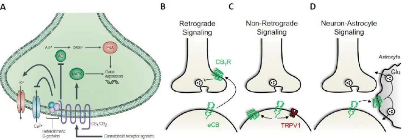
Other metabotropic and ionotropic receptors have been reported to respond to the endogenous agonists of classical cannabinoid receptors. Transient receptor potential vanilloid 1 (TRPV-1) is a nonselective cation channel with high calcium permeability that belongs to the transient receptor potential superfamily (Caterina et al., 1997). They are involved in the transduction of signals such as temperature, electrical charge, light, olfactive and taste stimuli and endogenous lipids (Pertwee et al., 2010b). Although TRPV-1 channels in the digestive track are most known for the mediation of the burning sensation elicited by the molecule capsaicin (present in “chili peppers”) (Caterina et al., 1997), they can also be present in the brain (Cristino
endocannabinoid-mediated forms of synaptic plasticity (Chavez et al., 2010, Marsch et al., 2007).
Another example of proteins that might act as potential cannabinoid receptors includes the deorphanized GPCRs GPR55 and GPR119. GPR55 was initially isolated from the human striatum (Sawzdargo et al., 1999) and it has been reported to respond to 2-AG and Anandamide (Sharir and Abood, 2010). GPR119 is found predominantly in the pancreas and gastrointestinal tract (Fredriksson et al., 2003) and it can respond to the endocannabinoid oleoylethanolamide (OEA) (Overton et al., 2006). Although both receptors can be expressed in the brain, their function and potential action as cannabinoid receptor 3 (CB3) remains mostly unknown (Godlewski et al., 2009). Pertwee and colleagues (2010) established a range of criteria to classify a potential candidate protein as CB3 receptor. These include: 1) the candidate receptor should be activated by CB1/CB2 agonist at the orthosteric site with similar potency, 2) endogenous ligands at physiological conditions should elicit a response via this receptor, 3) it should display an amino acid similarity with CB1/CB2, 4) it should have specific functions elicited by classical agonist and 5) it should not be a receptor with other already identified functions (Pertwee et al., 2010b). The evidence of potential CB1 receptor-independent targets of endocannabinoids in the brain underlines the importance of the use of specific pharmacological methods together with genetic knock-out (KO) strategies to understand specific functions of CB1 receptors.
Because CB1 receptors are known to mediate the majority of the cannabinoid-induced psychotropic effects, studying the role of CB1 receptors in brain physiology and pathology is a major topic in cannabinoid research. In accordance with the aim of this thesis, I will thereby concentrate in the next section of the introduction on CB1 receptor and how they modulate brain functions, how their biology is strategically involved in behavior and what are the currently important unsolved questions regarding the role of this receptor in brain function.
III – Distribution of CB1 receptors in the brain
CB1 receptors are widely, but not exclusively, expressed in the CNS (Hu and Mackie, 2015, Marsicano and Kuner, 2008). Being likely the most abundant GPCR in the brain (Herkenham et al., 1990, Howlett et al., 1990), CB1 receptors have been extensively described in regions involved in key brain functions such as learning and memory, pain perception, reward, motor coordination and energy and metabolism (Di Marzo et al., 2004, Piazza et al., 2017). Consequently, CB1 receptors are present in important parts of the CNS such as the retina, the neocortex, the olfactory system, the amygdala, the hippocampus, the striatum, the cerebellum, the thalamus, the substantia nigra, the ventral tegmental area (VTA), the periaqueductal gray and the spinal cord (Busquets Garcia et al., 2016, Hu and Mackie, 2015, Marsicano and Kuner, 2008, Soria‐Gomez et al., 2017). As the characterization of CB1 receptors is crucial to understand where and how they modulate the diverse brain functions, I will describe the cellular and subcellular localization of CB1 receptors in the hippocampus as it is the main region of interest of this thesis.
III.A – CB1 RECEPTOR DISTRIBUTION IN THE HIPPOCAMPUS
The hippocampus, a key brain region for learning and memory, has one of the broadest and highest expressions of CB1 receptors in the brain (Figure 1) (Herkenham et al., 1990, Marsicano and Kuner, 2008).
Among the different cell-types that compose this region, CB1 receptors have been initially characterized in hippocampal interneurons, more precisely in the terminals of cholecystokinin (CCK)-positive cells. It is mostly absent in parvalbumin(PV)-positive GABAergic basket cells in the pyramidal cell layer, the molecular layer and also the granule cell layer of the dentate gyrus (Katona et al., 1999, Marsicano and Lutz, 1999, Marsicano and Kuner, 2008, Tsou et al., 1999). This mutual exclusion has been consistently observed and fosters hypotheses on how PV- and CCK-positive interneurons might interact in the modulation of hippocampal network activity (Klausberger et al., 2005, Klausberger and Somogyi, 2008). Besides its presence in gamma-aminobutyric acid (GABA)-ergic neurons, CB1 receptors have been described anatomically and functionally in the glutamatergic pyramidal neurons of the CA1 and CA3 regions, although in considerably less amount as compared to GABAergic neurons (Katona et al., 2006, Marsicano and Lutz, 1999, Marsicano et al., 2003). Mossy cells of the dentate gyrus, which
(Kawamura et al., 2006, Monory et al., 2006). Expression of CB1 receptors have also been reported in glial cells such as astrocytes (further reviewed in the part 3 – V). Additionally, CB1 receptors have been shown to be present in hippocampal cells expressing acetylcholine (Degroot et al., 2006) and dopamine type-2 (D2) receptor, 5-hydroxytryptamine type-1B(5-HT1B) and 5-Hydroxytryptamine type-3(5-HT3) receptors (Hermann et al., 2002) suggesting functional crosstalk between the ECS and the cholinergic, the dopaminergic and the serotonergic systems (Marsicano and Kuner, 2008).
III.B
– SUBCELLULAR DISTRIBUTION OF CB1 RECEPTORS
The lipidic nature of the (endo)cannabinoids suggests that they can act on intracellular targets. Notably, besides the classical distribution at the cellular membrane (Dudok et al., 2015, Katona et al., 1999), CB1 receptors have been recently described in intracellular compartments, such as endosomes (Dudok et al., 2015) and brain mitochondria (mtCB1) (Figure 1F) (Benard et al., 2012, Hebert-Chatelain et al., 2014, Hebert-Chatelain et al., 2016, Koch et al., 2015). Mitochondria are highly dynamic organelles that act as powerhouses of eukaryotic cells by generating adenosine triphosphate (ATP), the universal cellular energy substrate, using a process called oxidative phosphorylation (OXPHOS) (Yin and Cadenas, 2015). In neurons, besides mitochondrial contribution to energy supply via generation of ATP to support intracellular processes (e.g. active transport, endocytosis and neurotransmitter production), they can contribute to phospholipids synthesis, production of intermediate metabolites and intracellular signaling molecules (Picard and McEwen, 2014). Furthermore, the apoptotic function of mitochondria, which normally leads to programmed cell death, can be responsible at synaptic level for the physiological induction of Long-term Depression (LTD) of synaptic transmission in hippocampal neurons (Li et al., 2010) showing that traditional functions of these organelles can previously unknown roles under certain conditions. Recently it has been shown that chronic THC administration (10 mg per kg, twice a day for 6.5 days) decreases overall CB1 receptor content in GABAergic axon terminal, with increased CB1 receptor internalization (Dudok et al., 2015). Although the authors suggest that CB1 receptors are possibly internalized in endosomes, one cannot exclude that these intracellular CB1 receptors might rather be in the mitochondria. Further examination will elucidate how CB1 receptors in intracellular compartments can impact the synaptic function and how they are functionally related to plasma membrane CB1 receptors.
.
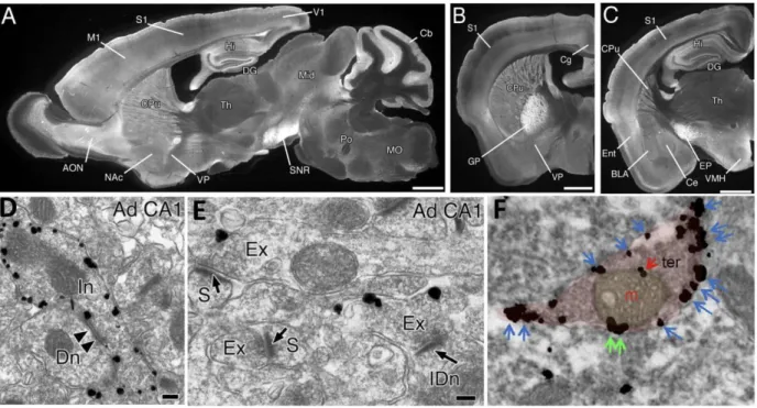
FIGURE 1 – DISTRIBUTION OF CB1 RECEPTORS IN THE ADULT MOUSE BRAIN
A-C. CB1 receptor protein distribution in the brain shows high immunoreactivity in the
structures of the temporal lobe: hippocampus (Hi), dentate gyrus (DG) and entorhinal cortex (Ent). Other regions of high CB1 receptor expression include the anterior olfactory nucleus (AON), neocortex, caudate putamen (CPu), thalamus (Th) basolateral (BLA) and central (Ce) amygdaloid nuclei (C), cerebellum (Cb). D-E. In the hippocampus, CB1 receptors are mainly present in the presynaptic terminal (Ad CA1: Adult CA1; Dn, dendrite; Ex, Excitatory terminal; IDn, interneuronal dendrite; In, Inhibitory terminal; S, Synapse). F. CB1 receptors are also found in intracellular organelles such as mitochondria (m) present in the presynaptic terminals (ter). M1, primary motor cortex; S1, primary somatosensory cortex; V1, primary visual cortex; Cg, cingulate cortex; Ent, entorhinal cortex; DG, dentate gyrus; NAc, nucleus accumbens, GP, globus pallidus; VP, ventral pallidum; Mid, midbrain; SNR, substantia nigra pars reticulata; PO, pons; MO, medulla oblongata; EP, entopedoncular nucleus; VMH, ventromedial hypothalamus; DH, dorsal horn; DLF, dorsolateral funiculus. Bars: 1 mm (A-E), 100 nm (D-E), 0.5 µm (F) [(A- E) Adapted from(Kano et al., 2009); (F) Adapted from (Hebert-Chatelain et al., 2014).
IV – Metabolism of Endocannabinoids
The discovery of the cannabinoid receptors prompted the search for endogenous ligands that could act as agonists (Mechoulam and Parker, 2013). These endocannabinoids are lipidic signaling molecules that act as endogenous agonists of CB1 and CB2 receptors (Lu and Mackie, 2016). Classically, electrically charged signaling molecules can be actively stored in synaptic vesicles, which are transported and docked near the synaptic terminals and released during neuronal activity. In the case of endocannabinoids, their lipophilic nature makes a similar scenario not plausible. Rather than being classically stored in synaptic vesicles to posterior release, endocannabinoids are thought to be produced “on demand”, a process controlled by a tight regulation of synthesis and degradation via specific enzymes (Piomelli, 2003).
In the following section, I will briefly discuss the metabolic pathways for the synthesis and degradation of the major endocannabinoids and some background on how the enzymatic machinery location might modulate the endocannabinoid signaling.
IV.A – THE MAJOR ENDOCANNABINOIDS, ANANDAMIDE
AND2-AG
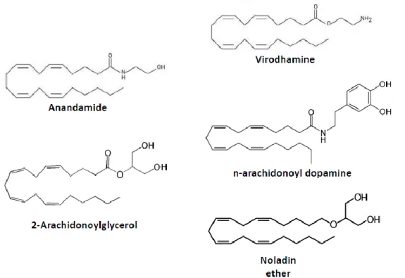
The first endocannabinoid to be identified was the lipid molecule of arachidonoyl ethanolamide (AEA), named Anandamide from the Sanskrit word “Ananda” which means “bliss” (Devane et al., 1992). Anandamide is a derivative of arachidonic acid that acts as a partial agonist for CB1 and CB2 receptors in the brain as well as in the periphery (Pertwee et al., 2010a). Soon after the discovery of Anandamide, a second endogenous lipid ligand called 2-arachidonoylglycerol (2-AG) was isolated, from the canine gut (Mechoulam et al., 1995) and from the brain (Sugiura et al., 1995). 2-AG, also a derivative of arachidonic acid, was found to be a full agonist of the CB1 receptors with high potency and selectivity (Pertwee, 2008).
Besides these two well-characterized endocannabinoids, there are other molecules that act as endogenous cannabinoid modulators with different selectivity and potency for CB1 receptors (Figure 2). These include: noladin ether, virodhamine and N-arachidonoyldopamine (Pertwee, 2008). The functional relevance of these molecules is not yet well characterized (Pertwee, 2008) and therefore I will focus only on the main two eCBs: 2-AG and Anandamide.
Apart from the endogenous CB1 receptor ligands, endogenous allosteric modulators that can modify the CB1 receptor activity have also been identified (Morales et al., 2016). The
A4 (Pamplona et al., 2012), the neurosteroid pregnenolone (Vallee et al., 2014) and the hemopressin-like polipeptide pepcan-12 (Hofer et al., 2015).
FIGURE 2 – CHEMICAL STRUCTURES OF ENDOGENOUS MOLECULES THAT BIND TO THE CANNABINOID RECEPTORS
IV.B – SYNTHESIS, TRANSPORT AND DEGRADATION OF THE 2-AG AND ANANDAMIDE
ANANDAMIDE
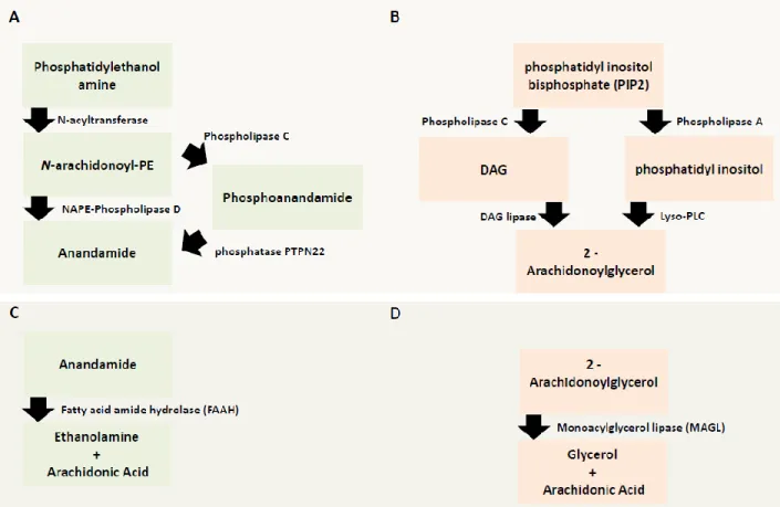
Anandamide synthesis starts with the conversion of the phosphatidylethanolamines by N-acetyltransferase (NAT) into the precursor N-arachidonoyl phosphatidyl ethanol (NAPE) (Piomelli, 2003). Currently, there is evidence of multiple synthesis pathways for the production of Anandamide which, most likely, depend on several factors such brain region, local circuitry or local neuronal and/or glial activity (Ahn et al., 2008, Lu and Mackie, 2016). Two of the most well characterized are NAPE-phospholipase D (PLD) (Di Marzo et al., 1994), the first to be discovered, and the NAPE-phospholipase D pathway. In the first case, NAPE is converted to Anandamide by the action of NAPE-phospholipase D (Lu and Mackie, 2016). This pathway has been extensively studied and is present in the CNS (Lu and Mackie, 2016). In the second pathway, NAPE is first converted to phosphoanandamide via Phospholipase C (PLC) and then dephosphorylated by a protein tyrosine phosphatase non-receptor type-22 (PTPN22) to produce Anandamide (Figure 3A)(Liu et al., 2006).
After release to the extracellular space most likely by passive diffusion, Anandamide exerts its effects by retrograde signaling at CB1 receptors located at the presynaptic terminals (Piomelli, 2003). After the activation of CB1 receptors, Anandamide is cleared from the extracellular space and quickly degraded by the enzyme fatty acid amid hydrolase (FAAH) (Figure 3C) (Cravatt et al., 1996). In the hippocampus, FAAH has been shown to be integral membrane bound protein found in the soma and dendrites of pyramidal cell that are innervated by CB1 receptor-positive axon terminals, most likely from CCK-positive interneurons (Gulyas et al., 2004, Hu and Mackie, 2015). Interestingly, intracellular membrane systems such as mitochondria and the endoplasmic reticulum are highly enriched in FAAH (Ahn et al., 2008, Gulyas et al., 2004). A catalytically silent variant of FAAH, the FAAH-1 (named FLAT by the authors) has been described (Fu et al., 2011). This membrane-bound protein, which lacks catalytic activity due to alternative splicing, has high affinity to Anandamide and has been shown to lead to an accumulation of Anandamide in the cytosol thus being suggested to act as an endocannabinoid transporter (Lu and Mackie, 2016).
2-ARACHIDONOYLGLYCEROL (2-AG)
The endocannabinoid 2-AG is synthetized mainly by two principal mechanisms: 1) a calcium-dependent release (CaER) mechanism and 2) calcium-assisted receptor regulated endocannabinoid release (RER) (Figure 3B) (Ahn et al., 2008). The calcium-dependent release is likely initiated by the activation of postsynaptic metabotropic type-one glutamate (mGlu1) receptors (Maejima et al., 2005) and also muscarinic type-1 (M1), type-3 (M3) receptors (Ohno-Shosaku et al., 2003) or orexin receptors (Kukkonen and Leonard, 2014). Following activation of these receptors, which induces PLC activity, will produce inositol trisphosphate (IP3) and diacylglycerol (DAG) from phosphatidyl inositol bisphosphate (PIP2). Next, DAG is converted by a DAG lipase, isoform α and β, into 2-AG (Di Marzo, 2008). It is currently believed that isoform DAGα is the one responsible for the synthesis of the majority of 2-AG necessary for the retrograde suppression of neurotransmitter release in the cerebellum, the hippocampus and the striatum (Tanimura et al., 2010). The second main mechanism found to regulate 2-AG production is via the generation of the intermediate molecule phosphatidyl inositol by the action of phospholipase A, which is then converted into 2-AG by the enzyme lyso-PLC (Lu and Mackie, 2016).
2-AG, similarly to Anandamide, is thought to be transported by facilitated transport across the membranes to the extracellular space. However, such transport protein has not yet been identified (Di Marzo, 2008).
The degradation of 2-AG is mainly processed by the enzyme monoacylglycerol lipase (MAGL) (Figure 3D) (Dinh et al., 2002b). This enzyme belongs to the family of the serine hydrolase, highly expressed in the CNS (Dinh et al., 2002a), and it converts 2-AG into arachidonic acid and glycerol (Ahn et al., 2008). In hippocampal neurons, MAGL is expressed mainly presynaptically in glutamatergic and GABAergic terminals, in contrast to FAAH, which is mainly postsynaptic (Dinh et al., 2002a). MAGL is localized in close proximity to CB1 receptors to ensure a tight regulation of CB1 receptor activity by 2-AG (Gulyas et al., 2004). At the subcellular level, MAGL have also been functionally and anatomically identified in the mitochondria (Alger and Tang, 2012, Marsicano and Kuner, 2008).
The idea that ECS is tightly regulated, together with recent characterization of CB1 receptors in intracellular compartments (i.e. mitochondria), raises questions regarding the functional relevance of the presence of both the degradation enzymes and the receptors at the same locations.
V – Methodologies to dissect the function of CB1 receptors
The broad distribution of CB1 receptor in several brain regions, circuits and close associated cells illustrates the complexity of this system. In order to understand the specific contribution of CB1 receptors to the modulation of synaptic plasticity and behavior both in physiology and pathology, a combination of genetic and pharmacological approaches is needed. Advanced methods such as cutting edge microscopy and/or electrophysiological approaches will allow further dissection of the specific role of the CB1 receptor in brain function. In the following section, I will review some of the current main pharmacological and genetic tools used to dissect the role of CB1 receptors in brain function.
V.A – PHARMACOLOGICAL TOOLS
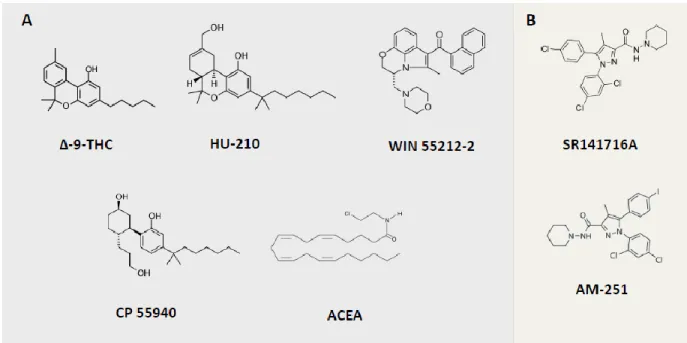
The use of pharmacological tools that are based on the structure of natural exogenous and endogenous cannabinoid molecules is very important to identify and dissect CB1 receptor specific functions from the molecular to the behavioral level. There are currently several synthetic molecules that can act as full agonists with high activity and partial agonists with mild affinity of the receptor. Antagonists that block the action of the receptor and inverse agonists decrease the activation of the receptor below a threshold of basal activity. There are also allosteric modulators that through binding in allosteric rather than orthosteric sites can modify the function of the receptor (Mackie, 2008).
Besides the natural agonists (e.g. THC), there are several synthetic ligands that are currently used to address specific functions of CB1 receptors (Figure 4). So far, the best characterized are: the agonist HU-210, with high affinity and potency; CP55940, a potent agonist with high affinity (though inferior to HU-210), WIN 55212-2 and arachidonyl-2′-chloroethylamine (ACEA), both highly selective and potent agonists (Pertwee et al., 2010b) . As selective CB1 receptor antagonists, the best characterized are SR141716A (also known as Rimonabant) and AM-251 (Pertwee et al., 2010b). As many known CB1 receptor agonists have also high affinity for CB2 receptors (e.g. HU-210), before claiming that an effect is CB1 receptor-dependent it is important to demonstrate that it can be blocked by specific antagonists of CB1 receptors (e.g. Rimonabant) or that the phenotype of interest is absent in full CB1 receptor KO models.
FIGURE 4 – CHEMICAL STRUCTURES OF EXOGENOUS NATURAL AND SYNTHETIC MOLECULES THAT BIND TO THE CANNABINOID RECEPTORS
V.B – GENETIC TOOLS TO STUDY THE ECS
The generation of mouse models ubiquitously lacking CB1 receptors provided a major step to study the specific functional of these receptors at the molecular, cellular and behavioral level (Ledent et al., 1999, Marsicano et al., 2002, Zimmer et al., 1999). However, as CB1 receptors are expressed in brain cells and circuits with apparent functional opposing effects (e.g. glutamatergic and GABAergic neurons in the hippocampus) the constitutive deletion of CB1 receptor does not allow to study its specific contribution to brain functions (Castillo et al., 2012).
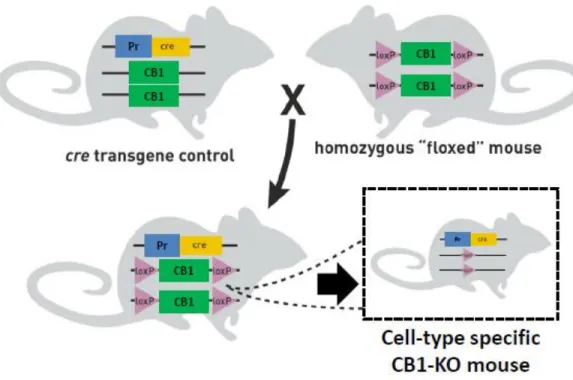
In order to dissect the role of CB1 receptors in specific neuronal and glial cells, the use of the Cre recombinase(CRE)/loxP system of genetic recombination to generate cell type-specific conditional KOs provided a valuable tool. CRE is a protein that allows the targeted excision of genes in the genome that are located between two artificially introduced 34-bp sequences, known as loxP (Orban et al., 1992, Sauer and Henderson, 1988). The loxP sequences (generally introduced into the genome by homologous recombination) are very small and do not have impact on the normal animal phenotype. Thus mice carrying loxP sequences flanking the gene of interest (named floxed mice) are considered as WT animals (Nagy, 2000).
In order to achieve the specific deletion of the gene of interest, “floxed” mice (i.e. with the gene of interest flanked by the LoxP sequences) are crossed with a mouse that expresses the Cre recombinase under the control of a promoter specific for the cell-type to be targeted (Nagy, 2000, Orban et al., 1992). Once the breeding is done, the offspring will express the CRE in the cell-type of interest, allowing it to modify the genome by excision of the “floxed” gene, thereby generating a cell-type specific KO mouse (Figure 5)(Nagy, 2000).