Université de Montréal
Arterial stiffness and brain health: investigating the impact
of sex-related differences
par : Dalia Sabra
Programme des Sciences biomédicales Faculté de Médecine
Mémoire présentée
en vue de l’obtention du grade de Maitrise en sciences biomédicales, option sciences du vieillissement
Août, 2019
Résumé
Introduction: Il est bien établi que les maladies vasculaires, cérébrovasculaires et
cardiovasculaires se manifestent différemment chez les hommes que chez les femmes. La rigidité artérielle (RA), un prédicteur indépendant de la maladie cardiovasculaire (MCV), a été associée à des changements de la réactivité cérébrovasculaire (RCV) et à un déclin cognitif lors du vieillissement. Plus précisément, les personnes âgées ayant une RA plus élevée présentent un déclin plus marqué au niveau des tâches exécutives. Une diminution des fonctions exécutives (FE) est également liée à une réduction de la RCV chez les personnes âgées. Cependant, il est important de noter que la relation entre la RA et la RCV est plus complexe. Certaines études montrent une diminution de la RCV associée avec une RA plus élevée, tandis que d’autres rapportent une RCV préservée avec une RA élevée. De plus, des travaux récents suggèrent que les différences de concentration en hématocrit (HCT) pourraient avoir une incidence sur les mesures de RA. Ici, nous avons étudié le rôle possible du sexe et de l'HCT sur ces relations hémodynamiques.
Méthodes: Des acquisitions ont été effectuées chez 48 adultes âgés en bonne santé (31 femmes,
63 ± 5 ans) dans un scanneur d’imagerie par résonance magnétique (IRM) 3T. Des données de marquage de spin artériel pseudo-continu utilisant des lectures à double écho ont été collectées pendant un défi d'hypercapnie (changement de CO2 de 5mmHg, pendant deux blocs de 2
minutes). La RCV a été calculée comme étant le % de changement du signal de débit sanguin cérébral (% ∆CBF) par changement de mmHg dans le CO2 à la fin de l’expiration. Les données
de vitesse d’onde de pouls (VOP) aortique ont été acquises à l’aide d’une série de contraste de phase cine encodée par la vitesse durant 60 phases cardiaques avec un encodage en vélocité de 180cm/s dans le plan. La VOP dans l'arcade aortique a été calculée entre l'aorte ascendante et descendante. Les analyses statistiques ont été effectuées à l'aide de SPSS.
Résultats: Un test de modération contrôlant pour l’âge et le volume des hyperintensités de la
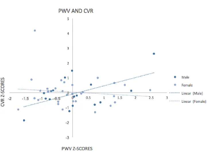
matière blanche a révélé un effet direct significatif de la VOP sur la RCV (β = 1,630, IC à 95% [.654, 2,607), ainsi que de la VOP sur la FE (β = -. 998, IC 95% [-1,697, -,299]). Le sexe a modéré la relation entre VOP et RCV (β = -1,013, IC 95% [-1,610, -,4169]), et VOP et FE (β =
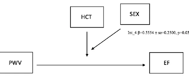
.447, IC 95% [.020, .875]). En outre, il existait un effet significatif de l’HCT sur les différences de sexe observées dans l’effet de modération (VOP * SEXE) sur la FE (β = -0,7680, SE = 0,3639, IC 95% [-1,5047, -0,0314], p = 0,0414).
Conclusion: Nos résultats indiquent que les relations entre la VOP, la RCV et la FE sont
complexes et que le sexe et l’HCT modulent ces relations. L’influence des variations hormonales (p. ex. la ménopause) sur ces relations devrait être étudiée dans le futur et pourrait permettre de personnaliser les stratégies de prévention des MCV.
Mots-clés : rigidité artérielle, réactivité cérébrovasculaire, imagerie par résonance magnétique,
Abstract
Introduction: It is well established that sex differences exist in the manifestation of vascular,
cerebrovascular and cardiovascular disease. Arterial stiffness (AS), an independent predictor of cardiovascular disease (CVD), has been associated with changes in cerebrovascular reactivity (CVR) and cognitive decline in aging. Specifically, older adults with increased AS show a steeper decline on executive function (EF) tasks. Decreased EF is also linked with reduction in CVR among older adults. Interestingly, the relationship between AS and CVR is more complex, where some works show decreased CVR with increased AS, and others demonstrate preserved CVR with higher AS. In addition, recent work suggests that measurements of AS may be affected by differences in the concentration of hematocrit (HCT). Here, we investigated the possible role of sex and HCT on these hemodynamic relationships.
Methods: Acquisitions were completed in 48 healthy older adults (31 females, 63 ± 5 years) on
a 3T MRI. Pseudo-continuous arterial spin labeling using dual-echo readouts were collected during a hypercapnia challenge (5mmHg CO2 change, during two, 2 min blocks). CVR was calculated as the %∆CBF signal per mmHg change in end-tidal CO2. Aortic PWV data was acquired using a cine phase contrast velocity encoded series during 60 cardiac phases with a velocity encoding of 180cm/s through plane. PWV in the aortic arch was computed between ascending and descending aorta. Statistical analyses were done using SPSS.
Results: A moderation model test controlling for age and white matter hyperintensity volume
revealed a significant direct effect of PWV on CVR (β=1.630, 95% CI [.654, 2.607), as well as PWV on EF (β=-.998, 95% CI [-1.697, -.299]). Sex moderated the relationship between PWV and CVR (β=-1.013, 95% CI [-1.610, -.4169]), and PWV and EF (β=.447, 95% CI [.020, .875]). In addition, there was a significant effect of HCT on the sex differences observed in the moderation effect (PWV*SEX) on EF (β=-0.7680, SE = 0.3639 ,95% CI [-1.5047, -0.0314], p=0.0414).
Conclusion: Together, our results indicate that the relationships between PWV, CVR and EF is
. Future work should investigate the role of hormone variations (e.g., menopause) on these relationships to better personalize CVD prevention strategies.
Keywords : arterial stiffness, cerebrovascular reactivity, magnetic resonance imaging, arterial
TABLE OF CONTENTS
RÉSUMÉ 1 ABSTRACT ... 3 TABLE OF CONTENTS ... 5 LIST OF TABLES ... 8 LIST OF FIGURES ... 9 LIST OF ABBREVIATIONS ... 10 DEDICATION ... 12 ACKNOWLEDGMENTS ... 13 CHAPTER 1 INTRODUCTION ... 14 1.1IMAGING MODALITIES ... 141.2AGE-RELATED CHANGES IN CBF AND CVR ... 15
1.3SEX DIFFERENCES IN CBF AND CVR ... 16
CHAPTER 2 ARTERIAL STIFFNESS AND BRAIN INTEGRITY: A REVIEW OF MRI FINDINGS ... 19
2.1ABSTRACT ... 21
2.2INTRODUCTION ... 22
2.3ARTERIAL STIFFNESS MEASUREMENTS ... 24
2.4OVERVIEW OF HISTOPATHOLOGICAL CHARACTERISTICS OF CEREBRAL WHITE MATTER AND GRAY MATTER AGING ... 27
2.5MACRO AND MICROSTRUCTURAL CHANGES OF THE CEREBRAL WHITE MATTER RELATED TO ARTERIAL STIFFNESS... 29
2.6MACRO AND MICROSTRUCTURAL CHANGES OF THE CEREBRAL GRAY MATTER RELATED TO ARTERIAL STIFFNESS ... 33
2.7BRIDGING THE GAP BETWEEN ARTERIAL STIFFNESS, CEREBRAL BLOOD FLOW AND MICROSTRUCTURAL INTEGRITY. ... 36
2.7ARTERIAL STIFFNESS AND BRAIN INTEGRITY:INSIGHT FROM OTHER MODALITIES ... 38
2.8ARTERIAL STIFFNESS AND COGNITION ... 40
CHAPTER 3 SEX MODERATES THE RELATIONSHIP BETWEEN AORTIC STIFFNESS, COGNITION AND
CEREBROVASCULAR REACTIVITY IN HEALTHY OLDER ADULTS ... 44
3.1INTRODUCTION ... 46
3.2METHODS ... 49
3.2.1 Participants ... 49
3.2.2 Cognitive Composite Score... 49
3.2.3 Hypercapnia ... 50 3.2.4 MRI Acquisition ... 50 3.2.5 Aortic Exam ... 51 3.2.6 Data Analysis ... 51 3.2.7 Resting CBF Analysis ... 51 3.2.8 CVR Analysis ... 52
3.2.9 Vascular Lesion Quantification ... 52
3.2.10 Pulse Wave Velocity Data ... 53
3.2.11 Blood Tests ... 53
3.2.12 Statistical Analysis ... 53
3.3RESULTS... 53
3.3.1 Moderation Analysis ... 54
3.3.2 Moderated Moderation Analysis ... 58
3.4DISCUSSION ... 60
3.4.1 Main Results ... 60
3.4.2 Sex Differences ... 61
3.4.3 Arterial Stiffness and Cerebrovascular Reactivity ... 61
3.4.4 The Association between pulse wave velocity and cognitive function relative to sex ... 63
3.4.5 The Association between cognitive function and cerebrovascular reactivity relative to sex ... 64
3.4.6 The effect of Hematocrit ... 65
3.4.7 Limitations ... 66
3.5CONCLUSION ... 67
CHAPTER 4 CONCLUSION ... 69
REFERENCES ... 71
1.1 HIGHER CARDIOVASCULAR FITNESS LEVEL IS ASSOCIATED WITH LOWER CEREBRAL VASCULAR REACTIVITY AND PERFUSION IN HEALTHY OLDER ADULTS ... 103 1.2 SCATTERPLOTS ... 118
List of Tables
Table 1 - Arterial stiffness measures. ... 26 Table 2 - Participant Demographics. ... 54
List of Figures
Figure 1 - Normal values for pulse wave velocity: average according to age (1455 subjects). 24
Figure 2 - Arteries and veins of the white and gray matter. ... 28
Figure 3 - White matter regions denoted vulnerable to arterial stiffness. ... 32
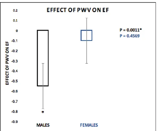
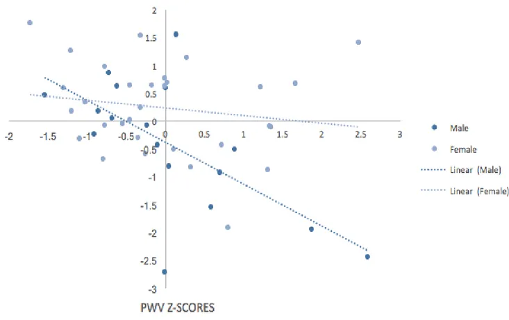
Figure 4 - Moderation Effect (PWV*SEX) on CVR. ... 55
Figure 5 - Effect of PWV on CVR... 55
Figure 6 - Moderation Effect (PWV*SEX) on EF. ... 56
Figure 7 - Effect of PWV on EF. ... 57
Figure 8 - Moderation model showing the effect (CVR*SEX) on EF. ... 58
Figure 9 - Moderated moderation model depicting the effect of HCT on the relationship PWV on CVR among sexes. ... 58
Figure 10 - Moderated moderation model depicting the effect of HCT on the relationship PWV on EF among sexes. ... 59
List of Abbreviations
MRI: Magnetic resonance imaging WMH: White matter hyperintensities DTI: Diffusion tensor imaging FA: Fractional anisotropy RD: Radial diffusivity ASL: Arterial spin labeling AS: Arterial stiffness PWV: Pulse wave velocity
cfPWV: Carotid-femoral pulse wave velocity GMV: Grey matter volume
FLAIR: Fluid-attenuated inversion recovery MT: Magnetization transfer
NODDI: Neurite density and orientation dispersion
CHARMED: Composite hindered and restricted model of diffusion BOLD: Blood-oxygen-level-dependent
fMRI: Functional magnetic resonance imaging MEG: Magnetoencephalography
ADNI: Alzheimer’s disease neuroimaging initiative TEdDI: TE dependent diffusion imaging
MMSE: Mini-mental state examination MTL: Medial temporal lobe
CVR: Cerebrovascular reactivity CVD: Cardiovascular disease HCT: Hematocrit CSF: Cerebrospinal fluid VBM: Voxel-based morphometry PP: Pulse pressure
CBF: Cerebral blood flow EF: Executive function
PET: Positron-emission tomography TCD: Transcranial doppler
PS: Processing speed
MMSE: Mini-mental state examination CWIT: Color-word interference test TMT-B: Trail making test part B SNR: Signal-to-noise ratio
pCASL: Pseudo-continuous arterial spin labeling HRT: Hormone replacement therapy
RF: Radiofrequency
CO2: Carbon dioxide
EtCO2: End-tidal carbon dioxide
CMRO2: Cerebral metabolic rate of oxygen
OEF: Oxygen extraction fraction CBV: Cerebral blood volume
Dedication
This thesis is dedicated to my mother, my father and my brother, for their continuous support, love and patience. This work would not have been possible without you by my side.
Acknowledgments
I would like to take this opportunity to thank my supervisors Dr. Claudine Gauthier and Dr. Louis Bherer, for their guidance and mentorship throughout the years. I am extremely privileged and grateful you have given me the chance to be a graduate student in both of your labs. With both your expertise and dedication to your students, I have grown both professionally and personally. You have gone above and beyond the basic requirements of any research supervisor and have made this unlikely dream turn into a reality. I would also like to thank my committee members for accepting to evaluate this thesis.
Secondly, I would like to take this opportunity to acknowledge the guidance and support that I received throughout my Master’s degree from my lab mates. I would like to thank Brittany Intzandt for always being there and giving me advice and ideas that allowed me to complete my thesis, Julia Huck for teaching me numerous new techniques, and Atef Badji for being a great leader, researcher, and friend.
Finally, the completion of this thesis would not have been possible without the great LESCA team. I would like to thank them all for their hard work. I am extremely honored that I have had the opportunity to work with such a great team.
Chapter 1
Introduction
1.1 Imaging Modalities
Aging is associated with an increasing incidence of cerebrovascular, cardiovascular and neurodegenerative diseases (Niccoli and Partridge 2012; Harman 1990; Najjar et al. 2005). Recent advances in neuroimaging techniques have now begun to improve our understanding of the effects of aging on the brain, and how this is linked to the pathogenesis of diseases of aging. Age-related cerebrovascular alterations have been reported with the use of arterial spin labeling (ASL), a magnetic resonance imaging based-technique used for quantifying local perfusion in the brain (Leoni et al. 2017; Badji et al. 2019; Gauthier et al. 2013; Grade et al. 2015). Noninvasive ASL techniques directly estimates cerebral blood flow (CBF), the rate of delivery of arterial blood to the capillary bed in brain tissue, by applying a magnetic tag to inflowing arterial blood at the carotid level and measuring the delivery of tagged blood into each imaging voxel (Zhang et al. 2017). The magnetic tag is applied by inverting the magnetization of blood water using radiofrequency (RF) pulses that are similar to those used in the MR imaging process (Leoni et al. 2017; Williams; Intzandt et al. 2019; Grade et al. 2015). In addition, blood oxygenation level dependent (BOLD) MR imaging has been used in the aging literature as a common means of observing changes in the functional state of the brain (D'Esposito et al. 2003). This technique is based on the T2*‐weighted signal, to indirectly measure flow changes by observing changes in the local concentration of oxygenated and deoxygenated hemoglobin in the brain (Golestani et al. 2016; Williams). The BOLD signal is however ambiguous in nature, as it simultaneously reflects changes in oxidative metabolism, blood flow and blood volume (Gauthier et al. 2013; Gauthier and Fan 2019). This ambiguity can be harnessed however to obtain measures of vascular health. Since the BOLD contrast is highly dependent on the local blood volume and flow, it can be used in combination with a vasoactive challenge such as breathholding or inhalation of carbon dioxide (CO2) to map quantitative cerebrovascular
reactivity (CVR), a measure of vascular elasticity in aging (Rostrup et al. 2000; Kastrup et al. 2001). Indeed, inhalation of CO2 increases the concentration of CO2 in the lungs, quantified by
end-tidal CO2 (EtCO2) which is at equilibrium with arterial CO2 partial pressure in healthy
people (Ibler and Lage 1984; Yezhuvath et al. 2009). Increased concentrations of CO2 will then
result in the dilation of small arteries, venous vessels, arterioles and increase CBF (Ito et al. 2003; Davis et al. 1998; Hoge et al. 1999; Yezhuvath et al. 2009). CVR is therefore an indirect measure of vasodilatory capacity as it measures the vascular response to a well-controlled vasodilation challenge. CVR is calculated as the percent change in BOLD signal per change in end-tidal partial pressure of CO2 and expressed as %S/mm Hg.
1.2 Age-related changes in CBF and CVR
A decrease in whole brain CBF in older individuals in comparison to younger individuals has been demonstrated in several ASL studies (Ambarki et al. 2015; Amiri et al. 2014; Wagner et al. 2012). Similarly, previous work reports CBF declines at about 0.38 ∼ 0.45% per year (Parkes et al. 2004; Biagi et al. 2007; Ambarki et al. 2015; Wagner et al. 2012; Vis et al. 2018; Zhang et al. 2018). Indeed, among healthy older adults, CBF reductions have been found to be more pronounced in the frontal, temporal and parietal lobes (Chen et al. 2011; Parkes et al. 2004). Moreover, aging is accompanied with impaired neuronal and glial mitochondrial metabolism (Tarumi and Zhang 2018). As such, the age related reductions in cerebral perfusion may be due to decreased cerebral metabolic rate (Marchal et al. 1992; Tarumi and Zhang 2018) and cerebrovascular dysfunction (Zhu et al. 2011; Tarumi and Zhang 2018). Indeed, age decreases the metabolic rates for oxygen and glucose by 5% per decade (Tarumi and Zhang 2018). Notably, the reductions in metabolic rate observed across the lifespan have been associated with decreases in CBF (Leenders et al. 1990; Petit-Taboué et al. 1998; Tarumi and Zhang 2018). In addition, decreases in cerebrovascular responses to hypercapnia have been reported in the aging brain (Flück et al. 2014; Gauthier et al. 2013; Lu et al. 2011; Reich and Rusinek 1989; Yamaguchi et al. 1979; McKetton et al. 2018). For example, in a previous calibrated fMRI study using a 5mmHg hypercapnia block, it was found that older adults exhibited lower BOLD and flow responses in hypercapnia compared to younger adults (Gauthier et al. 2013). Similarly, De Vis et al found reduced BOLD CVR among older adults in cortical frontal, temporal and occipital grey matter, using two 10 mmHg hypercapnia blocks (Vis et al. 2015; McKetton et al. 2018). More importantly, the CVR BOLD signal is dependent on blood flow changes observed
in local T2* decay which reflects the interaction between cerebral metabolic rate of oxygen (CMRO2), oxygen extraction fraction (OEF), CBF and cerebral blood volume (CBV) (Kim and
Ogawa 2012). As such, a possible explanation for decreased CVR in elderly may be related to decreased CMRO2, oxygen, OEF and CBF with age (Vis et al. 2015; McKetton et al. 2018). In
addition, chronic dilation of cerebral vessels due to age-related inflammation can also be possible explanation for decreased CVR in the elderly. Nonetheless, CVR remains as one of the most reliable neuroimaging predictor of brain vascular health (Pillai and Mikulis 2015; Mandell et al. 2008).
1.3 Sex differences in CBF and CVR
Studies on structural brain development have shown that there are large sex differences in CBF across all ages. For instance, females show higher CBF than males during childhood, mid-puberty and mid-late adolescence (Tontisirin et al. 2007; Satterthwaite et al. 2014; Robison et al. 2019). Although previous work report a decline in CBF among box sexes during adulthood (Rodriguez et al. 1988; Robison et al. 2019), recent work suggests that females maintain a higher flow throughout adulthood. A study examining CBF among adults ranging from 18-72 years of age found that women consistently maintain a 11% increase in CBF compared to men across the lifespan (Rodriguez et al. 1988; Robison et al. 2019). This is also in line with previous work showing a 15% increase in CBF across young adulthood compared to males (Gur et al. 1982; Robison et al. 2019). Indeed, women have thinner blood than men, which allows the arterial blood to travel faster and has a shorter arterial transit time, increasing CBF (Robison et al. 2019). Furthermore, there is evidence that female sex hormones play a role in regulating CBF (Brackley et al. 1999; Diomedi et al. 2001; Robison et al. 2019). Indeed, a study examining the role of female sex hormones on cerebral hemodynamics found that post-menopausal women taking hormone replacement therapy (HRT) had increased levels of whole brain CBF compared to controls (Ohkura et al. 1995).
Sex differences have also been demonstrated in CVR using ultrasound imaging. Indeed, females exhibit greater cerebral autoregulation, the ability of the brain to maintain relatively constant CBF despite changes in blood pressure, during adolescence (Tontisirin et al. 2007; Robison et al. 2019) and older adulthood (Deegan et al. 2011). In addition, animal studies have shown that
middle cerebral arteries in female rodents are more dilated across a range of pressures compared to males. Similarly, it has been demonstrated that the response to angiotensin II, a vasoconstrictor that contributes to the development of arterial stiffness, is decreased in female human cerebral arteries in comparison to males (Ahnstedt et al. 2013). Similarily, estrogen has been associated with higher nitric oxide (NO) bioavailability and therefore lower vascular tone (Robinson et al. 2019). Taken together, these studies show strong evidence that sexual dimorphism exists in CVR.
With the overwhelming evidence that sex differences exist in the manifestation of vascular, cerebrovascular and cardiovascular diseases, a better understanding of the impact of sex on brain hemodynamics is needed to identify sex-specific preventive strategies to mitigate CVD risk. As such, this thesis first provides and overview of the current neuroimaging literature demonstrating the impact of arterial stiffness on the aging brain, followed by experimental work focusing on the impact of sex-related differences on the link between arterial stiffness, cognitive performance and cerebrovascular reactivity using magnetic resonance imaging in a group of healthy older men and women. Furthermore, the relative contribution of hematocrit on these relationships is investigated. We hypothesize that sex will moderate the relationships between pulse wave velocity and cerebrovascular reactivity and the relationship between pulse wave velocity and executive function. Furthermore, we anticipate that there will be asignificant effect of hematocrit on the sex differences observed in the moderation effects of sex on the relationships between pulse wave velocity and cerebrovascular reactivity and the relationship between pulse wave velocity and executive function
Chapter 2
Arterial Stiffness and Brain Integrity: a review of MRI findings
Atef Badji 1, 2, 3 *; Dalia Sabra 2,5 *; Louis Bherer 2,8,9 ; Julien Cohen-Adad 1, 2, 3 ; Hélène Girouard 2, 4; Claudine J. Gauthier 6,7
* Atef Badji and Dalia Sabra have equally contributed to this work
Manuscript accepted 2 May 2019: https://doi.org/10.1016/j.arr.2019.05.001
1 NeuroPoly Lab, Institute of Biomedical Engineering, Polytechnique Montréal, Montréal, QC,
Canada
2 Centre de recherche de l’Institut Universitaire de Gériatrie de Montréal (CRIUGM),
Montréal, QC, Canada
3 Department of Neurosciences, Faculty of Medicine, Université de Montréal, Montréal, QC,
Canada
4 Department of pharmacology and physiology, Faculty of Medicine, Université de Montréal,
Montréal, QC, Canada
5 Department of Biomedical Science, Faculty of Medicine, Université de Montréal, Montréal,
QC, Canada
6 Physics Department, Concordia University, Montréal, QC, Canada 7 PERFORM Centre, Concordia University, Montréal, QC, Canada 8 Research Center, Montreal Heart Institute, Montréal, QC, Canada
Corresponding Author:
Claudine J. Gauthier, PhD
Title: Assistant Professor, Physics
Office: L-SP 365-06 Richard J.Renaud Science Complex, 7141 Sherbrooke W. Phone: (514) 848-2424 ext. 2193
2.1 Abstract
Background: Given the increasing incidence of vascular diseases and dementia, a better
understanding of the cerebrovascular changes induced by arterial stiffness is important for early identification of white and grey matter abnormalities that might antedate the appearance of clinical cognitive symptoms. Here, we review the evidence from neuroimaging demonstrating the impact of arterial stiffness on the aging brain.
Method: This review presents findings from recent studies examining the association between
arterial stiffness, cognitive function, cerebral hypoperfusion, and markers of neuronal fiber integrity using a variety of MRI techniques.
Results: Overall, changes associated with arterial stiffness indicates that the corpus callosum,
the internal capsule and the corona radiata may be the most vulnerable regions to microvascular damage. Changes in gray matter structure have also been found to be associated with arterial stiffness and are present as early as the 4th decade. Moreover, decrease in cerebral perfusion has been associated with arterial stiffness as well as lower cognitive performance in age sensitive domains such as executive function.
Conclusion: Considering the established relationship between arterial stiffness, brain and
cognition, this review highlights the need for future studies of brain structure and function in aging to implement measurements of arterial stiffness in parallel with quantitative imaging.
Keywords = arterial stiffness, diffusion, white matter microstructure, arterial spin labelling,
2.2 Introduction
Several cerebrovascular risk factors, such as hypertension and arterial stiffness have been associated with the pathogenesis of cognitive decline and dementia, in particular Alzheimer’s disease and vascular dementia (Hanon et al., 2005; Henskens et al., 2008; Singer et al., 2014). Among those cerebrovascular risk factors, arterial stiffness is a common condition that arises with aging (Najjar et al., 2005) figure 1 and is apparent as a 2-fold increase in aortic pulse wave velocity (PWV) in healthy individuals between the ages of 20 and 80 years (Vlachopoulos et al., 2011). Arterial stiffness refers to the loss of elasticity mainly in large arteries such as the aorta and carotids. The elasticity of large arteries allows the dampening of the arterial pressure waveform, transforming the pulsatile flow at the heart level into steady blood flow in the micro-vessels (Iulita et al., 2018; Scuteri et al., 2011). Consequently, arterial stiffening increases flow pulsations through the carotid and vertebral arteries which eventually extend deep into the microvasculature of the brain and may result in haemorrhages, endothelial denudation and thrombotic obstruction (Henskens et al., 2008; O’rourke and Hashimoto, 2007). Moreover, arterial stiffening elevates arterial pressure wave propagation, causing the reflected arterial wave to arrive back at the aorta during the systolic rather than the diastolic phase of the cardiac cycle (Laurent et al., 2005; Pase et al., 2012; Vlachopoulos et al., 2011), thereby escalating systolic blood pressure and contributing to a widening of pulse pressure (Pase et al., 2012).
Chronically elevated arterial stiffness and pulse pressure transmitted into the brain are known to contribute to cerebrovascular changes such as cerebral white matter parenchymal damage via an alteration of cerebral blood flow (Iulita et al., 2018; Mitchell, 2008; O’Rourke and Safar, 2005). In particular, areas perfused by arterioles supplied by the anterior and middle cerebral arteries are more vulnerable to cerebral hypoperfusion because of their geographic localization within areas with few interconnections (Badji et al., 2018; Rosano et al., 2013; Tarumi et al., 2015). For instance, it has been shown that abnormal elevations in central and cerebral pulsatility promote the development of white matter hyperintensities (WMHs), a marker of white matter degradation reflecting small vessel diseases (Singer et al., 2014). However, despite the acknowledged importance of arterial stiffness in the genesis of WMHs, little is known about the microstructural correlates of arterial stiffness. In addition, arterial stiffness has been also shown
to impact gray matter structure as well as cerebral perfusion, which in turn can impact cognitive function (Tarumi et al., 2011; Tarumi and Zhang, 2017).
Knowing how arterial stiffness is implicated in the pathogenesis of cognitive impairment lays the groundwork for devising better strategies to prevent cognitive decline. Thus, a better understanding of the cerebrovascular changes induced by arterial stiffness is important for early identification of the white and gray matter abnormalities that might antedate the appearance of clinical cognitive symptoms. Advances in neuroimaging techniques have now begun to improve our knowledge of the effects of arterial stiffness on the brain (Badji et al., 2018; Maillard et al., 2017; Tarumi et al., 2015, 2011; Tarumi and Zhang, 2017). For instance, diffusion tensor imaging (DTI) is particularly useful for the investigation of microstructural changes in white neuronal fiber tracts by means of semi-quantitative metrics such as Fractional anisotropy (FA) and Radial diffusivity (RD) (Mori and Zhang, 2006), while ASL can be used to estimate cerebral perfusion and cerebrovascular reactivity, a measure of vascular brain health in grey matter. Understanding the relationship between arterial stiffness and advanced neuroimaging markers such as DTI and ASL can help identify biomarkers of subclinical brain abnormalities which will in turn have tremendous public health implications considering the established relationship between these markers and the prognostic factors of cognitive decline and dementia (Dufouil et al., 2009; Pantoni, 2002; Pantoni et al., 2007; Rosano et al., 2005; Sachdev et al., 2005). In this review, we analyze the latest MR literature using advanced neuroimaging techniques to investigate the impact of arterial stiffness in the white and gray matter of the brain. We summarize their main findings as well as bridge the gap between arterial stiffness, cerebral blood flow and microstructural integrity. Finally, we close this review with an overview of the pathological problem involving arterial stiffness, cognitive impairment and cutting-edge neuroimaging.
NB: Boxes contain 50% of the data and bars contain the remainder, horizontal lines indicate
medians and the circle indicates outliers.
2.3 Arterial stiffness measurements
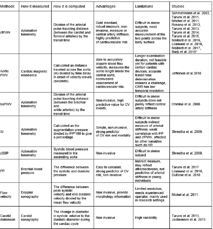
An extensive overview of all means to measure arterial stiffness and recommendation for their clinical application has been published by the European Network for Noninvasive investigation of Large Arteries (Laurent et al., 2006) and by an American Heart association panel (Townsend et al., 2015). Among all type of measurements, the methods used to establish a correlation with cognitive performance have been recently highlighted in the special issue on Vascular Dementia in the Journal of Neurochemistry (Iulita et al., 2018). The carotid-femoral pulse wave velocity (cfPWV) is currently the gold standard measure for central arterial stiffness. cfPWV reflects the time taken for the transmission of the arterial pulse wave from the carotid to the femoral artery andcan be assessed by the non-invasive procedure of applanation tonometry, following the Van Bortel protocol (Van Bortel et al., 2012). Briefly, cfPWV is measured twice by dividing arterial pulse traveling distance by the transit time and expressed in meters per second. The arterial pulse Figure 1 - Normal values for pulse wave velocity: average according to age (1455 subjects).
traveling distance is measured as the straight distance between the carotid and femoral measurement sites using a tape ruler whereas the transit time is determined from the time delay between proximal and distal “foot” waveforms assessed by placing a tonometer both in the carotid and the femoral artery (Millasseau et al., 2005). The cfPWV is then calculated by taking the mean of the two measurements. However, other measures of arterial stiffening have been used in the literature. In table 1 we provide an overview of the arterial stiffness measures used in all papers we refer to in the following sections of this review.
N.B. : cfPWV= carotid-femoral pulse wave velocity, baPWV= brachial ankle pulse wave
elocity, AI= augmentation index, cSBP= central systolic blood pressure, PP= pulse pressure, CV= cardiovascular, CMR= cardiac magnetic resonance.
2.4 Overview of histopathological characteristics of cerebral white matter and gray matter aging
As we age, the dampening of pulsatile flow becomes less efficient (O’Rourke and Hashimoto, 2007). The progressive increase in collagen coupled with the degradation and fragmentation of elastin fibers contribute to stiffer blood vessels, leading to reduced microvascular reactivity through atherogenic, hypertrophic and inflammatory responses (Iulita et al., 2018; Pase, 2012). The effects of these changes on arterial walls are amplified by the early returns of pulse wave reflection leading to an increase in arterial pulse pressure which extends into smaller vessels and impacts the integrity of the brain and kidneys (O’Rourke and Safar, 2005). The brain and kidney vulnerability to central hemodynamic alterations could be accounted by their low resistance and impedance in addition to the short distance between arterioles and large arteries (Ito, 2012; O’Rourke and Safar, 2005).
Higher aortic stiffness is also associated with lower cerebral blood flow and global cerebrovascular reserve in humans (DuBose et al., 2018; Jefferson et al., 2018). This may be critical in brain regions more vulnerable to lower perfusion such as the white matter. Indeed, small white matter lesions are (WML) often localized in watersheds regions between the tissues supplied by the anterior, posterior and middle cerebral arteries, which correlate with the presence of symptomatic cerebrovascular insufficiency (Minkner et al., 2005). In addition, the deep white matter is essentially perfused by long medullary arterioles arising from the anterior and middle cerebral artery (Brown and Thore, 2011) which become tortuous with age (Brown et al., 2002; Hassler, 1965; Moody et al., 1991; Thore et al., 2007). This tortuosity appears as early as the 5th decade (Akima et al., 1986) and is present in all individuals above 80 years old (Beskow et al., 1971). In addition, arteries in the white matter arise from medullary arteries, which are end arteries coming from pial arteries that penetrate the cortical layers before entering the periventricular white matter regions (Takahashi and Others, 2013). Thus, it is not surprising that vascular changes affect both the white as well as gray matter in the brain.
Although cerebral aging is a complex process associated with a high degree of inter-individual variability, structural MRI can be used to identify non-disease related aging of the cerebral white and gray matter (Gunning-Dixon et al., 2009). On MRI, there is little doubt that the brain shrinks with age and that this shrinkage accelerates after the age of 50 (Gunning-Dixon et al.,
2009; Raz and Rodrigue, 2006). However, there is still a debate on whether the decline is more pronounced in white or gray matter. Indeed, several studies have shown a greater age-associated decline in white matter volume in the absence of gray matter loss (Allen et al., 2005; Bartzokis et al., 2003; Gunning-Dixon et al., 2009; Guttmann et al., 1998; Jernigan et al., 2001), whereas other studies have shown the opposite effect, with greater age-associated gray matter loss compared to the white matter (Blatter et al., 1995; Sullivan et al., 2004; Thompson et al., 2003). A number of factors could explain these conflicting results, such as the presence or absence of cardiovascular risk factors.
Figure 2 - Arteries and veins of the white and gray matter.
N.B.:
a) Illustration of intracortical arteries (red) and veins (blue) in relation to brain tissue. The arteries originate from the cortical surface. Some arteries penetrate the gray matter and perfuse white matter exclusively whereas other perfuse both the gray and white matter. b) Ink-labelled vascular network in striate cortex illustrating reduced vasculature from gray to
white matter. Layer IV of the gray matter has the most intensive vascular density (green arrows). Gray and white matter boundary is indicated by yellow lines.
2.5 Macro and Microstructural changes of the cerebral white matter related to arterial stiffness
Our understanding of the vascular pathogenesis of cognitive decline has improved in recent years. Arterial stiffness has previously been found to be associated with several neuroimaging features of small vessel disease such as silent cerebral infarcts (Tsao et al., 2013), lacunes (Hatanaka et al., 2011), microbleeds (Ochi et al., 2010) and WMHs , as assessed by T2-weighted or fluid-attenuated inversion recovery (FLAIR) images (Singer et al., 2014). For instance, Rosano et al. found that arterial stiffness, as assessed by cfPWV, is associated with WMH volume in the left superior longitudinal fasciculus in older adults (Rosano et al., 2013). This association remained significant after adjustment for blood pressure, the presence of diabetes mellitus and other markers of vascular conditions, suggesting that arterial stiffness is an independent predictor of white matter abnormalities. Moreover, Shrestha et al. reported an association between higher arterial stiffness and WMHs in periventricular and deep white matter (Shrestha et al., 2009) whereas Ohmine et al. found an association only in periventricular areas (Ohmine et al., 2008). These conflicting results could be due to the fact that both studies used different approaches to measure arterial stiffness. Shrestha et al. used measures of augmentation index which is an indirect measure of arterial stiffness that could be affected by ventricular ejection and peripheral hemodynamic changes, whereas Ohmine et al. used brachial-ankle PWV which reflects central artery stiffness (cf table 1).
Despite discordance in the spatial distribution of WMHs in the relationship with arterial stiffness, several brain regions have been highlighted in the reviewed DTI literature to be vulnerable to hemodynamic changes and to precede the development of WMHs. These regions include the corpus callosum, the internal capsule, the corona radiata, and the superior longitudinal fasciculus (Badji et al., 2018; Maillard et al., 2017, 2016; Tarumi et al., 2015). Indeed, conventional MRI techniques such as FLAIR only dichotomize white matter tissue into abnormal and normal tissue and do not quantitatively assess the white matter microstructure (Haller et al., 2013). In contrast, DTI provides a semi-quantitative evaluation of the underlying white matter tissue alteration at the voxel level (Wardlaw et al., 2015) by measuring water diffusion in white matter tracts. Interestingly, when the diffusion of water molecules is restricted by the presence of physical barriers (e.g axonal membranes and myelin), the signal becomes
imprinted with signatures of the confining geometry. Therefore, the diffusion signal carries valuable physiological information about tissue microstructure such as axonal membranes and myelin (Mori and Zhang, 2006; Palombo et al., 2018). For instance, FA measures the anisotropy of water diffusion (e.g. close to 0 if water diffuses equally in all directions like in pure water, close to 1 if water diffuses preferentially along a single direction like along white matter axons), while RD reflects the apparent diffusion coefficient orthogonal to the principal diffusion direction axis (e.g. perpendicular to the main axon bundle). In a study looking at DTI changes within WMHs over a three-year period, Maillard et al. showed significant DTI changes over time in these WMHs, but no change using FLAIR imaging (Maillard et al., 2014). This could indicate that microstructural white matter changes precede the development of WMHs visible in FLAIR imaging. In a more recent study, with 18 cognitively normal and 36 mild-cognitively impaired participants (55-80 years) showing no significant difference in DTI metrics, Tarumi et al. reported not only an association between PWV and WMHs but also an association between higher cfPWV, with lower FA and higher RD in the corpus callosum, the internal capsule, the corona radiata, and the superior longitudinal fasciculus (Tarumi et al., 2015). In another larger study, Maillard et al. reproduced the association between cfPWV and white matter injury as assessed by DTI in the corpus callosum, internal capsule and corona radiata (Maillard et al., 2016) in 903 participants from the Framingham Heart Study Third Generation (mean age, 48 ±8.7 years). Such associations were found to be accentuated by age and attenuated by antihypertensive treatment, and were present as early as the fifth decade (Maillard et al., 2016). Recently, Maillard et al. investigated for the first time the potential of Free water imaging as a biomarker of subtle cerebrovascular injury. Free water reflects the fraction of water molecules that diffuse equally in all directions of space (an equivalent of FA=0). Using mediation analysis, Maillard et al. shed light on a potential pathophysiological cascade triggered by arterial stiffness and elevated blood pressure. Arterial stiffness and elevated blood pressure were associated with increased Free water content. Moreover, Free water had a direct effect on WMHs, but this effect was mediated by FA (Maillard et al., 2017). Interestingly, Tarumi et al. and Maillard et al. formulated two hypotheses: The first one is that alterations in white matter fiber tracts related to increased arterial stiffness are due to axonal demyelination (Maillard et al., 2017; Tarumi et al., 2015) and the second one is that the increase of free water following arterial stiffening may
result in axonal dispersion, lessening the constraint of water directionality along axons, as reflected by FA (Maillard et al., 2017). However, both groups used diffusion imaging for their experiment, which does not provide a specific measure of myelin integrity. Indeed, with an image resolution on the order of millimetres, diffusion MRI conflates information not only from thousands of neurons but also from various cell types such as myelin-producing cells (e.g. oligodendrocytes). Since the diffusion MRI signal averages, the contributions emerging from all compartments, the signal might reflect a number of physiological changes including demyelination (Assaf and Pasternak, 2008). Therefore, diffusion imaging provides only an indirect way to measure demyelination (Song et al., 2005), as this technique can be affected by confounding factors such as tract architecture and axon degeneration (Wheeler-Kingshott and Cercignani, 2009). As such, Tarumi et al. and Maillard et al. were not able to test their hypotheses.
In contrast, other MRI contrasts such as magnetization transfer (MT) imaging sensitize the MR signal to hydrogen atoms bound to macromolecules and thus provide a means to estimate myelin volume fraction (Edzes and Samulski, 1977; Kucharczyk et al., 1994; Wolff and Balaban, 1989). By using DTI and MT to assess axonal integrity and myelination of nerve fibers respectively, Badji et al. showed that arterial stiffness as assessed by cfPWV was associated with axonal degeneration rather than demyelination (Badji et al., 2018). Furthermore, despite their sensitivity to tissue microstructure, DTI metrics lack specificity for individual tissue microstructure feature (Zhang et al., 2012). Indeed, it can be hypothesized that the observation of a reduction in FA may be due to various microstructural tissue changes such as a reduction in neurite density or an increase in the dispersion of neurite orientation distribution (Beaulieu, 2009). In contrast, newer diffusion MRI techniques such as orientation dispersion and density imaging (NODDI) based on multi-shell protocol provide a model of white matter microstructure that consists of individual compartments for glial cells, axons and extracellular space that can be used to infer information about neurite density and orientation dispersion (Zhang et al., 2012).
Although NODDI has not been used yet to investigate the effects of arterial stiffness in the brain, Suzuki et al. recently compared NODDI metrics in hypertensive vs non-hypertensive participants and found a difference in both neurite density and orientation dispersion (Suzuki et
al., 2017). Moreover, Suzuki et al. also compared NODDI metrics between non-hypertensive and pre-hypertensive participants and found a higher orientation dispersion in the right superior longitudinal fasciculus and the right superior thalamic region. However, Suzuki et al. did not control for arterial stiffness, which may be the underlying parameter leading to alterations in white matter microstructure. This suggests that changes in orientation dispersion might be the earliest microstructural alterations related to hemodynamic changes. Further studies using the gold standard measure of arterial stiffness might need to rely on more advanced models to map the white matter microstructure (e.g. NODDI) in order to help researchers better understand the impact of arterial stiffness on axonal integrity.
Despite all these encouraging avenues, in-vivo probing of tissue microstructure with MRI has its caveats. Indeed, Novikov et al. raised concerns regarding the distinction between a biophysical model and its relevance to an actual physical phenomenon (Novikov et al., 2018). While more work is needed in the realms of microstructural modelling to accurately probe the white matter microstructure, these new biophysical models might help us better understand the effects of arterial stiffness on the white matter microstructure. Considering the fact that microstructural changes following arterial stiffening may be reversible as opposed to WMH (Fernando et al., 2006; Maillard et al., 2014), their early detection may be an important target for biomarker discovery.
N.B.:
White matter regions based on the ICBM-DTI-81 atlas are overlaid on an FA map of one healthy older adult. 1: corpus callosum, 2: internal capsule, 3: corona radiata, 4: superior longitudinal fasciculus. These white matter regions have been identified as vulnerable to Figure 3 - White matter regions denoted vulnerable to arterial stiffness.
arterial stiffness based on the reviewed literature. This means that these regions are more susceptible to microstructural changes following an increased arterial stiffness in the elderly.
2.6 Macro and Microstructural changes of the cerebral gray matter related to arterial stiffness
Several cross-sectional studies have shown a link between increased arterial stiffness and white matter changes in the brain. However, limited research has attempted to understand the impact of arterial stiffness on gray matter structure. Gray matter changes following arterial aging can be assessed using T1-weighted MRI and manifest essentially as reduced gray matter volume (GMV) or density. Schmahmann et al. and Maillard et al. found a significant association between higher PWV and lower gray matter density in young adults, particularly in the bilateral thalamic regions (Maillard et al., 2016; Schmahmann, 2003). This association was found to be accentuated by age and attenuated by antihypertensive treatment (Maillard et al., 2016). In addition, Maillard et al. found that these thalamic structural brain changes were present as early as the 5th decade (Maillard et al., 2016). By contrast, no relationship was found between higher cfPWV and GMV among older adults, in the Reykjavik Study (Mitchell et al., 2011). However, the authors looked also at the impact of carotid flow pulsatility on brain integrity using ultrasonography, and found that higher carotid pulsatility was associated with lower GMV(Mitchell et al., 2011). This result is consistent with the important role of carotid stiffness on brain health and the idea that excessive pulsatility following arterial stiffening leads to microvascular dysfunction and alteration in the brain (Mitchell et al., 2011). This hypothesis is further supported by work from Jochemsen et al. examining the associations between carotid distension, total brain volume, WML volume, and cortical volume among 526 older participants (Jochemsen et al., 2015). Jochemsen et al. found that lower carotid distension was related to a lower brain volume (both total and cortical) and a larger WML volume. However, after a mean follow-up of 4.1 years (in average), further stiffening of the carotid arteries did not lead to changes in total GMV or WML volume (Jochemsen et al., 2015). Although this is the first study to examine the relationship between arterial stiffness, the progression of brain atrophy and WML volume at the same time longitudinally, a potential explanation for these findings could be that changes over 4 years are modest compared to changes that occur across the lifespan to
older age. The authors further argue that arterial stiffness was mild in their population compared to other older cohorts and may not have been severe enough to lead to brain pathology (Jochemsen et al., 2015). In contrast, a previous study found that increased aortic stiffness was associated with larger WML volume in an older population 10 years later but the authors did not report on change in WML volume over time (Rosano et al. 2013).
However, it is still unclear what the local changes in T1-weighted signal intensity reflect at the microscopic scale (Tardif et al., 2016). Considering the fact that T1 is sensitive to several physiological features (e.g. myelin, fat, protein-rich fluid, melanin etc.), it is unlikely that a single cellular mechanism leads to the macroscopic brain changes detected using conventional T1-weighted images, making these images mainly qualitative and physiologically non-specific (Tardif et al., 2017, 2016). Furthermore, it has been shown that differences in grey matter volume could be caused by vasodilation or differences in blood volume (Tardif et al., 2017). This could be especially problematic in aging or diseases such as vascular conditions and dementia, where reductions in vascular density (and therefore blood volume) have been demonstrated (Montagne et al., 2016; Tardif et al., 2017). As such, GMV is biologically ambiguous and should be interpreted with caution as a marker of structural integrity (Tardif et al., 2016).
To address some of these issues, more quantitative MRI techniques have recently been used to better understand the link between arterial stiffness and gray matter integrity, in particular ASL. This technique provides a non-invasive, highly repeatable quantitative measure of human brain perfusion by manipulating the magnetic resonance signal of inflowing blood in feeding arteries before it is delivered to the capillary bed of different brain areas (Golay and Petersen, 2006). Using ASL, Tarumi et al. examined the association between cfPWV and regional cerebral perfusion within gray matter regions including the hippocampus, thalamus and caudate nucleus in 35 middle-age adults, but did not find evidence of significant associations (Tarumi et al., 2011). However, participants with higher cfPWV, defined as >1,090 cm/s, showed significantly lower cerebral perfusion in the hippocampus compared to participants with lower cfPWV (Tarumi et al., 2011). Considering the fact that participants from the Tarumi et al. study were between 40-60 years old, a potential explanation for the null results could be that changes in
gray matter structure following arterial stiffening follow a protracted time course and were thus not yet visible in this cohort (Maillard et al., 2016).
Additionally, in a more recent study, Jefferson et al. (Jefferson et al., 2018) quantified thoracic aortic stiffening from cardiac magnetic resonance and found reduced cerebral blood flow as assessed by ASL in cognitively normal older adults free from clinical stroke and dementia. More specifically, higher aortic PWV was related to lower frontal cerebral blood flow and higher cerebrovascular reactivity (CVR), in the frontal, temporal and occipital lobes, as well as in the whole brain (Jefferson et al., 2018). The latter suggests that aortic stiffening may be associated with cerebral hypoperfusion in the presence of preserved CVR (Jefferson et al., 2018). Interestingly, APOE-ε4 carriers were found to have higher PWV, lower cerebral blood flow in the whole brain and in the temporal lobe, as well as a higher CVR in the temporal lobe compared to non APOE-ε4 carriers (Jefferson et al., 2018). This is particularly relevant as individuals who inherit the APOE-ε4 allele have a genetic predisposition making them at higher risk of developing Alzheimer’s disease (Liu et al. 2013). Moreover, the relationship between CVR and PWV was regionally specific to the temporal lobes, where Alzheimer’s disease pathology first appears (Braak and Braak, 1991). Furthermore, Lilamand et al. looked at the association between PWV, and medial temporal lobe (MTL) atrophy in older adults (Lilamand et al., 2016). By categorizing participants of their study into three groups (no atrophy, mild atrophy, severe atrophy), the authors found that PWV was significantly associated with severe MTL atrophy (Lilamand et al., 2016), suggesting that arterial stiffness can potentially increase hypoperfusion of the MTL.
Despite these results, the relationship between cfPWV and cerebral blood flow remains unclear with some studies showing no reductions with cerebral blood flow, in particular in young individuals (Tarumi et al., 2014, 2011). Similarly, in a subset of older adults, aortic stiffness, brachial systolic blood pressure and pulse pressure were not correlated with resting global cerebral blood flow even after adjusting for age and sex (DuBose et al., 2018). While this could be indicative of a lack of association between arterial stiffness and cerebral blood flow in some populations at least, ASL sequences also suffer from some technical limitations. The contrast afforded by the subtraction of tagged images is only a fraction of a percent of the functional MRI contrast, providing a limited signal-to-noise ratio (SNR). Furthermore, the more commonly
used single delay ASL cannot measure transit times directly which further limits SNR (Golay and Petersen, 2006). As such, most ASL measurements are unable to determine whether the changes detected are true reflections of changes in flow or the result of alterations in transit times. Multi-delay implementations can, however, alleviate some of these problems (Golay and Petersen, 2006), and recommendations from a recent paper on ASL in aging and disease can help optimize parameter selection for the population studied (Alsop et al., 2015). Finally, coverage of the brain is typically incomplete, limiting the ability to draw conclusions on the entire brain. Multi-band approaches are however promising in this regard (Li et al., 2015). Taken together, it is important to note that ASL measurements should be interpreted with some caution, taking into account the methodological quality of the implementation (e.g. what post-label delay times were used, whether there are multiple delays, etc.). As such, to better understand the relationship between arterial stiffness and cerebral blood flow, longitudinal studies using the gold standard measure of arterial stiffness and multi-delay ASL are necessary.
2.7 Bridging the gap between arterial stiffness, cerebral blood flow and microstructural integrity.
As mentioned in previous sections, arterial stiffness has been consistently associated with white as well as gray matter changes in the elderly population (Gunning-Dixon et al., 2009; Singer et al., 2014; Tarumi and Zhang, 2017). Interestingly, the interplay between cerebral blood flow and white matter health have also been investigated. In general, a good agreement in the literature exists, highlighting a strong interplay between cortical blood flow and white matter microstructure (Chen et al., 2013; Tarumi and Zhang, 2017). In particular, Tarumi et al. have shown that participants with a higher 24h ambulatory measurement of pulse pressure had a lower diastolic cerebral blood flow, which accounted for 15-20% of the reduction in white matter fiber integrity as assessed by DTI (Tarumi et al., 2017). In addition, white matter cerebral blood flow has also been shown to be significantly associated with white matter microstructural integrity (Aslan et al., 2011; Giezendanner et al., 2016). However, conflicting results exist in the literature regarding the direction of the correlation. Aslan et al. showed that cerebral blood flow measured in various white matter tracts was inversely correlated with FA (Aslan et al., 2011) whereas Giezendanner et al. found a positive correlation between cerebral blood flow in the white matter and FA (Giezendanner et al., 2016). A potential mechanism for the observed negative
correlations found by Aslan et al. is that white matter cerebral blood flow may be related to the axonal diameter of white matter tract (Aslan et al., 2011). Indeed, Aslan et al. showed that the cerebral blood flow of these white matter tracts was negatively correlated with FA and Axial diffusivity, but was positively correlated with RD (Aslan et al., 2011). However, as mentioned previously, DTI is not well suited to disentangle the exact underlying microstructural properties that contribute to the observed relationships between anisotropy and cerebral blood flow (Beaulieu, 2002; Giezendanner et al., 2016; Miller et al., 2007). Advances in diffusion models such as NODDI (Zhang et al., 2012), CHARMED (composite hindered and restricted model of diffusion) (Assaf and Basser, 2005) or TE dependent Diffusion Imaging (TEdDI) (Veraart et al., 2018) might be of great interest to probe the nature of the complex relationship between cerebral blood flow and the underlying microstructural characteristics in the white matter. Considering the fact that white and gray matter share a common vascular blood supply, a potential explanation underlying the mechanisms between arterial stiffness, white matter microstructural integrity and cerebral blood flow is that arterial stiffening of large arteries impacts both white and gray matter structure based on their respective cerebral blood flow. Indeed, sustained inadequate cerebral blood flow may reduce the delivery of oxygen and glucose to neuronal cells, thereby slowly initiating a pathway of progressive alteration in brain integrity, neuronal metabolism and cognitive decline (de la Torre, 1999, 1997, 1994; de la Torre and Mussivand, 1993). Studies with arterial stiffness measures (cfPWV), advanced diffusion model (NODDI, CHARMED, TEdDI etc), and measures of cerebral blood flow both in the white and gray matter (ASL) in healthy individuals are needed to test this hypothesis.
However, one should note that despite the recent advances in ASL technologies, the SNR of ASL in the white matter is known to be relatively poor due to small perfusion fraction and long transit times (Van Gelderen et al., 2008). Experimental data shows that it might be possible to measure white matter perfusion by using an appropriate tagging duration and post-labelling delay in healthy individuals (Wu et al., 2013) but a further extension of pulse labelling delay might be needed when ASL is acquired in the elderly populations and/or individuals experiencing ischemia as prolonged transit time is expected (Wu et al., 2013). Another alternative to improve the accuracy of the quantification of the white matter cerebral blood flow is the use of multiple pulse labelling delays which however requires longer scan time.
2.7 Arterial stiffness and brain integrity: Insight from other modalities
Although MRI appears to be the modality of choice to study the impact of arterial stiffness on brain integrity, other modalities such as positron emission tomography (PET), and transcranial Doppler (TCD) have been used (Jefferson et al., 2018; Zhu et al., 2013). For instance, Dubose et al. quantified global cerebral blood flow in 205 adults using quantitative [15O] water PET to
look at its relationship with cfPWV, as well as cerebrovascular reserve measured as the change in global cerebral blood flow after intravenous infusion of acetazolamide. Interestingly, Dubose et al. reported not only a lower global cerebral blood flow in older adults compared with younger adults but also an association between higher cfPWV and lower cerebrovascular reserve. This association remained significant after adjusting for age, sex, and mean arterial pressure. In contrast, aortic stiffness (as assessed by cfPWV) was not related to global cerebral blood flow but the authors argue that increased arterial stiffness may impair the cerebrovasculature’s ability to dilate in response to a vasodilatory challenge and therefore reduce its ability to increase global cerebral blood flow in older adults (DuBose et al., 2018). In another study, Jaruchart et al. replicated these findings in 28 healthy sedentary young (mean age, 25±1 years) and older adults (mean age,67±1 years) using brachial-ankle PWV and TCD (Jaruchart et al., 2016) which was used to measure cerebrovascular conductance and reactivity in response to a physiological CO2 stimulus, and basal CBF. The authors reported a reduction in cerebral blood flow velocity and cerebrovascular conductance in older adults compared with younger adults. In addition, arterial stiffness was significantly and inversely associated with cerebral blood flow velocity and cerebrovascular conductance among older adults (Jaruchart et al., 2016). These findings suggest that CVR may be an important measure to implement in future studies to better understand the physiological changes underpinning the relationship between higher arterial stiffness and brain integrity. However, one must note that the TCD technique also suffers from several limitations. For instance, although this modality assesses CVR by measuring the change in cerebral blood flow velocity in the middle cerebral artery following a hypercapnic challenge, it assumes that hypercapnia does not induce a diameter change in the middle cerebral artery (Coverdale et al., 2014). As such, TCD may underestimate cerebral blood flow velocity.
Other modalities than PET and TCD and have been used to study the impact of arterial stiffness in the brain such as resting-state magnetoencephalography (MEG). For instance, Nieboer et al.
examined the association between carotid stiffness (as assessed by ultrasound imaging) and functional connectivity, computed from the phase lag index of resting-state magnetoencephalography (MEG) in 230 young healthy adults (mean age= 42±0.7) (Nieboer et al., 2016). The phase lag index estimated the asymmetry in the distribution of phase differences between two-time series and was assessed in six frequency bands (δ–γ)(Nieboer et al., 2016). Interestingly, carotid stiffness was found to be significantly associated with increased functional connectivity in the α2 band in men and β band in woman after adjusting for a set of covariates (participants’ height, mean arterial pressure, body fat percentage and level of triglyceride to HDL-C)(Nieboer et al., 2016). Although the authors did not find a significant association between cognitive performance and functional connectivity, they suggested that the increase in connectivity following arterial stiffening might be due to: i) either the effect of a compensatory mechanism in order to maintain brain homeostasis such as the one reported in individuals with mild cognitive impairment (Bajo et al., 2012) or ii) the effect of a pathological mechanism leading to neurodegeneration (de Haan et al., 2012; Nieboer et al., 2016). Given the absence of other literature on the link between connectivity and arterial stiffness using MEG or other, MRI-based measures of connectivity (BOLD connectivity, structural connectivity etc), future longitudinal studies looking at the relationship between changes in arterial stiffness and brain connectivity are needed to confirm these hypotheses.
Taken together, other imaging modalities than MRI have provided valuable knowledge towards a better understanding of the underlying mechanisms underpinning brain changes associated with vascular aging. However, contradictory results also exist, in particular related to the relationship between PWV and CVR. Indeed, some authors have reported reductions in CVR among older adults with greater aortic stiffness using PET (DuBose et al., 2018) and TCD (Jaruchart et al., 2016) while others demonstrate preserved CVR in the presence of higher aortic PWV using ASL (Jefferson et al., 2018; Zhu et al., 2013). Although these modalities (TCD, PET, ASL) measure CVR differently, these results indicate that the relationship between PWV and CVR is highly complex. As such, additional studies using different imaging modalities should further investigate the impact of other markers of cerebral health to better understand the impact of hemodynamic changes in the aging brain.
2.8 Arterial stiffness and cognition
In general, there is a good agreement in the literature showing that higher PWV is associated with alterations in cognitive performance and cognitive decline independently of other cardiovascular risk factors (Cui et al., 2018; Iulita et al., 2018; Pase et al., 2012; Singer et al., 2014). Cross-sectional evidence suggests that greater arterial stiffness is associated with poorer performance in age-sensitive domains such as processing speed, executive skills, working memory, and episodic memory (Iulita et al., 2018; Pase et al., 2012; Singer et al., 2014). However, some discrepancies exist in the literature, mainly due to different study designs, target populations and heterogeneity of cognitive screening tools (Iulita et al., 2018). For instance, Hanon et al. (Hanon et al., 2005) looked at the relationship between arterial stiffness and global cognitive function by means of the Mini-Mental State Examination (MMSE) in a population of elderly adults experiencing memory loss and found PWV to be inversely correlated with MMSE. Although these results are in line with other studies (Benetos et al., 2012; Fukuhara et al., 2006; Scuteri et al., 2005; Zhong et al., 2014), the Rotterdam study provides contradictory findings (Poels et al., 2007). Indeed, this longitudinal population-based cohort study ongoing since 1990 investigates the factors that determine the occurrence of cardiovascular and neurological diseases in the elderly and did not reveal any associations between PWV and cognitive decline as assessed by the MMSE (Poels et al., 2007). However, after adjustment for cardiovascular risk factors, an association between increased PWV and poorer performance on the Stroop test was found (Poels et al., 2007). While the MMSE is a useful screening tool for advanced cognitive decline and dementia, it is a non-specific indication of the general mental state that has important limitations such as the lack of standardization and sensitivity to mild cognitive impairment (J. Larner, 2012). Thus, it should be interpreted with caution as it may not be sensitive enough to measure subtle cognitive changes. In contrast, the literature has shown associations between arterial stiffness and individualized domains of cognitive function including executive function, memory, processing speed, verbal learning, and visual-spatial function. For instance, although the Baltimore study did not find aortic stiffness to predict MMSE scores, significant associations were found between PWV and a decline in verbal learning and memory performance (Waldstein et al., 2008). In addition, Elias et al (Elias et al., 2009) found PWV to be associated with poorer performance in visual-spatial organization, and
memory and verbal episodic memory after adjusting for cardiovascular risk factors. These results are also strengthened with other cross-sectional studies showing progressively lower performance on processing speed and executive function in relation to increased arterial stiffness (Tarumi et al., 2013; Tsao et al., 2013; Waldstein et al., 2008).
Taken as a whole, these results suggest that arterial stiffness impacts multiple brain regions as it has been associated with a decrease in various cognitive domains. However, the pathophysiological mechanisms through which arterial stiffness impacts the brain are complex and poorly understood. A variety of neuroimaging techniques such as DTI and ASL allow us to gain some insight into the functional and structural brain changes related to arterial stiffening, and interestingly the literature has already provided evidence of a strong interplay between white matter changes and cognitive performance (Badji et al., 2018; Tarumi et al., 2015). Indeed numerous studies have reported that WMH burden correlates with poorer performance in age-sensitive domains, including executive skills and memory (Cook et al., 2004; Gunning-Dixon and Raz, 2000; Kramer et al., 2007; Pase et al., 2012; Singer et al., 2014). Moreover microstructural integrity of the regions known to be vulnerable to the effects of arterial stiffness (e.g.., corpus callosum, internal capsule, superior longitudinal fasciculus) have also been found to be associated with cognitive performance. For instance, Sasson et al. reported that executive function is correlated with DTI changes in the superior longitudinal fasciculus (Sasson et al., 2013) and Madden et al. reported that the best predictor of response time in a visual task was a higher FA in the corpus callosum for older adults and a higher FA in the internal capsule for the elderly (Madden et al., 2004). In addition, Tarumi et al. (Tarumi et al., 2015) and Badji et al. (Badji et al., 2018) showed that cognitive flexibility as assessed by the Trail making test B-A was positively correlated with FA and negatively correlated with RD in the regions known to be vulnerable to age-related hemodynamic changes, in particular the corpus callosum and the corona radiata (Badji et al., 2018). These results are not surprising considering the established relationship between higher arterial stiffness, white matter changes and cognitive decline (Singer et al., 2014). Furthermore, MRI measures of brain gray matter structure as assessed by T1-weighted have also found a strong association between GMV and cognitive function. For instance thalamic volume, which has been denoted to be vulnerable to arterial stiffness (Maillard