HAL Id: dumas-01826169
https://dumas.ccsd.cnrs.fr/dumas-01826169
Submitted on 29 Jun 2018
HAL is a multi-disciplinary open access archive for the deposit and dissemination of sci-entific research documents, whether they are pub-lished or not. The documents may come from teaching and research institutions in France or abroad, or from public or private research centers.
L’archive ouverte pluridisciplinaire HAL, est destinée au dépôt et à la diffusion de documents scientifiques de niveau recherche, publiés ou non, émanant des établissements d’enseignement et de recherche français ou étrangers, des laboratoires publics ou privés.
Electro-clinical correlation between seizures induced by
direct electrical cortical stimulation and spontaneous
seizures: relevance to define the epileptogenic zone
Nastasia Tardy
To cite this version:
Nastasia Tardy. Electro-clinical correlation between seizures induced by direct electrical cortical stim-ulation and spontaneous seizures: relevance to define the epileptogenic zone. Human health and pathology. 2017. �dumas-01826169�
AVERTISSEMENT
Ce document est le fruit d'un long travail approuvé par le
jury de soutenance et mis à disposition de l'ensemble de la
communauté universitaire élargie.
Il n’a pas été réévalué depuis la date de soutenance.
Il est soumis à la propriété intellectuelle de l'auteur. Ceci
implique une obligation de citation et de référencement
lors de l’utilisation de ce document.
D’autre part, toute contrefaçon, plagiat, reproduction illicite
encourt une poursuite pénale.
Contact au SID de Grenoble :
bump-theses@univ-grenoble-alpes.fr
LIENS
LIENS
Code de la Propriété Intellectuelle. articles L 122. 4
Code de la Propriété Intellectuelle. articles L 335.2- L 335.10
http://www.cfcopies.com/juridique/droit-auteur
1
UNIVERSITÉ GRENOBLE ALPES
UFR DE MÉDECINE DE GRENOBLE
Année : 2017
Electro-clinical correlation between seizures
induced by direct electrical cortical stimulation and
spontaneous seizures: relevance to define the
epileptogenic zone
THÈSE PRÉSENTÉE POUR L’OBTENTION DU TITRE DE DOCTEUR EN MÉDECINE DIPLÔME D’ÉTAT
TARDY Nastasia
Thèse soutenue publiquement le 20 octobre 2017 à la Faculté de médecine de Grenoble, devant un jury composé de:
Monsieur de Professeur Philippe Kahane, Président du jury Monsieur le Professeur Fabrice Bartolomei
Monsieur le Professeur Stéphan Chabardes Monsieur le Professeur Olivier David Monsieur le Professeur François Dubeau
Madame le Docteur Lorella Minotti, directrice de thèse
L’UFR de Médecine de Grenoble n’entend donner aucune approbation ni improbation aux opinions émises dans les thèses ; ces opinions sont considérées comme propres à leurs auteurs.
2
Remerciements à messieurs Fabrice Bartolomei, Stephan Chabardes, Olivier David et François Dubeau pour avoir accepté d’expertiser ce travail et d’en composer le jury.
Recevez toute ma gratitude.
J’exprime particulièrement ma sincère reconnaissance à Lorella Minotti et Philippe Kahane, qui m’ont encadrée chaleureusement tout au long de ce travail, et pendant mon cursus.
7
Introduction
The primary aim of epilepsy surgery is to remove the epileptogenic zone (EZ), i.e. ‘the
minimum amount of cortex that must be resected (inactivated or completely disconnected)
to produce seizure freedom’ (Rosenow and Lüders, 2001). In this context, intracranial EEG
(iEEG) can be performed when non-invasively obtained data remain insufficiently
concordant, when they are discordant or inconclusive, or when they suggest an early
involvement of highly eloquent areas (Jayakar et al., 2016). Such recordings aim not only
at precisely identifying the cortical areas to be removed, but also at evaluating their
functional integrity. This needs on the one hand the recording of spontaneous seizures, and
on the other hand the use of cortical electrical stimulation (CES) to map eloquent cortical
areas that will have to be spared during surgery.
Although the iEEG definition of the EZ mainly relies on the recording of spontaneous
seizures (SS), some teams, mainly in France and Italy, routinely use CES during stereotactic
iEEG (SEEG) recordings to aid in delineating the EZ, providing that the patient's typical
electro-clinical pattern is produced (Kahane et al., 2006; Trébuchon and Chauvel, 2016).
This approach, promoted by the Hospital Sainte Anne school (Bancaud and Chauvel,
1986), is considered as a necessary complement of SS recording, particularly when
spontaneous ictal discharges are widely extended from the onset, when they apparently arise
independently from different brain structures, or when regions that are initially silent at
seizure onset abruptly activate in the course of the ictal discharge. The clinical utility of
CES-induced seizures (CESS), however, remains controversial and most of north-american
centers using subdural electrodes do not rely on such information because they consider and
CES-induced afterdischarges and seizures as a by-product of cortical mapping that can
disrupt the procedure (Bank et al., 2014), may inaccurately localize cortical functions
8
and the SS onset area (Blume et al., 2004). This controversy is partly linked to the great
variability in the methodologies used across centers, to the lack of a consensual definition of
CESS, and to the differences in the conceptual approach to epilepsy surgery.
Suprisingly and despite decades of intra- and extraoperative CES procedures in epilepsy
surgery candidates, only a few studies have investigated the relevance of CESS in clinical
practice. In a recent review, 14 studies were identified during a 30 years period (1974-2004),
of which 9 used SEEG electrodes, 3 intraoperative CES, and 2 subdural electrodes (Kovac
et al., 2016). Overall, the percentage of patients in whom seizures could be elicited varied
from 37 to 100%, with a large predominance of CES-elicited auras. The concordance rate
between the electrodes exhibiting CESS and those overlying the site(s) of SS onset,
evaluated in only 6 of these 14 studies, varied from 26% to 100%, with the best results
obtained in patients with focal cortical dysplasia or suffering from temporal lobe epilepsy
(Bernier et al., 1990; Chassoux et al., 2000; Chauvel et al., 1993; Landré et al., 2004;
Schulz et al., 1997; Wieser et al., 1979). How the information obtained from CESS might
influence seizure outcome was assessed in only one study, that did not show any differences
according to whether CESS site(s) were included in the resection or not (Schulz et al. 1997).
Based on these existing data, it thus appear that further studies are needed to better evaluate
the clinical and iEEG overlap beween SS and CESS, and to determine what is the precise
added contribution of CESS over SS in terms of seizure outcome. The present retrospective
study aimed at answering these issues in a series of 30 consecutive patients who underwent
SEEG recordings at our institution. Whether general features might influence the
9
Patients and methods
Patients selection
Between October 2013 and November 2015, 32 patients suffering from drug-resistant focal
seizures underwent a SEEG study at Grenoble-Alpes University Hospital as part of their
presurgical evaluation. For the purpose of the study were selected the 30 patients in whom at
least one complete spontaneous electro-clinical seizure was recorded during SEEG
investigation.
Patients's characteristics
The 30 patients were all suffering from drug-resistant focal epilepsy, the operability of which
could not be decided on the basis of non-invasive procedures only. These latter included in all
cases high-resolution MRI, scalp-video-EEG monitoring, and neuropsychological tests. Some
patients underwent also functional neuroimaging.
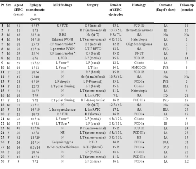
A summary of the patients’ characteristics is given in Table 1. Briefly, there were 15 males
and 15 females, whose mean age at SEEG investigation was 28 years (range 9-59 years).
Mean age at seizure onset was 15 years (range 0.5-38 years), and mean duration of epilepsy
was 14 years (range 3-40 years). MRI demonstrated different kind of lesions in 21 patients, of
whom 6 had previously undergone unsuccessful resective surgery before SEEG procedure.
Table 1: Patient’s characteristics.
are highlighted in grey (n=24). Pt: patient number; M: male; F:female; N: normal; R: right; L: left; F: frontal; T: temporal; P: parietal; C:central; Ins: insular; O: occipital ;
focal cortical dysplasia; HS: hippocampal sclerosis; MMCDOH: mild malformation of cortical developement with oligodendroglial cells; PVNH: pervicentricular nodular heterotopia; RFTC: radiofrequency thermocoagulation; NA: not applicable;
F cavernoma; #10: L F resection/disconnection (negative histology); #21: L F resection with negative MRI; #22: L T disconnection with negative MRI).
Table 1: Patient’s characteristics. Patients who experienced at least one CES-elicited electro
are highlighted in grey (n=24). Pt: patient number; M: male; F:female; N: normal; R: right; L: left; F: frontal; T: temporal; P: parietal; C:central; Ins: insular; O: occipital ; DNET: dysembryoplasic neuroepithelial tumor; FCD: focal cortical dysplasia; HS: hippocampal sclerosis; MMCDOH: mild malformation of cortical developement with oligodendroglial cells; PVNH: pervicentricular nodular heterotopia; RFTC: radiofrequency agulation; NA: not applicable; 1 previously operated (#5: R F oligodendroglioma; #7: R F DNET; #8: L
F cavernoma; #10: L F resection/disconnection (negative histology); #21: L F resection with negative MRI; #22: L T disconnection with negative MRI).
10
elicited electro-clinical seizure are highlighted in grey (n=24). Pt: patient number; M: male; F:female; N: normal; R: right; L: left; F: frontal; T: DNET: dysembryoplasic neuroepithelial tumor; FCD: focal cortical dysplasia; HS: hippocampal sclerosis; MMCDOH: mild malformation of cortical developement with oligodendroglial cells; PVNH: pervicentricular nodular heterotopia; RFTC: radiofrequency previously operated (#5: R F oligodendroglioma; #7: R F DNET; #8: L F cavernoma; #10: L F resection/disconnection (negative histology); #21: L F resection with negative MRI; #22:
11
SEEG investigation
Twelve to 18 (mean: 14.3) multicontact semirigid intracerebral electrodes were implanted in
each patient in stereotaxic conditions, according to the SEEG methodology developed in our
group (Kahane et al., 2004). Electrodes placement and number was tailored in each
individual depending on the suspected origin of seizures. Preoperative targeting was
performed using 3-D T1 brain MRI computed with a stereotactic software (ROSA stereotactic
assistance®), and using a stereotaxic and stereoscopic digitalized arteriography to determine
avascular trajectories of the electrodes. Insertion of the electrodes (DIXI Medical, Besançon,
France; diameter of 0,8 mm; 10 to 18 contacts, 2 mm length, 1.5 mm apart) was guided by a
robotic arm (Medtronic O-Arm®) that was connected to the stereotactic frame and driven by
the stereotactic software. Electrodes contacts were visualized directly on post-implantation
MRI, and/or after a computer-assisted matching of post-implantation CT-scan with a
pre-implantation 3D MRI.
SEEG recordings were conducted extra-operatively for about 2 weeks with reduced
medication in 23 of the 30 patients, using an audio-video-EEG monitoring system
(MicromedTreviso, Italy) that allowed to record simultaneously up to 128 contacts, with a
sampling rate of 512 Hz and an acquisition band-pass filter between 0.1 and 200 Hz. Data
were acquired using a referential montage, with a reference electrode chosen in the white
matter. SEEG activity was displayed between contiguous contacts at different levels along the
axis of each electrode, and interictal and ictal analysis of SEEG traces was done visually.
CES were performed for a few days during one to three hour sessions, according to our
routine procedure (Kahane et al., 1993). The aim was to reproduce part or the totality of the
ictal clinical symptomatology, and to localize functionally eloquent areas that had to be
spared during surgery. Bipolar stimuli were delivered using a constant current rectangular
pulse generator designed for a safe diagnostic stimulation of the human brain (Micromed,
12
and 50 Hz (pulse width: 0.5-1ms; ES duration: 5 sec), and applied between all contiguous
contacts located in the cortex, with stepwise increasing intensities up to 5 mA and until
clinical responses, after-discharges or seizures were obtained.
Following SEEG results, 17 patients had a left-sided, 10 a right-sided, and 3 a bilateral focal
epilepsy (Table 1). The epilepsy was unilobar in 21 cases (frontal: 11, temporal: 8, insular: 2),
multilobar in 6 (temporo-parietal: 2, temporo-central: 1, temporo-insular: 1,
temporo-insulo-opercular: 1, fronto-parietal: 1), bitemporal in 2, and bilateral multifocal in 1.
Surgery
Surgery, including radiofrequency thermocoagulation (RFTC), was performed in all but 3
cases, according to SEEG results (Table 1). The extent of the resection/RFTC was assessed on
a 3-D T1 operative MRI usually performed 3 months after surgery. After a mean
post-operative follow-up of 15 months (range: 2-38 months), 17 patients (63%) were in class I
according to Engel's classification, 2 in class II (7.4%), 2 in class III (7.4%), and 6 in class IV
(22.2%). Pathological examination, available in all the 24 resective cases, found different kind
of lesions in 13 of the 16 patients whose MRI proved positive, and in 7 of the 8 patients
without any abnormality on MRI.
Data analysis
Spontaneous seizures
All SEEG-recorded SS were reviewed, but neither subclinical seizures, nor seizures in which
auras occurred without any SEEG modification were kept for analysis.
The EZ was defined according to its electrophysiological definition, i.e. the ‘‘site of the
beginning and of the primary organization of the epileptic seizures’’ (Munari and Bancaud,
1987). The EZ therefore included the seizure onset zone (the region exhibiting the first SEEG
13
the region of early seizure spread (that was arbitrarily defined as the region involved by the
SEEG discharge within the first 20 seconds following the ictal onset). SEEG changes at
seizure onset were classified, whatever possible, according to the seizure-onset patterns
proposed by Perucca et al. (2014).
The anatomical extent of the EZ was evaluated in each patient by the number of electrode
contacts involved during the first 20 seconds of the SS. If different seizure patterns were
recorded, all the contacts involved within the first 20 seconds, whatever the seizures, were
included in the EZ.
Seizure semiology was analyzed according to a working definition of clinical phenomena
derived from the classification of Lüders et al. (1998). Briefly, a total number of 6 core
clinical signs were systematically evaluated, including aura, autonomic signs, loss of contact,
simple motor signs, complex motor signs and negative phenomena. They were quoted as
present or absent in each SS, and they were then compiled in a single list for all SS.
CES-elicited seizures
Only CES-induced electro-clinical seizures were analysed, and neither elicited isolated aura
(without SEEG discharge), nor isolated after-discharges (without clinical accompaniment),
were considered in the present work.
CESS thus referred to electro-clinical events that occurred as an immediate result of CES, and
in which both clinical signs and SEEG discharge outlasted the electrical stimulus. They were
considered as habitual (non habitual) when elicited clinical manifestations were (were not)
recognized by the patient and/or his/her relatives.
The parameters used to elicit CESS were first evaluated. Then, the CESS onset patterns,
anatomical extent of the elicited discharges, and CESS semiology were assessed using the
same criteria than those used for SS analysis. Whether the CES location within or outside the
14
Comparison between SS and CESS
A rate of electrical concordance was calculated for each patient by comparing the extent of
the EZ with the mean extent of all elicited discharges. Similarly, a rate of clinical concordance
was evaluated in each patient by comparing the mean number of clinical signs observed
during CESS with the list of SS clinical signs.
Still, whether other factors might influence the rate of electrical and/or clinical concordance
was assessed. These factors included the age at seizure onset, epilepsy duration, MRI findings
(negative versus positive), and epilepsy type (temporal versus extra-temporal).
CESS and outcome
All the post-operative MRI were reviewed to evaluate, for each patient, the percentage of
CESS contacts that were located in the resective (or thermo-coagulated) area. This percentage
was then compared with post-operative seizure outcome.
Other potential prognosis factors were also evaluated, including the number of CESS, the
percentage of contacts that elicited CESS within the EZ, the SEEG LVFA pattern at CESS
onset, and the rates of electrical/clinical concordance.
Statistics
We used Fisher’s exact test for categorical data, and Mann-Whitney's U test or Spearman's
Results
Twenty four of the 30 patients (80%) exhibited at least
characteristics did not seem to differ from the ones of the 6 patients who did not present any
CESS (Table 1). None of these later, however, were suffering from temporal lobe
(TLE), as compared with 10 of the
The following sections will refer only to these 24 patients, who exhibited both SS and CESS.
Table 2: Comparison between stimulation experienced at least one complete CES
patient; SS: spontaneous seizures; EZ: epileptogenic zone; CESS: electro (cortical electrical stimulation); CS: complete electro
available; na: not applicable.
Twenty four of the 30 patients (80%) exhibited at least one CESS
characteristics did not seem to differ from the ones of the 6 patients who did not present any
(Table 1). None of these later, however, were suffering from temporal lobe
(TLE), as compared with 10 of the 24 patients with CESS.
The following sections will refer only to these 24 patients, who exhibited both SS and CESS.
Table 2: Comparison between stimulation-induced and spontaneous electro-clinical seizures.
experienced at least one complete CES-elicited electro-clinical seizure are highlighted in grey (n=16). Pt: patient; SS: spontaneous seizures; EZ: epileptogenic zone; CESS: electro-clinical seizures elicited by CES tion); CS: complete electro-clinical seizures elicited by CES; * no video material
15
CESS. Their general
characteristics did not seem to differ from the ones of the 6 patients who did not present any
(Table 1). None of these later, however, were suffering from temporal lobe epilepsy
The following sections will refer only to these 24 patients, who exhibited both SS and CESS.
clinical seizures. Patients who clinical seizure are highlighted in grey (n=16). Pt: clinical seizures elicited by CES clinical seizures elicited by CES; * no video material
16
Spontaneous seizures (SS) (Tables 1 and 2)
A total number of 402 SS (mean: 16.7/patient; range: 1-193) was recorded in the 24 patients.
Six to 74 contacts per patient were part of the EZ (mean: 29), which was temporal in 10
patients (mesial: 7, temporo-basal: 1, bitemporal: 2), frontal in 7 patients (mesial: 4,
fronto-basal: 2, lateral: 1), insular in 2 patients, and multilobar or multifocal in 5 patients.
Most SS started with a low-voltage fast activity (LVFA, n=270, 67%) (Figure 1a). The other
seizure onset patterns included a fast discharge or spikes or polyspikes (FDS, n=68, 17%), a
mixture of LVFA and FDS, (n=50, 12%), a rythmic spikes or spike-and-waves discharge
(RSD, n=13, 3%), and a rythmic alpha-theta sharp activity (RSA, n=1, <1%).
CES-elicited electro-clinical seizures (CESS) (Tables 1, 2 and 3)
A total number of 99 electro-clinical seizures (mean: 4.1/patient; range: 1-14) were elicited by
CES in the 24 patients, of which 76 (in 22 patients, mean: 3.4/patient) were clinically
characterized by auras (77%), and 23 (in 16 patients, mean: 1.4/patient) by complete seizures
(CS, 23%). CES-elicited aura was the main seizure type in TLE (48/59), insular epilepsy
(ILE, 8/10), and frontal epilepsy (FLE, 17/24), but not multilobar/multifocal epilepsy (MLE,
3/6) epilepsy.
Five of the 76 auras (in 5 patients) and 4 of the 23 CS (in 4 patients) were reported by the
patients as non habitual seizures (NHS). (Figure 2b) All but one were observed in TLE cases,
while the remaining one occurred in an ILE case. Six of the 9 NHS, however, were
considered by the observers as quite similar to the SS recorded during the video-(S)EEG
monitoring. Thus, only 3 of 99 CESS (3%) raised the issue of 'false positive' electro-clinical
events; they were elicited in 2 patients with TLE (#24, #25) and in one patient with ILE (#19).
CESS were more frequently induced using 50Hz CES (n=77, 77.8%) than using 1Hz CES
(n=22, 22.2%), but the difference did not reach statistical significance (p=0.27, Fisher's exact
test). Also, the intensity used to elicit CESS was lower with 50Hz than with 1Hz stimulation,
commonly CS (20/77, 26%) than 1
to elicit NHS (8/9 NHS after 50 Hz CES
test).
In comparison with SS, the elicited discharges started more frequently with a FDS (n=78,
65%) than with a LVFA (n=13
LVFA+FDS (n=5, 14%), and RSA (1, 1%).
Sixty nine of the 99 CESS (70%), and 13 of the 23
contact located within the EZ.
Table 3: General characteristics of stimulation CES: cortical electrical stimulation; CESS: electro elicited by CES; CS: complete electro
seizure elicited by CES; * 5 (in 5 pts) were part of auras, and 4 (in 4 pts) were part of CS; ** 5 (in 5 pts) were part of auras, and 3 (in 3 pts) were part of CS; *** this event was part
frontal lobe epilepsy; ILE: insular lobe epilepsy; MLE: multilobar/multifocal epilepsy.
Electro-clinical comparison between
SEEG findings were available in all the 24 patients.
within the first 20 seconds of all the CESS (mean number/patient: 3.2
(70.4%) (mean number/patient: 0
concordance varied from 0 to 100% per patient, w
than 1Hz CES did (3/22, 13.6%), but they were also more prone
after 50 Hz CES versus 1/9 after 1 Hz CES, p=0.68, Fisher's exact
In comparison with SS, the elicited discharges started more frequently with a FDS (n=78,
65%) than with a LVFA (n=13, 16%) (Figure 1bc). The other ictal patterns i
, and RSA (1, 1%). Elicited RSD was never observed.
(70%), and 13 of the 23 CS (13/23, 56.5%),
.
General characteristics of stimulation-induced electro-clinical seizures. Pts: patients; n°: number; CES: cortical electrical stimulation; CESS: electro-clinical seizure elicited by CES; : aura: electro
elicited by CES; CS: complete electro-clinical seizure elicited by CES; NHS: non habitual electro
ted by CES; * 5 (in 5 pts) were part of auras, and 4 (in 4 pts) were part of CS; ** 5 (in 5 pts) were part of auras, and 3 (in 3 pts) were part of CS; *** this event was part of CS; TLE: temporal lobe epilepsy; FLE: frontal lobe epilepsy; ILE: insular lobe epilepsy; MLE: multilobar/multifocal epilepsy.
between SS and CESS (Tables 2 and 3)
SEEG findings were available in all the 24 patients. Overall, 1312 contacts were involved
within the first 20 seconds of all the CESS (mean number/patient:
3.2-(70.4%) (mean number/patient: 0-48) overlapped with the EZ. The rate of electrical
concordance varied from 0 to 100% per patient, with an average of 77.4%. This concordance
17
3/22, 13.6%), but they were also more prone
, p=0.68, Fisher's exact
In comparison with SS, the elicited discharges started more frequently with a FDS (n=78,
The other ictal patterns included
Elicited RSD was never observed.
(13/23, 56.5%), were elicited by a
Pts: patients; n°: number; clinical seizure elicited by CES; : aura: electro-clinical aura ted by CES; NHS: non habitual electro-clinical ted by CES; * 5 (in 5 pts) were part of auras, and 4 (in 4 pts) were part of CS; ** 5 (in 5 pts) were of CS; TLE: temporal lobe epilepsy; FLE:
Overall, 1312 contacts were involved
-48), of which 854
48) overlapped with the EZ. The rate of electrical
18
rate was even better when considering only the CS, with a mean of 86.7% per patient
(30-100%) (Figure 1abc).
Seizure semiology was available in all but one patients, in whom the video material of the
unique seizure recorded could not be visualized (#19). For the remaining 23 patients, 126
(85.1%) of the 148 clinical signs identified during CESS (1-21/patient) were concordant with
those identified during SS. As for electrical findings, the rate of clinical concordance was
higher for CS (n=15 patients, mean: 95.3%, range: 25-100%) than for the whole group of
CESS (mean: 85.1%, range: 0-100%).
No statistically significant association was found between the rates of electrical/clinical
concordance and (i) the age at seizure onset (p=0.14/0.09, Spearman's correlation), (ii)
epilepsy duration (p=0.44/0.33, Spearman's correlation), (iii) MRI findings (p=0.67/0.33,
Mann-Whitney's U test), and (iv) epilepsy type (p=0.15/0.67, Mann-Whitney's U test).
CESS and seizure outcome
Overall, the percentage of contacts that elicited seizures and which were located in the
resective (or thermo-coagulated) area varied from 0% to 100% per patient (mean: 43.7%) for
all CESS, and from 0% to 100% (mean: 48%) for CS only. These percentages did not
significantly correlated with the post-operative seizure outcome (respectively p=0.24 and
p=0.74, Mann-Whitney's U test).
Otherwise, no statistically significant association was found between seizure outcome and (i)
the number of CESS (p=0.26, Mann-Whitney's U test, (ii) the percentage of contacts that
elicited CESS within the EZ (p=0.24, Mann-Whitney's U test), (iii) the SEEG pattern of
LVFA at CESS onset (p=1, Fisher's exact test), and (iv) the rates of electrical/clinical
Figure 1a. Spontaneous seizure (patient 29). Low voltage fast activity (LVFA) involving first the amygdala (*), followed by a mixture of LVFA with a fast discharge of spikes within mesial temporal structures 12 seconds later (arrow). The discharge then propagate to the insula an
operculum; STG: superior temporal gyrus; MesioT: mesio
Figure 1b. 1Hz-stimulated seizure, anterior hippocampus, 3mA (same patient as in Figure 1a).
similar to the spontaneous one. Clinical semiology is the same. Fop: frontal operculum; STG: superior temporal gyrus; MesioT: neocortex: temporal neocortex. EKG: electrocardiogram.
Low voltage fast activity (LVFA) involving first the amygdala (*), followed by a mixture of LVFA with a fast discharge of spikes within mesial temporal structures 12 seconds later (arrow). The discharge then propagate to the insula an
operculum; STG: superior temporal gyrus; MesioT: mesio-temporal structures; T neocortex: temporal neocortex. EKG: electrocardiogram.
stimulated seizure, anterior hippocampus, 3mA (same patient as in Figure 1a). The spatial distribution and shape of the elicited discharge is similar to the spontaneous one. Clinical semiology is the same. Fop: frontal operculum; STG: superior temporal gyrus; MesioT:
electrocardiogram.
19
Low voltage fast activity (LVFA) involving first the amygdala (*), followed by a mixture of LVFA with a fast discharge of spikes within mesial temporal structures 12 seconds later (arrow). The discharge then propagate to the insula and temporal neocortex. Fop: frontal
temporal structures; T neocortex: temporal neocortex. EKG: electrocardiogram.
The spatial distribution and shape of the elicited discharge is similar to the spontaneous one. Clinical semiology is the same. Fop: frontal operculum; STG: superior temporal gyrus; MesioT: mesio-temporal structures; T
Figure 1c. 50Hz-stimulated seizure, entorhinal cortex, 2mA (same patient as in Figure 1ab).
elicited seizure is obscured by artefacts, the SEEG pattern consists from the onset in a fast discharge of spikes, and seizur discrepencies (loss of contact from the onset and amnesia of the episode).
Figure 1d: 50Hz-stimulated seizure, temporal pole, 2mA: patient referred a chest warmth recognized as a part of her habitual seizure. The afterdischarge involves the mesi
the anterior insula and the temporal neocortex with different electrical patterns than the spontaneous one, more tonic on the temporal pole (arrow).
stimulated seizure, entorhinal cortex, 2mA (same patient as in Figure 1ab). As compared with the spontaneous seizure (Fig.1a), the initial part of the elicited seizure is obscured by artefacts, the SEEG pattern consists from the onset in a fast discharge of spikes, and seizure spread faster, therefore explaining some clinical
crepencies (loss of contact from the onset and amnesia of the episode).
20
stimulated seizure, temporal pole, 2mA: the patient referred a chest warmth recognized as a part of her habitual seizure. The afterdischarge involves the mesial temporal structures, the anterior insula and the temporal neocortex with different electrical patterns than the spontaneous one, more tonic on the
As compared with the spontaneous seizure (Fig.1a), the initial part of the e spread faster, therefore explaining some clinical
Figure 2a: spontaneous seizure: the discharge involves first (*) the left mesial temporal structures (mesioT), becoming more tonic and spreading (arrow) 54 s after the onset, to the left insular cortex, the left temporal neocortex (aMTG, pMTG, PSTG) and to the right mesial temporal
the right insula. L: left. R/right. MesioT: mesial temporal structures. aMTG: anterior middle temporal gyrus. pMTG: posterior first temporal gyrus. Amy: amygdala. Hc: hippocampus. EKG: electrocardiogram.
the discharge involves first (*) the left mesial temporal structures (mesioT), becoming more tonic and spreading (arrow) 54 s
after the onset, to the left insular cortex, the left temporal neocortex (aMTG, pMTG, PSTG) and to the right mesial temporal structures (amygdala and hippocampus) and the right insula. L: left. R/right. MesioT: mesial temporal structures. aMTG: anterior middle temporal gyrus. pMTG: posterior middle temporal gyrus. pSTG : posterior
ampus. EKG: electrocardiogram.
21
the discharge involves first (*) the left mesial temporal structures (mesioT), becoming more tonic and spreading (arrow) 54 seconds structures (amygdala and hippocampus) and
Figure 2b: right amygdala 50Hz stimulation insula, as in the second part of the spontaneous seizure.
: the afterdischarge involves the right amygdala and the right hippocampus and in a lesser extent the right insula, as in the second part of the spontaneous seizure.
22 : the afterdischarge involves the right amygdala and the right hippocampus and in a lesser extent the right
23
Discussion
Since the pioneering works of Penfield and Jasper (Penfield and Jasper, 1954), CES has
appeared as a powerful technique to study network mechanisms in epilepsy, including
functional mapping of brain functions, estimation of functional connectivity, assessment of
cortical excitability, and elicitation of seizures (David et al., 2010). This later aspect,
however, has been poorly investigated (Kovac et al., 2016) and the main purposes of the
present work were to evaluate how CESS and SS overlapped, both electrically and clinically,
and whether the information obtained from CESS might help for the surgical decision. To this
aim, attention was focused on electro-clinical events in order to rule out all the ambiguities
raised by the elicitation of isolated auras (i.e., auras that are generated during the stimulation
without concomitant nor further iEEG change), or by the induction of isolated afterdischarges
(i.e., asymptomatic discharges of spikes, polyspikes or sharp waves that are directly related to
and outlast CES, without evolving in frequency and spatial location).
CESS is not a rare phenomenon
In a recent review, the rate of CESS was shown to vary from 37 to 100%, depending on the
type of CES procedure, on the criteria used to define CESS, and on the studied population
(Kovac et al., 2016). Our study, conducted in an heterogenous population of consecutive
patients, confirms that CESS ressembling to SS (i.e., electro-clinical CESS) can be found in a
vast majority of patients submitted to SEEG recordings (24/30 patients, 80%), a rate which is
close to the overall rate of CESS (343/449 patients, 76%) found in other studies conducted by
means of intracerebral electrodes (Bernier et al., 1990; Chassoux et al., 2000; Chauvel et
al., 1993; Halgren et al. 1978; Kahane et al., 1993; Landré et al., 2004; Munari et al.,
1993; Wieser et al., 1979). By comparison, the overall rate of CESS observed
intraoperatively (Salanova et al., 1992; Salanova et al., 1995a,b) or using subdural
24
(65/173, 38%), a finding that might be related to the use of electrodes that penetrate the cortex
rather overlay it, as well as to the use of bipolar CES which delivers more localized current
flows than unipolar CES does (Nathan et al., 1993).
As showed by others (Landré et al., 2004), our study also found that the temporal lobe was
more sensitive than the frontal lobe for eliciting CESS, since all of the 10 stimulated TLE
patients (of whom 8 were suffering from mesio-TLE) exhibitedCESS, as compared with 7 of
the 10 patients with FLE (see Table 1). Additionnally, CES of the insular lobe resulted in
CESS in the 2 patients stimulated so far. Limbic structures therefore appear as particularly
responsive to CES, which may in turn lead more frequently to NHS, as suggested by our
results (all the NHS were elicited either in TLE or ILE patients).
Previous studies have showed that a large percentage of CESS clinically resulted in auras, that
could be associated or not with iEEG changes (Halgren et al., 1978; Kahane et al., 1993;
Salanova et al., 1992; Salanova et al., 1995a,b; Schulz et al., 1997). Most were elicited
from the temporal lobe (Halgren et al., 1978; Kahane et al., 1993) or from the posterior
cortex (Salanova et al., 1992; Salanova et al., 1995a,b). CS were less frequent and were
observed in any locations, in particular when stimulating the frontal lobe (Bancaud et al.,
1974). Using more strict criteria for defining CESS, we also found a large predominance of
auras (77%) as compared with CS (23%). This over-representation of auras were found in all
forms of epilepsies, except in MLE.
Whereas literature has showed that most CESS were elicited using high frequency (50-60Hz)
CES, a few studies have reported the interest to use low frequency (1Hz) stimulation to elicit
seizures (Chassoux et al., 2000; Chauvel et al., 1993; Kahane et al., 1993; Landré et al.,
2004; Munari et al., 1993). 1Hz CES, indeed, allows a better visualization of the SEEG
recording at the onset of the induced discharge and during the stimulation itself (see Figure
1b), though with a more uncertain effectiveness (Kahane et al., 1993; Munari et al., 1993).
25
versus 22%), especially for CS. However, 50Hz CES also accounted of most NHS (89%),
which could thus limit its utility.
Are CES-induced seizures similar to the spontaneous ones?
A few studies have tried to evaluate the electrical and/or clinical similarities between CESS
and SS, with an estimation of the concordance rate that varied between 26% and 100%
(Bernier et al. 1990, Chassoux et al., 2000; Chauvel et al., 1993; Landré et al., 2004;
Schultz et al., 1997; Wieser et al., 1979). Data analysis, however, face with many difficulties
such as the lack of information on the number of stimulated electrodes (Bernier et al., 1990;
Chassoux et al., 2000; Chauvel et al., 1993; Landré et al., 2004; Wieser et al., 1979), CES
parameters (Wieser et al., 1979), type of epilepsy (Bernier et al., 1990), iEEG changes
(Schulz et al., 1997), and type (auras versus complete seizures) of CESS (Bernier et al.,
1990; Chassoux et al., 2000; Chauvel et al., 1993; Landré et al., 2004; Wieser et al., 1979).
Still, some studies evaluated the concordance rate only in terms of seizure laterality (Wieser
et al., 1979), or in specific situations such as focal cortical dysplasias (Chassoux et al.,
2000).
Our study, despite some limitations, compared carefully both electrical and clinical correlates
of SS and CESS. It showed that a strong electrical (77.4%) and clinical (85.1%) concordance
with SS did exist for the whole group of CESS, that was even better (86.7% and 95.3%,
respectively) after having excluded the elicited auras which were likely to activate only part
of the EZ. This high rate of electro-clinical concordance, which was neither influenced by the
age at seizure onset, duration of epilepsy, MRI data, and epilepsy type, therefore suggests that
CES can reactivate accurately the cortical areas involved in spontaneous ictogenesis, and thus
could be reliable enough to define the EZ. This assumption, however, must be softened based
on the following elements: i) we chose arbitrarily the first 20 seconds of SEEG discharges to
26
we thus have possibly missed some late discordances between SS and CESS, or late
activations of initially silent areas that were more relevant than those occurring as the
immediate result of CES (Figure 2a,b) ii) As compared with SS, which started more
frequently with a LVFA (i.e., the most common iEEG seizure onset pattern, see Singh et al.,
2015), CESS started more frequently with a FSD, even when they involved the same
anatomical sites (Figure 1c,d); the way neural synchronization is produced could be therefore
different in SS and CESS, although providing similar information about the spatial
organisation of the EZ; iii) 30% of CESS and 43.5% of CS were elicited from a cortical
region located outside the EZ, which can limit the utility of CESS for defining the extent of
the resection; worse results were found by Jacobs et al. (2010): they found a maximum of
34% of stimulated contacts inducing an ictal response and located in the SOZ. On the other
hand, interpretation may be less straightforward, and analysis of CESS must take into account
the way the ictal discharge is generated, both spatially and temporally, rather than to simply
consider that the seizure onset zone lies where the seizure is produced (Trébuchon and
Chauvel, 2016); iv) 9/99 CESS were reported by the patients as non habitual, of which only 3
definitely showed unusual electro-clinical patterns after having reviewed the previously
recorded episodes; the rate of such 'false positive' seizures therefore remains low (3%), but
clinicians should be aware that such episodes do exist, especially in mesio-temporal lobe
structures and when applying 50Hz CES; whether they are only a by-product of CES, or
whether they need an abnormal underlying hyperexcitable cortex to be elicited remains
debatable.
Can CES-elicited seizures contribute in improving surgical outcome ?
Despite a strong electro-clinical concordance between CESS and SS, the resections that
included CESS-eliciting sites did not lead to any superior outcome, compared to resections
27
CESS-eliciting sites, however, were fully resected in only a few patients of our series (4/24),
so that the prognostic significance of removing CESS-eliciting sites cannot be definitely ruled
out.
30 % of CESS and 43.5% of the CS were induced by a stimulation performed outside of the
epileptogenic zone. This finding probably reflects the existence of cortical regions more
extended than the considered epileptogenic zone which could participate in the seizure
production. The limits of this region and the consequences of its resection on the post surgical
prognostic were not evaluated. However, its lack of consideration could explain the
recurrence of seizures after a transient post operative seizure-free period.
Finally, no association was found between the electroclinical correlations and the post
operative outcome (neither electrical p=0.96 – nor clinical p=0.45). The epileptogenic zone
appeared not to be better identified with a higher correlation rate. In our study, the small size
of the sampling doesn’t enable us to draw any conclusions. However, a high number of
seizures was not associated to a worse prognostic, demonstrating that CESS do not disturb the
presurgical evaluation and the cortical mapping, as it was assumed by Bank et al. Bank et al
29
References
Bancaud J, Chauvel P. Commentary: acute and chronic intracranial recordings and
stimulation with depth electrodes. In: Engel J Jr., ed. Surgical treatment of the
epilepsies. New-York: Raven Press, 1986: 289-96.
Bancaud J, Talairach J, Morel P, Bresson M, Bonis A, Geier S, et al. ‘‘Generalized’’
epileptic seizures elicited by electrical stimulation of the frontal lobe in man.
Electroencephalogr Clin Neurophysiol 1974; 37: 275-282.
Bank AM, Schevon CA, Hamberger MJ. Characteristics and clinical impact of
stimulation-evoked seizures during extraoperative cortical mapping. Epilepsy Behav
2014; 34: 6-8.
Bernier GP, Richer F, Giard N Bouvier G, Mercier M, Turmel A, Saint-Hilaire JM,
Electrical Stimulation of the Human Brain in Epilepsy. Epilepsia 1990; 31: 513-520.
Chassoux F, Devaux B, Landré E, Turak B, Nataf F, Varlet P, et al.
Stereoelectroencephalography in focal cortical dysplasia: a 3D approach to delineating
the dysplastic cortex. Brain 2000; 123: 1733-1751.
Chauvel P, Landré E, Trottier S, Vignel JP, Biraben A, Devaux B, et al. Electrical
stimulation with intracerebral electrodes to evoke seizures. Adv Neurol 1993; 63: 115–
121.
David O, Bastin J, Chabardès S, Minotti L, Kahane P. Studying network mechanisms
using intracranial stimulation in epileptic patients. Front Syst Neurosci 2010 Oct 20; 4:
30
De Salles AA, Swartz BE, Lee TT, Delgado-Escueta AV. Subdural recording and
electrical stimulation for cortical mapping and induction of usual seizures. Stereotact
Funct Neurosurg 1994; 62: 226-231.
Engel JJ, Van Ness Paul C, Rasmussen TB et al. Outcome with respect to epileptic
seizures. In: Engel JJr ed. Outcome with respect to epileptic seizures. Philadelphia:
Lippincott-Raven; 1996: 609–621.
Halgren E, Walter RD, Cherlow DG, Crandall PH. Mental phenomena evoked by
electrical stimulation of the human hippocampal formation and amygdala. Brain 1978;
101:83-117.
Jacobs J, Zijlmans M, Zelmann R, Olivier A, Hall J, Gotman J, Dubeau François, Value
of electrical stimulations and high frequency oscillations (80-500Hz) in identifying
epileptogenic areas during intracranial EEG recordings, Epilepsia, 51 (4): 573-582, 2010
Jayakar P, Gotman J, Harvey AS, Palmini A, Tassi L, Schomer D, Dubeau F,
Bartolomei F, Yu A, Kršek P, Velis D, Kahane P. Diagnostic utility of invasive EEG for
epilepsy surgery: Indications, modalities, and techniques. Epilepsia 2016; 57: 1735-1747.
Kahane P, Minotti L, Hoffmann D, Lachaux JP, Ryvlin P. Invasive EEG in the
definition of the seizure onset zone: depth electrodes. In : Rosenow F, Lüders HO, eds.
Handbook of Clinical Neurophysiology, Vol.3. Presurgical assessment of the epilepsies
with clinical neurophysiology and functional imaging. Amsterdam : Elsevier BV, 2004 :
109-133.
Kahane P, Tassi L, Francione S, Hoffmann D, Lo Russo G, Munari C. Electroclinical
manifestations elicited by intracerebral electric stimulation ‘‘shocks’’ in temporal lobe
31
Kahane P, Landré E, Minotti L, Francione S, Ryvlin P, The Bancaud and Talairach
view on the epileptogenic zone: a working hypothesis, Epileptic Disord 2006; 8 (Suppl.
2): S16-26.
Kovac S, Kahane P, Diehl B. Seizures induced by direct electrical cortical stimulation.
Mechanisms and clinical considerations. Clinical Neurophysiology 2016; 127: 31–39.
Landré E, Turak B, Toussaint D, Trottier S. Intérêt des stimulations électriques
intracérébrales en stéréoélectroencéphalographie dans les épilepsies partielles.
Epilepsies 2004;16: 213-225.
Lüders H, Acharya J, Baumgartner C, Benbadis S, Bleasel A, Burgess R, Dinner DS,
Ebner A, Foldvary N, Geller E, Hamer H, Holthausen H, Kotagal P, Morris H, Meencke
HJ, Noachtar S, Rosenow F, Sakamoto A, Steinhoff BJ, Tuxhorn I, Wyllie E.
Semiological seizure classification. Epilepsia. 1998; 39: 1006-1013.
Munari C, Kahane P, Tassi L, Francione S, Hoffmann D, Lo Russo G, Benabid AL.,
Intracerebral low frequency electrical stimulation: a new tool for the definition of the
"epileptogenic area"?. Acta Neurochir Suppl (Wien) 1993; 58: 181-185.
Nathan SS, Sinha SR, Gordon B, Lesser RP, Thakor NV. Determination of current
density distributions generated by electrical stimulation of the human cerebral cortex.
Electroencephalogr Clin Neurophysiol 1993; 86: 183-192.
Penfield, Jasper. Epilepsy and the functional anatomy of human brain. Boston: 1954;
Little, Brown.
Perucca P, Dubeau F, Gotman J. Intracranial electroencephalographic seizure-onset
32
Salanova V, Andermann F, Olivier A, Rasmussen T, Quesney LF. Occipital lobe
epilepsy: electroclinical manifestations, electrocorticography, cortical stimulation and
outcome in 42 patients treated between 1930 and 1991. Surgery of occipital lobe
epilepsy. Brain 1992; 115: 1655-1680.
Salanova V, Andermann F, Rasmussen T, Olivier A, Quesney LF. Parietal lobe
epilepsy. Clinical manifestations and outcome in 82 patients treated surgically between
1929 and 1988. Brain 1995a; 118: 607-627.
Salanova V, Andermann F, Rasmussen T, Olivier A, Quesney LF. Tumoural parietal
lobe epilepsy. Clinical manifestations and outcome in 34 patients treated between 1934
and 1988. Brain 1995b;118:1289–304.
Schulz R, Lüders HO, Tuxhorn I, Ebner A, Holthausen H, Hoppe M, et al.
Localization of epileptic auras induced on stimulation by subdural electrodes.
Epilepsia 1997; 38: 1321-1329.
Singh S, Sandy S, Wiebe S. Ictal onset on intracranial EEG: Do we know it when we see
it? State of the evidence. Epilepsia 2015; 56: 1629-1638.
Trébuchon A, Chauvel P. Electrical Stimulation for Seizure Induction and Functional
Mapping in Stereoelectroencephalography. J Clin Neurophysiol 2016; 33: 511-521.
Wieser HG, Bancaud J, Talairach J, Bonis A, Szikla G. Comparative value of
spontaneous and chemically and electrically induced seizures in establishing the
34
Remerciements
Merci à Lorella pour ces longues heures de lecture de SEEG, humoristiques d’un côté (« comme un malade! »), et source d’inspiration de l’autre, comme peut l’être votre
personnalité, à la fois maternelle et directive. On en a souvent besoin. Merci d’avoir compris que mettre en avant les qualités des personnes permet de soutenir, rassurer et faire avancer. Merci à Philippe pour ses visites inspirantes et son humour décalé, dont nous avons souvent besoin pour soulager la pression. Ce fut un honneur de travailler avec vous, le soutien moral dont vous nous faites preuve est précieux. Merci également pour l’inspiration clinicienne, la dyschronométrie, l’aphasie anomique, l’akinésie de l’AMS. C’est tout ce dont nous avons besoin pour nous rappeler pourquoi nous pratiquons la neurologie. L’apprentissage de l’urinal compartimenté pourra évidemment aussi nous être utile…
A la Neurologie :
Tout d’abord merci à Pierre Marie Gonnaud qui m’a donné le goût de la neurologie, et à Vincent Tarel qui a pris la suite. Pour leur générosité et gentillesse démesurées.
A mes chefs tant appréciés :
Tout d’abord à Anne Sophie Job, dont j’aurais voulu être la petite sœur, je me contenterai d’être ton camionneur, merci pour ton calme rassurant et ton intégrité, ta capacité à mettre les gens en valeur, merci aussi de n’avoir pas (trop) peur de montrer tes faiblesses. Tu es notre exemple à tous et j’espère que tu le sais.
A Elena Moro, notre maman de la neuro. Merci pour votre personnalité parfois déconcertante et le souci que vous avez pu mettre pour notre formation. Merci aussi pour ce restaurant du 1er semestre, qui a contribué au fait que je continue la médecine.
A Mathieu Vaillant, pour sa gentillesse, et son partage, ses remises en question perpétuelles. A Olivier Casez pour avoir été présent et m’avoir tant rassurée, et prolongé les contre visites humoristiques où avec un point d’honneur à ne pas écouter. Toc toc.
A Gérard Besson, pour sa personnalité et son grand cœur même s’il le cache bien parfois. A Olivier Detante pour sa disponibilité au milieu de la nuit pour les thrombolyses
improbables, et Isabelle Favre pour son accessibilité et sa gentillesse.
A Marie Barré et Julien Gere, pour ce semestre mémorable, plein de chocolat et de coca cola, de débats anti/pro féministes, et perchés dans la galaxie des spice girls. On évitera les
thrombolyses à H+7 la prochaine fois Julien.
A l’équipe d’infectiologie de Chambéry, Emmanuel Forestier, Olivier Rogeaux, Claire Lecomte, Claire Marie Dutrop, et Nastasia El Zeenni dont j’ai tant apprécié l’humanité.
35
A Louise Cavat, qui a tellement embelli mes gardes, à la fois par sa bonne humeur, son professionnalisme, et ses fromages.
A toutes les équipes hospitalières, dévouées au service des patients, que je respecte tant et qui font un travail si difficile au quotidien, merci pour les prescriptions rattrapées, et votre
compréhension dans les moments difficiles.
A mes cointernes,
A Marie, merci d’être là quand on en a besoin, pour ta bonne humeur, et ta nonchalance qui me font tellement de bien, je suis heureuse d’avoir partagé cette période de nos vies avec toi. Merci aussi à Alexis, son humour sarcastique incomparable, et le winch bien sûr.
A Pauline, pour ce semestre difficile, et les semaines de DU, ton exigence a pu me pousser vers le haut, même si je pense qu’elle pourrait être revue à la baisse parfois, tant pour toi que pour les autres, et dans laquelle je me reconnais trop souvent.
A Clem pour sa gentillesse profonde, « un peu de mystère c’est bien parfois », et Seb pour les potins, et le soutien rassurant de celui « qui y est déjà passé ». A Jérémie, avec un « e » merci pour ton savoir vivre, et ta bonne humeur, à Fanny et Anne, les accolithes si généreuses et humaines par nature, à Guilaume l’être sensible de la neuro, à Lucie et Hélène avec qui j’aurais voulu partager plus, à Giovanni, pour sa curiosité envers tout et tous, Sara pour ce semestre difficile pour moi, merci d’avoir supporté mes sauts d’humeur, à Loïc, Hugo, pour leur calme tranquille, et à Thomas pour avoir forcé tout le monde à manger à l’heure! A Victor Cubillé, Anne Laure Destrem, et Chloé Moulin pour ce semestre à Chambéry mémorable…
A ma famille,
Plus particulièrement à ma mère qui m’a tant soutenue, et a vécu autant que moi les moments difficiles, de la P1 à l’accident de voiture pendant l’internat. Merci de l’exemple que tu me montres chaque jour, de tolérance avant tout. Les années toutes les deux n’ont pas toujours été roses, et pourtant elles sont d’autant plus lumineuses que tu les as éclairées de ta philosophie et ton amour, mais pour tellement d’autre chose encore. Merci aussi de l’exemple de femme indépendante que tu représentes. Malgré mes tentatives, je ne le suis probablement pas autant que toi.
A Franck, que j’aime tant, un autre exemple dans ma vie, de simplicité, de courage et d’humilité, tu as été un pilier dans la tempête. Merci d’avoir embelli nos vies, à maman et à moi.
A Claude et Catherine, merci de votre présence votre soutien et votre amour, je pense que vous pouvez arrêter de croire qu’il faut encore rattraper le temps perdu à présent. A mon frère Paul, dont je suis si fière, de son ouverture envers les autres, sa capacité à apprécier
36
A Gérard pour son amour et son éducation, se perpétrant malgré l’absence et
l’incompréhension de mes choix de vie, merci pour m’avoir poussée au début, et tenté de me ralentir à la fin, merci pour les bain-douches, les cours de math, de dessin, pour la musique classique et tout le reste... A Luan et Neil Vinh, pour ne pas m’avoir oubliée, et pour la fusée-bouteille ! Je souhaite de tout cœur faire d’avantage partie de vos vies.
A mes grands parents, Yvonne et Guy, pour leur amour et leur soutien inconditionnel. Merci pour les allers-retours à la fac et les repas chronométrés pendant les révisions. Les révision d’histoire et merci mamy pour ton énergie et ton amour, inépuisables, comme tes recettes de cuisine.
A ma marraine Cath, pour tout ce que tu es, ton humilité, ton soutien, ton amour et ta présence.
A mamy Helga, d’avantage ma grand-mère que bien d’autre. Merci pour ton soutien malgré l’absence, pour tes histoires du soir et les semaines de vacances, les coccinelles sur la neige. On ne peut pas le dire à toutes les grandes mères mais toi oui : merci d’être ma mamy. A tous mes cousins cousines, et oncles et tantes, si nombreux et si présents dans ma vie, pour leur soutien et leur bonne humeur.
A Béa pour son côté décalé et féministe, ses plans foireux, et son ouverture sur le monde qui m’a beaucoup appris, et l’importance qu’elle met à être présente dans ma vie.
A Elodie pour sa bonne humeur, son insistance téléphonique et son hippocampe surnaturel. A Chris, ma maman de substitution pour certaines périodes de ma vie.
A mamy Dedette, pour ton émotivité à toute épreuve, et pour le mois à l’usine…
Aux Bozons, pour m’avoir accueillie parmi les leurs, et avoir également supporté mes heures de travail, pour leur amour des montagnes, et toutes les petites attentions.
A mes amis :
A Léa, que je respecte tant, merci pour ton amitié sans faille, et ton exigence qui m’ont poussée vers le haut. Je sais que nous allons encore partager tant de chose encore, tant sportives que familiales…
Je n’oublie pas Jason et Laloum qui me font tant rire. Si vous pouviez être un tout petit moins macho, je vous aimerais encore plus…
A Féréole, ma zouev, les 2 distraites de service, merci pour tous les moments passés et futurs partagés, merci d’être aussi bon public, et de l’importance que tu accordes à notre amitié et à la vie en générale.
A Manu pour les révisions de l’internat surtout, et son amitié, ses préoccupations de fille qui m’autorise à l’être aussi un peu et Clem, pour les rigolades, et le rosé, sa présence n’importe quand si ça ne va pas.
A Camila pour son côté dégenté, les soirées lycéennes, les vacances au Brésil, les hébergements à Paris, et Julia et Maud, dont l’amitié compte tellement pour moi.
37
A Aurélie pour les footings du matin, et ce semestre en neuro gé transformé grâce à toi. A Margaux, que j’admire tant et que j’aurai voulu voir plus souvent.
A Anais, pour son peps, et sa joie de vivre qui transcende les gens alentours.
A Eve pour cette année de coloc, le chocolat dans le placard et le fromage dans le frigo, que je mangeais autant que toi, les virées à carrefour à 21h et surtout ces dernières semaines où tu m’as supportée et chouchouter.
Et toute la clique, Victor pour me faire relativiser alors que c’est le plus stressé, et Nico celui sur qui on peut compter, sauf pour compter l’estran, à la belle Lucille, Quentin son leadership en soirée, et sa chambre abracadabrante, Charles et Linéa les intrépides, Thomas et son placard vide et sa Céline que j’apprécie tant, à Aude pour sa gentillesse et son bonheur de vivre, à Fx pour la participation vaisselle à la coloc…
A Elise et Mariane, les star wars girls, merci pour les quelques rencontres qui ont eu le pouvoir de me transcender tant par les discussions philosophiques que par votre humour inépuisable, non épuisant…
A Bruno et Enora, pour leur côté aventurier, et les soirées barbecue. A Cécile, Megan et Javi pour cette année à Madrid si mémorable.
A claire et Sam pour les éclats de rire, les fenecs et autres animaux nocturnes. A Manu et Virginie, Julien et Jess, Arsène et Manu de qui j’ai partagé la vie pendant un certain temps, et à leurs nombreux, actuels ou futurs enfants.
A Benjamin, mon amour. Merci de m’avoir attendue pendant toutes ces années, de ne pas avoir lâché malgré la distance et les coups durs, les concours blancs, les heures dans les bouquins à la lumière de ta petite lampe de bureau, puis les gardes, le tout malgré mon stress parfois ingérable. Merci de ton honnêteté et ton intégrité qui m’ont transformée, de ta
confiance en moi. Merci d’avoir traversé ma vie et d’avoir grandi avec moi. Notre histoire est déjà si belle, notre nouvelle vie qui commence risque de nous transporter, comme ce fameux train qui ne cessait d’accélérer...