University of Montreal
Identification of the peripheral niche controlling CD4
homeostatic proliferation.
Intesar Zaid
Department of Microbiology, infectiology and Immunology Faculty of medicine
Master thesis
Thesis submitted for Master Degree in Microbiology, infectiology and Immunology
June 2018
This brief entitled
Identification of the peripheral niche controlling CD4 homeostatic proliferation
presented by Intesar Zaid
was evaluated by a jury composed of the following persons
Dr Jean Sebastein Delisle President of Thesis
Dr Martin Guimond Director of research
Dr Jacques Thibodeau Member of jury
Résumé
La greffe de cellules souches hématopoïétiques représente la meilleure option thérapeutique pour plusieurs patients atteints de leucémie agressive. Malheureusement, la reconstitution immunitaire des lymphocytes T, particulièrement celle des lymphocytes T CD4+, peut prendre plusieurs mois ou plusieurs années à survenir. Pendant cette période de lymphopénie profonde, les patients sont susceptibles de développer des complications infectieuses ou rechuter de leur leucémie. La régénération des lymphocytes peut s’effectuer par thymopoïèse via la prolifération homéostatique (PH) des lymphocytes T matures du greffon. Cependant, dans la majorité des contextes cliniques de lymphopénie humaine, la thymopoïèse est dysfonctionnelle et la régénération des lymphocytes T doit s’effectuer par la PH. Alors que la PH est normalement suffisante pour régénérer les lymphocytes T CD8+, elle est insuffisante pour restaurer le nombre de lymphocytes T CD4+ en périphérie. L’interleukine-7 (IL-7) et le complexe majeur d'histocompatibilité de classe II (CMH II) exprimé par les cellules présentatrices d’antigènes (CPA) sont essentiels pour la PH des lymphocytes T CD4+ . Le but de mon projet de maîtrise était d'identifier le(s) type(s) de CPA qui régule(nt) la PH des lymphocytes T CD4+ naïfs. À l’aide de transferts adoptifs de lymphocytes T dans des souris lymphopéniques qui présentaient des déficiences pour certains types de CPA, nous avons montré que les cellules dendritiques (DC) CD11c+ étaient essentielles pour la PH de cellules T CD4+. Étonnamment, la déplétion sélective des DC CD8a+, ou DC plasmacytoïdes (pDC) ou des deux types de DC en même temps n’était pas suffisante pour diminuer la PH des lymphocytes T CD4+. Jusqu'à présent, nos données supportent un modèle où les DC CD8a+ et les pDC ne seraient pas essentielles à la PH des lymphocytes T CD4+ chez une souris
lymphopénique. Par contre, la déplétion sélective de ces 2 types de DC se traduit par une diminution significative du nombre de cellules T CD4+. Des études supplémentaires sont nécessaires pour déterminer le rôle potentiel des DC conventionnelles CD11b+CD11c+ dans l'homéostasie des lymphocytes T CD4+.
Mots-clés: Cellules dendritiques, interleukine-7, cellules présentatrices d'antigènes, lymphocytes, CD4.
Abstract
Hematopoietic stem cell transplantation represents the best therapeutic option for patients with aggressive leukemia. Unfortunately, side effects are numerous and lymphocytes regeneration can take several months or years to occur. During this period of profound lymphopenia, patients are at higher risk of developing infectious complications or relapse from their disease. Lymphocyte regeneration can occur via thymopoiesis which recapitulates T cell production as it occurs early in life or via homeostatic proliferation (HP) of mature T cells in the graft. However, in most clinical settings of human lymphopenia, thymopoiesis is dysfunctional and T cell regeneration largely depends on HP. While HP is typically sufficient for CD8 regeneration, it is insufficient for restoring naïve CD4 counts. Access to IL-7 and major histocompatibility complex class II (MHCII) expressed by antigen presentation cells (APCs) are critical for HP of CD4+ T cells. This project aims to identify the APC subset(s) that control CD4 HP during lymphopenia. Adoptive transfer of T lymphocytes into lymphopenic mice with target APC subsets were utilized in this study. We were able to observe how CD11c+ dendritic cells (DCs) are critical components for HP of CD4+ T cells. Interestingly, the depletion of CD8a+DCs or plasmacytoid DCs (pDCs), or both subsets did not have a significant impact on CD4 proliferation. However, the depletion of CD8a+ DCs or pDCs influenced the CD4 numbers. The findings ultimately support a model where CD8a+ DCs and pDCs are not needed for HP but may affect CD4+ T cells survival during lymphopenia. Further research is needed in order to verify the potential role of CD11b+CD11c+ DCs in CD4 HP.
Table of contents
Résumé ... i
Abstract ... iii
Table of contents ... iv
List of figures ... viii
List of abbreviations ... ix
Acknowledgments ... xii
Chapter 1: Introduction ... 14
1.1 The innate immune system ... 14
1.1.1 Cells of the innate immune system ... 14
1.2 Dendritic cells ... 15
1.2.1 Conventional DCs (cDCs) in non-lymphoid tissues ... 17
1.2.2 Tissues resident CD103+ cDCs ... 17
1.2.3 Tissues resident CD11b+ DCs ... 18
1.2.4 Langerhans cells ... 18
1.2.6 Lymphoid resident DCs ... 19
1.2.7 Lymphoid tissues resident CD8α+ DCs ... 19
1.2.8 Lymphoid tissues resident CD11b+ DCs ... 20
1.3 The adaptive immune system ... 21
1.4 Lymphocyte production ... 22
1.5 Naïve T cells ... 24
1.6 Activated lymphocytes ... 24
1.7 Memory lymphocytes ... 25
1.7.1 T cell homeostasis ... 25
1.7.2 Memory CD8+ T cell homeostasis ... 26
1.7.3 Memory CD4+ T cell homeostasis ... 27
1.7.4 Naïve T cell homeostasis ... 27
1.7.5 Interleukin-7 ... 28
1.7.6 IL-7-/-and IL-7Ra-/- ... 29
1.8 Homeostatic proliferation under lymphopenic conditions ... 31
Chapter 2: Rationale, Hypothesis and Objective ... 33
Chapter 3: Materials and Methods ... 35
3.1 Mice ... 35
3.2 Organ harvest ... 35
3.3 Bone marrow chimera ... 35
3.4 Depletion of endogenous DCs ... 36
3.5 CFSE Staining and adoptive transfer of lymphocytes ... 36
3.6 T cell co-culture with DCs ... 36
3.7 Cell sorting ... 37
3.8 Flow cytometry ... 37
3.9 Statistical analysis ... 38
Chapter 4: Results ... 39
4.1 Development of the mouse models to study the role of APCs in CD4 HP ... 39
4.2 CD11C+ DC controls HP of CD4+ T cells ... 41
4.3 DC1 cells are not essential for CD4 HP ... 43
4.5 The depletion of both pDCs and DC1 failed to reduce CD4 HP. ... 47
4.6 B cells do not support CD4+ T cell HP ... 49
4.7 CD11c+ DCs support HP of CD4 T cells in a co-culture system. ... 50
4.8 Co-culture of T cells with different DC subsets ... 52
Chapter 5: Discussion ... 54
Chapter 6: Conclusion ... 61
List of figures
Figure 1. Specificmarkersexpressed by humanandmousedendriticcells.……...………..21
Figure 2. Lymphocytedifferentiationduringpost-natalperiodbeginsinsidethymus ... 23
Figure 3. IL-7R signaling pathway. ... 30
Figure 4. CD11c-DTR and Batf3 -/- development and transgene/alteration detection. ... 40
Figure 5. Role of CD11C+ cells in CD4 HP………. . 42
Figure 6. Role of CD8α DCs in CD4 HP.. ... 44
Figure 7. Role of pDCs in CD4 HP.. ... 46
Figure 8. Role of pDCs and CD8+ DCs in CD4 HP.. ... 48
Figure 9. B lymphocytes do not support CD4 HP. ... 49
Figure10. Co-culture of CD4+ T lymphocytes with different types of APCs showed an effect of CD11c+ DC on CD4 HP……….50
List of abbreviations
AKT: Protein kinase B APC: Antigen presenting cellAPECED: Autoimmune polyendocrinopathy candidiasis ectodermal dystrophy AIRE: Autoimmune regulator
A-MPDCA: Anti-mPDCA-1 BCL-2: B-cell lymphoma 2
BM: Bone marrow
BATF: Basic leucine zipper transcription factor, ATF-like CCR7: C-C chemokine receptor type 7
cDCs: Conventional dendritic cells CDP : Common DC progenitors
CFSE: Carboxyfluorescein succinimidyl ester CLPs: Common lymphoid progenitor cells CMP: Common myloid progenitors CTV: Cell trace violet
cTEC: Cortical thymic epithelial cells CR3: Complement receptor
CD80: Cluster of differentiation 80, a protein found on activated antigen DAMPs: Damage-pathogen-associated molecular patterns
DC: Dendritic cell
DN: Double negative thymocytes DP: Double positive thymocytes DT: Diphtheria toxin
DTR: Diphtheria toxin receptor DUSP6: Dual specificity phosphatase 6 ETP: Early thymic progenitors FBS: Fetal bovine serum
Flt3-L: FMS-like tyrosine kinase 3 ligand Flt3-R: FMS-like tyrosine kinase 3 receptor FoxP3: Forkhead box protein 3
GvHD: Graft vs. host disease
HC: Homeostatic cycling
HSC: Hematopoietic stem cell
HSCT: Hematopoietic stem cell transplantation HP: Homeostatic proliferation
HPE: Homeostatic peripheral expansion IL-7: Interleukin-7
IL-15: Interleukin-15
IL-7Rα: Interleuckin-7 receptor alpha IL-12: Interleukin-12
INF-1: Type I interferon
JAK: Janus kinase
LN: Lymph node
Ly6: Lymphocyte antigen 6
MCL-1: Myeloid cell leukemia sequence-1 MDP: Macrophage and DC progenitors.
MHC I: Major Histocompatibility Complex class one MHC II: Major Histocompatibility Complex class two
MoAb : Monoclonal antibodies
mTEC: Medullary thymic epithelial cells NK: Natural killer cell
PBS: Phosphate buffered saline
PalsL: Periarteriolar lymphoid sheath of the spleen PAMPs: Pathogen-associated molecular patterns pDC: Plasmacytoid dendritic cell
PI3K: Phosphoinositide 3-kinase PRRs: Pattern recognition receptors
STAT5: Signal transducer and activator of transcription 5 SCT: Stem cell transplantation
Sirpa: Signal-Regulatory Protein Alpha SP: Single positive
TCR: T cell receptor TLRs: Toll Like receptors TH1: T cell helper 1
TSLP Thymic stromal lymphopoeitin
Acknowledgments
I would like to take this opportunity to extend my appreciation to the many people without whom the completion of this thesis would not have been possible. Indeed, I must acknowledge that there are other people that either contributing directly or indirectly to enable the completion of this thesis. While I salute all, I find it imperative to mention a few directly.
Firstly, I would like to specially mention my supervisor, Dr. Martin Guimond. Writing this thesis has been an amazing and engaging exercise that I could not have undertaken without the able supervision of Dr. Guimond. Other than always creating time to attend to my questions and to offer guidance on how to proceed, Dr. Guimond has gone out of his way many times to provide additional resources that helped me complete this thesis project. Further, he unwaveringly reviewed my work in progress and ensured that I did not deviate from the main aim of the research. For these contributions, I salute Dr. Guimond and I greatly appreciate both his professional and personal help, as they played a pivotal role in this thesis’s completion. I hugely appreciate Dr. Guimond, especially for sharing his expertise so willingly, and for being so dedicated to his role as my supervisor.
I would also like to thank my staff, Moutuaata M. Moutuou. Moutaata played a major part in completion of this thesis, as she consistently offered his help from the onset of this project to its completion. Her efforts were important to this research. To her, I extend my immense appreciation.
Most importantly, I cannot fail to mention my loving husband, Aboubaker Altaher. Completing a research like this not only takes time but also consumes an immense amount of resources. I have been privileged to have my husband’s understanding during the entire process, as not only did he give me adequate time to write but he also went out of his way to ensure that I worked in an environment that would enable me to complete my thesis. Additionally, I want to thank my lovely son Adam even when Mummy was far away and working on the thesis, you were always a source of support.
My list of special people to acknowledge would be incomplete without mentioning my brother Zaid Zaid, my father, and my mother, all of whom not only encouraged me to pursue further studies but also offered advice and encouragement that helped me through the difficult times in writing this thesis Without them, it would have been a tedious and an entirely discouraging task. Last but not least, to my youngest brother Mohammed – you waited for me to finish my thesis and go back home, but I was not able to see you one last time before you left us. You will forever be in our hearts.”
Finally, my appreciation goes to the entire Department of Microbiology, Infectiology, and Immunology, which offered me the opportunity to complete this study.
Chapter 1: Introduction
The immune system protects the body from dangerous foreign microorganisms. In mammals, the immune system arises from the differentiation of hematopoietic stem cells located inside the bone marrow (BM). Our immune system is composed of innate and adaptive components. While most infections are normally stopped by the innate immune system, the activation of the adaptive immune system is essential for the development of immune memory that will provide long lasting protection against infectious pathogens. This introduction will provide a brief overview about the innate and adaptive immune system with an emphasis on dendritic cells (DCs) and T lymphocytes.
1.1 The innate immune system
The innate immune system serves as the first defensive barrier to protect against infections. This system is important for preventing the dissemination of pathogens and use of variety of mechanisms to trap, kill and degrade microorganisms. The innate immune system is also important for the development of adaptive immunity and help to the capture and presentation of foreign antigens to lymphocytes to induce long lasting protection. Finally, the innate immune system participates in wound healing.
1.1.1 Cells of the innate immune system
Myeloid cells originate from a common myeloid progenitor that gives rise to neutrophils, basophils, eosinophils, mast cells, monocytes, DCs, megakaryocytes, and platelets (Janeway, 2001). Neutrophils, basophils and eosinophils constitute the polymorphonuclear cells. Following infection, mast cells found in most conjunctive tissues secrete a vast array of
molecules and chemotactic cytokines that act on blood vessels to increase the permeability and facilitate the recruitment of polymorphonuclear cells to the wound (Urb & Sheppard, 2012). Polymorphonuclear cells are stocked with cytotoxic granules possessing bactericidal properties that kill microorganisms. The recognition of pathogens is mediated by a set of receptors called pattern recognition receptors (PRRs). PRRs are expressed by most innate immune cells and serve to recognize pathogen-associated molecular patterns (PAMPs) expressed by microbes or damage-associated molecular patterns (DAMPs) released by cells upon cell death. Toll like receptors (TLRs) are PRRs that are expressed on the cytoplasmic membrane or in the cytosol and serve to recognize extracellular or intracellular PAMPs or DAMPs (Mogensen, 2009). Monocytes and DCs also express TLRs and can be activated by PAMPs and DAMPs. While monocyte activation induces the differentiation in macrophage and the acquisition of phagocytosis, the activation of DCs induces their migration toward the draining lymph node (LN) where they present antigens to lymphocytes (Flores-Romo, 2001).
1.2 Dendritic cells
DCs are critical for initiating the adaptive immune responses and help facilitate interactions between the innate and the adaptive immune systems. These cells are found in most tissues, and they create a heterogeneous population with similar morphology but distinct functions (Audiger, Rahman, Yun, Tarbell, & Lesage, 2017). Several studies have shown that cDCs have a short half-life and are constantly being replaced by hematopoietic progenitors (Liu & Nussenzweig, 2010; Merad, Sathe, Helft, Miller, & Mortha, 2013). A progenitor differentiating only in DC has been characterized (Liu et al., 2009). These common DC Progenitors (CDP) (Lin-Sca-1- IL-7Rα-CD16/32lowc-kitintCD11b-CD115+CD135+) are produced in bone marrow from macrophage
and DC progenitors (Macrophage and DC Progenitors, MDP), themselves derived from common lymphoid progenitors (Common Lymphoid Progenitors, CLP) or from common myeloid progenitors (Common Myeloid Progenitors, CMP). Indeed, the transfer of these progenitors (MDP, CLP or CMP) into irradiated mice allows development, but not only, of DC (Manz, Traver, Miyamoto, Weissman, & Akashi, 2001; Traver et al., 2000).
The CDP differentiates subsequently into pre-cDC (CD11c+CMHII-) which migrate to the lymphoid organs to finish their development in cDC CD8α + and CD11b +, but not in pDC (Liu et al., 2009; Naik et al., 2007). In fact, unlike cDCs, pDCs mature in the bone marrow before migrating to secondary lymphoid organs (Schlitzer et al., 2011). Additionally, The development of DC is strongly dependent on the expression of the Flt3 (CD135) receptor on the surface of the hematopoietic progenitors but also on their potential to respond to Flt3 ligand, Flt3-ligand (Flt3-L). Indeed, there is very little DC in Flt3 - / - or Flt3-L - / - mice (Karsunky, Merad, Cozzio, Weissman, & Manz, 2003; Waskow et al., 2008). GM-CSF is commonly used to differentiate hematopoietic progenitors to DC in vitro (Caux et al., 1996). Batf3 is atranscription factor reguired for development of CD8a+ classical dendritic cells ( cDCS) and related CD103+ DCs that cross present antigen to CD8 + T cells and produce IL12 in response to pathogen (Tussiwand et al., 2012).
The classical role of DCs is to pick up and present antigens they acquire from the blood or tissues. While most DC subsets are excellent at presenting antigens, other DCs, particularly plasmacytoid DCs (pDCs) which are not classical DCs, are more specialized at secreting interferon type I (IFN-1) rather than presenting antigens to T lymphocytes. Classical DCs are
located in non-lymphoid tissues or in the marginal zone of the spleen (Merad et al., 2013). They are highly efficient at processing antigens and upon activation, they migrate to the LN to present antigens to naïve and/or memory T lymphocytes. Finally, other DC subsets are located in lymphoid organs where they can capture particles and initiate T cell activation in LNs or induce the negative selection of autoreactive thymocytes in the thymus. In this section, we will discuss about DCs in mice.
1.2.1 Conventional DCs (cDCs) in non-lymphoid tissues
Based on CD11c expression, 2 subsets of cDCs are found in non-lymphoid tissues and express CD103+CD11b- or CD11b+ cDCs. The third subset of cDC, found in non-lymphoid tissues, comprises the Langerhans cells.
1.2.2 Tissues resident CD103
+cDCs
CD11c+CD103+CD11b− are found in most connective tissues and represent roughly 25% of cDCs found in non-lymphoid tissues. CD11c+CD103+CD11b- express CD8 receptor with reduced expression of major histocompatibility complex class two (MHC II) on their surface. Flt3 receptor (Flt3-R) is highly expressed by most DCs, and CD103+ DCs are almost absent in mice lacking Flt3-R (Ginhoux et al., 2009). CD103+ cDCs express the C-type lectin receptor langerin that binds to mannose. Certain microorganisms express at their surface glycan with terminal mannose(s) that can bind C-type lectin. C-type lectin expression by cDCs could be important for the non-classical antigenic presentation of endosomal antigens by major histocompatibility complex class one (MHC I) molecules. Interestingly, CD103+ cDCs excel at cross-presenting antigens to CD8+ T cells and secrete type II interferon and interleukin-12
(IL-12) to promote T cell helper 1 (TH1) cellular cytotoxicity (Bedoui et al., 2009; Mashayekhi et al., 2011). Given their ability to cross-present antigens to CD8+ T cells, CD103+ DCs are poor activators of CD4+ T cells (Bedoui et al., 2009; Sanchez-Paulete et al., 2017).
1.2.3 Tissues resident CD11b
+DCs
CD11c+CD11b+ DCs do not express CD103 and they are highly abundant in the lamina propria of the gut (Merad et al., 2013; Muzaki et al., 2016; Persson, Scott, Mowat, & Agace, 2013). CD11b+ cDCs are produced from cDC-precursors and recent studies have demonstrated that they can also originate from monocytes (Bajana, Turner, Paul, Ainsua-Enrich, & Kovats, 2016; Merad et al., 2013). These cells do not express the C-type lectin receptors and they are important for the classical endosomal antigenic presentation through MCH class II to CD4+ T cells.
1.2.4 Langerhans cells
Langerhans cells are the most abundant subset of DCs found in the skin, more specifically in the epidermis. These cells express high levels of C-type lectin langerin while the expression of MHC II is low. Similar to CD103+ cDCs, the expression of C-type lectin provides accessibility to captured antigens to non-classical antigen presentation through MHC I. In contrast with most DC subsets that require FLT3 Ligand (Flt3-L) for their development, Langerhans cell numbers are normal in mice that lack Flt3R or Flt3-L (Karsunky et al., 2003; Merad et al., 2013; Pulendran et al., 1997).
1.2.5 Function of non-lymphoid tissues cDCs
The primary function of non-lymphoid tissue cDCs is to pick-up and bring antigens to the LN for antigenic presentation to T lymphocytes. TLR stimulation and inflammation can induce the activation of cDCs. As these cells undergo maturation, they reduce their capture of antigens and increase antigenic presentation (Hemmi & Akira, 2005). During this process, cDCs augment expression of co-stimulatory molecules (CD80 and CD86), and upregulate expression of MHC I and II, C-C chemokine receptor type 7 (CCR7), and other chemokine receptors to guide their migration to the nearest draining LN. Inside the LN, cDCs will reach the paracortex to present antigenic peptides to T lymphocytes. Once activated, CD8+ lymphocytes will migrate to the inflammatory site to eliminate infected cells whereas CD4+ T cells will migrate to the cortex to provide help to B cells.
1.2.6 Lymphoid resident DCs
Lymphoid resident DCs spend their entire life in lymphoid tissues. In the LNs, resident DCs normally express lower levels of MHCs and co-stimulatory molecules compared to migratory cDCs (Audiger et al., 2017; Chen et al., 2016; Merad et al., 2013; Ohl et al., 2004). In the spleen, only resident DCs are detected (Elpek et al., 2011). Lymphoid tissue-resident cDCs are composed essentially of two subsets, CD8a+ and CD11b+ cDCs.
1.2.7 Lymphoid tissues resident CD8α
+DCs
CD8+ DCs express the alpha chain of CD8 and not the beta chain normally found on CD8ab+ T cells. These cells are abundant in the spleen, LNs and thymus. While splenic and LN CD8a+ DCs originate from a common DC progenitor that responds to Flt3-L, the thymic CD8a+
cDCs share a common origin with thymocytes as they both differentiate from an early thymocyte progenitor (Merad et al., 2013). In the thymus, CD8a+ cDCs are located primarily in the medulla and they are important for the negative selection of autoreactive thymocytes. In the spleen and LNs, CD8a+ cDCs express low levels of MHC I and II as well as co-stimulatory molecules. However, following TLR stimulation or upon exposure to inflammatory cytokines, expression of MHC I and II and co-stimulation are dramatically increased on CD8a+ cDCs. Finally, like their counterpart CD103+ cDCs found in non-lymphoid tissues, CD8a+ cDCs express a unique set of lectins (CD205, Clec9A, and langerin) that contribute to their ability to cross-present endosomal antigens on MHC I molecules.
1.2.8 Lymphoid tissues resident CD11b
+DCs
The CD11b+ cDC subset does not express the CD8a receptor and while CD11b+ DCs make up the most important subset found in the spleen and LN, CD8a+ cDCs is the most abundant subset found in the thymus. Like most cDC subsets, Flt3-L treatment can induce dramatic expansion in vivo and in mice deficient for Flt3-L or Flt3-R, cDC numbers are reduced (McKenna et al., 2000; Merad et al., 2013; Waskow et al., 2008).
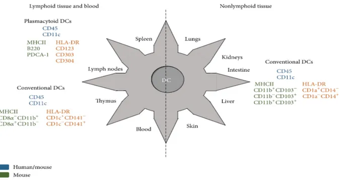
Figure 1. Specific markers expressed by human and mouse dendritic cells. Figure reprinted with permission from (Amorim, Chagas, Sulczewski, & Boscardin, 2016).
1.3 The adaptive immune system
For invertebrates, the innate immune system has proved to be largely sufficient for maintaining health. As the complexification of organisms intensified over time, the immune system adapted by developing lymphocytes to provide long lasting immunity to pathogens. Infants born with severe dysfunctions of the adaptive immune system cannot survive unless extraordinary measures are taken in order to correct and prevent infectious complications. B and T lymphocytes are the most important actors of the adaptive immune system and both cell types can generate long lasting memory cells to protect against reinfection by microbes. This second line of defense is more sophisticated and specific. Billions of lymphocytes with distinct
specificity are needed in order to recognize the large array of potential antigens that could harm the host.
1.4 Lymphocyte production
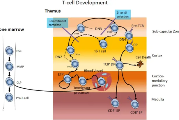
Lymphocyte differentiation requires a specialized microenvironment and occurs in the thymus. Common lymphoid progenitor cells (CLPs) are found in BM and can be identified as Lin−Il7Rα+ c-Kit+ Sca-1+ (Kondo, Weissman, & Akashi, 1997). Inside the thymus, CLPs become early thymic progenitors and undergo intensive proliferation before passing through several CD4-CD8- double negative (DN) stages. DN cells do not express CD4 or CD8 receptor and the expression of the beta chain of the T cell receptor (TCR) occurs at the DN3 stage (Starr, Jameson, & Hogquist, 2003). After the expression of the alpha chain, thymocytes become double positive (DP) and express both CD4 and CD8 receptors. DP thymocytes undergo positive selection by interacting with thymic epithelial cells in the cortex (cTEC) (Klein, Kyewski, Allen, & Hogquist, 2014). During this process, thymocytes that fail to transmit signal through their TCR after interacting with MCH I or II are eliminated. As DPs reach the medulla, they lose the expression of CD4 or CD8 receptors to become single positive (SP) thymocytes. In the medulla, SP thymocytes undergo negative selection by interacting directly with medullary thymic epithelial cells (mTECs) or thymic DCs (Hu, Lancaster, Sasiponganan, & Ehrlich, 2015; Li et al., 2007). mTECs express a vast repertoire of self-antigens used to eliminate autoreactive thymocytes (Klein et al., 2014; Oh & Shin, 2015). The expression of organ specific self-antigens is controlled by the transcription factor called autoimmune regulator (AIRE). Mutations of AIRE have been documented in mouse and human and results in a severe autoimmune syndrome called autoimmune polyendocrinopathy candidiasis ectodermal dystrophy (APECED)
(Nagamine et al., 1997). Thus, T cells reacting too strongly with these autoantigens are either eliminated or turn into regulatory T cells in order to prevent the production of self-reactive T lymphocytes (Klein et al., 2014). Following their journey in the thymus, thymocytes are released into the blood to provide immunocompetence to the host.
Figure 2. Lymphocyte differentiation during post-natal period begins inside thymus. Hematopoietic stem cells (HSCs) migrate in the thymus after they differentiate into early thymic progenitors (ETPs) and develop into CD4+CD8+ DN thymocytes. After the recombination of the beta and alpha chains of the TCR, thymocytes become DP as they express both CD4 and CD8 co-receptors. At the DP stage, positive selection allows thymocytes to escape death upon TCR engagement and signaling. Finally, thymocytes will downregulate the expression of either CD4 or CD8 co-receptor to become SP thymocytes. Before leaving the thymus, CD4+ and CD8+ thymocytes undergo negative selection to eliminate autoreactive thymocytes. (This figure was adapted with permission from (Aifantis, Raetz, & Buonamici, 2008).
1.5 Naïve T cells
Naïve T cells are lymphocytes that have never reacted against their cognate antigen. These cells are found in the blood and in secondary lymphoid organs. In mice, naïve T cells can be defined based on low expression levels of CD44 and high expression levels of CD62L and CCR7 (Berard & Tough, 2002; Surh & Sprent, 2008; Takada & Jameson, 2009). In humans, CD45RA and CCR7 are currently used to identify naïve T cells. CD4+ and CD8+ T lymphocytes are the most abundant cell types produced by the thymus. While CD4+ T cells are restricted to MHC II, CD8+ T lymphocytes are restricted to MHC I molecules. Naïve T cells can survive for several years without being activated and their peripheral maintenance requires specific signals that will be discussed later.
1.6 Activated lymphocytes
The activation of naïve T lymphocytes occurs in the LN where they meet with DCs (Kastenmuller, Gerner, & Germain, 2010). Following insults or infection, DCs become active and bring potential antigens to the LN. Antigens are captured and processed in the endosome and the resulting peptide can bind to MHC II (classical presentation pathway) or MHC I (cross-presentation) to activate CD4+ or CD8+ T cells respectively. Importantly, the sole recognition of MHC I or II by lymphocytes is normally insufficient for T cell activation and requires co-stimulation delivered by CD80 or CD86 (Mueller, Jenkins, & Schwartz, 1989). Following CD8 T cell activation, CCR7 and CD62L expression will be diminished to prevent access to lymphoid tissues and promote their migration to inflamed tissues where they will eliminate cells expressing the relevant peptide. For CD4+ T cells, their activation will promote the secretion of
cytokine that will help cytotoxic CD8+ T cells but also B lymphocytes. Activated CD4+ T cells can also migrate to the cortex of the LN to provide help to B cells in order to promote their full activation to become plasmocytes.
1.7 Memory lymphocytes
The production of memory lymphocytes is an important aspect of the adaptive immune system to provide long lasting protection of the host. Memory cells are lymphocytes that have previously encountered their cognate antigen and have undergone activation and differentiation into memory cells. However, it is still unclear how memory cells are formed after T cell activation. While the binaries model supports the acquisition of memory phenotype immediately after T cell priming (Pennock et al., 2013; Restifo & Gattinoni, 2013), the linear differentiation theory supports a model wherein memory lymphocytes arise from effector T cells (Reiner, Sallusto, & Lanzavecchia, 2007). Once memory lymphocytes are formed, they can live for several years, recirculating between the blood and lymphoid tissues searching for their cognate antigens (Alberts, 2002).
1.7.1 T cell homeostasis
T lymphocytes normally undergo very low-levels of proliferation called homeostatic cycling (HC) that serves to maintain T cell number and diversity in the periphery. Following T cell depletion, T lymphocytes undergo intense proliferation called homeostatic proliferation (HP) or homeostatic peripheral expansion (HPE). During the last 10-20 years, studies have identified several factors implicated in the regulation of HC and HP (Sprent & Surh, 2011). In addition, studies have revealed substantial differences between HC and HP of naïve and memory
T lymphocytes. In this section, factors controlling naïve CD4+ and CD8+ versus memory CD4+ and CD8+ T cell homeostasis will be discussed.
1.7.2 Memory CD8
+T cell homeostasis
The proportion of memory lymphocytes tends to increase as we age (Y. Lin et al., 2016). Involution of thymic one of the most significant contributors to the phenomenon of aging with resulting in atrophied thymus this lead to a steady decline in production of naïve t cell numbers leading to restricted TCR repertoire. As more naïve T cell turn into memory cells and accumulation of memory T cells that are specific of persisting pathogens. (also known as memory inflation). (Nikolich-Zugich, 2014; Salam et al., 2013). Surprisingly, the number of antigenic encounters does not have a dramatic effect on the number of memory CD8+ T cells, suggesting that intrinsic factors control T cell numbers. Memory CD8+ T cells do not require TCR stimulation by MCH I since these cells can survive upon transfer in hosts lacking MHC I molecules (Murali-Krishna et al., 1999). IL-15 is the critical factor controlling the survival and HP of memory CD8+ T cells in mice and humans. Although memory CD8+ T cells can be generated in IL-15-/- hosts, they cannot survive (Kaech, Wherry, & Ahmed, 2002). IL-15 controls both the survival and HP of memory CD8+ T cells (Judge, Zhang, Fujii, Surh, & Sprent, 2002). The administration of recombinant IL-15 can expand memory CD8+ T cells but its effect on naïve CD8+ T cells is modest (Melchionda et al., 2005). In addition, transgenic mice overexpressing IL-15 have greater numbers of memory CD8+ T cells (Fehniger & Caligiuri, 2001). While IL-15 appears essential for memory CD8+ T cell homeostasis, under lymphopenic conditions, IL-7 levels are high enough to induce survival of memory CD8+ T cells (Kieper et
al., 2002; Tan et al., 2002). However, in normal hosts, IL-7 levels are too low to exert any effects on memory CD8+ T cells. Thus, while IL-15 is essential for memory CD8 homeostasis, IL-7 could be sufficient to promote the survival of this subset during lymphopenia.
1.7.3 Memory CD4
+T cell homeostasis
For memory CD4+ T cells, it is not as clear whether IL-7 and tonic interaction with MHC II are both required for controlling the homeostasis of this subset. For instance, Swain et al. showed that peripheral maintenance of memory CD4+ T cell did not require contact with peripheral MHC II (Dorfman, Stefanova, Yasutomo, & Germain, 2000; Swain, Hu, & Huston, 1999), and others showed that IL-7 signaling was sufficient for memory CD4 survival (Lantz, Grandjean, Matzinger, & Di Santo, 2000). In contrast, Seddon et al. showed that both TCR stimulation by MHC II and IL-7 signaling on T cells were required for HP and survival of memory CD4+ T cells (Seddon, Tomlinson, & Zamoyska, 2003). Thus, the requirement for TCR stimulation and IL-7 signaling could be important for memory CD4+ T cells and their effects may vary in lymphopenic versus non lymphopenic conditions.
1.7.4 Naïve T cell homeostasis
The interaction between TCRs and self-peptide-MHCs is essential for the survival of naïve T cells in the periphery. For instance, naïve CD8+ T cells disappear after transfer in hosts lacking MHC I (Kieper & Jameson, 1999). Similarly, naïve CD4+ T cells fail to survive after transfer in hosts lacking MHC II (Clarke & Rudensky, 2000). Other studies used TCR transgenic lymphocytes to demonstrate the loss of naïve CD4+ and CD8+ T cells upon TCR ablation by tetracycline (Labrecque et al., 2001; Leignadier, Hardy, Cloutier, Rooney, & Labrecque, 2008).
Surprisingly, loss of naïve T cells was gradual after TCR ablation, suggesting that other factors may contribute to the survival of this subset in the periphery. The contribution of co-stimulation B7 molecules has been studied but failed to reveal any evidence of the involvement of co-stimulatory molecules in T cell homeostasis. IL-7 however, represents the second factor absolutely required to allow naïve T cells to survive and persist in the periphery (Surh & Sprent, 2008). The model put forth to explain peripheral T cell maintenance relates to two primary factors: 1) TCR stimulation by self-peptide MHC I or II for CD8+ and CD4+ T cells respectively and 2) IL-7 signaling on T cells that regulates cell survival. As a result, naïve T cells require continuous access to TCR stimulations or IL-7 and interfering with either of these factors could disrupt T cell homeostasis and induce lymphopenia.
1.7.5 Interleukin-7
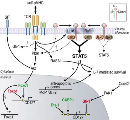
IL-7 belongs to a family of hematopoietins that comprises class I cytokines. It is produced by the epithelium of the thymus and stromal cells of BM (Jiang et al., 2005). IL-7 signaling on T cells is mediated by IL-7 receptor alpha (IL-7Rα) chain (CD127) and the common gamma chain, γc CD132 (Jiang et al., 2005; J. X. Lin & Leonard, 2017). The γc chain is detected on most hematopoietic cells (ElKassar & Gress, 2010; Leonard, Noguchi, & Russell, 1994), whereas IL-7Rα is expressed mainly by cells in the lymphoid lineage; this includes a variety of cells, such as immature B cells, thymocytes, and lymphocytes (Gao, Zhao, Wan, & Zhu, 2015). IL-7Ra is also expressed by Forkhead box protein 3 (FoxP3) regulatory T cells. However, the expression levels are substantially lower (Niedbala et al., 2007). The binding of IL-7 on IL-7Rα chain heterodimerizes the IL-7Rα and the common γc chain. This reaction subsequently activates Janus kinase (JAK) 3 and stimulates the phosphorylation of JAK1 as well as of signal
transducers and activators of transcription 5 (STAT5) (Palmer et al., 2008; Repass et al., 2009). This process creates binding sites for the signal transducers and activators of transcription STAT5, which translocate to the nucleus once JAK protein is phosphorylated (Kiu & Nicholson, 2012). The phosphorylation of STAT5 induces the expression of B-cell lymphoma 2 (BCL-2) and myeloid cell leukemia sequence-1 (MCL1) genes which are essential for the survival of naïve T cells (Ertel, Nguyen, Roulston, & Shore, 2013). In addition, when IL-7 levels are elevated, they can signal through phosphoinositide 3-kinase (PI3K), activated by JAK1, to recruit protein kinase B (AKT) and induce proliferation of lymphocytes by stabilizing c-myc and cyclin-D (Kittipatarin & Khaled, 2007). In addition, AKT can inhibit the transcription of genes coding for factors involved in apoptosis and cell cycle (Zhang, Tang, Hadden, & Rishi, 2011). As a result, IL-7 can induce survival of T cells under normal physiologic conditions but can promote proliferation when systemic IL-7 increases during lymphopenia (Fry, Christensen, Komschlies, Gress, & Mackall, 2001). Thus, IL-7 is a master regulator of T cell homeostasis in humans and mice.
1.7.6 IL-7
-/-and IL-7Ra
Mice with mutation in IL-7 are IL-7Ra genes are profoundly lymphopenic and present severe thymic dysfunction. Studies using adoptive transfer of congenic T lymphocytes in IL-7-/-and IL-7Ra-/- confirmed that, in addition to the severe thymic hypoplasia, IL-7 was required for the survival and homeostatic proliferation of naïve T cells (Tan et al., 2001). IL7-/- mice are characterized by a severe blockade of thymocyte maturation as IL-7 is essential for the survival and expansion of immature double negative thymocytes and later, IL-7 promote the survival of thymocytes that escaped negative selection. IL-7Rα-/- mice have a similar phenotype to IL-7
-/-mice but the dysfunction of the thymus is even more severe in IL7α-/- mice. Interleukin-7 signals through IL-7Rα but thymic stromal lymphopoeitin (TSLP) also uses IL7Rα as its receptor which explain the partial maturation of T cell in IL-7-/- mice . It can be explained that TSLP1 is able to overlap with IL-7 functions during thymopoiesis but in only 1% of pre-T cell become mature T-cells in IL-7-/-mice (Beq, Delfraissy, & Theze, 2004).
Figure 3. IL-7R signaling pathway. The binding of IL-7 to IL-7Rα chain leads to the herterodimerization of IL-7Rα and the common gc chain which activate JAK3 and induce the phosphorylation of JAK1 and the signal transduce and activation of transcription 5 (STAT5), resulting in increased BCL-2 and MCL-1 gene expression which promote the survival of T cells. In addition, when IL-7 is elevated, it can signal through phosphoinositide3-kinase (PI3K) and activate JAK1 to induce protein kinase B to induce the proliferation of T lymphocytes. Figure reprinted with permission from (Swainson et al., 2007).
1.8 Homeostatic proliferation under lymphopenic conditions
When lymphocytes are transferred into lymphopenic hosts, they undergo substantial proliferation and adopt a phenotype resembling those of memory cells (Goldrath, Luckey, Park, Benoist, & Mathis, 2004). This type of proliferation that is normally absent in non-lymphopenic hosts is called HP. Other studies have used the term HPE to describe this exaggerated form of proliferation that occurs in lymphopenic hosts (Mackall et al., 1996; Reading et al., 2015). IL-7 and TCR stimulation are both required for HP as they are for HC in a non-lymphopenic hosts. Despite these similarities, HP can induce significant changes to the T cell repertoire as lymphocytes with the highest affinity for the self tend to proliferate and accumulate more (Goldrath & Bevan, 1999). In contrast, TCRs with weak affinity for the self tend to proliferate less, resulting in a TCR repertoire more autoreactive under lymphopenic conditions.
1.8.1 Immune reconstitution after lymphopenia
Following T cell depletion, lymphocyte regeneration can occur via two distinct pathways: a thymic-dependent pathway in which lymphocytes are produced by the thymus, and a thymic-independent pathway where residual mature lymphocytes undergo brisk proliferation in an attempt to increase their number. In most clinical settings of human lymphopenia, thymic involution is too severe for thymic-dependent immune reconstitution and HP is the predominant pathway to regenerate naïve T cells. While HP of CD8+ T cells is efficient with normalization of CD8 counts in 1-3 months, HP of CD4+ T cells is inefficient and patients experience profound lymphopenia that can persist for several months or years. Differential expression of MHC I versus MHC II has been invoked to explain differences in CD4 versus CD8 immune reconstitution. MHC II expression is largely limited to antigen presenting cells and we still do
not know which type of antigen presenting cells (APC) controls CD4 homeostasis. This work seeks to identify the subset of APCs involved in CD4 HP.
Chapter 2: Rationale, Hypothesis and Objective
It is well known that CD4 immune reconstitution is slower that CD8 regeneration after post-natal T cell depletion. While the distribution of MHC I is ubiquitous, MHC II expression is largely restricted to antigen presenting cells, perhaps limiting access to TCR triggering compared with naïve CD8+ T cells. Our in vivo preliminary data in mice showed conclusively the requirement of CD11c+ DCs in CD4 HP during lymphopenia. In several clinical settings of human lymphopenia, a correlation between CD4 count and pDCs has been observed (Giraud et al., 2005). In addition, pDCs express IL-7Ra and a lack of IL-7 signaling on these cells could affect MHC II expression, perhaps contributing to the more efficient CD4 HP in IL-7Ra-/- mice. However, during graft-versus-host disease (GvHD, DC regeneration is impaired and our laboratory has demonstrated that increasing CD8a+ DCs with SDF-1a therapy was sufficient for improving CD4 HP (Gauthier, Leboeuf, Manuguerra-Gagne, Gaboury, & Guimond, 2015). Finally, while we had mouse models to deplete specifically pDCs and CD8a+ DCs, the CD11c+IRF4-/- mouse model to deplete DC2 subset (CD11b+CD11c+) was still under development and could not be used in this study. The goal of this project is to identify which APC type provides TCR stimulation to naïve CD4+ T cells. Our hypothesis is that a specific subset of CD11c+ DC provides TCR stimulation of naïve CD4+ T cells to promote HP of these cells under lymphopenic condition. In this work, we focused on the potential role of pDCs and CD8a+ DCs in CD4 HP.
The objective of this work:
To deplete individual subset of CD11c+ DCs and study the effect on CD4 HP in lymphopenic mice. Our preliminary data showed that CD11c+ cells are absolutely required for CD4 HP in lymphopenic mice. Using a variety of genetically mouse models, we evaluated the requirement of specific DC subset(s) on CD4 HP.
Chapter 3: Materials and Methods
3.1 Mice
The mice used in this project were between 6-20 weeks old. Male and female mice of C57BL/6.SJL (B6.SJL: H-2b, Ptprca Pep3b, CD45.1+), C57BL/6.129S7-Rag1tmMom/J (RAG-/-), H-2b, CD45.2), RAG-/-CD45.2+ mice were purchased from The Jackson Laboratory (Bar Harbor, ME), IL7Ra-/- CD45.2+, Batf 3-/- IL7Ra-/- CD45.2+ mice were provided by Dr. Olivier Lantz (Institute Curie), and IL-7Ra-/-CD11c DTR were generated at the animal facility by breeding IL-7Ra-/- mice with CD11c DTR. Animals were housed at the Maisonneuve-Rosemont hospital animal facility, and were treated in accordance with the regulations of the Maisonneuve-Rosemont hospital animal care committee.
3.2 Organ harvest
Spleen, thymus, LNs, and BM were homogenized and cells were suspended in Roswell Park Memorial Institute (RPMI 1640) medium containing 2% FBS and 1% of penicillin and streptomycin. Cell enumeration was performed with an hemocytometer and Trypan blue to exclude death cells.
3.3 Bone marrow chimera
1X10⁷ cells were obtained from the BM of CD11c+DTR IL7Ra-/- mice were injected intravenously into tail vein of sublethally irradiated recipient RAG-/- mice. Recipient mice were then treated with antibiotic paytril (enrofloxacin 100mg/ml) from ( Bayer, Mississauga, ON,
Canada) for 10 days. Five weeks post-stem cell transplantation (SCT), BM chimera were used for experiments.
3.4 Depletion of endogenous DCs
Diphtheria toxin (4ng/g of body weight) and/or anti-MPDCA-1 (250µg) monoclonal antibodies (Miltenyi Biotec, Bergisch Gladbach, Germany) was administered to mice to deplete CD11c or pDCs , respectively. DT and MPDCA-1 antibody were IP administered every other day starting from day -1 to +7. At day +8, mice were sacrificed and the spleen was harvested to evaluate by flow cytometry CD4 proliferation and DC depletion (Fortessa, BD). Flow cytometry analyses were performed using FlowJO software (Treestar, Ashland, OR, USA).
3.5 CFSE Staining and adoptive transfer of lymphocytes
TCR transgenic CD4+ lymphocytes were isolated by negative selection from the spleen and LN using a T cell enrichment kit (Stem cell technology, Vancouver, CA). Enriched lymphocytes were incubated with 1µl/ml of 5µM CFSE (at concentration of 1x 10⁷ cells/ml and incubated for 15 minutes at room temperature (Hennion et al, blood 2013). Cells were then washed twice and resuspended in PBS. 1x10⁶ CD4+ cells were IV injected in recipient mice. Seven days later, mice were sacrificed and lymphocytes were evaluated by flow cytometry for evidence of proliferation based on CFSE dilution.
3.6 T cell co-culture with DCs
T cells were isolated from the spleen or LN using a T cell enrichment kit (Stem cell technologies). The purity of enriched T cells was evaluated by flow cytometry. Lymphocytes
were then labeled with cell trace violet (CTV) (Invitrogen, Burlington, ON, Canada) or CFSE for 15 minutes, washed and resuspended in complete medium: RPMI 1640 10% FBS, 1% P/S (Penicillin/ streptomycin), 1% L-Glutamate, 1% sodium pyruvate, 1% non-essential amino acids.
DCs were isolated from the spleen of female B6SJL, CD45.1+. For co-culture experiments, 1x10⁵ T cells were co-culture with total CD11c+ DCs or CD11c+CD11b+ DC2 subset or CD11c+ CD8a+ DC1 subset or CD19+ B lymphocytes and incubated at 37C in complete media + IL-7 (30 ng/ml) for 6 to 8 days. At the end of the experiment, cells were harvested, enumerated and analyzed by flow cytometry using a FACS Fortessa.
3.7 Cell sorting
5x10⁷ splenocytes were labeled with specific monoclonal antibodies (MoAb) to B lymphocytes and DCs and cells were sorted using a FACS ARIA. The antibody cocktail used to isolate B lymphocytes and DCs were the following: pacific blue anti-IA/IE (M5/114.15.2), and PECY7-anti-CD19 (6D5). For CD11c+ DC subtypes, FITC anti-CD11C (N418), pacific blue anti-IA/IE (M5/114.15.2), PerCP5.5-anti-CD11b (M1/70), and PE-anti-CD8a (53-6.7). These antibodies were obtained from Biolegend (San Diego, California, United States).
3.8 Flow cytometry
1x10⁶ cells were incubated with diluted MoAb, washed and resuspended in FACS buffer for cytometric acquisition with a FACS Fortessa. For T cell labelling, we used APC/CY7 and APC anti-CD4 (GK1.5), PerCP/CY5.5 and PE anti-CD8a (53-6.7) and BV786 anti-CD8a (RA3-6B2), FITC anti-TCRb (H57-597), PerCP/CY5.5 anti-CD45.1(A20),
PerCP/CY5.5-anti-CD45.2 (104), APC anti-CD62L (MEL-14), PE/CY7 anti-CD44 (IM7). For DCs labelling, the following MoAb were used: FITC and PerCPCY5.5- anti-CD11b (M1/70), PE and FITC-CD11c (N418), Pacific Blue Anti-IA/IE (M5/114.15.2), PE LY6C (HK1.4), BV605 anti-B220 (RA3-6B2), FITC anti-CD80 (16-10A1), APC/CY7 anti-CD86(GL-1). For B lymphocytes, we used PE/CY7 anti-CD19 (6D5) MoAb. These antibodies were obtained from Biolegend (San Diego, California, United States).
3.9 Statistical analysis
We used prism 5.0 for statistical analysis (Graphpad, La Jolla, CA, USA). Non-parametric Mann-Whitney tests were used to compare pairs of data while, the Kruskal-Wallis tests were used to compare 3 or more groups. A value equal or lower to 0.05 was considered statistically significant.
Chapter 4: Results
4.1 Development of the mouse models to study the role of APCs in CD4 HP
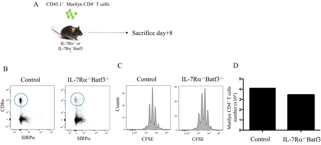
To deplete CD11c+ DCs, we used a mouse model that expresses the diphtheria toxin receptor (DTR) under the CD11c promoter (Hochweller, Striegler, Hammerling, & Garbi, 2008). In these mice, the injection of diphtheria toxin leads to DC depletion. For mice lacking DC1 subset, we used Batf3 -/- mice which lack CD8a+ DCs. Finally, for pDC depletion, we used the MPDCA-1 MoAb to deplete this subset. Since Rag-/- mice do not support efficiently CD4 HP, we used IL-7Ra-/- mice that possess a peripheral CD4 niche highly supportive for HP and interbreed IL-7Ra-/- with CD11c-DTR and Batf3-/- mice (Guimond et al., 2009) (Figure 4A). The presence of the DTR transgene was confirmed by PCR on genomic DNA. Figure 4B shows PCR for Batf3 gene.
Figure 4. CD11c-DTR and Batf3 -/- development and transgene/alteration detection. A) IL-7Rα-/- mice were interbred with CD11c-DTR or Batf3 -/- mice to obtain IL-7Rα-/- CD11c-DTR or IL-7Rα-/- Batf3 -/- double knockout mice. B) DNA from mice was extracted, and subjected to PCR to identify transgenic mice. DNA controls include wild type and mutated allele of Batf3 and IL-7Rα genes as well as DTR transgene.
4.2 CD11c
+DC controls HP of CD4
+T cells
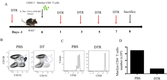
To evaluate the role of antigen presenting cells in CD4+ T cell homeostasis, we used IL-7Rα-/-CD11c-DTR. CD11c-DTR mice cannot tolerate multiple injections of DT, required to maintain DC depletion, since neurotoxicity occurs in this model, we engineered BM chimeras in which Rag-/- mice were transplanted with BM cells obtained from CD11c-DTR, IL-7Rα-/-. At Day -1, mice received DT (4ng/g of body weight). At day 0, mice received 1X10⁶ CD45.1+ Marilyn CD4+ T cells (Marilyn mice are transgenic mice on background C57BL/6 mice and where all T cells carry the same transgenic TCR specific for a Y chromosome derived antigen presented by MCH class II. male mice at least male do not have CD4+ T cell in peripheral compartment after negative selection in thymus to prevent autoimmunity, while female mice express CD4+ T cell due to absence of HY peptide). DT treatments were performed at day -1, +1, +3, +5 and +7 (Figure 5A). DC depletion and T cell proliferation were evaluated at day +8. Following DT administration, we found that most DC subsets were depleted compared with mice receiving vehicle (Figure 5B). However, a small portion of the CD11c+CD11b- still persisted after DT treatment. Most importantly, CD4 HP was completely abrogated in mice treated with DT (Figure 5C). These results confirmed that CD11c+ DCs are essential for CD4 HP and their depletion is sufficient to constrain HP of this subset.
Figure 5. Role of CD11c+ cells in CD4 HP. A) Schematic representation of the experiment. BM chimera received DT at day -1, +1, +3, +5 and +7 to maintain CD11c+ DC depletion. At day 0, BM chimeras received 1X10⁶ Marilyn CD4+ T cells and at day +8, mice were sacrificed and transferred T cells were evaluated by flow cytometry. B) Evaluation of CD11c+ DC depletion by flow cytometry. C) Evaluation of lymphocyte proliferation +/- DT treatment. D) The absolute numbers of CD4+ T cells after 7 days. Data are representative of 2-3 independent experiments of 3-4 mice in figure (A, B and C) and 1 mice per group in figure D.
4.3 DC1 cells are not essential for CD4 HP
Since the ablation of CD11c+ DCs constrains CD4 HP, we sought to investigate which DC subset(s) supported CD4 HP. To investigate the role of CD8a+ DCs, we used Batf3 -/-knockout mice which lack DC1 subset. In order to have a peripheral niche permissive for CD4 HP, we interbred Batf3-/- mice with IL-7Ra-/- mice (Guimond et al., 2009). We transferred 1X10⁶ CFSE-labelled Marilyn CD4+ T cells into Batf3-/- IL-7Ra-/- recipients to evaluate the potential role of DC1 subset on CD4 HP (Figure 6A). At day +8 post-lymphocyte transfer, Batf3-/- IL-7Ra-/- mice were sacrificed and HP of CD4+ T cells was evaluated. As predicted, Batf3-/- IL-7Ra-/- mice had very few, if any, cells expressing CD11c+CD8a+Sirpa- compared to IL-7Ra-/- control mice (Figure 6B). Surprisingly, despite very few cells with a phenotype corresponding to the DC1 subset, we did not observe a decrease in CD4 HP. (Figure 6B and C). This result demonstrated that CD8α+ DCs are likely not essential for CD4 HP.
Figure 6. Role of CD8α DCs in CD4 HP. A) Schematic representation of the experiment. Congenic CD45.1+CD4+ Marilyn T cells were enriched and transferred into IL-7Rα-/- Batf3 -/- mice. B) Flow cytometric analysis of the spleen of IL-7Rα-/- Batf3-/- mice to evaluate CD8α+ DCs. C) Flow cytometric analysis showing the proliferation and TCR transgenic Marilyn CD4+ 8 days after their transfer into IL-7Rα-/- Batf3-/- mice. D) The absolute numbers of CD4+ T cells after 7 days. Data are representative of 2-3 independent experiments of 3-4 mice in figure (A,B and C) and 1 mice per group in figure D.
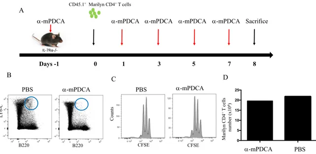
4.4 pDCs are not essential for CD4 HP
We then examined the potential role of pDCs in CD4 HP. To remove pDCs, we treated IL-7Ra-/- mice with the MoAb mPDCA-1 (250µg). At day -1, mice receive 250µg of anti-mPDCA-1 to deplete pDCs prior to the adoptive transfer of CD4+ T cells. At day 0, mice received 1X10⁶ enriched CD45.1+ Marylin CD4+ T cells. Anti-pDCs MoAb treatment were then pursued at day +1, +3, +5 and +7 post-T cell transfer (Figure 7A). pDCs depletion and T cell proliferation were evaluated at day +8. Anti-pDCs treatments were highly effective at depleting pDCs compared with control mice (Figure 7B). However, despite the depletion of pDCs, CD4 HP still occurred. Based on these results, we concluded that pDCs were not essential for CD4 HP. (Figure 7C).
Figure 7. Role of pDCs in CD4 HP. A) Schematic representation of the experiment. At day -1, IL-7Ra-/- mice were treated with anti-MPDCA-1 to deplete pDCs. At day 0, CD45.1+ Marilyn CD4+ T cells were enriched and transferred into IL-7Ra-/- mice. From day 1 to +7, mice continue to receive anti-pDCs antibodies to maintain pDC depletion. B) Flow cytometry for the evaluation of B220 and LY6C pDCs. C) Flow cytometry showing HP of Marilyn CD4+ T cells in mice lacking pDCs. D) The absolute numbers of CD4+ T cells after 7 days. Data are representative of 2 independent experiment of 3-4 mice in figure (A,B and C) and 1 mice per group in figure D.
Days -1
Sacrifice
0 1 3 5 7
a-mPDCA
8
a-mPDCA a-mPDCA a-mPDCA a-mPDCA IL-7Rα-/-CD45.1+Marilyn CD4+T cells B220 B220 CFSE CFSE LY 6-C PBS a-mPDCA PBS a-mPDCA C ount s A B C 0 5 10 15 20 25 D PBS a-mPDCA M ar il yn C D 4 +T c el ls num be r (x10 4)
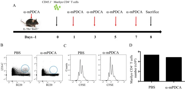
4.5 The depletion of both pDCs and DC1 failed to reduce CD4 HP.
Although individual DC1 and pDC subsets appear dispensable for HP of CD4+ T cells, together, their role on CD4 HP may be superior. To understand the role of both subsets on CD4 HP, we used IL-7Rα-/- Batf3-/- mice and depleted pDCs with anti-mPDC1 monoclonal antibody. At day -1, mice received anti-pDC treatment and the next day, they received 1X10⁶ CD45.1+ Marilyn CD4+ T cells. Anti-pDCs MoAb treatment were then given at day +1, +3, +5 and +7 post-lymphocyte transfer (Figure 8A). At day +8, mice were sacrificed and pDC depletion and T cell proliferation were evaluated. We confirmed the effectiveness of pDC depletion in IL-7Rα-/- Batf3-/- mice by showing a significant reduction of pDCs (Figure 8B). Surprisingly, the lack of DC1 combined with the depletion of pDCs did not result in diminished CD4 HP. (Figure 8C). Therefore, we concluded that both DC1 and pDCs are not essential of CD4 HP, and other CD11c subset(s) must provide sub-optimal TCR stimulation when these cells are absent. Since CD11c+CD11b+ are the remaining DC subset to be found in our mice, it is possible that DC2 cells might be sufficient for CD4 HP when other pDCs and CD8a+ are absent depletion, Additional studies in lymphopenic mice lacking CD11b+CD11c+ DCs are needed in order to determine the role of DC2 in CD4 HP.
Figure 8. Role of pDCs and CD8+ DCs in CD4 HP. A) Schematic representation of the experiment. At day -1, 1, 3, 5 and 7, IL-7Ra-/- Batf3-/- mice were treated with anti-mPDCA-1 to deplete pDCs. At day 0, CD45.anti-mPDCA-1+ Marilyn T cells were enriched and transferred into IL-7Ra-/- Batf3-/- mice. B) Flow cytometry for pDCs in IL-7Ra-/- Batf3-/- mice. C) Flow cytometry showing CFSE dilution of Marilyn CD4+ T cells. D) The absolute numbers of CD4+ T cells after 7 days. Data are representative of 2 independent experiment of 3-4 mice in figure (A,B and C) and 1 mice per group in figure D.
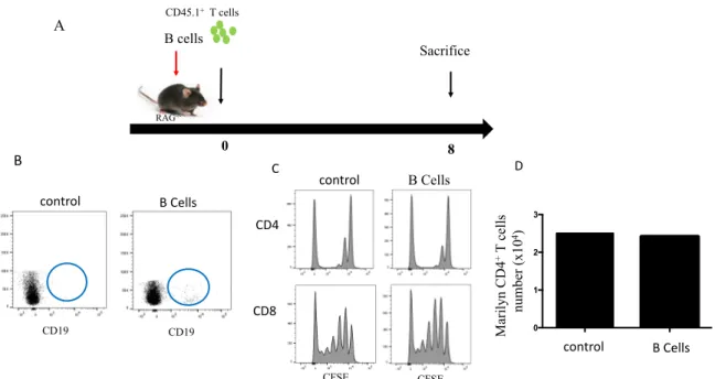
4.6 B cells do not support CD4
+T cell HP
Rag-/- and IL-7Ra-/- mice do not have B cells, yet IL-7Ra+ mice support CD4 HP. B lymphocytes express MHC II and it is not clear whether B cells could provide homeostatic signal(s) to CD4 T cells. To address the potential role of B cells in CD4 HP, we sorted 10 x106 B lymphocytes from C57BL/6 mice and transfer them in Rag-/- mice along with 1x10 6 of polyclonal CD4+ T cells. Seven days later, mice were sacrificed and CD4 HP was evaluated by flow cytometry (Figure 9A and B). As predicted, the transfer of B lymphocytes in lymphopenic RAG-/- mice did not add to the proliferating of CD4+ T cells (Figure 9C). Together, our data confirmed that B cells have no effect on CD4 HP.
Figure 9. B lymphocytes do not support CD4 HP. A) Schematic representation of the experiment. Congenic CD45.1+ T cells were enriched and transferred with purified CD45.1+ B cells into RAG-/- CD45.2+ mice. B) Flow cytometry showing B cells in RAG-/- mice. C) Evaluation of the proliferation of CD4+ T cells. D) The absolute numbers of T cells after 7 days. Data are representative of 2 independent experiments of 2 mice in figure (A, B and C) and 1 mice per group in figure D.
CD19 CFSE CFSE B Cells Sacrifice 0 8 RAG -/-CD45.1+T cells A B cells CD4 CD8 B C control B Cells CD19 0 1 2 3 D M ar il yn C D 4 +T c el ls num be r (x10 4) control control B Cells
4.7 CD11c
+DCs support HP of CD4 T cells in a co-culture system.
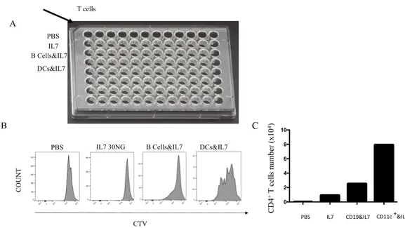
We tried to recapitulate in vitro the proliferation of CD4+ T cells induced by CD11c+ DCs. CD11c+ DCs from C57BL/6 CD45.2+ mice were isolated from the spleen and co-culture with T cells obtained from C57BL/6 CD45.1+ mice. Enriched T cells were labelled with CTV staining and co-cultured in 96 wells plate with DCs at a ratio of 2.5 DCs:1Lymphocyte. Cultures were supplemented with mouse recombinant IL-7 (30 ng/ml) and 8 days later, T cells were harvested, enumerated and evaluated by flow cytometry for proliferation based on CTV dilution (Figure 10B). The same experiment was also repeated with B cells to understand their potential effect on T cell HP. While we observed T cell proliferation in co-culture with DCs, we did not observe CD4 proliferation when co-culture with B cells (Figure 10B). Proliferation of T cells was accompanied with an increase in T cell numbers. Surprisingly, T cell numbers were also higher when co-culture with B cells compared with IL-7 alone. These results confirmed that DCs provide the requisite stimulus to promote CD4 HP whereas for B cells, although they might exert an effect on T cell survival, B cell effect on CD4 HP is very weak compared with DCs.
Figure 10. Co-culture of CD4+ T lymphocytes with different types of APCs showed an effect of CD11c+ DCs on CD4 HP. A) CD4+ T cells were co-cultured with CD11c+ DC or B cells. B) Flow cytometry showing the proliferation of CD4+ T cells under different culture conditions. C) The absolute numbers of CD4+ T cells recovered after 8 days of cell culture. Data are representative of 2 independent experiments .
T cells
IL7 PBS
IL7 30NG B Cells&IL7 DCs&IL7 PBS A B C CTV CO U N T DCs&IL7 B Cells&IL7
PBS IL7 CD19&IL7 CD11c +&IL7
0 2 4 6 8 10 CD4 +T cel ls num be r (x10 4)
4.8 Co-culture of T cells with different DC subsets
Given that CD11c+ DCs can induce HP of CD4+ T cells in vitro, we evaluated whether CD4 HP can be induced by individual DC subsets. We sorted DC subsets (CD11c+, CD11b+ and CD8α+) from C57BL/6 CD45.2+ mice for co-culture with enriched T cells. Polyclonal lymphocytes (CD45.1+) were labelled with CTV staining and co-cultured with individual DC subsets at a ratio of 2.5:1 in a 96-well round plate for 6 days. Recombinant mouse IL-7 (30 ng/ml) was also added to cell cultures. At day 6, cells were harvested, enumerated and analyzed by flow cytometry. Unfortunately, the number of cells recovered after 6 days was too low to evaluate the proliferation of T cells based on CTV dilution. However, we were able to evaluate T cell numbers in most cell cultures. Surprisingly, the recovery of total T cells and particularly CD4+ T cells was the highest when co-cultured with CD8α+ DCs. T cells were potentially more abundant than the other components in the co-cultures (Figure 11). Total CD11c+ DCs as well as CD11b+ cells allowed similar T cell recovery, yet T cell numbers were lower compared with lymphocytes cultured with CD8α+ DCs. This result agrees with our previous results suggesting that DCs can induce the survival and possibly the proliferation of T cells.
Figure 11. T lymphocyte expansion is stronger when incubated with DCs. A) Absolute numbers of T cells exposed to different APC subsets. B) Absolute number of CD4+ T cells exposed to different APC subsets. Data are representative of 1 independent experiment .
0 1 2 3 4 PBS CD19+ + IL-7 B+C+ + IL-7 IL-7 CD8a+C+ + IL-7 B-C+ + IL-7 0 1 2 3 4 T Cel ls num be rs (x10 4) PBS IL-7 CD19+ + IL-7 B+C+ + IL-7 B-C+ + IL-7 CD8a+C+ + IL-7 A B CD 4 +T c el ls num be r (x10 4)
Chapter 5: Discussion
In most clinical settings of human lymphopenia, T cell regeneration depends primarily on HPE which is normally insufficient for restoring CD4 counts (Rosenberg et al., 2006). As a result, patients often experience chronic CD4 depletion that can persist for several months or years (Mackall et al., 1997). Several works have demonstrated that IL-7 and TCR stimulation are both essential to HC and HP of naïve T cells. While MHC I expression is found in most cells, including hematopoietic and non-hematopoietic cells, MHC II is expressed on APCs, including B cells, monocytes, and DCs (as professional antigen presenting cells). However, under certain condition, non hematopoietic derived cells such as endothelial and certain epithelial cells express MHC II and Some have reported that MHCII-expressing non-hematopoietic cells can initiate proliferation of CD4+ T cell clones (Kreisel et al., 2010; Londei, Lamb, Bottazzo, & Feldmann, 1984). In this study, we showed that CD11c+ DCs supported naïve CD4+ T cell HP during lymphopenia. However, DCs can no longer be considered a large homogeneous family since several types of DCs that have been characterized during the last 10-20 years (Merad et al., 2013; Mildner & Jung, 2014). These different subsets of DCs likely have different functions. The different DCs subsets serve to protect the host against harmful pathogens but can also prevent self-reactivity through the activation of regulatory T cells (Tregs) (Kushwah & Hu, 2011; Zanoni & Granucci, 2011). Despite our progressing understanding of DCs, it is still unclear how DCs work together and whether they are multitasked or they simply perform a unique genetically programmed task. Depending on their location and the state of activation, DC function(s) may change (Villadangos & Schnorrer, 2007). While activated DCs can prime T cells, resting DCs could induce tolerance by triggering anergy (Hawiger et al., 2001). For CD4
homeostasis, despite the critical role played by CD11c+ to promote HP, it is still unclear whether HP is controlled by a specific subset of DCs or DCs are equivalent and interchangeable in this process.
To our knowledge, very few studies have investigated the contribution of specific DC subsets in CD4 HP (Do & Min, 2009), probably because HP of CD4+ T cells is typically inefficient in lymphopenic mice and measuring the effect of each individual DC subset on CD4 HP may yield only small differences and inconclusive results. In fact, when polyclonal CD4+ T cells are transferred into lymphopenic hosts, three distinct patterns of proliferation are normally observed; 1) few cells undergo brisk proliferation, 2) few cells undergo very slow proliferation, and 3) most of the cells fail to divide after 7 days in lymphopenic hosts. As a result, the cells which are briskly proliferating rapidly outcompete the rest of the CD4 population, adding to the already complex task of studying CD4 HP. Most importantly, the majority of lymphopenic mouse models available fail to support HP of TCR transgenic CD4+ T cells (Guimond 2009). However, recent studies have demonstrated that IL-7Ra-/- mice efficiently supported HP of polyclonal and TCR transgenic naïve CD4+ T cells. Therefore, we used this model to study the role of CD11c+ in CD4 HP.
Firstly, in vitro We investigated the proliferation CD4+ T cell induced by CD11c+ DCs and B cell enriched by IL7 and we observed T cell proliferation in co-culture with DCs and we did not find CD4 proliferation when co-culture with B cell (figure 10B). Therefore, we evaluated whether CD4 HP can induced by individual DC subsets. we sorted DC subsets (CD11c, CD11b and CD8a). As result, co-culture of CD4+ T cell with CD8a+ DC yielded the best recovery of