Abstract. The aim of this study was to assess the useful-ness of fluorine-18 fluoro-2-deoxy-D-glucose positron emission tomography (FDG PET) in the post-therapy surveillance of endometrial carcinomas. Forty-one fully corrected whole-body PET studies were performed in 34 women with previously treated endometrial cancers as a part of their follow-up programme. In 28 studies, FDG PET was indicated to localise a recurrence suspected at the control visits on the basis of clinical examination and/or radiological abnormalities (chest X-ray, CT or MRI) and/or elevated tumour marker levels (CA125, CEA). Another 13 studies were performed as a simple surveillance procedure. Overall, in 26 studies PET de-tected recurrent disease, which was confirmed either by histology (n=7) or by clinical and radiological outcomes (n=19). In 88% of the cases, the PET findings confirmed recurrence suggested by routine follow-up. In the maining 12% of cases, PET detected asymptomatic re-currences that were unsuspected at the control visits. Whole-body PET accurately localised the site of con-firmed recurrences as being above and below the dia-phragm in 50%, only below the diadia-phragm in 35% and only above the diaphragm in 15%. In one patient, how-ever, PET missed microscopic lung metastases shown on thoracic CT, and in three studies, metabolic imaging re-sults were not confirmed. In 11 of 12 negative PET stud-ies, no subsequent clinical or radiological recurrences were observed with a median follow-up of 10 months. Overall, the results of PET agreed well with the final di-agnosis (Cohen’s kappa coefficient =0.78). In 9/26 pa-tients (35%) with confirmed recurrences, the PET find-ings significantly altered the treatment choice by detect-ing either clinically or radiologically unsuspected distant metastases. The sensitivity, specificity, diagnostic accu-racy and positive and negative predictive values of FDG PET imaging in the post-therapy surveillance of
endo-metrial carcinomas were 96%, 78%, 90%, 89% and 91%, respectively. Indeed, the high likelihood ratio for a posi-tive test result (4.5) and the low likelihood ratio for a negative test result (0.05) demonstrated the clinical utili-ty of metabolic imaging in “ruling in” disease as well as “ruling out” recurrence. In conclusion, whole-body FDG PET appears useful in the post-therapy surveillance of endometrial cancers, both for the accurate localisation of suspected recurrences and for the detection of asymp-tomatic recurrent disease.
Keywords: Endometrial carcinomas – FDG PET – Post-therapy surveillance
Eur J Nucl Med (2002) 29:1132–1139 DOI 10.1007/s00259-002-0878-2
Introduction
The post-therapy surveillance of women previously treated for an endometrial carcinoma routinely includes complete physical examination at the control visits and conventional imaging procedures when clinically indi-cated [1]. Patients with localised recurrences are classi-cally treated by surgery and/or radiation, while those with disseminated recurrent disease are good candidates for hormonal therapy or systemic chemotherapy regi-mens [2, 3]. With the routine protocol, however, the de-tection of recurrences has been shown to be suboptimal, particularly in asymptomatic patients [4, 5, 6]. Indeed, soon after treatment and sometimes later in follow-up, the accuracy of morphological imaging techniques such as computed tomography (CT) or magnetic resonance imaging (MRI) for the diagnosis of recurrences may be limited by the post-surgical or post-radiation changes. As a consequence, tissue sampling may be required to defi-nitely differentiate treatment-related scarring or fibrosis from recurrent disease [7, 8, 9, 10, 11]. Post-treatment serum levels of CA125 and, to a lesser extent, CA19.9 have been shown to be useful indicators of active recur-Tarik Belhocine (
✉
)Division of Nuclear Medicine, University Hospital of Liège, Sart Tilman – Bât.35, 4000 Liège, Belgium
e-mail: tarik.bel@swing.be
Tel.: +32-4-3667199, Fax: +32-4-3667933
Original article
Usefulness of
18
F-FDG PET in the post-therapy surveillance
of endometrial carcinoma
Tarik Belhocine, Caroline De Barsy, Roland Hustinx, Jacqueline Willems-Foidart Division of Nuclear Medicine, University Hospital of Liège, Sart Tilman – Bât.35, 4000 Liège, Belgium Received 14 February and in revised form 8 May 2002 / Published online: 19 June 2002
(4.5 mCi). Patients were kept at rest and were asked to void just before starting the acquisition. Scanning was carried out from the inguinal region to the neck. In all cases, a whole-body segmented attenuation correction was performed using single photon trans-mission scans with a caesium-137 external point source. Images were reconstructed using an iterative algorithm based on OS-EM (ordered subset – expectation maximisation). The total scanning time was 45–60 min. UGM software was used for both data acqui-sition and reconstruction. For visual interpretation, the images were displayed on transversal, sagittal and coronal slices, and also in rotating fashion.
Morphological imaging procedures. Following our routine
surveil-lance policy, only those women presenting with suspicion of re-currence at the physical examination and/or elevated tumour marker levels underwent conventional imaging procedures. For this purpose, MRI was performed using a 1.0-T Signa MR/Hispeed (GE Medical Systems; Milwaukee, Wis., USA) before and after in-travenous injection of 0.1 mmol/kg of gadolinium (Magnevist, Schering Laboratories, Germany). CT of the thorax and/or the abdominopelvic region was performed using contrast agents (PQ 1500-PQ 2000, 4th generation; Picker, Cleveland, USA).
Image interpretation. In all cases, the clinical records of the
pa-tients were retrospectively reviewed and the results of each imag-ing modality (PET, CT or MRI) were faithfully reported as init-ially interpreted by the nuclear physicians and radiologists. For PET analysis, only 18F-FDG uptake abnormalities were considered
as pathological, while the physiological distribution of tracer was not taken in account. Overall, the lesions detected by FDG-PET imaging were reported according to the anatomical sites and then grouped with regard to their location above and/or below the dia-phragm. Accordingly, recurrences located below the diaphragm included pelvic lesions involving central organs or recurrent dis-ease extending to the pelvic wall and also nodal foci, peritoneal deposits and liver metastases. Recurrences located above the dia-phragm included lung metastases as well as mediastinal and supra-clavicular nodal disease.
Evaluation of the PET results. The recurrences detected by FDG
PET imaging were most often confirmed by clinical and radiologi-cal outcomes (19 PET studies), particularly in cases of disseminat-ed disease. Otherwise, in those patients who underwent surgical resection for recurrences confined to the pelvis and targeted biop-sies of superficial nodes or liver metastases, the results of PET were confirmed histologically (seven PET studies). In three stud-ies, the PET findings were invalidated after comparison with mor-phological imaging results. On the other hand, patients with nega-tive PET (12 studies) had routine post-therapy surveillance with a median follow-up of 10±4 months. The results were considered as true-negatives after a minimum follow-up period of 6 months without any clinical or radiological recurrences.
Statistics. To assess the performances of PET imaging in the
post-therapy surveillance of endometrial carcinomas, we calculated the sensitivity, the specificity, the diagnostic accuracy and the predic-tive values according to the classical definitions. The likelihood ratios were then inferred from the following formulae:
● Likelihood ratio for a positive test result =
sensitivity/1–speci-ficity
● Likelihood ratio for a negative test result =
1–sensitivity/speci-ficity rences. So far, however, these tumour markers are unable
to localise the site of disease [12, 13].
On the other hand, the early detection of recurrences in asymptomatic patients may have a significant impact on survival [2, 3]. Additionally, the appropriate use of combined therapy modalities could improve the treat-ment efficacy for recurrences confined to the pelvis or showing extension beyond the pelvic wall [14, 15, 16, 17]. This provides an important rationale for the intro-duction of more effective diagnostic tools in the post-therapy management of women with endometrial carci-nomas.
Fluorine-18 fluoro-2-deoxy-D-glucose (FDG) positron emission tomography (PET) is a high technology imag-ing method usimag-ing a glucose analogue as a metabolic tracer. The value of FDG PET in oncology has been firmly established [18, 19]. In particular, its performance in the post-treatment evaluation of many cancers, includ-ing gynaecological malignancies such as breast cancer, ovarian cancer and cervical cancer, is well documented [20, 21, 22, 23, 24, 25, 26]. To date, however, nearly no data have been reported on the usefulness of metabolic imaging in endometrial cancer. In this study, we retro-spectively assessed the contribution of FDG PET in the post-therapy surveillance of women with endometrial carcinoma.
Materials and methods
Patient population. Between September 1997 and November
2001, 34 women (mean age 65±10 years) previously treated for proven endometrial carcinoma underwent 41 FDG-PET studies in their follow-up programme. Five women had two PET studies and one had three PET studies. According to the International Federa-tion of Gynaecology and Obstetrics (FIGO) staging system, the disease was initially classified from stage IB to stage IVA. In 12 asymptomatic women, the PET studies (n=13) were performed as a part of the post-therapy surveillance, which routinely included complete physical examination at the control visits and oriented morphological imaging procedures (chest X-ray, CT or MRI). In 22 patients, the PET studies (n=28) were indicated to localise a re-currence suspected on the basis of clinical and/or radiological ab-normalities and/or elevated tumour marker levels (CA125, CEA). The mean follow-up time after performance of the PET studies was 10±4 months.
PET imaging procedure. All of the patients underwent fully
cor-rected whole-body FDG PET studies. A Penn Pet 240H scanner (UGM Philadelphia, Pa., USA) was used in 24 patients and a C-Pet scanner (Adac, Philips Medical System, Milpitas, Calif., USA) in ten. Patients fasted for at least 4–6 h before injection to reduce serum glucose and insulin levels to near basal concentra-tions. In order to avoid bladder and ureteral artefacts, intravenous injection of diuretics (Lasix, 10 mg) was preferred to the use of Foley catheters and continuous hydration. The PET studies were performed an average of 65±10 min (46–96 min) after intravenous injection of a mean activity of 240.5 MBq (6.5 mCi) of 18F-FDG.
On average, women explored on the Penn Pet received 259 MBq (7 mCi) while those studied on the C-Pet received 166.5 MBq
Estimates are presented with the 95% confidence interval (95% CI).
The agreement of PET with the final diagnosis was also deter-mined by means of the Cohen’s kappa coefficient (κ). The closer the value of κto 1, the better the agreement.
For statistical analysis, we used the latest website version pro-vided by the Department of Gynaecology and Obstetrics from the University of Hong Kong located at: http://www.obg.cuhk. edu.hk/.
Results
Whole-body FDG PET imaging was able to detect a con-firmed recurrence in 26 out of the 41 studies performed in 34 women previously treated for endometrial carcino-mas. In 23/26 studies (88%), there was also suspicion of recurrence using the routine surveillance protocol, which included clinical and/or radiological abnormalities and/or elevated tumour marker levels. In the three re-maining PET studies demonstrating a recurrence, the women were asymptomatic (12%). In the present series,
85% of the women (22/26) presenting with confirmed re-currences had FIGO stages II–IV. More specifically, five patients had FIGO stage II (two IIA and three IIB), nine had FIGO stage III (four IIIA and five IIIB) and eight had FIGO stage IVA. Additionally, four women (15%) with FIGO stage IB had recurrent disease detected by FDG PET imaging.
The site of recurrence was both above and below the diaphragm in 50% of cases (Fig. 1), and only above the diaphragm in 15% (Fig. 2). Recurrences confined to be-low the diaphragm (i.e. abdominopelvic recurrences) were detected in 35% of studies (Fig. 3).In 13 patients with disseminated recurrent disease, PET detected 55 metastatic sites including 37 nodal metastases and 18 visceral recurrences (Table 1). Four women had only su-pradiaphragmatic metastatic sites (Table 2) and nine had localised infradiaphragmatic recurrences(Table 3).
In terms of patient management, PET had an impact on treatment options in 9 of the 26 patients with con-firmed recurrences (35%). This included six women with symptomatic disease in whom the conventional imaging
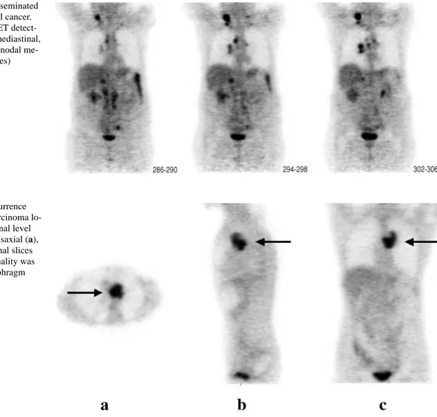
Fig. 1. A case of disseminated
recurrent endometrial cancer. Whole-body FDG PET detect-ed supraclavicular, mdetect-ediastinal, hilar and para-aortic nodal me-tastases (coronal slices)
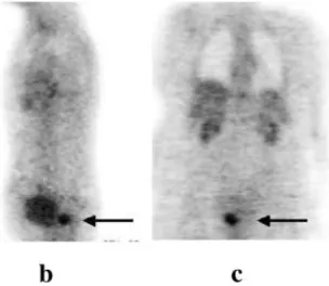
Fig. 2. A case of recurrence
from endometrial carcinoma lo-cated at the mediastinal level (arrow), seen on transaxial (a), sagittal (b) and coronal slices (c). No other abnormality was found below the diaphragm
work-up showed recurrence confined to the pelvis whereas whole-body PET detected extrapelvic foci. The treatment strategy was thus changed from a surgery-based approach to chemotherapy regimens ± radiation. In three patients, only PET was able to detect recurrent
disease, the recurrence being unsuspected both clinically and radiologically. Interestingly, PET was also useful in five of ten women who presented with a strong suspicion of recurrence on the basis of the physical examination and/or elevated tumour marker levels. In these cases, it provided valuable information by localising the sites of disease above and below the diaphragm. The impact of PET on staging and patient management is summarised in Table 4.
On the other hand, in 11 of 12 PET studies, a negative result allowed confirmation of complete remission with a minimum follow-up period of 6 months. However, PET yielded three false positive results corresponding to focal abdominal 18F-FDG uptake not confirmed to represent malignancy by histology and/or by follow-up. Further-more, in one patient, metabolic imaging missed micro-scopic lung metastases detected by thoracic CT. As indi-cated by the kappa coefficient value (κ=0.78), the results of PET agreed quite well with the final diagnoses. Over-all, in the post-therapy surveillance of women with endo-metrial cancers, FDG PET had a 96% sensitivity, a 78% specificity, a 90% diagnostic accuracy, an 89% positive predictive value and a 91% negative predictive value. The likelihood ratio for a positive test result of 4.5 (1.6–12.2) and the likelihood ratio for a negative test re-sult of 0.05 (0.007–0.31) demonstrated the capability of PET to “rule in” disease as well as to “rule out” recur-rence. The results (with 95% CI) are detailed in Table 5.
Discussion
Adenocarcinoma of the endometrium is the most fre-quent gynaecological cancer in post-menopausal women [2]. Although the FIGO recommendations provide a practical framework for the staging of newly diagnosed primary tumours [3], to date no consensual guidelines exist regarding the appropriate post-therapy surveillance of patients previously treated for endometrial carcino-mas. As a consequence, routine follow-up strategies are
Fig. 3. A case of endometrial
cancer presenting with a recur-rence (arrow) confined to the pelvis (a, b, c: transaxial, sagit-tal and coronal slices)
Table 1. Sites of recurrence
de-tected by FDG PET in patients with disseminated recurrent disease (n=13)
Anatomical area No. Lung/pleura 5 Liver 3 Bone 4 Peritoneum 3 Central pelvis 2 Ureter 1 Supradiaphragmatic nodes Supraclavicular 8 Mediastinal 6 Hilar 5 Infradiaphragmatic nodes Para-aortic 10 Iliac 5 Inguinal 3
Table 2. Sites of recurrence
de-tected by FDG PET in patients with recurrent disease located only above the diaphragm (n=4)
Anatomical area No.
Lung 1
Mediastinal nodes 1 Supraclavicular nodes 2
Table 3. Sites of recurrence
de-tected by FDG PET in patients with recurrent disease located only below the diaphragm (n=9)
Anatomical area No. Central pelvis 3 Pelvic wall 4 Abdominopelvic nodes 3
Liver 3
based on institutional experiences, but their effectiveness is a matter of controversy. While most recurrences from endometrial cancer occur within the first 2 years follow-ing treatment, the conventional work-up at control visits, including gynaecological examination ± vaginal Pap smears, chest X-rays and ultrasonography, is rarely ef-fective in diagnosing asymptomatic recurrences [4, 5, 6]. After recent surgery or radiation therapy, the detection of recurrent disease only on the basis of CT or MRI may be extremely difficult given the acute oedema or inflamma-tion surrounding the site of primary tumour. Later in the course of the follow-up, the persistence of
treatment-related fibrosis may also hamper accurate evaluation of the disease status [7, 8, 9, 10, 11]. An elevated serum level of CA125 after initial treatment has been shown to be highly specific in detecting recurrences from endome-trial carcinoma [12, 13]. However, tumour markers are unable to localise the site of the disease. For all these reasons, more effective diagnostic methods are required for the post-treatment surveillance of endometrial carci-nomas.
FDG PET imaging has been shown to be of great value for the diagnosis, staging, re-staging and post-treatment monitoring of various malignancies [27, 28]. Many gynaecological cancers, such as breast cancer, ovarian cancer,and cervical cancer, are now recognised as good indications for PET imaging using 18F-FDG. The value of FDG PET has been verified for the pre-treatment evaluation of primary cancers as well as for the surveillance of previously treated tumours [20, 21, 22, 23, 24, 25, 26]. Nonetheless, very few data are avail-able on the utility of metabolic imaging in the explora-tion of endometrial cancers. Lapela et al. first reported the feasibility of PET using carbon-11 methionine for the imaging of uterine carcinomas. These authors studied 14 such carcinomas by this means, including eight endome-trial cancers and six cervical carcinomas. However, they Table 4. Impact of FDG PET findings on staging and management of patients with confirmed recurrent endometrial carcinoma (n=26) Patient no. FIGO stage Sites of PET findings Routine protocol results Treatment
1 IIIB Local Clin +/TM + Surgery
2 IIA Local + distant Clin +/Rx + (local) Chemotherapy + radiation
3 IIIA Local + distant Clin +/TM + Surgery + chemotherapy
4 IIB Local Clin + Surgery
5 IVA Local + distant Clin +/Rx + (local) Surgery + chemotherapy
6 IVA Distant Clin +/Rx + (distant) Surgery + chemotherapy
7 IVA Distant Clin +/Rx + (distant) Chemotherapy
8 IVA Local + distant Clin +/TM + Chemotherapy
9 IVA Local Clin –/TM –/Rx – Chemotherapy
10 IIB Distant Clin +/Rx + (distant) Chemotherapy
11 IIIB Local Clin + Chemotherapy
12 IIIB Local + distant Clin +/TM + Chemotherapy
13 IIIB Local + distant Clin +/TM +/Rx + (local) Chemotherapy + radiation
14 IIIA Local Clin +/TM + Chemotherapy
15 IIIB Local + distant Clin + Chemotherapy
16 IIA Local + distant Clin +/Rx + (local) Surgery + chemotherapy
17 IB Local Clin +/Rx + (local) Surgery + radiation
18 IB Local + distant Clin + Chemotherapy
19 IVA Local Clin +/Rx + (local) Chemotherapy
20 IVA Local Clin +/Rx + (local) Chemotherapy
21 IVA Distant Clin +/Rx + (distant) Chemotherapy
22 IB Local + distant Clin +/Rx + (local) Chemotherapy
23 IB Local + distant Clin +/Rx + (local) Chemotherapy
24 IIIA Local Clin –/Rx – Surgery + radiation
25 IIIA Local + distant Clin +/TM + Chemotherapy
26 IIB Local + distant Clin –/Rx – Chemotherapy
Local, Recurrences confined to the pelvis; distant, recurrences located beyond the pelvis; Clin, clinical symptoms; Rx, radiological sus-picion; TM, tumour markers (CA125 or CEA)
Table 5. Overall results
FDG PET 95% CI results
Sensitivity 0.96 0.89–1.03
Specificity 0.78 0.57–1.00
Diagnostic accuracy 0.90 0.81–0.99
Negative predictive value 0.91 0.76–1.07 Positive predictive value 0.89 0.78–1.00 Likelihood ratio for a positive test result 4.5 1.6–12.2 Likelihood ratio for a negative test result 0.05 0.007–0.31
underlined the poor quality of the images, which were sometimes impaired by urinary artefacts at the bladder level [29]. Recently, FDG PET was reported to be able to image both the primary tumour and lung metastases in a 60-year-old woman suffering from a histologically proven endometrial carcinoma [30]. In a series of nine women previously treated for endometrial carcinoma, Grigsby et al. also showed the usefulness of metabolic imaging in detecting lung metastatic sites that were un-detected by conventional imaging studies [31]. This led to significant changes in the management of individual patients. In an older study, Spellman et al. measured the levels of seven enzymes in endometrium from 208 women with regular cycles. A physiological increase in the activity of all seven enzymes was found during the secretory phase. The enzymes included hexokinase and glucose-6-phosphate dehydrogenase, two key enzymes of the glycolytic pathway and thus of the 18F-FDG tissue uptake process [32]. Other basic studies showed that oestrogens increase the activity of glucose-6-phosphate dehydrogenase and pyruvate kinase in rat uterus [33, 34]. These preclinical and clinical reports warranted fur-ther investigations in the use of FDG PET in women presenting with endometrial carcinoma, an oestrogen-dependent cancer. Additionally, tumour changes detected at the metabolic level usually precede macroscopic fea-tures evidenced by morphological imaging procedures [18, 19, 20, 21, 22, 23, 24, 25, 26]. The goal of the pres-ent study was to specifically address the issue of the role of 18F-FDG PET imaging in the post-therapy surveil-lance of endometrial carcinomas. The lack of a stan-dardised follow-up programme and the relatively low sensitivity of the protocols routinely used in gynaecolog-ical practice for the detection of recurrences stress the need for more efficient diagnostic tools.
Our results demonstrate the usefulness of PET imag-ing usimag-ing 18F-FDG in the post-therapy surveillance of patients with previously treated endometrial carcinoma. For instance, metabolic imaging confirmed the presence of recurrent disease in 88% of patients with suspicion of recurrence at control visits. However, unlike the routine follow-up protocol, whole-body FDG PET was able to accurately localise the sites of recurrence over the entire body within a single imaging procedure. Moreover, the value of PET was further illustrated by its ability to de-tect unsuspected recurrences in 12% of women with con-firmed recurrent disease: by contrast, the sensitivity of routine follow-up in detecting asymptomatic recurrences is known to be very low [4, 5, 6].
Although all stages of disease may recur locally and/or at distant sites, on the basis of pathological fac-tors a woman may be classified as having a low, interme-diate or high risk of recurrence [35]. In the present se-ries, 85% of the women (22/26) presenting with con-firmed recurrences had FIGO stages II–IV disease (Table 4). These findings suggest that the optimal role for PET could be in this subgroup of patients with a high
risk of recurrence (stage II, III or IV disease or serous or clear-cell carcinoma). Nonetheless, four women (15%) with FIGO stage IB also had recurrent disease detected by FDG PET imaging. This suggests that PET may also play a role in patients initially classified as having a “low or intermediate risk” of recurrence (stage I, grade 1 or 2 non-serous and non-clear-cell adenocarcinoma with invasion of the myometrium). It is likely, however, that the frequency of the PET controls should be modulated according to the risk of recurrence. Further controlled studies are warranted to specifically assess the contribu-tion of PET for each stage of the disease.
Owing to its limited spatial resolution, in one patient PET missed lung micrometastases detected by thoracic CT. Metabolic imaging also yielded three false-positive results, which rather impaired the specificity of the tech-nique. Although the clinical context and the medical his-tory of the patient can help in achieving an accurate di-agnosis, the inherent pitfalls of metabolic imaging can-not be avoided in all circumstances. For instance, some benign tumours or infectious diseases may also take up 18F-FDG, examples including leiomyoma, benign serous cystadenoma, benign fibroids, endometriosis and endo-metrioma [36, 37, 38]. Similarly, the inflammatory changes that follow the primary therapy (recent surgery or radiation therapy) can induce intense uptake of the metabolic tracer [39]. Other false-positive results are re-lated to the physiological retention of the tracer in the urinary system or the bowels. These situations are not in-frequent in the context of gynaecological malignancies. To help overcome the limited specificity of FDG PET, reference can be made to the post-treatment serum levels of CA125: increases in comparison with baseline have been shown to reflect, at an early stage, the presence of active recurrences of endometrial carcinoma [40]. In equivocal cases (ureteral uptake versus nodal metastases, or normal intestinal uptake versus tumour mass), recent-ly introduced PET-CT devices may also contribute in accurately distinguishing a “true” recurrence from physi-ological or benign uptake. Indeed, such combined devices may be of great utility in anatomically localising a metabolically active recurrence for the purpose of tar-geted radiation [41, 42, 43]. On the basis of these consid-erations, we believe that the judicious combination of a single whole-body imaging technique such as FDG PET with pre- and post-treatment measurement of the serum level of a highly specific tumour marker such as CA125 could avoid both the cost of repeated conventional imag-ing studies and the disagreement to which they give rise. Morphological imaging would be performed when PET results are negative but tumour marker levels are abnor-mally elevated, or to precisely define the anatomical margins of the disease for the purpose of targeted radia-tion therapy. Theoretically, this protocol should be fo-cussed particularly on the first 2 or 3 years following the primary tumour therapy, when the prevalence of re-currence is highest. Further multicentre studies are,
how-ever, necessary to confirm our encouraging results and also to determine the optimal timing of FDG PET during the post-therapy surveillance.
The results of this study, like those reported by Grigsby et al., indicate that whole-body FDG PET offers added value in terms of staging and treatment choices in endometrial carcinoma (Table 4). For instance, the utility of a whole-body technique is shown by the fact that 50% of the recurrences occurred both above and below the di-aphragm, while 15% occurred only above the diaphragm and 35% only below it. These results are in line with the pattern of recurrence commonly reported in the literature [1, 2, 3, 4, 5, 6, 34]. Currently, in patients with endome-trial carcinoma, systemic chemotherapy and/or proges-terone therapy is advocated for disseminated recurrent disease [2, 16, 17]. Localised pelvic recurrences may be treated by surgery and/or targeted radiation [2, 3, 14, 35]. Additionally, it has been shown that pelvic exentera-tion may be performed with relative success in a subset of patients with failure of prior radiation [15].
In the present study, PET had an actual treatment im-pact in 35% (9/26) of the women with confirmed recur-rent disease, either by detecting extrapelvic foci (6/9) or by detecting clinically and radiologically unsuspected re-currences (3/9). As a result, PET significantly altered the treatment options from surgery ± radiation to chemother-apy ± radiation. Metabolic imaging also allowed the ini-tiation of treatment in those asymptomatic women in whom it identified recurrent disease. Owing to the high rate of extrapelvic and metastatic recurrences in our se-ries, the majority of patients underwent systemic thera-pies rather than surgical treatment. Therefore, most of the recurrences were confirmed by the clinical and radio-logical outcomes rather than by systematic historadio-logical analyses. In other words, in these patients with multiple foci, the non-invasive nature of PET imaging and its high diagnostic accuracy could allow the avoidance of invasive surgical procedures. As recently reported by Beggs et al. in a study of 50 oncology patients, PET findings suggesting disseminated disease may be consid-ered as “a metabolic biopsy” [44]. On the other hand, the early detection by whole-body PET imaging of recur-rences confined to the pelvis would warrant more ag-gressive therapies. Last but not least, metabolic imaging has the capability to reliably exclude recurrent disease. As evidenced by the low likelihood ratio for a negative test result (<0.1), even negative PET had a clinical value since in 11 of the 12 negative PET studies no recurrence was observed with a median follow-up duration of 10 months.
In conclusion, whole-body FDG PET is a valuable tool in the post-therapy surveillance of women previously treated for endometrial carcinoma. The capability of PET to detect suspected and unsuspected recurrences, in one session, enables non-invasive follow-up of patients by means of a single procedure. The optimal timing of FDG PET during the follow-up programme and the way in
which it should be combined with other diagnostic modal-ities, such as tumour marker determinations and morpho-logical imaging, remain to be defined. Further prospective studies are also necessary to assess the cost-effectiveness of the technique and its impact on patient survival.
Acknowledgements. The authors thank Dr. Frederic Kridelka for
helpful discussions.
References
1. Salvesen HB. Routine follow up after treatment for gyneco-logical cancer. Tidsskr Nor Laegeforen 2001; 121:1253–1265. 2. Pazdur R, Coia LR, Hoskins WJ, Wagman LD. Cancer
man-agement: a multidisciplinary approach, 5th edn. New York:
PRR, 2001.
3. Benedet JL, Bender H, Jones H 3rd, Ngan HY, Pecorelli S. FIGO staging classifications and clinical practice guidelines in the management of gynecologic cancers. FIGO Committee on Gynecologic Oncology. Int J Gynaecol Obstet 2000; 70:209– 262.
4. Salvesen HB, Akslen LA, Iversen T, Iversen OE. Recurrence of endometrial carcinoma and the value of routine follow up.
Br J Obstet Gynaecol 1997; 104:1302–1307.
5. Shumsky AG, Stuart GC, Brasher PM, et al. An evaluation of routine follow-up of patients treated for endometrial carcino-ma. Gynecol Oncol 1994; 55:229–233.
6. Owen P, Duncan ID. Is there any value in the long term follow up of women treated for endometrial cancer? Br J Obstet
Gynaecol 1996; 103:710–713.
7. Ebner F, Kressel HY, Mintz MC, et al. Tumor recurrence ver-sus fibrosis in the female pelvis: differentiation with MR im-aging at 1.5 T. Radiology 1988; 166:333–340.
8. Kinkel K, Ariche M, Tardivon AA, et al. Differentiation be-tween recurrent tumor and benign conditions after treatment of gynecologic pelvic carcinoma: value of dynamic contrast- en-hanced subtraction MR imaging. Radiology 1997; 204:55–63. 9. Siegelman ES, Outwater EK. Tissue characterization in the
fe-male pelvis by means of MR imaging. Radiology 1999; 212:5–18.
10. Sovik E, Lien HH, Tveit KM. Postirradiation changes in the pelvic wall. Findings on MR. Acta Radiol 1993; 34:573–576. 11. Connor JP, Andrews JI, Anderson B, Buller RE. Computed
to-mography in endometrial carcinoma. Obstet Gynecol 2000; 95:692–696.
12. Cherchi PL, Dessole S, Ruiu GA, et al. The value of serum CA 125 and association CA 125/CA 19–9 in endometrial car-cinoma. Eur J Gynaecol Oncol 1999; 20:315–317.
13. Kurihara T, Mizunuma H, Obara M, et al. Determination of a normal level of serum CA125 in postmenopausal women as a tool for preoperative evaluation and postoperative surveillance of endometrial carcinoma. Gynecol Oncol 1998; 69:192–196. 14. Wylie J, Irwin C, Pintilie M, et al. Results of radical
radiother-apy for recurrent endometrial cancer. Gynecol Oncol 2000; 77:66–72.
15. Morris M, Alvarez RD, Kinney WK, Wilson TO. Treatment of recurrent adenocarcinoma of the endometrium with pelvic ex-enteration. Gynecol Oncol 1996; 60:288–291.
16. Dimopoulos MA, Papadimitriou CA, Georgoulias V, et al. Paclitaxel and cisplatin in advanced or recurrent carcinoma of the endometrium: long-term results of a phase II multicenter study. Gynecol Oncol 2000; 78:52–57.
32. Spellman CM, Fottrell PF, Baynes S, et al. A study of some enzymes and isoenzymes of carbohydrate metabolism in hu-man endometrium during the menstrual cycle. Clin Chim Acta 1973; 48:259–268.
33. Pepe GJ, Yochim JM. Glucose-6-phosphate dehydrogenase ac-tivity in the endometrium and myometrium of the rat uterus during the estrous cycle and progestation. Biol Reprod 1971; 5:161–171.
34. Singhal RL, Valadares JR. Estrogenic regulation of uterine py-ruvate kinase. Am J Physiol 1970; 218:321–327.
35. Rose PG. Endometrial carcinoma. N Engl J Med 1996; 335:640–649.
36. Hubner KF, McDonald TW, Niethammer JG, et al. Assessment of primary and metastatic ovarian cancer by positron emission tomography (PET) using 2-(F18) deoxyglucose. Gynecol
Oncol 1993; 51:197–204.
37. Shreve PD, Anzai Y, Wahl RL. Pitfalls in oncologic diagnosis with FDG PET imaging: physiologic and benign variants.
Radiographics 1999; 19:61–77.
38. Holder WD Jr, White RL, Zuger JH, Easton EJ Jr, Greene FL. Effectiveness of positron tomography for the detection of mel-anoma metastases. Ann Surg 1998; 227:764–765.
39. Nakamoto Y, Eisbruch A, Achtyes ED, et al. Prognostic value of positron emission tomography using F-18-fluorodeoxyglu-cose in patients with cervical cancer undergoing radiotherapy.
Gynecol Oncol 2002; 84:289–295.
40. Rose PG, Sommers RM, Reale FR, et al. Serial serum CA 125 measurements for evaluation recurrence in patients with endo-metrial carcinoma. Obstet Gynecol 1994; 177:231–236. 41. Kluetz PG, Meltzer CC, Villemagne VL, Kinahan PE, Chander
S, Martinelli MA, Townsend DW. Combined PET/CT imaging in oncology. Impact on patient management. Clin Positron
Imaging 2000; 3:223–230.
42. Charron M, Beyer T, Bohnen NN, Kinahan PE, Dachille M, Jerin J, Nutt R, Meltzer CC, Villemagne V, Townsend DW. Image analysis in patients with cancer studied with a com-bined PET and CT scanner. Clin Nucl Med 2000; 25:905–910. 43. Makhija S, Howden N, Edwards R, Kelley J, Townsend DW,
Meltzer CC. Positron emission tomography/computed tomo-graphy imaging for the detection of recurrent ovarian and fallopian tube carcinoma: a retrospective review. Gynecol
Oncol 2002; 85:53–58.
44. Beggs AD, Hain SF, Curran KM, O’Doherty MJ. FDG-PET as “metabolic biopsy” tool in non-lung lesions with indetermi-nate biopsy. Eur J Nucl Med 2002; 29:542–546.
45. Hart KB, Han I, Shamsa F, et al. Radiation therapy for endo-metrial cancer in patients treated for postoperative recurrence.
Int J Radiat Oncol Biol Phys 1998; 41:7–11.
46. Podczaski E, Kaminski P, Gurski K, et al. Detection and pat-terns of treatment failure in 300 consecutive cases of “early” endometrial cancer after primary surgery. Gynecol Oncol 1992; 47:323–327.
17. Thigpen T, Brady M, Alvarez R, et al. Oral medroxyproges-terone acetate in the treatment of advanced or recurrent endometrial carcinoma: a dose response study by the Gy-necologic Oncology Group. J Clin Oncol 1999; 17:1736– 1744.
18. Delbeke D. Oncological applications of FDG PET imaging.
J Nucl Med 1999; 40:1706–1715.
19. Delbeke D. Oncological applications of FDG PET imaging: brain tumors, colorectal cancer, lymphoma and melanoma.
J Nucl Med 1999; 40:591–603.
20. Eubank WB, Mankoff DA, Vesselle HJ, et al. Detection of lo-coregional and distant recurrences in breast cancer patients by using FDG PET. Radiographics 2002; 22:5–17.
21. Lonneux M, Borbath II, Berliere M, Kirkove C, Pauwels S. The place of whole-body PET FDG for the diagnosis of distant recurrence of breast cancer. Clin Positron Imaging 2000; 3:45–49.
22. Zimny M, Siggelkow W, Schroder W, et al. 2-[Fluorine-18]-fluoro-2-deoxy-D-glucose positron emission tomography in the diagnosis of recurrent ovarian cancer. Gynecol Oncol 2001; 83:310–315.
23. Jimenez-Bonilla J, Maldonado A, Morales S, et al. Clinical impact of 18F-FDG-PET in the suspicion of recurrent ovarian
carcinoma based on elevated tumor marker serum levels. Clin
Positron Imaging 2000; 3:231–236.
24. Sun SS, Chen TC, Yen RF, et al. Value of whole body 18
F-fluoro-2-deoxyglucose positron emission tomography in the evaluation of recurrent cervical cancer. Anticancer Res 2000; 21:2957–2961.
25. Grigsby PW, Dehdashti F, Siegel BA. FDG-PET evaluation of carcinoma of the cervix. Clin Positron Imaging 1999; 2:105–109.
26. Umesaki N, Tanaka T, Miyama M, et al. The role of
18F-fluoro-2-deoxy-D-glucose positron emission tomography
(18F-FDG-PET) in the diagnosis of recurrence and lymph
node metastasis of cervical cancer. Oncol Rep 2000; 7:1261– 1264.
27. Kubota K. From tumor biology to clinical Pet: a review of positron emission tomography (PET) in oncology. Ann Nucl
Med 2001; 15:471–486.
28. Czernin J, Phelps ME. Positron emission tomography scan-ning: current and future applications. Annu Rev Med 2002; 53:89–112.
29. Lapela M, Leskinen-Kallio S, Varpula M, et al. Imaging of uterine carcinoma by carbon-11-methionine and PET. J Nucl
Med 1994; 35:1618–1623.
30. Nakahara T, Fujii H, Ide M, et al. F-18 FDG uptake in endo-metrial cancer. Clin Nucl Med 2001; 26:82–84.
31. Grigsby PW, Dehdashti F, Siegel BA. FDG-PET evaluation of recurrent endometrial carcinoma. 7th Biennial Meeting of the International Gynecologic Cancer Society; Rome, Italy, 26–30 September 1999.