Review
Maria Fusaro*, Maurizio Gallieni, Maria Antonietta Rizzo, Andrea Stucchi, Pierre Delanaye,
Etienne Cavalier, Rosa M.A. Moysés, Vanda Jorgetti, Giorgio Iervasi, Sandro Giannini,
Fabrizio Fabris, Andrea Aghi, Stefania Sella, Francesco Galli, Valentina Viola
and Mario Plebani
Vitamin K plasma levels determination in human
health
DOI 10.1515/cclm-2016-0783
Received June 19, 2016; accepted September 6, 2016
Abstract: Vitamin K (phylloquinone or vitamin K1 and
menaquinones or vitamin K2) plays an important role as a cofactor in the synthesis of hepatic blood coagulation pro-teins, but recently has also aroused an increasing interest for its action in extra-hepatic tissues, in particular in the regulation of bone and vascular metabolism. The accurate
measurement of vitamin K status in humans is still a criti-cal issue. Along with indirect assays, such as the under-carboxylated fractions of vitamin K-dependent proteins [prothrombin, osteocalcin (OC), and matrix gla protein], the direct analysis of blood levels of phylloquinone and menaquinones forms might be considered a more inform-ative and direct method for assessing vitamin K status. Different methods for direct quantification of vitamin K serum levels are available. High-performance liquid chro-matography (HPLC) methods coupled with post-column reduction procedures and fluorimetric or electrochemical detection are commonly used for food and blood analysis of phylloquinone, but they show some limitations when applied to the analysis of serum menaquinones because of interferences from triglycerides. Recent advancements include liquid chromatography tandem mass spectrome-try (LCMS/MS) detection, which assures higher specificity. The optimization and standardization of these methods requires specialized laboratories. The variability of results observed in the available studies suggests the need for further investigations to obtain more accurate analytical results.
Keywords: human health; metabolism; plasma levels;
vitamin K.
Introduction
Vitamin K has important biological actions, some of which are still being discovered. Vitamin K plays a key role in the synthesis of several blood coagulation factors, but it is also strongly connected to bone metabolism and vascular calcifications [1].
Vitamin K exists in two main natural forms: K1 (or phylloquinone, PK) and K2 (including several differ-ent vitamers called menaquinones, MKs) (Figure 1). In addition to the naturally occurring phylloquinone and menaquinones, there is as a synthetic form of vitamin K:
*Corresponding author: Maria Fusaro, National Research Council
(CNR) – Institute of Clinical Physiology (IFC), Pisa Via G. Moruzzi 1, 56124, Pisa, PI, Italy; and Department of Medicine, University of Padova Italy; Via Giustiniani 2, 35128, Padova, PD, Italy, E-mail: dante.lucia@libero.it
Maurizio Gallieni: Nephrology and Dialysis Unit, Ospedale San
Carlo Borromeo, Department of Clinical and Biomedical Sciences “Luigi Sacco”, University of Milan, Milan, Italy
Maria Antonietta Rizzo: Nephrology and Dialysis Unit, Ospedale di
Circolo di Busto Arsizio, ASST Valle Olona, Italy
Andrea Stucchi: Nephrology and Dialysis Unit, IRCCS Multimedica,
Sesto San Giovanni (Milano), Milan, Italy
Pierre Delanaye: Department of Nephrology, Dialysis, and
Transplantation, University of Liège, Centre Hospitalier Universitaire du Sart Tilman (ULg CHU), Liège, Belgium
Etienne Cavalier: Department of Clinical Chemistry, University
of Liège, Centre Hospitalier Universitaire du Sart Tilman, Liège, Belgium
Rosa M. A. Moysés: Universidade Nove de Julho, UNINOVE,
São Paulo, Brazil; and Universidade de Sao Paulo, São Paulo, Brazil
Vanda Jorgetti: Universidade de Sao Paulo, São Paulo, Brazil Giorgio Iervasi: Institute of Clinical Physiology, National Council of
Research, Pisa, Italy
Sandro Giannini, Fabrizio Fabris, Andrea Aghi and Stefania Sella:
Department of Medicine, Clinica Medica 1, University of Padova, Padova, Italy
Francesco Galli: Department of Pharmaceutical Sciences, University
of Perugia, Italy
Valentina Viola: Azienda Ospedaliera Universitaria Policlinico Tor
Vergata, Rome, Italy
Mario Plebani: Laboratory Medicine Unit, Department of Medicine,
University of Padova, Padova, Italy. http://orcid.org/0000-0002-0270-1711
menadione, or vitamin K3 (2-methyl-1, 4-naphthoquinone nucleus), representing the basic structure common to phylloquinone and menaquinones.
Phylloquinone is mainly found in green vegeta-bles. The source of menaquinones is more uncertain, and although the contribution of the intestinal bacte-rial flora to vitamin K status is still incompletely under-stood, the literature indicates that menaquinones are derived mainly from intestinal bacteria [2, 3] and fer-mented food (e.g. cheeses and the Japanese soybean product known as ‘natto’) [3]. Liver is also a rich source of menaquinones [4].
Menaquinones are classified according to the length of their unsaturated side chains. Up to 12 different types have been described, from MK-4 to MK-15 [5]. The most common MKs in humans are the short-chain MK-4, which is the only MK produced by systemic conversion of phyl-loquinone to menaquinones, and the long-chain vitamers,
Figure 1: Structures of phylloquinone (vitamin K1) and
menaqui-nones (MKs or vitamin K2).
Menadione (vitamin K3) represents the basic structure common to K1 and K2. Menadione is available as a synthetic form of vitamin K.
MK-7 through MK-10, which are synthesized by bacteria in humans.
The predominant dietary form of vitamin K in the USA, Europe, and most Western countries is phylloqui-none, while the major form in Japan is menaquinones, especially menaquinone 7 (MK-7), which is a component of natto [6]. Natto is most popular in the eastern regions of Japan, including Kanto, Tohoku, and Hokkaido, but it is consumed in all areas.
Different phylloquinone and menaquinones plasma concentrations have been reported, with a possible influ-ence of dietary intake, and a possible analytic interfer-ence from triglycerides [7]. Mean plasma concentrations ranging from 0.22 to 8.88 nmol/L have been reported, although in most studies phylloquinone had a concentra-tion below 2 nmol/L [8].
Daily intake of vitamin K in a Western diet range is estimated from 60 μg to 200 μg, of which phylloqui-none is the larger component (about 90%, versus 10% of menaquinones) [9].
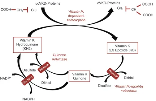
Recommendations for daily intake of vitamin K are inconsistent. The Institute of Medicine (US) Panel on Micronutrients proposed an adequate intake (AI) for men and women of 120 and 90 μg/day, respectively, based on representative dietary intake data from healthy individu-als, because of the lack of data to estimate an average requirement [10]. Considering that no adverse effect has been reported for individuals consuming higher amounts of vitamin K, a tolerable upper intake level was not estab-lished. In 2012, the Italian LARN (Reference Assumption Levels for Nutrients and Energy), proposed by the Human Nutrition Italian Society (SINU), suggested an intake of vitamin K stratified for age (140 or 170 μg/day for 18–59 and > 60 years old, respectively). However, in the 2014 release of the same recommendations this group stated that the available evidence does not allow to define the adequate intake for vitamin K [11]. In the UK, a government backed panel of experts pointed out that although vitamin K is known to be essential, recommendations on adequate nutritional intakes have not been precisely established, because of the unquantified contribution made by the intestinal bacteria [12]. They reported previous data of daily intake suggested by the UK Department of Health’s Committee on Medical Aspects of Food Policy (COMA): 1 μg/kg body weight [13], which is probably adequate for blood clotting, but suboptimal for bone health. Vitamin K is required to introduce carboxyl groups into glutamic acid residues in four of the blood coagulation factors (II, VII, IX, X) to yield γ-glutamic carboxyl (Gla) residues (Figure 2), while vitamin K hydroquinone is transformed into vitamin K 2,3-epoxide [14]. Importantly, warfarin
interferes with regeneration of vitamin K 2,3-epoxide to vitamin K hydroquinone, thus impairing γ-carboxylation and the activity of vitamin K dependent proteins, includ-ing the extra-hepatic proteins OC (bone Gla-protein; BGP) and matrix Gla-protein (MGP), respectively involved in bone mineralization and inhibition of vascular calcifi-cations. If vitamin K is deficient, vitamin K dependent proteins cannot increase their carboxylation status (and they become significantly undercarboxylated), losing their capacity to bind calcium, so that bone metabolism may be impaired and the process of vascular calcification enhanced [15].
Both phylloquinone and MKs may activate the steroid and xenobiotic receptor (SXR). SXR is a nuclear receptor involved in the transcriptional regulation of enzymes such as cytochrome P450 (in particular the CYP3A4 isoform) [16]. SXR and its murine ortholog, pregnane X receptor (PXR), are nuclear receptors that are expressed at high levels in the liver and the intestine. They work as xenobi-otic sensors that induce expression of genes involved in detoxification and drug excretion, but they were recently shown to be also expressed in osteoblasts and involved in bone metabolism [17]. Hydroxylation of vitamin K is catalyzed by cytochrome P450 enzymes, which often are induced by their substrates themselves via the activation of the nuclear receptor PXR [18].
Azuma et al. described the osteopenic phenotype of systemic PXR knockout mice; they observed a remark-able reduction of width and an important gap between
femoral and tibial articular cartilages in PXR knockout mice, resulting in aging-dependent wearing of knee joints articular cartilage. These findings may indicate that SXR/ PXR protects against aging-dependent wearing of articu-lar cartilage and that ligands for SXR/PXR have a poten-tial role in preventing osteo-articular diseases caused by aging [19].
Metabolism of vitamin K: general
principles
Most of our knowledge on the metabolism of vitamin K – in particular about its intestinal absorption, trans-port, cellular uptake and catabolism – is related to phyl-loquinone, while data about menaquinones are more limited [2]. Vitamin K forms derived from plants and bac-teria have a poorer bioavailability than those contained in oil-based and processed foods. Low plasma concen-trations of phylloquinone reflect low tissue reserves. Vitamin K tissue concentrations have been determined. In adults, phylloquinone concentrations are about 10 pmol/g-wet tissue in liver, hearth and pancreas, while they are lower in brain, kidney and lung (> 2 pmol/g). Contrariwise, high MK-4 concentrations were found in human brain and kidney (6 pmol/g) and even higher in pancreas (22 pmol/g) than in other tissues, such as in plasma and liver [20].
ucVKD-Proteins cVKD-Proteins COOH
COOH CH2 Glu COOH CH Gla Vitamin K 2,3 Epoxide (KO) Vitamin K Quinone Dithiol Dithiol Disulfide Warf arin Warf arin Warf arin Disulfide NADP+ NADPH Vitamin K Hydroquinone (KH2) Vitamin K dependent carboxylase Quinone reductase Vitamin K-epoxide reductase
Figure 2: The vitamin K cycle and the interference of warfarin with vitamin K recycling.
Gla-proteins are carboxylated by a specific vitamin K dependent carboxylase, through several rounds of carboxylation. Here a single round of carboxylation is depicted. (Reprinted with permission from Fusaro et al. [15].) ucVKD-proteins, undercarboxylated vitamin K dependent proteins.
Considering that MK-4 derives from the endogenous conversion of phylloquinone, the specific tissue distribu-tion of MK-4 is suggestive of local synthesis from phyl-loquinone. MK-4 is synthetized in testes, pancreas and blood vessels. In particular, the conversion of phylloqui-none into MK-4 occurs either directly or by interconver-sion to menadione (K3), followed by prenylation to MK-4 [21]. Therefore, the biological activity of K3 is entirely dependent on its prenylation to MK-4.
However, the molecular mechanisms of conversion remain unclear. Nakagawa et al. identified a human MK-4 biosynthetic enzyme known as UbiA prenyltransferase containing 1 (UBIAD1), by screening the human genome database. UBIAD1 is localized in the endoplasmic reticu-lum and ubiquitously expressed in several mouse tissues. Short interfering RNA against the UBIAD1 gene inhibited the conversion of deuterium-labeled phylloquinone mol-ecules into deuterium-labeled-MK-4 (MK-4-d7) in human cells. An additional proof that the UBIAD1 gene encodes an MK-4 biosynthetic enzyme derives from its expres-sion in cells infected with UBIAD1 baculovirus, which can convert deuterium-labeled vitamin K derivatives into MK-4-d7 [22].
Phylloquinone is probably converted into MK-4 within the tissues themselves, rather than via hepatic meta-bolism. After phylloquinone administration, the MK-4 concentration increased much more slowly in each of the tissues than that of phylloquinone, and the MK-4 concen-tration in plasma and liver reached much lower levels than those observed in other tissues [23].
Vitamin K is transported in plasma by lipoproteins [24]. After digestion in the intestinal tract, dietary vitamin K and triglycerides (TG) are emulsified by bile salts to form mixed micelles in the enterocytes and processed into chy-lomicrons (CR), containing apolipoprotein A (apoA) and apoB, and then secreted into the lymph ducts and blood circulation. CR are modified peripherally, in adipose or muscular tissues, by the action of lipoprotein lipase (LPL) and re-enter in the circulation, but they continue trans-porting vitamin K in their lipophilic core [25].
The uptake of vitamin K into the liver seems to follow the same pathway of lipoprotein [26]. In fact, CR enter into the liver by endocytosis and they are processed to finally obtain smaller LDL molecules; vitamin K is presumed to remain still located in the lipophilic core of lipoproteins.
Concerning the uptake of vitamin K into bone tissue [27], it is known that osteoblasts obtain most of their phyl-loquinone by CR pathway and most of their MK-7 by LDL pathway. Osteoblasts express lipoprotein receptors, which interact with CR and LDL and start the process of endocy-tosis of the particles and the vitamin K.
The liver is also the site of vitamin K catabolic pathway, common to phylloquinone and MKs. The poly-isoprenoid side chains are shortened, undergoing ω-oxidation fol-lowed by β-oxidation leading to two major aglycone metab-olites with side chain lengths of five and seven carbon atoms (5C and 7C metabolites, respectively). Finally, after conjugation with glucuronic acid, the metabolites are excreted in the bile and urine, mainly as glucuronides [2].
Review of the assessment of
vitamin K status and the possible
role of vitamin K plasma levels
measurement
One barrier to gaining a better understanding of vitamin K function has been the difficulty in developing methods for the measurement of vitamin K. Vitamin K was the last of the four fat-soluble vitamins to be measured at endog-enous levels. The degree of this analytical challenge is a consequence of vitamin K being the most lipophilic and least abundant of the fat soluble vitamins. These proper-ties do not easily lend themselves to the development of assays suitable for current generation automated chemis-try platforms, unlike vitamin D.
Vitamin K status can be assessed by indirect func-tional tests such as the prothrombin time or by measure-ment of undercarboxylated proteins (Table 1), such as OC and matrix Gla protein (MGP), which are more sensitive in detecting subclinical vitamin K deficiency than pro-thrombin time [28]. The amount of undercarboxylated OC could represent a sensitive marker of vitamin K status in humans [29]. However, although a high percentage of undercarboxylated OC indicates poor vitamin K status, this value better reflects recent vitamin K intake and not the long-term vitamin K status.
Osteocalcin levels are also influenced by vitamin D, which is required for the production of undercarboxy-lated OC, whereas vitamin K is required for the conversion of undercarboxylated to mature OC. Data from healthy volunteers indicate a weak but significant correlation between undercarboxylated OC and vitamin D, suggest-ing that the serum level of undercarboxylated OC may be an insufficiently reliable marker for vitamin K status [30]. OC is also influenced by PTH, which is significantly elevated in many CKD patients. Therefore, CKD patients with hyperparathyroidism will present high serum uOC, but this does not necessarily mean that they are vitamin K deficient.
Undercaboxylated unphosphorylated MGP (ucdpMGP), the inactive form of MGP, may be a good alternative to evaluated vitamin K status of the subjects; Indeed, randomized controlled studies have shown that vitamin K therapy decreases ucdpMGP levels [31–34]. Moreover, AVK treatment increased the amount of inactive ucdpMGP [35, 36]. Stopping that treatment has also been shown to decrease ucdpMGP [37]. All these date suggest that ucdpMGO could be a good marker of vitamin K status. However, ucdpMGP determination is only available on the automated IDS iSYS instrument, which is not available everywhere. Moreover, measuring inactive MGP may only reflect the vitamin K status at the vascular level and not at other organ’s levels (in particular bone and liver). In this context, direct measurement of vitamin K could be of interest by its own, or in combination with ucdpMGP.
Moreover, protein induced in vitamin K absence (PIVKA-II) could be a sensitive marker to predict mild vitamin K deficiency, as reported among newborns. PIVKA-II is an inactive precursor of prothrombin and is elevated in vitamin K deficiency [38].
Urinary vitamin K metabolites can be measured in the urine. Harrington et al. tested this analytic approach in young adults [39] and studied its clinical significance in the pediatric population [40]. Urinary excretion of 7C-aglycone and 5C-aglycone, vitamin K metabolites common to both phylloquinone and the menaquinone series, were assessed following restriction or supple-mentation with phylloquinone. Results indicate a good relationship of urinary metabolites with dietary phylloqui-none intake. In newborns, vitamin K urinary metabolites excretion was 25 times lower than adults and improved
after prophylaxis. Infants mainly excreted 5C-aglycone, while increased excretion of the 7C-aglycone was associ-ated with metabolic overload because of the exposure to high-tissue phylloquinone concentrations. Measurement of the 5C- and 7C-aglycones was therefore proposed as a good approach to study vitamin K status in neonates and as an aid the development of improved prophylactic regi-mens [40]. Urinary vitamin K metabolites have not been studied in CKD patients, but deterioration of renal func-tion might interfere with the reliability of this marker of vitamin K status.
Determination of vitamin K plasma
levels: technical aspects
Providing a standardized measurement of plasma levels of vitamin K forms, in particular for some MKs, is challeng-ing. Measuring vitamin K in plasma is difficult because of the low circulating vitamin K levels and the non-polar characteristics of vitamin K as well as the interference of lipids [41].
Different methods for direct quantification of vitamin K serum levels have been developed and evalu-ated (Table 1). High-performance liquid chromatography (HPLC) is considered the elective technique to measure vitamin K subtypes.
HPLC has been developed starting from the second half of the last century [42], then extended to the meas-urement of long-chain MKs in the late 1980s [43]. Ini-tially, HPLC with ultraviolet detection (UV) was used to
Table 1: Measurement of vitamin K status in humans: summary of direct and indirect methods.
Methods Main characteristics
Indirect methods
Prothrombin time Cannot be used as a reliable indicator of vitamin K status Undercarboxylated osteocalcin (ucOC) or matrix Gla protein
(ucMGP) Vitamin K deficiency is associated with reduced carboxylation of vitamin K dependent proteins and higher levels of ucOC and ucMGP. Vitamin D regulates osteocalcin gene expression
PIVKA-II Elevated in vitamin K deficiency
Urinary vitamin K metabolites (7C-aglycone and 5C-aglycone) Vitamin K metabolites mainly tested in pediatric population Direct methods
High-performance liquid chromatography (HPLC) with
ultraviolet (UV) detection Lower sensitivity and selectivity
HPLC with fluorescence detection Provides greater sensitivity and selectivity than UV detection. Most common method used in laboratories
HPLC with electrochemical detection (ECD) Post-column reduction is used to convert the quinone structure of vitamin K in the corresponding hydroquinones, measured in oxidation mode Liquid chromatography tandem mass spectrometry
determinate PK plasma concentration. Afterwards, HPLC with fluorescence after post column reduction demon-strated greater sensitivity and selectivity than UV method [44, 45], but it required extensive sample pre-purification to reduce the chromatographic interference induced by lipids [41].
Other recent methods for determination of PK and MKs are based on liquid chromatography tandem mass spectrometry (LCMS/MS), which present higher sensi-tivity and selecsensi-tivity, but they require longer analytical times. Suhara et al. [46] developed a model of liquid chro-matography-tandem mass spectrometry (LC-APCI-MS/MS) method for the measurement of vitamin K plasma levels (PK, MK-4, and MK-7), characterized by high sensitivity and selectivity. However, the method is excessively long for routine analysis, because of the pre-purification pro-cedure is based on dual step extraction, chromatographic separation followed by a wash and re-equilibration period. Gentili et al. simplified the process and reduced the total run-time [47]. With this method, MK-4 and MK-7 levels were undetectedable in the analyzed serum samples of subjects on a Mediterranean diet [47]. Karl et al. [48] devel-oped a method employing HPLC-mass spectrometry with atmospheric pressure chemical ionization (LC-APCI-MS) for simultaneous quantification of 11 vitamin K vitamers, which was applied to biological samples such as serum, faeces and food. They suggested that the method can be applied in human and animal studies examining the role of vitamin K vitamers derived from the diet and gut bacte-ria synthesis in health and disease.
Riphagen et al. [49] also described a method based on LC-APCI-MS/MS with atmospheric pressure chemical ionization to detect plasma PK, MK-4 and MK-7 levels, sim-plifying the sample pre-purification process and reducing the total run-time without compromising sensitivity and selectivity. In a study population of 60 renal transplant recipients, the authors confirmed that plasma PK con-centrations were significantly associated with recent PK dietary intake and plasma MK-4 concentrations, and that plasma vitamin K levels were strongly correlated with plasma triglyceride concentrations [49].
It is known that vitamin K is mainly transported by triglyceride-rich lipoproteins [50]. This is a critical point to consider for obtaining a reliable determination of vitamin K serum levels, because the interaction of the lipoprotein and the HPLC-column may affect the sample purification and measurement.
Considering that UV and fluorimetric detectors appear to be largely inadequate, selective and sensitive analysis of all the vitamin K subtypes can be also achieved by electro-chemical detection (ECD), using small samples containing
low vitamin K concentrations [51]. During ECD, post-col-umn reduction is used to convert the quinone structure of vitamin K in the corresponding hydroquinones, measured in oxidation mode. This reduction is operated by zinc or platinum. Reduction can also be achieved electrochemi-cally, through a dual electrode approach [52].
In the VIKI study [53], taking into account the con-nection between the lipid plasma content and the chro-matography resolution, the authors who performed the laboratory analyses used a simple, sensitive and selective reversed phase HPLC method for determination of vitamin K subtypes in human plasma. The method followed the principle of redox mode electrochemical detection based on Wakabayashi’s technology [52]. It is characterized by a liquid-liquid extraction, followed by a solid-phase extrac-tion of human plasma using polymeric reversed phase cartridges; the MKs were measured by an electrochemi-cal detector after post-column reduction with platinum on alumina powder and using the MK-8 form as internal standard. The method was able to reduce the percent-age of bad chromatogram resolution in plasma of dialy-sis patients, which is often characterized by increased total cholesterol and triglycerides concentrations. The authors also observed that some vitamer concentrations resulted higher than the normal reference value previ-ously reported in literature [53].
Because of the complexity of plasma vitamin K determination and of possible analytical errors, external quality assurance services could be useful to verify and harmonize results from different laboratories. One exter-nal quality assurance service (KEQAS) for circulating phylloquinone analysis is already active [54].
Determination of vitamin K levels:
clinical issues
A large variability of vitamin K levels has been observed in humans [8]. In addition to the analytical variability, dietary and individual factors influence plasma levels of vitamin K subtypes. Most studies assessed circulating vitamin K in relation to dietary vitamin K intake, and a smaller but significant number of studies evaluated the association of vitamin K levels with chronic diseases. Cur-rently, there is not a consensus on a plasma vitamin K level indicating deficiency or insufficiency. Similarly, it is not clear which vitamer should be considered as reference for determining the vitamin K status. Most studies have addressed the coagulation system, but other outcomes, especially bone metabolism and vascular calcifications,
are clinically relevant and might behave differently with respect to plasma PK or MKs.
Data obtained in healthy subjects and osteoporotic patients supplemented with MK-4 showed a large vari-ability of vitamin K levels. In healthy subjects, levels of MK-4, PK and MK-7 (reported as ng/mL and mean ± SD) were 0.15 ± 0.17, 1.81 ± 1.10 and 16.27 ± 20.58, respectively, while in osteoporotic patients receiving MK-4, these levels were 46.83 ± 46.41, 0.62 ± 0.25 and 4.18 ± 6.28, respectively [42]. The influence of supplementation on MK-4 levels was also observed in another study [55], in contrast with the low MK-4 bioavailability reported in humans by Sato [56].
Variability of menaquinones levels has also been reported in a Japanese study in post menopausal women (5.26 ± 6.13 ng/mL in the Tokyo area, where natto is largely consumed vs. 1.22 ± 1.85 in the western Japanese area), indicating that the geographic difference in MK-7 levels may be ascribed to natto intake. In this study, the authors observed an inverse correlation between natto consump-tion and fracture risk, suggesting the possibility that natto intake might contribute to reduce fractures by increasing MK-7 levels [6].
Other authors measured phylloquinone levels, dem-onstrating that vitamin K deficiency affects 24% of the general population and 29% of hemodialysis patients [57, 58].
The impact of vitamin K on human health (Table 2) has become more and more relevant, considering the
Table 2: Main vitamin K actions in humans.
– Regulation of blood coagulation activity
– Bone protection; prevention of osteoporosis and bone fracture – Prevention of vascular calcifications
– Prevention of cancer – Prevention of inflammation
Table 3: Consequences of inhibition of vitamin K-dependent proteins.
Coagulation Prothrombin (factor II) Bleeding VII, IX, X factors
Protein C, S, Z
Vessels MGP Vascular calcifications
Osteocalcin
GAS-6
Bone Osteocalcin Inadequate bone mineralization
MGP Bone fractures
Periostin
Cellular proliferation GAS-6 Action on cellular proliferation, cellular adhesion, inhibition of apoptosis Others: Inflammation Not defined Increase of inflammation
MGP, matrix Gla protein; BGP, bone Gla protein; Gas, growth arrest specific gene.
evidence of vitamin K biological actions in bone metab-olism and cardiovascular disease, exceeding its better known involvement in the blood coagulation system [59], as reported in Table 3.
Several studies suggest that low vitamin K levels are related to osteoporosis, pathological fractures and vas-cular calcifications. Supplementing MK-7 at the dose of at least 200 μg per day might help protecting from vas-cular calcification, osteoporosis and cancer [60]. More-over, supplementation of 5 mg daily phylloquinone in 440 postmenopausal women with osteopenia for 2 years in a randomized, placebo-controlled, double-blind trial caused a > 50% reduction in clinical fractures vs. placebo, although no protection against the age-related decline in bone mineral density was observed [61].
A meta-analysis has shown that in seven Japanese trials reporting fractures, menaquinones administration significantly reduced the risk of hip (77% reduction), ver-tebral (60% reduction) and all non-verver-tebral fractures (81% reduction) [62].
Vitamin K administration also significantly delayed the progression of coronary artery calcifications and the deterioration of arterial elasticity [63]. A lower risk of coronary heart disease and severe aortic calcifications was observed with higher menaquinones intake, but not with phylloquinone intake. This finding suggests that the dietary phylloquinone intake, without menaquinones, may not be sufficient to suppress arterial calcifications [64, 65].
Menaquinones have been shown to play an important role also in cancer. In a small (40 patients) randomized study the administration of menaquinones 45 mg/day reduced the development of hepatocellular carcinoma in patients with liver cirrhosis: the risk ratio for the devel-opment of hepatocellular carcinoma in patients given menaquinones was 0.13 [66].
Menaquinones have additional properties in certain cell and tissue types, particularly in bone tissue and in the immune system. Much of the available evidence relates specifically to MK-4, which was found to have a role in bone health since the 1990s. Low circulating levels of menaquinones are associated with osteoporotic fractures in the elderly [67], and menaquinones improved bone mineral density in Japanese women [68]. In an experi-mental setting, MK-4 reduced bone losses caused by either oestrogen withdrawal or corticosteroid treatment in experimental model on rats [69, 70]. Moreover, other in vitro studies showed that MK-4 inhibits the synthesis of prostaglandin E2 (PGE2), a bone reabsorption-inducing agent, in cultured osteoblasts [71], and inhibits the for-mation of osteoclast-like cells in bone marrow-derived cultures [72]. Finally, experimental data suggests a possi-ble role of MK-4 on pancreatic exocrine cells metabolism. Stimulation of pancreatic acinar cells with secretagogues cholecystokinin-8 and secretin induces secretion of MK-4, along with phospholipase and the membrane trafficking protein caveolin-1 [73], although a well-defined function of MK-4 in this setting remains unclear.
The frequent use of warfarin enhances the problem of vitamin K deficiency and its role on bone and vascu-lar disease [74]. Warfarin may predispose to bone frac-tures and vascular calcification by different mechanisms: directly, by inhibition of γ-carboxylation of OC and other bone matrix proteins; indirectly, because patients treated with warfarin may limit their dietary intake of foods rich in vitamin K. New oral anticoagulant seems to have less influence on bone metabolism, but their long-term effects need more studies [75, 76].
In the VIKI study [53], a comprehensive assessment of vitamin K status was carried out in a cohort of hemodialysis patients and in healthy controls, including most vitamin K subtypes (in particular PK, MK-4, MK-5, MK-6, and MK-7), adjusted for triglycerides levels. Vitamin K deficiency was found in 35.4% of hemodialysis patients for MK-7, 23.5% for PK and 14.5% for MK-4. With the limitations of its observational nature, this is the first study to relate phyl-loquinone and menaquinones deficiency directly both to vertebral fractures and vascular calcification in the dialy-sis population. In particular, phylloquinone deficiency was the strongest predictor of vertebral fractures, while lower MK-4 and MK-7 levels were associated with vas-cular calcification. The results in hemodialysis patients may point out a possible role of vitamin K deficiency as a cause of bone and vascular disease also in the general population. We can hypothesize that a diet rich in vitamin K and/or vitamin K supplements might be of help in pre-venting bone disease and avoiding vascular calcifications,
opening interesting perspectives for research in human health.
Finally, vitamin K status was found to be inversely and significantly related to individual inflammatory markers and to the inflammatory process in a human population study based on the Framingham Offspring Study cohort [77]. This finding is supported by studies on rats demon-strating that animals with vitamin K-deficient diets had an enhanced expression of genes involved in acute inflam-matory response compared to those with normal or phyl-loquinone-supplemented diets and that a supplemented diet suppressed the inflammatory response [78].
Conclusions
There is no homogeneity of data about vitamin K plasma levels in healthy subjects. As dietary analytical, nutri-tional and metabolic factors play an important role, further investigations are necessary to provide more infor-mation on vitamin K status.
In this review, we analyzed the literature regarding the validity of direct measurement of vitamin K levels. Vitamin K is a molecule with non-polar characteristics and lipids, in particular triglycerides, interfere with vitamin K measurement. Sample preparation for vitamin K analysis remains difficult and requires highly specialized laborato-ries. In addition, circulating vitamin K levels are markedly lower than those of other lipophilic vitamins.
Indeed, not necessarily direct measurement of vitamin K plasma levels are superior to the assessment of vitamin K status through measurement of undercarboxylated frac-tions of vitamin K dependent proteins, which could better represent the functional status of the protein. However, if we recognize that phylloquinone and menaquinones have different actions, carboxylation status of vitamin K dependent proteins might not distinguish the respective roles of the two forms of vitamin K. Thus, developing better analytical techniques for direct vitamin K determination is certainly desirable for improving selective therapies for menaquinones associated bone and vascular diseases.
Currently, an effective analytical method for assess-ing circulatassess-ing vitamin K appears to be the one described by Riphagen et al. [49], a liquid chromatography tandem mass spectrometry method for determination of three vitamers (PK, MK-4, and MK-7) with a simplified sample pre-purification process and reduced total run-time. Hopefully, this assay should avoid the interference by cir-culating triglycerides and should allow the identification of all vitamin K subtypes.
Further efforts are needed for developing a single, rapid and standardized method for evaluating vitamin K plasma levels.
Author contributions: All the authors have accepted
responsibility for the entire content of this submitted manuscript and approved submission.
Research funding: None declared.
Employment or leadership: None declared. Honorarium: None declared.
Competing interests: The funding organization(s) played
no role in the study design; in the collection, analysis, and interpretation of data; in the writing of the report; or in the decision to submit the report for publication.
References
1. Booth SL. Roles for vitamin K beyond coagulation. Annu Rev Nutr 2009;29:89–110.
2. Shearer MJ, Newman P. Metabolism and cell biology of vitamin K. Thromb Haemost 2008;100:530–47.
3. Booth SL. Vitamin K: food composition and dietary intakes. Food Nutr Res 2012;56. doi: 10.3402/fnr.v56i0.5505.
4. .Elder SJ, Haytowitz DB, Howe J, Peterson JW, Booth SL. Vitamin k contents of meat, dairy, and fast food in the US diet. J Agric Food Chem 2006;54:463–7.
5. Sakano T, Nagaoka T, Morimoto A, Hirauchi K. Measurement of K vitamins in human and animal feces by high-performance liquid chromatography with fluorometric detection. Chem Pharm Bull (Tokyo) 1986;34:4322–6.
6. Kaneki M, Hodges SJ, Hosoi T, Fujiwara S, Lyons A, Crean SJ, et al. Japanese fermented soybean food as the major determi-nant of the large geographic difference in circulating levels of vitamin K2: possible implications for hip-fracture risk. Nutrition 2001;17:315–21.
7. Azharuddin MK, O’Reilly DS, Gray A, Talwar D. HPLC method for plasma vitamin K1: effect of plasma triglyceride and acute-phase response on circulating concentrations. Clin Chem 2007;53:1706–13.
8. Shea MK, Booth SL. Concepts and controversies in evaluat-ing vitamin K status in population-based studies. Nutrients 2016;8:pii:E8.
9. Booth SL, Suttie JW. Dietary intake and adequacy of vitamin K. J Nutr 1998;128:785–8.
10. Institute of Medicine (US) Panel on Micronutrients. Dietary refer-ence intakes for vitamin A, vitamin K, arsenic, boron, chromium, copper, iodine, iron, manganese, molybdenum, nickel, silicon, vanadium, and zinc. Washington (DC): National Academies Press (US); 2001. Available from: http://www.ncbi.nlm.nih.gov/ books/NBK222310/ doi: 10.17226/10026.
11. Available at: http://www.sinu.it/html/pag/10-VITAMINE-2.asp. Accessed 20 Mar 2016.
12. Expert Group on Vitamins and Minerals. Safe Upper Levels for Vitamins and Minerals. Vitamin K. 2003; pp 154–161. Available at: http://cot.food.gov.uk/sites/default/files/vitmin2003.pdf. Accessed 20 Mar 2016.
13. Committee on Medical Aspects of Food and Nutrition Policy (COMA). Dietary reference values for food energy and nutrients for the United Kingdom. Report of the panel on dietary reference values. HMSO, London; 1991.
14. Oldenburg J, Marinova M, Müller-Reible C, Watzka M. The vita-min K cycle. Vitam Horm 2008;78:35–62.
15. Fusaro M, Crepaldi G, Maggi S, Galli F, D’Angelo A, Calò L, et al. Vitamin K, bone fractures, and vascular calcifications in chronic kidney disease: an important but poorly studied relationship. J Endocrinol Invest 2011;34:317–23.
16. Traber MG. Vitamin E and K interactions: a 50-year-old problem. Nutr Rev 2008;66:624–9.
17. Tabb MM, Sun A, Zhou C, Grün F, Errandi J, Romero K, et al. Vitamin K2 regulation of bone homeostasis is mediated by the steroid and xenobiotic receptor SXR. J Biol Chem 2003;278:43919–27.
18. Landes N, Birringer M, Brigelius-Flohé R. Homologous metabolic and gene activating routes for vitamins E and K. Mol Aspects Med 2003;24:337–44.
19. Azuma K, Casey SC, Urano T, Horie-Inoue K, Ouchi Y, Blumberg B, et al. Pregnane X receptor knockout mice display aging-dependent wearing of articular cartilage. PLoS One 2015;10:e0119177. 20. Thijssen HH, Drittij-Reijnders MJ. Vitamin K status in human
tissues: tissue-specific accumulation of phylloquinone and menaquinone-4. Br J Nutr 1996;75:121–7.
21. Okano T, Shimomura Y, Yamane M, Suhara Y, Kamao M, Sugiura M, et al. Conversion of phylloquinone (vitamin-K1) into menaquinone-4 (vitamin K2) in mice: two possible routes for menaquinone-4 accumulation in cerebra of mice. J Biol Chem 2008;83:11270–9.
22. Nakagawa K, Hirota Y, Sawada N, Yuge N, Watanabe M, Uchino Y, et al. Identification of UBIAD1 as a novel human menaquinone-4 biosynthetic enzyme. Nature 2010;468:117–121.
23. Yamamoto R, Komai M, Kojima K, Furukawa Y, Kimura S. Menaquinone-4 accumulation in various tissues after an oral administration of phylloquinone in Wistar rats. J Nutr Sci Vita-minol (Tokyo) 1997;43:133–43.
24. Lamon-Fava S, Sadowski JA, Davidson KW, O’Brien ME, McNamara JR, Schaefer EJ. Plasma lipoproteins as carri-ers of phylloquinone (vitamin K1) in humans. Am J Clin Nutr 1998;67:1226–31.
25. Schurgers LJ, Vermeer C. Differential lipoprotein transport path-ways of K vitamins in healthy subjects. Biochim Biophys Acta 2002;1570:27–32.
26. Cooper AD. Hepatic uptake of chylomicron remnants. J Lipid Res 1997;38:2173–92.
27. Niemeier A, Niedzielska D, Secer R, Schilling A, Merkel M, Enrich C, et al. Uptake of postprandial lipoproteins into bone in vivo: impact on osteoblast function. Bone 2008;43:230–7.
28. Gundberg CM, Nieman SD, Abrams S, Rosen H. Vitamin K status and bone health: an analysis of methods for determination of undercarboxylated osteocalcin. J Clin Endocrinol Metab 1998;83:3258–66.
29. McKeown NM, Jacques PF, Gundberg CM, Peterson JW, Tucker KL, Kiel DP, et al. Dietary and nondietary determinants of vitamin K biochemical measures in men and women. J Nutr 2002;132:329–34.
30. Buranasinsup S, Bunyaratavej N. The intriguing correlation between undercarboxylated osteocalcin and vitamin D. J Med Assoc Thai 2015;98(Suppl 8):S16–20.
31. Schlieper G1, Westenfeld R, Krüger T, Cranenburg EC, Magdeleyns EJ, Brandenburg VM, et al. Circulating
nonphosphorylated carboxylated matrix gla protein predicts survival in ESRD. J Am Soc Nephrol 2011;22:387–95.
32. Westenfeld R, Krueger T, Schlieper G, Cranenburg EC, Magdeleyns EJ, Heidenreich S, et al. Effect of vitamin K2 supplementation on functional vitamin K deficiency in hemodialysis patients: a randomized trial. Am J Kidney Dis 2012;59:186–95.
33. Caluwé R, Vandecasteele S, Van Vlem B, Vermeer C, De Vriese AS. Vitamin K2 supplementation in haemodialysis patients: a randomized dose-finding study. Nephrol Dial Transplant 2014;29:1385–90.
34. Boxma PY, van den Berg E, Geleijnse JM, Laverman GD, Schurgers LJ, Vermeer C, et al. Vitamin k intake and plasma desphospho-uncarboxylated matrix Gla-protein levels in kidney transplant recipients. PLoS One 2012;7:e47991.
35. Cranenburg EC, Koos R, Schurgers LJ, Magdeleyns EJ, Schoonbrood TH, Landewé RB, et al. Characterisation and potential diagnostic value of circulating matrix Gla protein (MGP) species. Thromb Haemost 2010;104:811–22. 36. Delanaye P, Krzesinski JM, Warling X, Moonen M, Smelten N,
Médart L, et al. Dephosphorylated-uncarboxylated Matrix Gla protein concentration is predictive of vitamin K status and is correlated with vascular calcification in a cohort of hemodialysis patients. BMC Nephrol 2014;15:145.
37. Delanaye P, Dubois BE, Lukas P, Peters P, Krzesinski JM, Pottel H, et al. Impact of stopping vitamin K antagonist therapy on concentrations of dephospho-uncarboxylated Matrix Gla protein. Clin Chem Lab Med 2015;53:e191–3.
38. Teruya M, Soundar E, Hui SR, Eldin K, Adcock D, Teruya J. PIVKA-II correlates with INR but not protein C or protein S concentra-tions in cord blood among newborns. J Neonatal Perinatal Med 2016;9:139–43.
39. Harrington DJ, Booth SL, Card DJ, Shearer MJ. Excretion of the urinary 5C- and 7C-aglycone metabolites of vitamin K by young adults responds to changes in dietary phylloquinone and dihydrophylloquinone intakes. J Nutr 2007;137:1763–8. 40. Harrington DJ, Clarke P, Card DJ, Mitchell SJ, Shearer MJ. Urinary
excretion of vitamin K metabolites in term and preterm infants: relationship to vitamin K status and prophylaxis. Pediatr Res 2010;68:508–12.
41. Ducros V, Pollicand M, Laporte F, Favier A. Quantitative deter-mination of plasma vitamin K1 by high-performance liquid chromatography coupled to isotope dilution tandem mass spectrometry. Anal Biochem 2010;401:7–14.
42. Olson RE. The function and metabolism of vitamin K. Annu Rev Nutr 1984;4:281–337.
43. Shino M. Determination of endogenous vitamin K (phylloqui-none and menaqui(phylloqui-none-n) in plasma by high-performance liquid chromatography using platinum oxide catalyst reduction and fluorescence detection. Analyst 1988;113:393–7.
44. Kamao M, Suhara Y, Tsugawa N, Okano T. Determination of plasma vitamin K by high performance liquid chromatography with fluorescence detection using vitamin K analogs as internal standards. J Chromatogr B Analyt Technol Biomed Life Sci 2005;816:41–8.
45. Paroni R, Faioni EM, Razzari C, Fontana G, Cattaneo M. Determination of vitamin K1 in plasma by solid phase extraction and HPLC with fluorescence detection. J Chromatogr B Analyt Technol Biomed Life Sci 2009;877:351–4.
46. Suhara Y, Kamao M, Tsugawa N, Okano T. Method for the determination of vitamin K homologues in human plasma using high-performance liquid chromatography-tandem mass spectrometry. Anal Chem 2005;77:757–63.
47. Gentili A, Cafolla A, Gasperi T, Bellante S, Caretti F, Curini R, et al. Rapid, high performance method for the determination of vitamin K (1), menaquinone-4 and vitamin K (1) 2,3-epoxide in human serum and plasma using liquid chromatography-hybrid quadrupole linear ion trap mass spectrometry. J Chromatogr A 2014;1338:102–10.
48. Karl JP, Fu X, Dolnikowski GG, Saltzman E, Booth SL. Quantifi-cation of phylloquinone and menaquinones in feces, serum, and food by high-performance liquid chromatography-mass spectrometry. J Chromatogr B Analyt Technol Biomed Life Sci 2014;963:128–33.
49. Riphagen IJ, Van der Molen JC, Van Faassen M, Navis G, De Borst MH, Muskiet FA, et al. Measurement of plasma vitamin K1 (Phylloquinone) and K2 (Menaquinones-4 and -7) using HPLC- tandem mass spectrometry. Clin Chem Lab Med 2016;54:1201–10.
50. Cham BE, Smith JL, Colquhoun DM. Interdependence of serum concentrations of vitamin K1, vitamin E, lipids, apolipoprotein A1, and apolipoprotein B: Importance in assessing vitamin status. Clin Chim Acta 1999;287:45–57.
51. Wakabayashi H, Nakajima M, Yamato S, Shimada K. Determination of idebenone in rat serum and brain by high-performance liquid chromatography using platinum catalyst reduction and electrochemical detection. J Chromatogr 1992;573:154–7.
52. MacCrehan WA, Durst RA. Dual-electrode, liquid chromato-graphic detector for the determination of analytes with high redox potentials. Anal Chem 1981;53:1700–4.
53. Fusaro M, Noale M, Viola V, Galli F, Tripepi G, Vajente N, et al. Vitamin K, vertebral fractures, vascular calcifications and mor-tality: VItamin K Italian (VIKI) dialysis study. J Bone Mineral Res 2012;27:2271–8.
54. Card DJ, Shearer MJ, Schurgers LJ, Harrington DJ. The external quality assurance of phylloquinone (vitamin K (1)) analysis in human serum. Biomed Chromatogr 2009;23:1276–82. 55. Koitaya N, Ezaki J, Nishimuta M, Yamauchi J, Hashizume E,
Morishita K, et al. Effect of low dose vitamin K2 (MK-4) supple-mentation on bio-indices in postmenopausal Japanese women. J Nutr Sci Vitaminol (Tokyo) 2009;55:15–21.
56. Sato T, Schurgers LJ, Uenishi K. Comparison of menaquinone-4 and menaquinone-7 bioavailability in healthy women. Nutr J 2012;11:93.
57. Neogi T, Booth SL, Zhang YQ, Jacques PF, Terkeltaub R, Aliabadi P, et al. Low vitamin K status is associated with osteoarthritis in the hand and knee. Arthritis Rheum 2006;54:1255–61.
58. Pilkey RM, Morton AR, Boffa MB, Noordhof C, Day AG, Su Y, et al. Subclinical vitamin K deficiency in hemodialysis patients. Am J Kidney Dis 2007;49:432–9.
59. DiNicolantonio JJ, Bhutani J, O’Keefe JH. The health benefits of vitamin K. Open Heart 2015;2:e000300.
60. Vermeer C. Vitamin K: the effect on health beyond coagulation – an overview. Food Nutr Res 2012;56. doi: 10.3402/fnr.v56i0.5329. 61. Cheung AM, Tile L, Lee Y, Tomlinson G, Hawker G, Scher J, et al.
Vitamin K supplementation in postmenopausal women with osteopenia (ECKO trial): a randomized controlled trial. PLoS Med 2008;5:e196.
62. Cockayne S, Adamson J, Lanham-New S, Shearer MJ, Gilbody S, Torgerson DJ. Vitamin K and the prevention of fractures: system-atic review and meta-analysis of randomized controlled trials. Arch Intern Med 2006;166:1256–61.
63. Braam LA, Hoeks AP, Brouns F, Hamulyák K, Gerichhausen MJ, Vermeer C. Beneficial effects of vitamins D and K on the elastic properties of the vessel wall in postmenopausal women: a follow-up study. Thromb Haemost 2004;91:373–80. 64. Geleijnse JM, Vermeer C, Grobbee DE, Schurgers LJ, Knapen
MH, van der Meer IM, et al. Dietary intake of menaquinone is associated with a reduced risk of coronary heart disease: the Rotterdam Study. J Nutr 2004;134:3100–5.
65. Villines TC, Hatzigeorgiou C, Feuerstein IM, O’Malley PG, Taylor AJ. Vitamin K1 intake and coronary calcification. Coron Artery Dis 2005;16:199–203.
66. Habu D, Shiomi S, Tamori A, Takeda T, Tanaka T, Kubo S, et al. Role of vitamin K2 in the development of hepatocellular carcinoma in women with viral cirrhosis of the liver. J Am Med Assoc 2004;292:358–61.
67. Hodges SJ, Pilkington MJ, Stamp TC. Depressed levels of circulating menaquinones in patients with osteoporotic fractures of the spine and femoral neck. Bone
1991;12:387–9.
68. Orimo H, Shiraki M, Fujita T, Onomura T, Inoue T, Kushida K. Clinical evaluation of menatetrenone in the treatment of invo-lutional osteoporosis-a double blind multicenter comparative study with 1α hydroxyvitamin D3. J Bone Miner Res 1992(Suppl 1);7:S122 (Abstract).
69. Akiyama Y, Hara K, Ohkawa I, Tajima T. Effects of menatetrenone on bone loss induced by ovariectomy in rats. Jpn J Pharmacol 1993;62:145–53.
70. Hara K, Akiyama Y, Ohkawa I, Tajima T. Effects of menatetrenone on prednisolone- induced bone loss in rats. Bone 1993;14:813–8. 71. Koshihara Y, Hoshi K, Shiraki M. Vitamin K2 (menatetrenone)
inhibits prostaglandin synthesis in cultured human osteoblast-like periosteal cells by inhibiting prostaglandin H synthase activity. Biochem Pharmacol 1993;46:1355–62.
72. Akiyama Y, Hara K, Tajima T, Murota S, Morita I. Effect of vitamin K2 (menatetrenone) on osteoclast-like cell formation in mouse bone marrow cultures. Eur J Pharmacol 1994;263:181–5.
73. Thomas DD, Krzuykowski KJ, Engelke JA, Groblewski GE. Exocrine pancreatic secretion of phospholipid, menaqui-none-4, and caveolin-1 in vivo. Biochem Biophys Res Commun 2004;319:974–9.
74. Danziger J. Vitamin K-dependent proteins, warfarin, and vascu-lar calcification. Clin J Am Soc Nephrol 2008;3:1504–10. 75. Tufano A, Coppola A, Contaldi P, Franchini M, Minno GD. Oral anticoagulant drugs and the risk of osteoporosis: new anticoagulants better than old? Semin Thromb Hemost 2015;41:382–8.
76. Fusaro M, Dalle Carbonare L, Dusso A, Arcidiacono MV, Valenti MT, Aghi A, et al. Differential effects of dabigatran and warfarin on bone volume and structure in rats with normal renal function. PLoS One 2015;10:e0133847.
77. Shea MK, Booth SL, Massaro JM, Jacques PF, D’Agostino RB Sr, Dawson-Hughes B, et al. Vitamin K and vitamin D status: associations with inflammatory markers in the Framingham Offspring Study. Am J Epidemiol 2008;167:313–20. 78. Ohsaki Y, Shirakawa H, Hiwatashi K, Furokawa Y,
Mizu-tani T, Komai M. Vitamin K suppresses lipopolysaccharide-induced inflammation in the rat. Biosci Biotechnol Biochem 2006;70:926–32.