i
Université de Montréal
The relevance of preoperative ultrasound cervical mapping in thyroid cancer
by
Davit Kocharyan
Research Director: Edgard Nassif
Department of Surgical Oncology (CHUM) Faculty of Medicine
This thesis is being presented in order to obtain a Master’s Degree in Biomedical Sciences
Option: Clinical Research
December 2016
ii
RÉSUMÉ
Pendant les trente dernières années, le taux d'incidence du cancer de la thyroïde chez l'homme et la femme a considérablement augmenté partout dans le monde. Cependant, on estime que d'ici à 2019 le cancer de la thyroïde deviendra le troisième cancer le plus répandu chez les femmes dans tous les groupes d'âge en raison de la tendance d’augmentation plus dramatique chez elles. En général, il n'y a aucune raison claire qui explique l'augmentation mondiale de l'incidence du cancer de la thyroïde et il est émis l'hypothèse que cette recrudescence de l'incidence a une étiologie multifactorielle. Bien qu'il soit clair que le progrès technique des modalités de l’imagerie diagnostique telle que l'échographie peut amener à une augmentation du taux de détection du cancer de la thyroïde secondaire au sur-diagnostic des maladies sous-cliniques, il existe des preuves fortes indiquant une vraie augmentation du cancer de la thyroïde. La cartographie cervicale échographique préopératoire est un outil important dans l'algorithme diagnostic du cancer de la thyroïde. Elle aide à identifier l’étendue des métastases ganglionnaires cervicales afin de guider la dissection chirurgicale anticipée. La dissection chirurgicale du cou orientée selon les compartiments anatomiques et guidée par la cartographie cervicale échographique peut amener à une réduction des risques des complications postopératoires et des récidives tumorales locorégionales.
Nous avons effectué une analyse qualitative et quantitative de la cartographie cervicale échographique afin d'évaluer la fiabilité diagnostique de ce test. Nos résultats ont démontré une valeur prédictive positive assez élevée de cette modalité diagnostique ainsi q’une association quantitative forte entre les données de la cartographie échographique et les résultats de l’histopathologie. Nous suggérons que l’utilisation de la cartographie cervicale échographique
iii
cible les patients présentant un risque plus important d’une maladie persistante / récidivante.
Mots-clés : cancer de la thyroïde, cartographie cervicale échographique, valeur prédictive positive, analyse qualitative et quantitative.
iv
ABSTRACT
Over the last 30 years, the incidence rate of thyroid cancer has drastically increased in both genders all over the world. However, due to a more dramatic pattern in females, it is estimated that by 2019 it will become the third most prevalent cancer in women of all ages.
Overall, there are no clear reasons behind the worldwide increase in thyroid cancer incidence and it is hypothesized that this upsurge has a multifactorial etiology. Despite the fact that recent advances in imaging modalities such as ultrasound can lead to thyroid cancer overdiagnosis by improving the detection rate for subclinical disease, there is strong evidence indicating a true increase in the occurrence of thyroid cancer as well.
Preoperative ultrasound cervical mapping, an important tool in the diagnostic algorithm of thyroid cancer, helps to identify metastatic spread in cervical lymph nodes and guides the surgeon for subsequent surgical dissection. Compartment oriented neck dissection directed by ultrasound mapping decreases locoregional tumor recurrence and lowers the risk of postsurgical complications.
We conducted a qualitative and quantitative analysis of ultrasound mapping to evaluate this test’s diagnostic reliability. Our results demonstrated that the positive predictive value of this diagnostic modality was sufficiently high and that there was a strong quantitative association between ultrasound mapping and histopathology results. We therefore recommend that ultrasound mapping be used to target patients with a higher risk of persistent or recurrent thyroid cancer.
v
Keywords: Thyroid cancer, ultrasound cervical mapping, positive predictive value, qualitative and quantitative analysis.
vi
Contents
RÉSUMÉ... ii
ABSTRACT ... iv
List of Tables ... viii
List of Figures ...ix
List of Acronyms ... x Historical background (1) ... 1 Embryology (2) ... 1 Histology (3) ... 4 Anatomy (4) ... 6 Blood Supply (4.1)... 7 Lymph Drainage (4.2)... 8 Nerves (4.3) ... 10 Physiology (5) ... 11 Epidemiology (6) ... 15 Epidemiology: Canada (6.1) ... 17
Increasing incidence hypothesis (7) ... 19
Overdiagnosis of subclinical disease reservoir (7.1) ... 19
Arguments in favor of a true increase in thyroid cancer incidence (7.2) ... 22
Risk factors causing a true increase in thyroid cancer incidence (7.3) ... 23
Histologic classification (8) ... 25
Papillary thyroid carcinoma (PTC) (8.1) ... 26
Follicular thyroid carcinoma (FTC) (8.2) ... 28
Hurthle cell carcinoma (8.3) ... 29
Poorly differentiated thyroid carcinoma (PDTC) (8.4) ... 30
Medullary thyroid cancer (MTC) (8.5) ... 30
Anaplastic thyroid carcinoma (ATC) (8.6) ... 31
Thyroid cancer staging (9) ... 32
Molecular Pathogenesis (10) ... 34
vii
Preoperative ultrasound cervical mapping (12) ... 39
ARTICLE ... 42 Background ... 43 Methods ... 43 Results ... 43 Conclusion ... 44 Introduction ... 45
Materials and methods ... 47
Results ... 49 Discussion ... 51 Conclusion ... 53 Discussion ... 59 Conclusion ... 61 References ... i
viii
List of Tables
Table I: AJCC 7th Edition/TNM Classification System for Differentiated Thyroid Carcinoma ... 54
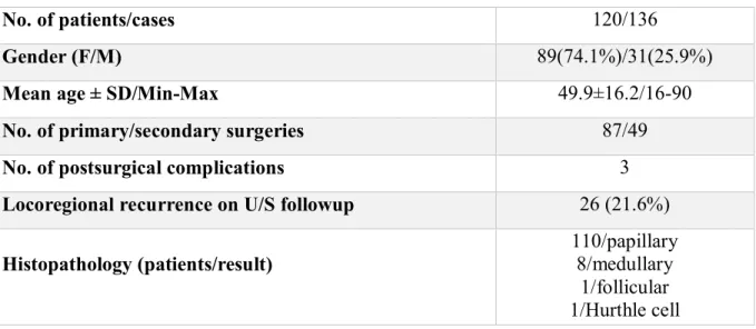
Table II: Surgical and histopathological characteristics of patients who underwent ultrasound mapping followed by neck dissection ... 55
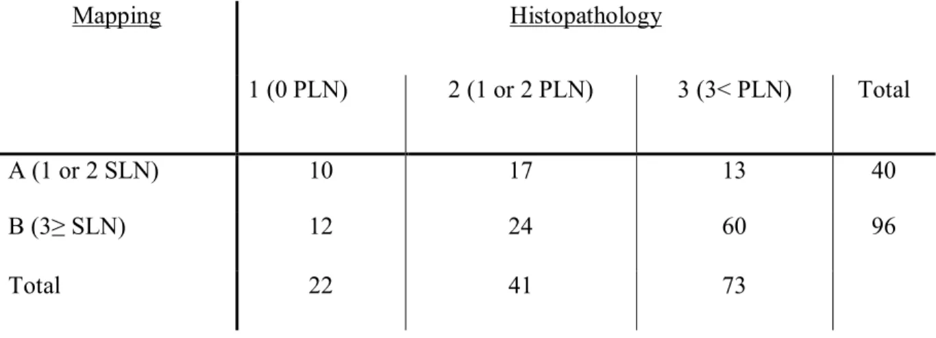
Table III: Cross-tabulation of the results among the groups of U/S mapping and
histopathology ... 56
ix
List of Figures
Figure 1: (A) Ultrasonography of the neck. (B) Cancerous nodule detected on ultrasound (arrow). (C) Microcalcifications in the cancerous nodule ... 58
x List of Acronyms
AAPC: Average annual percentage change
AJCC: American Joint Committee on Cancer
APC: Annual percentage change
ASIR: Age-standardized incidence rate
ATC: Anaplastic thyroid carcinoma
BPA: Bisphenol A
CC: Central compartment
CEA: Carcinoembryonic antigen
CGRP: Calcitonin gene-related peptides
CHUM: Centre hospitalier de l’Université de Montréal (University of Montreal hospital center)
CI: Confidence interval
CT: Computerized tomography
DIT: Diiodotyrosine
DSVPTC: Diffuse sclerosing variant of papillary thyroid carcinoma
DTC: Differentiated thyroid carcinoma
xi FNA-Tg: Fine needle aspiration – thyroglobulin
FTC: Follicular thyroid carcinoma
FV-PTC: Follicular variant of PTC
LC: Lateral compartment
LN: Lymph node
M: Metastasis
MEN: Multiple endocrine neoplasias
MIT: Monoiodotyrosine
MRI: Magnetic resonance imaging
MTC: Medullary thyroid cancer
N: Node
NIS: Sodium/iodine symporter
P/R: Persistent/recurrent
PCB: Polychlorinated biphenyls
pCND: Prophylactic central neck dissection
PDTC: Poorly differentiated thyroid carcinoma
PHAH: Polyhalogenated aromatic hydrocarbon
xii PMC: Papillary microcarcinoma
PPV: Positive predictive value
PTC: Papillary thyroid cancer
RLN: Recurrent laryngeal nerve
SD: Standard deviation
SEER: Surveillance, Epidemiology, and End Results
SES: Socioeconomic status
SLN: Suspicious lymph node
T: Tumor
T3: Triiodothyronine
T4: Thyroxine
TBG: Thyroxine-binding globulin
TFC: Thyroid follicular cell
Tg: Thyroglobulin
TPO: Thyroid peroxidase
TRH: Thyrotropin-releasing hormone
TSH: Thyroid-stimulating hormone
xiii TTR: Transthyretin
U.S.: United States
U/S: Ultrasound
1
Historical background (1)
The thyroid gland was first described by anatomist Andrea Vesalius in 1543 in his inauguration of modern anatomy “De Humani Corporis Fabrica” published in Switzerland.
Later, Julius Casserius (1545–1616) drew the first distinct image of the thyroid gland in a classical horseshoe shape. In 1656, Thomas Wharton named it “glandula thyreoidea” not because of its shape (thyreos in Greek means “shield”) but because it resembles nearby anatomical structures such as the thyroid cartilage of the larynx [1, 2]. The first surgical removal of the thyroid gland was described by German surgeon Lorenz Heister in 1742 [2].
Embryology (2)
The thyroid gland is the first endocrine structure to appear during the embryological development of the human body starting around the 4th week of gestation [1, 3]. It is a composite of two different cell types which have two distinct embryological origins. The major part of the thyroid is composed of epithelial cells which have an endodermal origin and arise from the primitive pharynx. Later in development, these cells become thyroid follicular cells (TFCs) which are responsible for producing thyroid hormones such as triiodothyronine (T3) and thyroxine (T4). The thyroid gland’s second embryological component is the neural ectoderm from the ultimobranchial body (fifth pharyngeal pouch) which gives rise to parafollicular cells or calcitonin-producing C cells [4-6]. Precursors of the C cells migrate from the neural crest to
2
the fourth pharyngeal pouch which is symmetrically located on both sides of the neck [5]. C cells subsequently reach their final position within the upper third of the thyroid lobes where they comprise only 1-2% of the thyroid cell mass [4].
Several developmental steps take place before mature thyroid gland formation [5]. Around the 20th embryonic day, the thyroid anlage develops as part of the endodermal epithelium in the midline of the primitive pharynx [5].
The foramen caecum, which is the endodermal bud between the first and second branchial arches, gives rise to the principle cells responsible for the thyroid gland’s structural and functional integrity [3]. The foramen caecum lies at the intersection of the tongue’s midline and the sulcus terminalis which defines the border between the oral (anterior two-thirds) and pharyngeal (posterior third) parts of the tongue [2, 3]. In adults, the circumvallate papillae delineate this border which also corresponds to the tongue’s ectodermal-endodermal boundary [3]. The TFC proliferation results in the formation of the initial thyroid primordium. The formation then starts to thicken and invaginates inferiorly, and this transformation forms a tubular structure called the thyroid diverticulum [3, 7]. As it continues to migrate caudally, it transforms into a bilobed and solid structure which eventually reaches its final position on the inferior aspect of the anterior neck. The migration process is completed by the 45th day and the thyroid gland lies inferiorly to the larynx, around the anterior and lateral sides of the trachea [3]. The migratory route from the foramen caecum to the final cervical position is called the thyroglossal tract [8]. During late embryological development, the thyroglossal duct regresses and becomes fibrotic. The foramen caecum then involutes into a vestigial pit that can be visualized in 60% of adults at the base of the tongue [3]. During the process of migration,
3
different deviations from the normal development route may occur. Occasionally, the thyroglossal duct fails to atrophy, leaving accessory or ectopic thyroid tissue along the migratory path. These islands of ectopic tissue transform into neoplasia in rare cases. More commonly, they may become metabolically active but the amounts of produced hormones are minimal without the main gland [3]. Rarely, the thyroid gland fails to descend into the neck, resulting in a lingual thyroid [7]. In this condition, a mass is noted in the posterior oral tongue, and the patient may complain of dysphagia, dysphonia, and dyspnea [3, 8]. Although a maldescended thyroid may not function appropriately, it is essential to keep in mind that a lingual thyroid may be the only functional thyroid tissue present [3, 8]. If it is removed due to its mass effect, subsequent replacement therapy should be administered to prevent further aggravation of the patient’s hypothyroid state. Cysts and sinuses are other morphological patterns of failed involution of the thyroglossal duct. These lesions are typically located in the midline, around the level of the hyoid bone. Infections and inflammatory processes are common pathologic manifestations of those congenital malformations, and antibiotics along with planned surgical intervention are the standard of care in advanced cases.
Simultaneously to the thyroid’s migration process, the C cells in the 4th pharyngeal pouch are positioned in the ultimobranchial body. First, the ultimobranchial bodies migrate from their lateral origin to a medial position on either side of the neck. By week 10, the main histophysiologic units of the mature and differentiated thyroid gland such as TFCs and C cells have merged at the front of the cricoid cartilage on the trachea. The thyroid gland then starts to enlarge and the TFCs, which notably outnumber the interspersed C cells, begin to organize into follicles. The final dispersion of C cells within the thyroid is not uniform, as they are mostly
4
localized in the middle and upper thirds of the gland’s lateral lobes [5]. The C cells comprise only 1-2% of the thyroid cell mass [4]. The final stage in normal organogenesis of the gland is a functional differentiation of the TFCs, which later lead to hormone production [5].
Functional differentiation of TFCs and C cells occurs along with the thyroid migration process and is regulated by relevant controller genes. The natural drive of thyroid morphogenesis is closely related to transcription factors such as TTF-1, TTF-2, Foxe1, Hhex, and PAX-8, which are essential for the thyroid’s normal development and migration [5, 8]. The simultaneous presence of these genes is essential for recruiting TFCs and organizing the thyroid bud. They continue to promote further development of the thyroid until it has completed migration and starts to enlarge [5]. At this point, other genes take over the functional development, and pathways for hormone synthesis start to activate. There are three functional stages in thyroid development: precolloid, colloid, and follicular. They occur approximately at 7 weeks to 12 weeks, 13 weeks to 14 weeks, and after 14 weeks. Hormone production in the thyroid starts at around 12 weeks [8].
Histology (3)
Histologically, the main architectural units of the thyroid gland are numerous hollow sacs called thyroid follicles. The connective tissue septa separates the thyroid gland into groups of 20-40 follicles [9]. The follicles contain colloid at their center and a simple cuboidal epithelium composes their main lining. Staining with hematoxylin and eosin produces a uniformly pink
5
color in the colloid lumen. The follicular cells’ principal role is to produce thyroid colloid, which contains high quantities of thyroglobulin and iodide.
The colloid is then extruded into the follicular lumen for future use. Subsequently, thyroid-stimulating hormone (TSH), which is the main regulator of the gland’s functional activity, stimulates the conversion of the colloid precursor into mature thyroid hormone. Although follicular cells are single layered and usually cuboidal in shape, their architecture may be significantly affected by the general metabolic state of the gland. In the event of high metabolic demand for thyroid hormones, the amount of colloid goes down as a consequence of its increased conversion into mature thyroid hormones. The follicular cells may also change their shape and become columnar. In cases of relative hypoactivity of the gland, the follicular lumen becomes larger as more colloid is stored in preparation for future use. Decreased metabolic activity causes the follicular cells to flatten and become smaller in size. Although most authors believe the monoclonal origin of thyroid cells, there is some evidence showing the polyclonal origin of thyroid cells with different malignant potential for each group of cells [10]. The next level of the thyroid’s functional and structural organization is the parafollicular cells (C cells) which produce calcitonin. They are situated in the stroma, between thyroid follicles. To better view these cells, immunostains such as immunoperoxidase are the preferred method [3].
6 Anatomy (4)
The mature thyroid gland is an H- or U-shaped bilobed, butterfly-shaped endocrine organ which is located on the anterolateral aspect of the cricothyroid membrane, cricoid and trachea [3, 5, 9]. Depending on gender and age, the normal thyroid in an adult weighs around 12-25 g, which corresponds to a volume of 12-20 ml [4, 5]. Usually, it is slightly larger in men. The isthmus of the gland connects the right and left lobes of the thyroid. On ultrasonography the average size of each lobe is 4 cm long vertically and 1-1.5 cm wide with a thickness of 1 cm. [3]. In some cases there is an extension of thyroid tissue which arises from the isthmus. This tissue, known as the pyramidal lobe, goes superiorly along the midline and is a remnant of the embryologic thyroglossal duct [3].
There are three main anatomical borders separating the thyroid from neighboring structures. The trachea and esophagus are the demarking posteromedial boundaries; the carotid sheath and sternocleidomastoid muscles are the limiting lateral and anterolateral boundaries respectively [5]. The thyroid extends from the level of the fifth cervical vertebra to the first thoracic vertebra.
An extension of the pretracheal fascia (Berry’s ligament) composes the fibrous capsule of the thyroid which serves as a supporting structure. The septae of the capsule divide the gland into macroscopic lobules. Arterial and venous branches run along the septae within the connective tissue. The visceral part of pretracheal fascia is attached anteriorly to the crycoid and thyroid cartilage [5, 8]. The muscular part of the pretracheal fascia encloses three pairs of infrahyoid muscles, namely the sternohyoid, sternothyroid, and thyrohyoid [8]. Because of its firm attachments, the thyroid may move superiorly as a consequence of tracheal elevation during
7
swallowing [3]. This has some diagnostic value as it helps to differentiate thyroid masses (such as thyroid nodules) and congenital malformations (such as thyroglossal duct cysts). In contrast to lymphadenopathy or branchial cleft cysts which remain stationary during swallowing, thyroid nodules and thyroglossal duct cysts are freely movable [3].
Blood Supply (4.1)
The arterial blood supply to the thyroid is the richest per gram of tissue [3, 4]. The external carotid artery and the thyrocervical trunk give rise to the superior and inferior thyroid arteries respectively. The superior thyroid artery supplies the upper pole and passes in a caudal direction from the external carotid artery [4, 8]. The inferior thyroid artery supplies the posterior part of the gland and runs in a cranial direction [4]. There may be a third, relatively uncommon, arterial supply from the brachiocephalic trunk called the arteria thyroidea ima (3%-10%) [2, 5, 8]. While arterial blood is circulated by two pairs of arteries, venous outflow is through three main venous pathways named the superior, middle, and inferior thyroid veins. The superior thyroid vein accompanies the superior thyroid artery and, along with the middle thyroid vein, drains into the internal jugular vein [2, 3]. There is some risk of rupture of the middle thyroid vein during thyroidectomy as it has a posterior course and can be damaged in forward traction of the gland [2]. Usually, there are several inferior thyroid veins that drain either into the internal jugular or the brachiocephalic veins.
8 Lymph Drainage (4.2)
The thyroid has a very rich lymphatic system. It consists of an intra and extraglandular network with developed lymph vessel anastamoses that drain each part of the gland in multiple different directions [4, 8, 11]. This can explain the frequent intraglandular spreading and multifocal distribution of thyroid neoplasias.
The lateral and medial aspects of the gland have some topographic differences in their lymphatic drainage. The lymphatic vessels of the gland’s lateral part pass along the arteries. They may ascend across the superior thyroid artery or descend across the inferior thyroid artery before reaching their final pool in the jugular chain of cervical nodes. Between the superior and inferior thyroid arteries, lymphatic vessels may drain directly into the jugular nodes. The lymphatic vessels of the gland’s medial part also drain in two directions: superiorly to the digastric nodes and inferiorly to the paratracheal and brachiocephalic nodes [2, 6]. In the midline, just above the isthmus of the thyroid, lies the Delphian node (named after the Oracle of Delphi). An abnormal looking Delphian node may have some diagnostic value as nodal metastases are common in thyroid cancer [8].
There are two main drainage pathways for the periglandular lymph network. The first pathway originates from lymph nodes and lymph vessels in the central zone of the neck. It is positioned between the carotid sheaths and extends in a downwards direction including pre and paratracheal regions. From this point, the lymph drainage may branch with the mediastinal lymphatic system.
The second lymph drainage pathway (secondary drainage) arises from the lateral region of the neck. It involves the lymph vessels and lymph nodes along the jugular vein as well as lymphatic
9
structures extending up towards the submandibular region. Lymph nodes in the supraclavicular triangle are also part of the secondary drainage network [4].
The thyroid’s dense lymphatic network is closely linked to the concept of a stepwise progression of nodal metastasis. This has enormous clinical relevance, as a nodal metastasis from one nodal station to another is directly correlated with the extent of the neck dissection and radiation therapy in patients with thyroid cancer [11]. Sentinel lymph node studies about the pattern of thyroid lymphatic drainage revealed some consistency in the dispersion of metastasis in different malignancies. The first metastases extend into the nodes of the central compartment of the neck (pre and paratracheal stations) followed by the nodes of the superior mediastinum and the lateral cervical nodes. The neck’s lymphatic mapping is often described in terms of “levels” ranging from level I to level VI. Level VI nodes are located in the central compartment which is bounded by the hyoid bone superiorly, the suprasternal notch inferiorly, and the carotid arteries laterally. There are three nodal groups in the level VI compartment that are responsible for thyroid drainage: paralaryngeal, paratracheal, and prelaryngeal (Delphian). Levels III, IV, and V are part of the lateral cervical compartments which are commonly affected by bilateral metastasis. Level III consists of lymph nodes located around the middle third of the internal jugular vein. It extends from the lower side of the body of the hyoid bone (superiorly) to the lower side of the cricoid cartilage arch (inferiorly). From anterior to posterior this zone is limited by the lateral border of the sternohyoid muscle and the posterior border of the sternocleidomastoid muscle[12, 13]. Level IV consists of LNs located around the lower third of the internal jugular vein. It extends from the lower side of the cricoid cartilage arch (superiorly) to the level of clavicle (inferiorly). The anterior boundary is the lateral border of the sternohyoid muscle, and the
10
posterior boundary is the posterior border of the sternocleidomastoid muscle[12-14]. Level V is comprised of the LNs in the posterior triangle. The boundaries of the triangle are the anterior border of the trapezius muscle posteriorly, the posterior border of the sternocleidomastoid muscle anteriorly, and the clavicle inferiorly[12-14]. Lymph nodes lying superiorly to the innominate vein in the superior mediastinum compose level VII [11].
Nerves (4.3)
The recurrent laryngeal nerve (RLN) provides motor and sensory innervations of the larynx. The RLN is responsible for phonation as well as swallowing, and any significant damage to this nerve can cause dysphonia and varying degrees of dysphagia [4, 5]. Considering the close location of the thyroid gland to the RLN path, there is a risk of RLN infiltration or compression by thyroid cancers, followed by impairment of function [4]. The RLN is a branch of the vagus which has different courses on both sides of the neck. On the right, the nerve loops posteriorly to the subclavian artery, then extends up until it reaches the tracheoesophageal groove. In contrast, the nerve on the left side loops posteriorly to the arch of the aorta, followed by an ascending course in the tracheoesophageal groove [2, 7]. Along its path the RLN also innervates the trachea and esophagus by multiple small branches [2]. The RLN carries motor, sensory, and parasympathetic fibers. The external branch of the nerve is responsible for the motor function of 4 intrinsic muscles (lateral cricoarytenoid, posterior cricorytenoid, transverse and oblique interarytenoid and thyroarytenoid) of the larynx. The internal branch of the RLN supplies sensation to the vocal cords and subglottic region. The RLN also sends branches to the inferior
11
constrictor and cricopharyngeus muscles before entering the larynx [15]Another important branch of the vagus is the superior laryngeal nerve which runs in a caudal direction along the superior thyroid artery. After passing behind the internal carotid artery, it divides into external and internal branches. The motor/external branch supplies the cricothyroid muscle, and damage to this nerve also results in voice changes [4].
Physiology (5)
The principal purpose of the thyroid gland is to provide adequate amounts of L-thyroxine (T4) and, to a much lesser extent, L-triiodthyronine (T3) which are essential for normal body metabolism, growth, and development. Both T4 and T3 are iodinated hormones which require the thyroid to be able to extract circulating iodide from the bloodstream for further organification and incorporation into the thyroid hormone molecules [9]. The thyroid’s level of activity and the amount of produced hormones are tightly regulated on several levels, both within and outside the gland. Those regulating mechanisms may have first line clinical relevance in thyroid cancer as the cancerous cells originate from normal thyroid cells. Depending on the level of dedifferentiation, the cancerous cells may use cellular mechanisms that are identical to those of normal cells in order to remain viable. In clinical settings these similarities help to identify, characterize, and finally treat different malignancies of the thyroid [9].
In order for the thyroid gland to provide the necessary amount of physiological hormones, an adequate supply of dietary iodine is essential. Both high and low iodine-containing diets may cause a vast range of serious problems. One of the most detrimental complications of over or
12
undersupply of dietary iodine is thyroid cancer. There is some evidence showing a higher incidence rate of papillary thyroid cancer in areas where there is an abundance of dietary iodine [16]. Meanwhile, low iodine intake may increase the proportional incidence of follicular thyroid cancer [8]. Administration of excess pharmacologic doses of iodine may inhibit iodide organification and thyroglobulin (Tg) proteolysis followed by an acute decrease in thyroid hormone production and release. This phenomenon, known as Wolff–Chaikoff effect, is explained by oversaturation of the sodium-iodine symporter (NIS). However, this effect does not last long as the gland adapts quickly to the new conditions and downregulates the uptake of iodine despite its continuously high plasma levels [9, 11].
One of the most critical steps in thyroid hormone formation is the transportation of the iodine into TFCs against an electrochemical gradient [11]. This is a saturable, energy dependent process which requires oxidative phosphorilation [11]. The main system that provides the necessary influx of iodine into the TFCs is NIS, which is a transmembrane protein located in the basolateral membrane of the thyrocyte [9, 11]. NIS cotransports sodium and iodide in a 2:1 ratio using the transmembrane sodium gradient as a driving force in order to stimulate iodide uptake into follicles [3]. The expression level of NIS on TFCs is responsive to different regulatory factors including TSH, and this mechanism is widely used in clinical settings. After intense TSH stimulation, malignant thyrocytes of papillary, follicular, and Hurthle cell cancers increase the uptake of I-131 as NIS is also present in their membrane [11]. However, in some types of thyroid cancer, NIS either does not have enough sensitivity or is completely absent which makes treatment with radioactive iodine practically useless [11]. Apart from therapeutic purposes, the NIS transporter concept can serve as a valuable diagnostic tool in patients with
13
differentiated thyroid cancers (papillary and follicular cancers) who underwent a thyroidectomy. In this case, the whole body scan with radiotracer can discover residual functioning thyroid tissue or actively functioning metastasis which can be retreated with larger doses of radioactive iodine [8]. Another way to use NIS for diagnostic purposes is by staining removed thyroid specimens with labeled antibodies against the symporter. This can provide valuable information in terms of the cancer’s aggressiveness and response to radioactive iodide [8].
It is worth mentioning that similar iodide transport mechanisms can be found in different tissues, including the stomach, salivary glands, and occasionally breasts. This can explain some side effects of radioactive iodine such as radiation-induced sialadenitis or xerostomia in patients treated for thyroid cancer [8, 9].
The next step in the synthesis of thyroid hormones is the organification of the iodide, which is carried out by the thyroid peroxidase (TPO) enzyme along with hydrogen peroxide. After the oxidation process, newly formed organic iodine iodinates the tyrosyl residues in thyroglobulin (Tg) [2, 9]. The whole process takes place in the opposite surface of TFCs where the apical membrane faces the colloid. The final stage of thyroid hormone formation is the coupling of the iodotyrosine molecules within thyroglobulin. Depending on the number of coupled iodine atoms, different types of thyroid hormone can be formed. While T4 is formed by coupling two molecules of diiodotyrosine (DIT), T3 is formed by coupling one molecule of DIT and one molecule of monoiodotyrosine (MIT). Antithyroid medications target the thyroperoxidase enzyme, which in turn blocks the formation of mature, end stage hormones.
14
The primary storage of thyroid hormones is the thyroid follicle colloid where the completely formed hormone is linked to thyroglobulin (Tg) [2, 9, 11]. Since it is one of the largest, active biological molecules in the human body and plays a central role in thyroid hormone synthesis and storage, thyroglobulin tends to have high diagnostic value in patients with different thyroid disorders including metastatic differentiated thyroid cancers [8]. High Tg levels indicate an increased probability of metastatic disease, especially in bones, lungs, and lymph nodes. Undetectable Tg levels combined with a negative whole body iodine scan are very reliable followup tests showing the absence of residual cancerous tissue [8].
The primary stimulator for thyroid hormone secretion is TSH which promotes proteolysis of thyroglobulin, followed by the release of free hormones into circulation [2, 9]. The equilibrium between the unbound (metabolically active) and bound (metabolically inactive) fractions of the released hormone is regulated by carrier proteins such as thyroxine-binding globulin (TBG), transthyretin (TTR), albumin, and lipoproteins [11].
The thyroid’s growth and functional activity, starting with the formation of thyroid hormones and ending with their secretion, is tightly regulated by the hypothalamo-pituitary-thyroid axis. The primary signal comes from the hypothalamus: thyrotropin-releasing hormone (TRH) stimulates TSH synthesis and release from the anterior pituitary which in turn stimulates the growth and hormone secretion of the thyroid itself. This mechanism is widely used in treating thyroid cancer as chronic suppression of TSH mitogenic activity by exogenous thyroxine is crucial in preventing cancerous cell activity [9].
15
The second hormone produced by the thyroid gland (C cells) is calcitonin which is a 32-amino acid polypeptide belonging to a family of related molecules called the calcitonin gene-related peptides (CGRP) [5, 9]. Calcitonin is one of the compounds regulating calcium levels in the blood. In conditions causing elevated levels of blood calcium, C cells secrete calcitonin which downregulates osteoclast-mediated bone resorption and by doing so it decreases high serum calcium levels [5]. Calcitonin may also serve as a tumor marker in medullary thyroid carcinoma which is part of Sipple syndrome (MEN type 2A) and is related to RET proto-oncogene.
Epidemiology (6)
With 140,000 newly diagnosed cases worldwide, thyroid cancer is the most common endocrine tumor and accounts for 2.5% of all human cancers [17-19]. Its incidence was relatively stable until the early 1990s. During the last three decades the incidence of thyroid cancer has increased drastically all over the world [20-24]. This trend is observed on all continents except Africa which probably is related to insufficient detection [24]. Between 1973 and 2009 the incidence of thyroid cancer more than doubled worldwide including such developed countries as the U.S., Canada, France and Australia [17, 22]. During the last 20 years the overall incidence rate has gone up by more than 6% annually [22]. While 20 years ago thyroid cancer was ranked only 14th, currently it is the 5th most common cancer in women. In Italy, it is the second most prevalent cancer among women in the age group below 45 [16, 24].
Rates of thyroid cancer are rising in both genders, but due to the more dramatic pattern in females it is estimated that by 2019 thyroid cancer will surpass the rates of common cancers and
16
will become the third most prevalent cancer in women of all age groups and the second most common cancer in women younger than 45 in the U.S. [21, 25]. In 2014, there were 62,980 new cases of thyroid cancer in the U.S. Every year thyroid cancer accounts for around 3.8% of all newly diagnosed cancer cases in the U.S. Overall, its incidence increased from 4.9 to 14.7 per 100,000 population in 2011. According to the Surveillance, Epidemiology, and End Results (SEER) program conducted in the U.S., the average annual percentage change (AAPC) for thyroid cancer is +5.2 in men and +5.7 in women for the 2002-2011 time interval [16]. The SEER database, which is a population-based cancer registry involving around 10% of the U.S. population, provides epidemiologic data on different characteristics of tumors as well as treatment approaches and survival rates [21]. It summarizes relevant epidemiologic information collected from 1973 to 2012. The higher incidence rate has been mostly attributable to papillary cancer, with only a slight increase in other histotypes [22]. Due to thyroid cancer’s indolent nature, the mortality rate is not as high as in other more common cancers. In 2014, estimated mortality from thyroid cancer was around 1,890 individuals. In comparison, the primary “killers” such as breast and lung cancers cause hundreds of thousands of deaths each year [20]. However, based on the SEER program’s results, the thyroid cancer mortality rate rose slightly for the 2001-2010 period with an AAPC of 0.9 [16]. In fact, the observed upshift in thyroid cancer mortality is not only statistically significant, it also surpasses the increase in mortality rate of any other cancer, except liver [26]. This increase in the mortality rate has occurred in spite of the introduction of better diagnostic and therapeutic measures [24].
17 Epidemiology: Canada (6.1)
Canada reflects the same trend: the overall incidence of thyroid cancer increased by 156% between 1991 and 2006 [27]. A study based on data from the Canadian Cancer Registry showed that age-adjusted incidence rates doubled between 1970-72 and 1994-96 [28]. Another study revealed that the rise in thyroid cancer incidence in Manitoba reached 373% from 1970-2010 [23]. The same trend was observed in an Ontario-based retrospective study indicating that the incidence rate was up 146% from 1990 to 2001 [29]. In 2007, more than 4,000 Canadians were diagnosed with thyroid cancer, or nearly 12 cases per 100,000 population, which accounts for around 2.5% of all malignant tumors [30]. In 2012, the numbers were even worse as an estimated 5,650 Canadians were diagnosed with thyroid cancer for the first time [23]. Characteristic features of thyroid cancer are an incidence rate that is three times higher in women than in men, along with the relatively young age of affected patients [30]. Around 75% of all cases are diagnosed before age 60, and nearly 40% of all thyroid cancers are found before the age of 45. With such a high incidence rate among the young population, thyroid cancer is currently ranked as the second most common cancer in Canadians aged 15 to 44 and is the most common cancer diagnosis in those aged 15 to 29 [30]. While papillary cancer is the most widespread histotype, composing 86% of all diagnosed cases, follicular, medullary, and anaplastic thyroid cancers represent 6%, 2%, and 1% respectively. Thyroid cancer has the most rapidly rising incidence rate in Canada. The age-standardized incidence rate (ASIR) jumped 5.7% per year in males (from 2.0 to 5.2 per 100,000 population) and 7.3% per year in females (from 6.8 to 17.9 per 100,000 population) for the 1992-2007 period. The most vulnerable category in terms of the highest spike in ASIR was women aged 30 to 59, with an annual ASIR of 8.2% [30]. The increasing ASIR trend was observed in all Canadian provinces and territories over the last 16
18
years. This progression varies widely across the various provinces. In 2007, the highest ASIR was in Ontario (15.2 per 100,000) and the lowest were in Saskatchewan (5.2) and British Columbia (5.8) [30]. For the same period, the annual percentage change (APC) for Quebec was 7.3% and the ASIR was around 11 per 100,000 population which is not much different from the Canadian average of 11.6 per 100,000.
The situation is completely different when it comes to the mortality rate from thyroid cancer: it has remained exceptionally low and stable [23, 30]. For the 1992-2007 period, there were an average of 142 deaths per year in Canada with even a slight decrease (less than 1% per year) in the age-standardized mortality rate from 0.5 per 100,000 in 1992 to 0.4 per 100,000 in 2007 [30]. Thyroid cancer’s low mortality rate is reflected in the five-year relative survival rate which is the highest of all cancers in Canada, reaching 97% for the 2001-2003 timeframe [30] and 98% in 2011 [23]. Increased incidence of thyroid cancer with low mortality causes increased proportional prevalence, with two- and five-year prevalence exceeding 9% per year from 1999/2000 to 2008 [31]. Only liver cancer has higher prevalence (8.5%) for a 10-year duration [31].
According to the Canadian Cancer Society, thyroid cancer is the 5th most common cancer in women and accounted for 5% of all cancers in 2015 [32]. In 2015, out of 6,300 newly diagnosed thyroid cancer cases, 4,800 were in women (ASIR 23.1 per 100,000) and 1,450 in men (ASIR 6.7 per 100,000). By comparison, in 1986 the ASIR for women was 5.2 per 100,000 population which proves once again the tremendous spike in thyroid cancer incidence [32].
19
The Canadian Cancer Society has also indicated that Quebec is second after Ontario with around 360 men and 1,200 women being newly diagnosed with thyroid cancer in 2015. These values correlate with an ASIR of 7 and 25 per 100,000 population in men and women respectively [32].
Increasing incidence hypothesis (7)
Overdiagnosis of subclinical disease reservoir (7.1)
There are multiple controversial explanations for the worldwide increase in thyroid cancer. Although various hypotheses have been put forward to pinpoint the principal mechanism behind such a dramatic upsurge in the thyroid cancer incidence rate, many authors agree that the main question that needs to be answered is whether the alarming increase reflects a true change in incidence or is simply due to the “overdiagnosis” of subclinical disease [18, 20, 22, 24, 33]. This distinction may have a very important clinical application in terms of prevention and management. For instance, a real upsurge in the incidence rate should raise concerns about environmental factors that may be responsible for this phenomenon. Meanwhile, the management approaches will have to be reviewed in case the incidence rate has been falsely pushed up by “overdiagnosing” a previously subclinical pool of the disease. This would help avoid unnecessary therapeutic interventions in an otherwise harmless, clinically insignificant disease [33].
Based on autopsies of people who died from other causes, the estimated prevalence range of papillary thyroid cancer varies from 3% to about 35% depending on the country and the pathology methods adopted to examine the specimens [16, 20, 22]. Moreover, incidental thyroid
20
cancers can be found in up to 50% of thyroid specimens removed for benign causes. These findings, along with the observation that the higher incidence is mostly ascribed to greater detection of cancers which are too small to be detected by conventional physical exams, indicate the existence of a large reservoir of subclinical papillary thyroid cancer. Overdiagnosis of this otherwise clinically insignificant reservoir of thyroid cancer can explain the “apparent” upsurge in the incidence rate [16].
Many recent studies show strong evidence in favor of the hypothesis that the rising incidence is due to increased diagnostic scrutiny and widespread access to highly sensitive radiological testing [34-36]. In the last 30 years there has been a dramatic increase in the use of all kinds of diagnostic imaging modalities including cross-sectional imaging, with a twofold increase in CT scans and a threefold increase in MRI scans [33, 37]. In their study, Udelsman and Zhang concluded that “the epidemic of thyroid cancer” in the U.S. is largely attributable to increased detection. They found a statistically significant direct linear correlation between the frequency of cervical ultrasonography and the thyroid cancer incidence rate [34]. In another study, Zevallos et al. presumed that the doubling of thyroid cancer incidence within the U.S. Veterans Affairs healthcare system between 2000 and 2012 could be related to a nearly fivefold and sevenfold increases in the use of thyroid ultrasound and fine needle aspiration respectively [36]. It should be noted that the prevalent use of ultrasound has boosted the detection of small thyroid nodules which otherwise would remain undiscovered without any further diagnostic or therapeutic interventions [38].
Data analysis from the National Cancer Institute’s SEER program showed that there is a large gender discrepancy in thyroid cancer detection. The incidence rate in women is 4 times greater
21
than in men even though the autopsy incidence of thyroid cancer is higher in men. This correlation is probably related to the overdiagnosing phenomenon in women [39]. Some authors suggest that even the age of the diagnosing physician may impact the thyroid cancer incidence rate, with more experienced doctors reporting fewer cases because they tend to use ultrasound and FNA less [40]. More aggressive approaches in terms of diagnosis and treatment, such as more frequent biopsies and an increase in the number and extent of surgeries especially total thyroidectomy [41], provide larger specimens and more opportunities for pathologists to find cancer [20]. The pathology guidelines for managing thyroid specimens have also changed, and thyroidectomy specimens are now examined in their entirety compared with the past practice of examining only representative sections of the specimen. More detailed examination of each thyroid specimen increases the chances of identifying small lesions thereby raising the incidence rate [20].
Other important factors that may have contributed to the increased incidence of thyroid cancer detection are access to healthcare services as well as socioeconomic status (SES) [20, 42]. Morris et al. showed a strong correlation between the markers of access to the healthcare system and the incidence of papillary thyroid cancer [22]. Countries with higher SES tend to have higher rates of screening procedures with a subsequent increase in thyroid cancer detection. The positive association between thyroid cancer and various socioeconomic indicators, such as high levels of income, education, and white-collar employment, would seem to indicate an increased screening effect [22, 24].
22
Arguments in favor of a true increase in thyroid cancer incidence (7.2)
Although most studies agree that the more widespread application of highly sensitive diagnostic modalities has greatly contributed to the detection of a previously “hidden” pool of subclinical disease, it is impossible to ignore the evidence indicating that such an upsurge in thyroid cancer incidence cannot be explained solely by the overdiagnosis phenomenon and that there must be other factors causing a true rise in the number of thyroid cancer cases [16, 20, 24].
The main argument against the overdiagnosis hypothesis is the stable or even decreasing incidence of other types of cancers diagnosed with highly specialized imaging techniques and diagnostic biomarkers [16]. According to data from the SEER registry, the rates of some common cancers, such as prostate, colorectal, and breast cancer, have dropped while the incidence rate of thyroid cancer increased by as much as 60% for the period of observation from 2002 to 2011.
Assuming that improved detection is the principal mechanism of increased incidence, then the diagnosis and treatment of early-stage, small tumors should be accompanied by a significant decrease in larger, more advanced tumors [24]. Although the upsurge in the detection rate for small thyroid tumors (≤1 cm) certainly surpasses the rate for larger, more advanced tumors (≥4 cm), many studies have shown a significant increases in thyroid cancer in all size categories [16, 17, 20, 33]. In their recent study, Malone et al. demonstrated that 38% of large thyroid cancers (≥4 cm) are found incidentally on imaging studies done for other reasons [43]. Since it seems very unlikely that 38% of all large tumors were being missed in the past, a real increase in the incidence rate could be the reason.
23
Another interesting observation is related to the histotypes of thyroid cancer. While improved detection would affect all histotypes, the rise in incidence is, in fact, almost exclusively due to papillary thyroid cancer (PTC) [17, 24].
Moreover, in their study, Cramer et al. state that “improved detection techniques may explain the 8.0%/year difference between the increase in microPTC compared with the average increase in neoplasms larger than 1 cm; however, changes in the methods of detection cannot explain the overall increase in all sizes of PTC” [17].
If all these hypotheses and studies are correct, there must be some underlying driving forces which could explain this profound increase in thyroid cancer.
Risk factors causing a true increase in thyroid cancer incidence (7.3)
Exposure to ionizing radiation is an extensively studied and well-documented risk factor for thyroid cancer due to the thyroid’s ability to concentrate iodine and the relatively higher irradiation because of its localization [16, 20, 24, 33]. The mechanisms behind this phenomenon are DNA strand breakages, the development of somatic mutations, chromosomal rearrangements, and intra-chromosomal inversions causing RET proto-oncogene to fuse with other genes such as H4 or ELE 1 [20, 33, 44]. Once exposed to ionizing radiation, young subjects (<20 y/o) have a higher probability of developing thyroid cancer due to greater radiosensitivity.
During the last 20 years, the lifetime dose of ionizing radiation for many U.S. residents has doubled as a result of medical and dental diagnostic procedures [20, 24, 33]. Although CT scans
24
account for only 15% of all radiological diagnostic interventions, they are the source of more than half of the radiation doses that U.S. residents are exposed to [24]. Some studies conducted in pediatric populations showed a weak but nonnegligible correlation between the risk of thyroid malignancies and CT scan exposure. Moreover, there is an inverse correlation between the risk of thyroid cancer and the age at exposure. Children aged <5 years have the highest risk and there is a significant decline in risk after the age of 20 [33, 45]. Nevertheless, the appreciable increase in thyroid cancer incidence cannot be ascribed solely to CT scans because most CT scans are performed in patients aged >50. Another major source of irradiation is nuclear medicine where radioactive iodine has been widely used recently for both diagnostic and therapeutic purposes in different thyroid diseases [24]. Diets containing supplemental iodine are another potential risk factor for thyroid cancer as they interfere with iodine organification and thyroid hormone synthesis [24]. Another possible mechanism is the increased incidence and severity of chronic thyroid conditions such as chronic lymphocytic thyroiditis and autoimmune thyroiditis [16].
Enviromental pollutants, such as asbestos, benzene, formaldehyde, pesticides, bisphenol A (BPA), polychlorinated biphenyls (PCBs), and polyhalogenated aromatic hydrocarbons (PHAHs), may also contribute as both genotoxic and nongenotoxic carcinogens [24]. In addition, chronic metabolic conditions, such as diabetes and obesity-related metabolic disorders, are among the proposed risk factors related to the higher incidence of thyroid cancer [20, 33]. Those conditions lead to low grade inflammation triggered by alterations in adipokines. This occurs along with increased circulating leptin and an adiponectin deficiency which is independently and inversely associated with thyroid cancer risk [46]. All these metabolic changes are linked to higher activity in the mitogen-activated protein kinase pathway which has
25
a crucial role in carcinogenesis by activating mitogenesis [33, 46, 47]. Other potential risk factors, including estrogen exposure and dietary nitrates, still need to be investigated since there is a lack of conclusive data in the literature [20, 33].
Overall, the reasons behind the worldwide increase in thyroid cancer are still unclear. Most likely, the observed exponential upsurge has a multifactorial etiology. Certainly, the increased detection rate can partly explain the phenomenon but, at the same time, there is strong evidence indicating a true increase in thyroid cancer incidence. In summary, further studies are warranted in order to investigate the real reasons and determine the potential carcinogens and their mechanisms of action [24].
Histologic classification (8)
Based on their cellular origin, characteristics, and prognosis, thyroid cancers are classified into several histological types and subtypes. Thyroid cancers are mostly epithelial tumors originating from thyroid follicular cells (TFCs) and parafollicular cells (C cells) or from nonepithelial stromal elements [48]. On the basis of architectural, cytologic, and histogenetic features, the World Health Organization (WHO) classifies primary thyroid tumors into epithelial or nonepithelial and benign or malignant categories. According to the same classification, lymphomas and miscellaneous tumors are defined as separate groups [48-51].
26 Papillary thyroid carcinoma (PTC) (8.1)
Papillary thyroid carcinoma (PTC) is the most common differentiated carcinoma, accounting for 80-90% of all primary thyroid malignancies [9, 48, 50]. Generally, papillary carcinomas are indolent, slowly growing tumors. Women aged 25-45 have a three times higher risk of developing PTC [8]. Because of the carcinoma’s indolent nature, most cases have an excellent prognosis but in up to 10% of cases recurrent disease may occur in the form of cervical lymph node metastasis and/or distal metastasis [48, 50]. The main mode of PTC spreading is via the lymphatic system. Dispersion of cancerous cells within the thyroid may lead to “multifocal” disease and cervical node metastasis. It has been estimated that in over 50% of cases, PTCs have nodal metastasis at initial diagnosis [48, 50]. Since venous invasion rarely occurs, metastasis outside the neck is unusual [52]. In 5-7% of cases, PTC may cause distant metastasis in lungs and bone [52]. PTC’s tendency to spread locally in the neck may cause tracheal compression and involve the recurrent laryngeal nerve [5]. Although most PTC show a papillary growth pattern, the main distinguishing feature is malignant cytologic (nuclear) changes which are important diagnostic hallmarks [11, 50, 52]. The typical nuclear appearance of PTC includes enlargement, hypochromasia, intranuclear cytoplasmic inclusions, nuclear grooves, and distinct nucleoli [11]. The PTC nuclei have also been described as clear, ground glass or “Orphan-Annie eyed” [50, 52]. Psammoma bodies, which are another microscopic characteristic of PTC, are laminated calcifications that may occur in about 40% of cases [9, 11, 52]. The psammoma bodies are formed when calcium is deposited on the dying cells after infarction of the papillae. Another theory relies on the intracellular accumulation of calcium by tumor cells which later leads to
27
their death and the calcium’s release. Further deposition of free calcium leads to lamellation [52].
Microscopically, papillary carcinoma has multiple histotypes, the most important of which are papillary microcarcinoma (PMC), follicular variant, tall cell variant, and diffuse sclerosing type [49, 52].
According to the World Health Organization (WHO), the term papillary microcarcinoma refers to thyroid cancer measuring 1 cm or less in diameter [53]. According to the American Thyroid Association, PMC detection accounts for almost 50% of the thyroid cancers diagnosed in the past three decades. The incidental detection of PMC in the thyroid gland is a frequent finding in autopsies and may indicate the indolent nature of this tumor without having any clinical relevance [53]. However, PMCs may lead to lymph-node metastasis in the central and lateral compartments of the neck [54]. Despite the incidence of locally metastatic disease, the prognosis for PMC is excellent and 10 year mortality rates in patients with this tumor are less than 1% according to the American Thyroid Association [54].
The follicular variant of PTC (FV-PTC) is one of the most common subtypes among all PTC histotypes, accounting for up to 41% of all PTC cases [55]. Although FV-PTC contains a mixed histology of both PTC and FTC, microscopically its architecture is predominantly follicular [55]. Despite some unique clinical behavior, FV-PTC seems to be quite a distinctive entity, sharing clinical features with both PTC and FTC. Having more favorable clinicopathologic features and a better tumor risk group profile does not impact the long-term outcome of FV-PTC, which is similar to that of conventional PTC [48, 56].
28
The diffuse sclerosing variant of PTC (DSVPTC) is an unusual variant, with prevalence ranging from 0.7-6.6% of all PTCs. A macroscopic feature of DSVPTC is the extensive involvement of the thyroid gland without forming a dominant mass [57]. Its hallmark on microscopic examination is the extensive fibrosis, squamous metaplasia and numerous psammoma bodies. Since DSVPTC has a higher incidence of lymph node metastasis at presentation than conventional PTC, along with a distant metastasis rate of around 5%, aggressive treatment protocols are required in managing DSVPTC [57]. Nevertheless, cancer recurrence and cancer-related mortality have been reported at 14% and 3% respectively [57].
The tall cell variant of PTC, which has 10% prevalence and a 10-year mortality rate of up to 25%, is the most common aggressive variant of PTC [50, 58]. Microscopically, tall cells are twice as tall as their width and should represent at least half of the papillary carcinoma cells [50]. This variant has a high prevalence among patients with radioactive iodine refractory disease and is associated with a high rate of B-RAF mutations, making the latter a potential target in terms of treatment strategies [58].
Follicular thyroid carcinoma (FTC) (8.2)
Follicular thyroid carcinoma (FTC) is the second most common malignant neoplasm arising from epithelial cells. It accounts for 5-10% of thyroid cancers in non-iodine deficient areas and ranges up to 30-40% in iodine deficient areas [48]. Because it is difficult to distinguish follicular carcinoma from follicular adenoma based on the results of fine needle aspiration (FNA), the
29
term “follicular neoplasm” is used for these tumors. Other methods, such as immunohistochemistry, morphometry, cytogenetic, and oncogen markers, have not provided reliable information regarding the distinction between follicular adenoma and follicular cancer on FNA [48]. Excisional biopsy (hemithyroidectomy) may be warranted in order to establish the precise histologic diagnosis on the basis of capsular and vascular invasion which helps discriminate between benign and malignant lesions [6]. On microscopic examination, follicular carcinoma is usually composed of microfollicles (minimal amount of colloid within the small follicles) or solid nests of tumor [8]. Depending on the degree of invasiveness, there are two major groups of follicular carcinomas: minimally invasive and widely invasive [48]. Follicular cells may invade either the tumor capsule and/or the blood vessels, with vascular invasion having a worse prognosis. Other recognized risk factors, such as age >45, gender, extrathyroid invasion, greater tumor size, and the presence of distant metastasis at presentation, are also associated with poorer prognosis [32]. Nevertheless, metastases to cervical lymph nodes are rare and are often accompanied by direct extrathyroidal extension of the cancer [9].
Hurthle cell carcinoma (8.3)
One of the variants of follicular carcinoma is Hurthle cell carcinoma which is composed predominantly (>75%) of oncocytic cells and is thought to have a worse prognosis than the usual follicular carcinoma [48, 50]. The benign variant of Hurthle cell lesions represented by Hurthle cell adenoma is generally indistinguishable from the malignant variant based only on the results
30
of cytological analysis. Thus, once a Hurthle cell lesion is detected by FNA, an excision biopsy may be indicated for further histological analysis [5]. Metastasis to cervical lymph nodes in Hurthle cell carcinoma is more common compared to the usual follicular carcinoma [9].
Poorly differentiated thyroid carcinoma (PDTC) (8.4)
Poorly differentiated thyroid carcinoma (PDTC) characterizes a heterogeneous group of malignant neoplasms. It was recognized as a distinct pathologic entity by the WHO in 2004 [59]. The macroscopic hallmarks of PDTC are large (>5 cm in diameter), solid, unencapsulated, nodular or multinodular, gray-white tumors which are likely to penetrate perithyroidal tissues. On microscopic examination, these tumors represent a trabecular, solid or insular growth pattern [48]. Due to its heterogeneity, PDTC has different biologic behavior and various histologic patterns of growth. Although PDTC accounts for only 1-15% of all thyroid cancers, it is the main cause of death from “nonanaplastic follicular cell-derived thyroid cancer” [59, 60].
Medullary thyroid cancer (MTC) (8.5)
Medullary thyroid cancer (MTC) is a neuroendocrine tumor of the thyroid gland’s parafollicular or C cells and it accounts for up to 10% of all malignant thyroid tumors [48]. Although approximately 80% of all MTCs are sporadic cases, there are also some hereditary cases resulting from RET proto-oncogene mutation which is the underlying defect in Multiple Endocrine Neoplasia type 2 (MEN2) [61]. Despite MTC’s morphological diversity, the
31
particular type of histology does not seem to influence the disease’s course [62]. The primary manifestation of sporadic MTC in most cases (75-95%) is a solitary nodule, usually located in the upper portion of the thyroid lobe [61, 63, 64]. In the majority of cases, the MTC has already metastasized at presentation, with 50% of patients having clinically detectable cervical lymph nodes, up to 15% having upper aerodigestive tract compression signs, and 5% having distal metastatic disease [63]. A characteristic feature of MTC is the production of calcitonin which can serve as a tumor marker because its concentration usually correlates with tumor mass and is almost always abnormally elevated in patients with a palpable tumor [63]. The MTC cells are also capable of secreting carcinoembryonic antigen (CEA) which may indicate advanced disease if levels are abnormally high [65].
Anaplastic thyroid carcinoma (ATC) (8.6)
Anaplastic thyroid carcinoma (ATC) is an extremely aggressive malignant thyroid tumor which accounts for less than 2% of all thyroid malignancies [66, 67]. Compared to differentiated thyroid cancers, ATC’s disease-specific mortality rate surpasses 90%, with an average survival duration of 6 months after diagnosis [66]. Having a mean age of 65 at initial presentation, most patients with ATC are older than those with differentiated thyroid carcinoma [67].
The major etiologic factors of ATC are pre-existing thyroid diseases or other types of thyroid cancer such as PTC, FTC, or poorly differentiated carcinoma. This observation supports the theory that ATC develops from more differentiated tumors following the dedifferentiating events [68]. This hypothesis might be supported by the fact that both well differentiated thyroid
32
cancers and ATC have the same underlying molecular development mechanism. In both cases, mutations in BRAF and RAS play a central role in the early events of the progression pathway. Although ATC has multiple histopathological features, there is no evidence showing the impact of a particular histotype on the prognosis [66]. Around 90% of patients have regional or distant metastasis at the time of initial presentation, with lungs being the most common site of distal metastasis [69, 70].
Thyroid cancer staging (9)
According to the American Cancer Society, the TNM system adopted by the American Joint Committee on Cancer (AJCC) is the most frequently used system to describe thyroid cancer stages. Based on three key parameters, the TNM system indicates the size of the primary tumor (T), describes the extent of metastatic disease to the regional lymph nodes (N) and specifies whether there are distant metastases to other organs (M).
Primary tumors (T) for nonanaplastic thyroid cancer might be subdivided into several subcategories, the first being Tx where the tumor cannot be assessed due to missing information and the most advanced being T4b where the tumor has invaded prevertebral fascia or encased the carotid artery or mediastinal vessels [71, 72]. All anaplastic thyroid carcinomas are considered T4 tumors at initial diagnosis, with T4a having only intrathyroidal expansion (surgically resectable) and T4b growing outside the thyroid gland (surgically unresectable) [71, 72].
33
The N parameter describes the metastasis into the regional lymph nodes, consisting of the central compartment, lateral cervical, and upper mediastinal lymph nodes. This parameter is also divided into several subcategories, with the most advanced being N1b which is associated with unilateral, bilateral, contralateral cervical, or superior mediastinal lymph node metastasis [71].
There are only three subcategories for M when evaluating distant metastasis. M1 indicates that the cancer has spread to distant lymph nodes and internal organs [71].
After the TNM values have been determined (table I, page 54), the combined results are used to assign the stage (ranging from I to IV). Differentiated thyroid cancers (PTC and FTC) are further grouped into stages in a way that the patient’s age is taken into consideration as a dichotomous variable. The cutoff value is age 45 and all patients below that age are either Stage I (no distant metastases) or Stage II (distant metastases present), which means they should have an excellent prognosis [11, 71, 72].
In contrast to PTC and FTC, age is not a factor in MTC staging, and all ATCs are considered Stage IV tumors, reflecting the poor prognosis for this type of cancer [71, 72].
34 Molecular Pathogenesis (10)
Several types of genetic alterations and epigenetic changes have been shown to play a fundamental role in the tumorigenesis of different variants of thyroid cancer [48, 73, 74]. An understanding of the pathways, such as gene mutation, aberrant gene methylation, and gene translocation, can open up new perspectives in terms of molecular diagnostics, treatment, and prognosis prediction [74].
One of the most common pathogenetic mutations of differentiated thyroid carcinoma (DTC) is the point missense mutation in exon 15 of BRAF which occurs in around 45% of sporadic PTCs [50, 73, 74]. This mutation is generated when glutamic acid is replaced by the amino acid valine at residue position 600. It leads to the expression of the BRAF-V600 mutant protein, causing constitutive activation of serine/threonine kinase [73, 74]. Over 90% of BRAF alterations are due to a V600 mutation. The most common subtype of PTC carrying a BRAF mutation is the tall cell variant (77%), with FVPTC having the lowest percentage (12%) of this particular mutation [50, 73].
Several studies have observed a correlation between BRAF-V600 mutation and the clinicopathological features of PTC [75]. BRAF mutation is more common in older patients and is associated with extrathyroidal invasion, advanced stages, and poorer clinical outcomes including higher recurrence rates and refractoriness to radioiodine treatment since BRAF mutation is responsible for suppressing the sodium/iodide symporter (NIS) [50, 73].
Rat sarcoma oncogene (RAS) mutations are second in prevalence after BRAF mutations. There are three RAS isoforms: H-RAS, K-RAS, and N-RAS [50, 73, 74]. The most frequent RAS