THESE
THESE
En vue de l'obtention du
DOCTORAT DE L’UNIVERSITÉ DE TOULOUSE
DOCTORAT DE L’UNIVERSITÉ DE TOULOUSE
Délivré par l'Université Toulouse III - Paul Sabatier
Discipline ou spécialité : Biologie cellulaire, Génétique et Développement
JURY
Professeur d'université SOULA Cathy
Dr. FAIVRE-SARRAILH Catherine, Rapporteur Dr. LLIMARGAS Marta, Rapporteur
Dr. ROCH Fernando, Directeur de thèse
... (préciser la qualité de chacun des membres)
Ecole doctorale : Biologie-Santé-Biotechnologie
Unité de recherche : Centre de Biologie du Développement, UMR 5547 Directeur(s) de Thèse : Dr. ROCH Fernando et Dr. WALTZER Lucas
Présentée et soutenue par ASSIA HIJAZI
Le 20 juin 2011
Titre : The role of two members of the Ly6 superfamily in the organization of septate junctions during Drosophila melanogaster development
“It is not the strongest of the species that survives… nor the most
intelligent that survives. It is the one that is the most adaptable to
change.”
"We can allow satellites, planets, suns, universe, nay whole systems
of universe to be governed by laws, but the smallest insect, we wish
to be created at once by special act.”
Charles Darwin
Acknowledgments
I would like to convey my gratitude to my PhD supervisor Fernando for his
nice supervision, also for his guidance, his valuable advices and his daily
presence during these four years.
He provided me unflinching encouragement and support in various ways.
His truly scientist intuition, passions and cheerful enthusiasm for science
exceptionally inspire and enrich my experience as a student, a researcher and
a scientist want to be. I am indebted to him more than he knows.
Thank you also for introducing me to the fascinating world of drosophila
and for teaching me the alphabet of flies genetics and development.
I consider myself lucky to have the opportunity of working with you and I
am happy also to be your first PhD student. All the best with your precious “
boudin family”. Thank you also to lend me many valuable books during my
PhD, but also for your precious gift “Le Chant d’Al Andalus”, it was so
touching for me!! I Hope we will meet again and I am waiting for you visit
to Lebanon!
I gratefully acknowledge Cathy Soula, the president of my thesis jury. Thank
you also to give me the opportunity of teaching in Paul Sabatier University.
It was a short, but a very nice exercice.
I am very greatful also to Catherine-Faivre Sarrailh and Marta
Llimargas, my thesis reviewers, for evaluating my my PhD work. I am very
honored by your participation in my jury committee and it was very nice to
meet you here in Toulouse.
My special thanks go also to Marc and Lucas for their careful interest on the
“Ly6 family”, a project completely different from the main thematic of the
team, but we had always with you many scientific interactions and
interesting discussions and advices. Thank you Lucas for your nice scientific
direction of the team. It was a pleasure to work beside you in the lab and
benefit from your experience. I also appreciate the corrections and the
precious comments you made to ameliorate this manuscript.
Marc thank you for many discussions we had about science, philosophy,
politics and many other topics … thank you also for many informatics helps!
Many thanks also go to all the team members for your help, advices,
discussions (scientific and non-scientific ones) and for providing an
agreeable atmosphere to work, discuss and share the divergent scientific
experiences of each one, which was very rewarding for all.
Benoit, it was pretty nice to work beside you in the lab, thank you for many
technical helps, thank you again for learning me how making a Western
Blot (la manipe du siècle)
.
Thank you also for the coffee and tea breaks at 16 p.m. in the hall, they will
miss me!
Thanks for Cedric, it was great to meet you. You are someone who is
friendly and always very close from students, shares his passion and
enthusiasm for science with them … Thank you for your encouragements
and support, but also for many and many advices! I hope for you a good
following and a lot of luck with your project.
Vanessa, it was nice to work beside you, thank you for different helps … I
hope for you a good luck and many more publications in the future.
Many thanks for all the PhD students who I met during these four years in
the lab. Special thanks for Dani for wise advices and many services,
especially when I arrived in Toulouse.
Thank you also for Karène, we had nice moments together, best of luck for
you!
Thank you Stephanie for many services, for your smile and your good mood
that you ever kept. Best of luck for the future and more success in your
favorite Rugby game.
Billel/Bilal (à la libanaise), “le Glandeur favori”, I really enjoyed working
with you, very gentle and kind, friendly and helpful, the best of luck and
don’t forget, you still have 2 years “ à glander” … So have a good fun and
a lot of publications !!
Osman, the second Lebanese I meet during my PhD. Thank you for nice
discussions we had, have great scientific adventures and see you maybe in
Lebanon !
Many thanks equally go to my collegues and friends with them we shared
unforgettable moments in the lab and elsewhere.
Thank you my Master 2 collegues specially (Marinou, Maeva, Dédé, Floflo,
Alex, Natalia, Céline, Romain …). But also, Hadi, Yac, Aurélie, Laurence,
chantal, Ahmad, Amir, momo, Rami and many others ... Hope to see you all
again …
Thanks also to Carine, Juju, Isabelle, sophie, Cathy Denesin, Sergio, Phiphi,
Hélène Foussard, Thomas and Gwen.
I am also very grateful to François Payre for all the help you done, especially
when I have arrived here in Toulouse, five years ago, now I am
remembering the moment when I got the hall of the CBD and asked for
your office ! Thank you for helping me to find housing and to take care of
many administrative procedures! Thank you also for your continuous
encouragement and your believe in my scientific capabilities. “François, on se
dit à bientôt!”
Thank you also for the people I met in Toulouse, with them I shared a good
time. Thanks to Rim, Lara, khello and Ado, but also for Youm, Camille,
Catherine, Osanna. Thank you for cooking, partying together, but also for
many trips we have done, even when the weather was not helpful
… Your
support was unlimited … I will miss you all!
Last but not least, I wish especially to thank my mom, dad and brothers for
their constant support and encouragement. My hard-working parents have
sacrificed their life for my brothers and myself and provided unconditional
care, but also gave me a good foundation to meet life… I love them so much,
and I would not have made it this far without them.
CONTENT
SUMMARY………...4 RÉSUMÉ………..6 ABBREVIATIONS LIST………...9 ILLUSTATIONS LIST………...13 I. INTRODUCTION……….15Part 1: Sealing junctions and the concept of cell barriers I. Biological barriers, a hallmark of life………..16
II. Cell junctions: a material basis for the barriers in multicellular organisms………...18
III. Sealing junctions: general structure and particular features………...20
a) Tight junctions………...20
b) Septate junctions………..22
c) Paranodal septate junctions………...24
IV. Molecular organization of the sealing junctions………...28
a) Molecular organization of Tight Junctions………...28
b) Molecular organization of Septate Junctions……….…33
c) Molecular organization of Paranodal Septate Junctions………....38
V. Drosophila: a model system to study sealing junctions………...40
Part 2: The Ly6 family of three-finger domain (TFD) proteins I. Structural conformation of Ly6/TFD proteins………48
III. Characteristic features of the Ly6 domain proteins……….. 50
IV. The roles of the Ly6 superfamily members in vertebrates………52
- Ly6 (lymphocyte antigen 6 complex)………..54
- CD59 (or Cluster of Differentiation 59)………...………54
- uPAR receptor (or urokinase Plasminogen Activator Receptor)……..………...54
- Lynx-1 (Ly6/neurotoxin-like protein 1)………..56
V. The Ly6 module is also present in invertebrate genomes………..56
II. RESULTS………..60
I. ARTICLE 1: boudin is required for septate junction organisation in Drosophila and codes for a diffusible protein of the Ly6 superfamily………61
II. ARTICLE 2: The Ly6 protein Coiled is required for septate junction and blood brain barrier organisation in Drosophila...80
III. SUPPLEMENTARY UNPUBLISHED RESULT...91
I. Could Drosophila Ly6 proteins Boudin and Coiled mediate cell adhesion?...93
II. Could bou and cold act cooperatively with the adhesion molecule Neuroglian? ...97
III. Materials and Methods (Supplementary results)………....98
IV. DISCUSSIONS AND PERSPECTIVES………..99
a) Discussions I. The role of bou and cold in epithelial septate junction organization ………100
II. Are Bou and Cold new septate junction components?...104
IV. Autonomous versus non autonomous requirements and the mode of action of cold and
bou...107
V. Functions of cold and bou in neural tissues...108
VI. Multiple roles for 4 Ly6 genes in SJ organization?...111
b) Perspectives
I. Studying in vivo the localization and the cellular trafficking of Bou and Cold ……...112
II. Identification of Bou and Cold molecular partners by phenotypic analysis and biochemical approaches………113
SUMMARY
Author: Assia HIJAZI
Title: The role of two members of the Ly6 superfamily in the organization of septate junctions
during Drosophila melanogaster development.
PhD supervisors: Dr. Fernando Roch and Dr. Lucas Waltzer
The Ly6 superfamily is a large family of genes present in most metazoan genomes, including 45 members in Humans. These genes mainly encode for extracellular glycoproteins attached to the cell membrane by a GPI anchor (Glycosylphosphatidylinositol), but also for soluble ligands. They are characterized by the presence of an extracellular domain, called Ly6 domain, whose structure is provided by 8 to 10 cysteines present in conserved positions. The great variability exhibited by the Ly6 primary sequences allows these proteins to exert highly divergent roles. Although their function has been elucidated in various organisms, we still know very little about their potential roles during animal development. During my PhD, I used the Drosophila model system to extend our knowledge about the functions of these proteins in a developmental context. Our work has permitted the identification of 36 members of the Ly6 superfamily in Drosophila melanogaster, and I have characterized at a functional level two of these genes during development. Phenotypic analysis of mutants for these two genes, called
boudin and coiled, has shown that both of them are required for tracheal morphogenesis and
organization of septate junctions in epithelial tissues. Septate junctions are cell adhesion structures analogous to vertebrate tight junctions. They allow epithelia to perform their barrier function and regulate the passage of solutes and ions through the paracellular space. Septate junctions in Drosophila are similar to the vertebrate paranodal junctions, present at the contact between axons and Schwann cells, and our results show that boudin and coiled are also required for the organization of septate junctions in the fly nervous system. On the other hand, we have shown that the protein Boudin is able to diffuse from one cell to another to regulate septate junction formation. This non cell autonomous mode of action had never been described for proteins involved in septate junction organization. Studying the diffusion mechanisms and the trafficking of Boudin is important to better understand how this protein performs its function. Finally, another challenge will be to identify functional partners of Boudin and Coiled to elucidate the molecular mechanisms by which these proteins control the maintenance and the organization of septate junction structures.
Key words: Ly6 superfamily, Drosophila development, septate junctions, paracellular barrier,
paranodal junctions.
Discipline: Cell biology, Genetics and Development. Laboratory address
Center of Developmental Biology
Paul Sabatier University, Toulouse III – CNRS UMR 5547 Bldg 4RIII- 118, rte de Narbonne - 31062 Toulouse cedex 09 - France
RÉSUMÉ
AUTEUR : HIJAZI Assia
Titre : Etude du rôle de deux gènes de la superfamille Ly6 dans l’organisation des jonctions
septées au cours du développement de Drosophila melanogaster
Directeurs de thèse : Dr. ROCH Fernando et Dr. WALTZER Lucas
La superfamille Ly6 est une famille de gènes présente dans le génome de la plupart des métazoaires, y compris l’Humain. Ces gènes codent principalement pour des glycoprotéines attachées à la membrane par une ancre GPI (Glycosylphosphatidylinositol), mais aussi pour des ligands solubles. Les membres de cette famille se caractérisent par la présence d’un domaine extracellulaire, appelé domaine Ly6, dont la structuration est assurée par 8 à 10 cystéines présents dans des positions conservées. La grande variabilité du reste de la séquence des protéines Ly6 leur permet d’exercer des fonctions divergentes, hautement spécialisées. Même si certaines fonctions des protéines Ly6 ont été élucidées chez divers organismes, on connait très peu sur leurs rôles potentiels pendant le développement animal.
Durant ma thèse, j’ai utilisé la drosophile comme système modèle afin d’étendre nos connaissances sur les fonctions de ces protéines dans un contexte développemental. Notre travail a permis d’identifier l’ensemble des 36 membres de la superfamille Ly6 chez
Drosophila melanogaster. J’ai étudié plus particulièrement le rôle de deux membres de cette
famille au cours du développement. La caractérisation fonctionnelle des mutants pour ces deux gènes, appelés boudin et coiled, a montré qu’ils sont tous les deux requis pour la morphogenèse trachéale et l’organisation des jonctions septées dans les tissus épithéliaux. Les jonctions septées sont des structures d’adhérence cellulaire, analogues aux jonctions serrées des vertébrés. Elles permettent aux épithéliums d’exercer leur fonction de barrière paracellulaire qui régule le passage des solutés et des ions. Les jonctions septées de la drosophile sont aussi similaires aux jonctions paranodales des vertébrés, présentes au contact entre axones et cellules de Schwann, et nos résultats montrent que boudin et coiled sont également requis pour l’organisation des jonctions septées dans le système nerveux. D'autre part, nous avons montré que la protéine Boudin est capable de diffuser d’une cellule à l’autre pour réguler la formation des jonctions septées. Ce mode d’action «cellulaire non-autonome » n’avait jamais été décrit pour des protéines qui participent à l’organisation des jonctions septées. L’étude du mode de
diffusion et du trafficking de Boudin permettra de mieux comprendre comment cette protéine exerce sa fonction. Enfin, un autre challenge sera d’identifier les partenaires fonctionnels de Boudin et Coiled pour élucider les mécanismes moléculaires par lesquels ces protéines contrôlent le maintien et l’organisation des jonctions septées.
Mots-clefs: la superfamille Ly6, développement de la drosophile, jonctions septées, barrière
paracellulaire, jonctions paranodales.
Discipline: Biologie cellulaire, Génétique et Développement.
Intitulé et addresse du laboratoire
Centre de Biologie du Développement
Université Paul Sabatier, Toulouse III – CNRS UMR 5547 Bât 4RIII- 118, rte de Narbonne-31062 Toulouse cedex 09 – France
LISTS OF ABBREVIATIONS
&
ILLUSTRATIONS
Abbreviations list
AChE: Acetylcholinesterase ARP2/3: Actin-Related Protein-2/3 ARS: Actin Rich structures
ASSET: Accelerated Segment Switch in Exons to alter Targeting Babo: Baboon
BBB: Blood Brain Barrier Bou: Boudin
CAMs: Cell Adhesion Molecules Caspr: Contactin-associated protein CD59: Cluster of differentiation 59 Cold: Coiled Cont: Contactin Cor: Coracle D-Contactin: Drosophila-Contactin Dlg: Discs Large
ECM: Extracellular Matrix EM: Electron Microscopy Fas III: Fasciclin III FBS: Foetal Bovine Serum
FERM: 4.1 Ezrin, Radixin, Moesin FITC: Fluorescein Isothiocyanate GFP: Green Fluorescent Protein
GPCR: G-protein coupled receptor GPI: Glycosylphosphatidylinositol
Ig L1-CAM: Immunoglobulin cell adhesion molecules IgLON: Immunoglobulin LAMP, OBCAM and Neurotrimin JAMs: Junctional Adhesion Molecules
Lach: Lachesin
Ly6: Lymphocyte Antigen 6
Ly6/uPAR/CD59: Lymphocyte Antigen 6/urokinase Plasminogen Activator/Cluster of Differentiation 59
Lynx-1: Ly6/neurotoxin-like protein 1 MAC: Membrane Attack Complex MDCK: Madin-Darby Canine kidney
MAGI: Membrane-Associated Guanylate kinase with Inverted domain orientation
MAGUKs: Membrane-Associated Guanylate Kinase Mega: Megatrachea
Mtf: Melanotransferrin
MUPP1: Multi-PDZ domain protein 1
Na+/K+ ATPase: Sodium/Potassium Adénosine Triphosphatase nAChR: nicotinic Acetylcholine Receptors
NCP1: Neurexin IV/ Caspr/Paranodin1 NF-155: Neurofascin 155
NlLynx-1: Nilaparvata lugens Lynx-1 NlLynx-2: Nilaparvata lugens Lynx-1
NMR: Nuclear Magnetic Resonance Nrg: Neuroglian
Nrx IV: Neurexin IV Nrv2: Nervana 2
PAF: Paraformaldehyde
Pdcyst-rich: Pocillopora damicornis cystein-rich
PDZ: PSD-95 (Postsynaptic density protein 95)/Discs large/ZO-1 PSJ: Paranodal Septate Junctions
Put: Punt Rtv: Retroactive S2: Schneider-2 Sax: Saxophone Sinu: Sinuous SJ: Septate Junctions SPG: Subperineurial Glia SSS: Sleepless
TevP: Tobacco mosaic virus protease TFD: Three-Finger Domain
TGF-β: Transforming Growth Factor-βeta TJ: Tight Junctions
Tkv: Thickveins
TRITC: Tetra methyl Rhodamine Iso Thio Cyanate uPA: urokinase-type Plasminogen Activator
uPAR: urokinase-type Plasminogen Activator Receptor Vari: Varicose
Wit: Wishful thinking ZA: Zonula Adherens ZOs: Zonula Occludens
ZO-1: Zonula Occludens-1
ZO-2: Zonula Occludens-2
ZO-3: Zonula Occludens-3
Illustrations list
Introduction
Figure 1. Junctional cell complexes in vertebrate epithelial cells………..17 Figure 2. Cartoon of tight junction strand and structure of tight junctions………19 Figure 3. TEM of Drosophila epidermis and Freeze fracture replica of septate junction
structures in arthropod epithelia………..21
Figure 4. Glial layers of the peripheral nervous system in Drosophila larvae………...23 Figure 5. Membrane domain organization of the vertebrate myelinated axons……….25 Figure 6. Localization of tight and septate junctions, respectively in vertebrate and invertebrate
epithelial cells………..27
Figure 7. Topology of tight junction integral membrane proteins……….29 Figure 8. Schematic representation of the basic molecular components of tight junctions in
epithelial cells………..31
Figure 9. Schematic representation of Septate junction molecular complex between Drosophila
epithelial cells………..35
Figure 10. Schematic model describing molecular interactions at the paranodal region of myelinated axons………...37
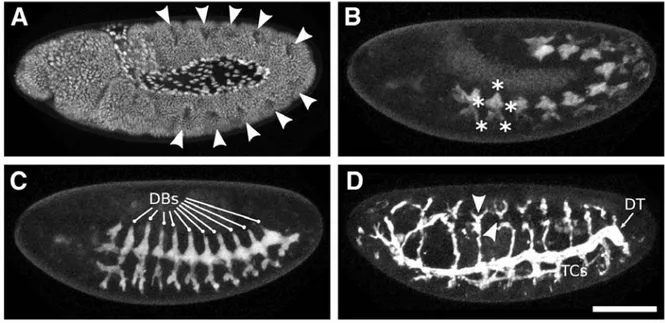
Figure 11. Branching morphogenesis of the Drosophila tracheal system during embryonic
development………....41
Figure 12. Septate junction components are required for tracheal morphogenesis…………...42 Figure 13. Septate junction genes are required for tracheal tube size control………...43 Figure 14. Septate junction integrity is required for epithelial paracellular barrier in Drosophila………..45 Figure 15. Ly6/TFD proteins share comparable tridimensional structure……….47
Figure 16. General structure of Ly6/TFD proteins and general structure of a typical GPI
anchored protein……….49
Figure 17. The Ly6/TFD protein loops contain molecular determinants that mediate the
interaction with their specific site receptors………...51
Figure 18. Complement system cascade pathway……….53 Figure 19. Schematic representation of the uPA/uPAR complex function………...55 Figure 20. Expression patterns of Ly6 genes in Drosophila embryos………..57
Supplementary results:
Figure 1. The Septate junction components Neuroglian, Lachesin, and Fasciclin III behave as
homophilic adhesion molecules in S2 aggregation assays………92
Figure 2. Overexpression of Bou or Cold does not elicit aggregation in S2 cells………94 Figure 3. Overexpression of Bou and Cold does not elicit aggregation in S2 cells………….95 Figure 4. Expression of of HA-Bou and FLAG-Cold does not enhance Nrg mediated
aggregation………96
Part 1
I. Biological barriers, a hallmark of life
In his wonderful essay “What is life?” appeared in 1944, the physicist E. Schrödinger enquired about the essential features that define living organisms and mark the transition between life and the inanimate matter. He found that one of the essential properties of living beings is that, despite their active metabolism, they are extremely organized structures capable of standing for a considerable amount of time against the universal course towards thermodynamic equilibrium. How do these “entropy islands” manage to temporarily avoid decay? He found a satisfactory explanation stating that life has the unique property of extracting order from the external environment, incorporate it into its structure and then release waste products in a constant exchange that, as he liked to put it, feeds the organism with negative entropy. Although Schrödinger did not venture into these grounds, it follows that such entropy islands need to be separated from the surrounding environment by some kind of barrier, which becomes thus one of the fundamental features of living organisms. So, it is tempting to propose that without efficient barriers, life simply would not be possible.
The most obvious and universal type of barrier existing in living organisms is the cell membrane that surrounds the protoplasm of each cell and separates the ordered intracellular components from the chaotic external environment. However, this cell barrier is selectively permeable, which means that is able to regulate the passage of different substances that enter and exit the cell, assuring the transport of materials needed for cell survival.
During evolution, multicellular organisms have in turn developed highly specialized cell types, the epithelial cells, whose main function is to act as barriers. However, epithelia not only insulate organisms from the external environment, but also allow body subdivision into physiologically distinct compartments, opening the way to both organogenesis and evolution of complex body plans. Thus, a crucial function of epithelia during animal development is to maintain the unique composition of different body compartments and to regulate the passage of different materials through the space separating adjacent cells, providing a control over which substances are allowed to enter or leave a particular tissue.
Fig 1. Junctional cell complexes in vertebrate epithelial cells. (A) Schematic drawing of
intestinal epithelial cells. Membrane epithelial cells display three types of cell adhesion complexes: 1) Occluding junctions embodied by the tight junctions in vertebrates and localized in the most apical part of the cell). 2) Anchoring junctions (including adherens junctions, desmosomes and hemidesmosomes). 3) Gap junctions. Apical cell domain is at the top, basal cell domain is at the bottom. Respective function established by different cell junction complexes is mentioned in the table, on the right. (B) Electron microscopy image showing different junctional complexes of mouse intestinal epithelial cells. (Mv, microvilli; TJ, tight junction, circled in black and localized most apically; AJ, adherens junction localized just below the TJ; DS, desmosome.) Scale bar, 200 nm. Ref. panel A: Alberts B. et al., 2002. Mol Biol of the Cell, Garland Science, 4th edition. Ref. panel B: (Tsukita et al., 2001).
How epithelial cells are organized and assembled and how they accomplish their physiological “barrier” function remains a fundamental issue in animal development, and has been for decades the object of multiple studies.
Interestingly, the concept of epithelial cell barrier has evolved over time, as different experiences and novel observations have accumulated. In fact, initial experiences performed on epithelial tissues using vital dyes, revealed a distinct region at the apical end of the lateral cell membrane, referred to as the “terminal bar.” This structure was thought to constitute an absolute barrier totally blocking the passage between cells. However, by the early 20th century, it became clear that some materials, like macrophages and water could indeed cross epithelia through the paracellular space, which is the space available between contiguous cells of the same epithelium. Current understanding shows that specialized cell junctions essentially form a selective permeability barrier across epithelial cells and behave as gates regulating the passage of solutes, ions and even small molecules from one side of the epithelium sheet to the other (Tsukita et al., 2001; Knust and Bossinger, 2002). But, which is the material basis for these selective gates?
II. Cell junctions: a material basis for the barriers in multicellular organisms
The solution that multicellular organisms have adopted to respond to the need for efficient paracellular barriers is to build up specialized cell adhesion contacts charged with this specific task. In fact, not all the known types of adhesion contacts contribute to the formation of paracellular barriers.
In general terms, epithelial cell junction complexes have been classified into three groups that assume different, albeit often related functions (Müller and Bossinger, 2003). We can distinguish (Fig 1):
- Sealing or occluding junctions, such as zonula occludens or tight junctions, which maintain the selective barrier of epithelia (Schneeberger and Lynch 1992; Anderson et al., 1993).
- Anchoring junctions such as adherens junctions (Niessen and Gottardi, 2008) and desmosomes (Holthofer et al., 2007; Garrod and Chidgey, 2008) which keep cells mechanically attached to each other’s by joining specific cytoskeleton components of
Fig 2. (A) Cartoon of a tight junction strand. At tight junctions, tightly aligned rows
of proteins, localized under cytoplasmic half of lipid bilayer, permit to join the tight junction strand in the apposed membranes, sealing the association between adjacent cells. This serves to block the movement of materials through the intercellular space, by forming the so-called kissing points. (B) Structure of tight junctions. Freeze-fracture replica electron microscopic image of intestinal epithelial cells. Tight junctions appear as a continuous, anastomosing particle fibrils, forming strands (arrowheads) with complementary vacant grooves (arrows). (Mv, microvilli; Ap, apical membrane; Bl, basolateral membrane.) Scale bar, 200 nm. (C) Ultrathin
transmission electron microscopy section of tight junction structures. This
electron micrograph shows that at kissing points of tight junctions (arrowheads), the intercellular space is obliterated. Scale bar, 50 nm. Ref. panels B and C: (Tsukita et al., 2001).
the adjacent epithelial cells, or the hemidesmosomes (reviewed by Jones et al., 1994) (Fig 1), permitting to attach the cytoskeleton of epithelial cells to the extracellular matrix.
- Communicating junctions, such as gap junctions which are channels that mediate communication of chemical or electrical signals between cells that are in direct contact with each other’s (Bennett et al., 1991; Kumar and Gilula, 1996).
In my thesis, I will particularly focus on the “sealing junctions” that control the paracellular flow of water, nutrients, ions, growth factors and even cells. However, this is not their only role, as they assure at the same time other important functions. For instance, they share with anchoring junctions the capacity to mediate cell adhesion and communication between adjacent cells.
III. Sealing junctions: general structure and particular features
Sealing junctions characterized in so far can be grouped in three main categories: the vertebrate tight junctions (TJ), the invertebrate septate junctions (SJ) and the paranodal septate junctions (PSJ), found in both vertebrates and invertebrates.
a) Tight junctions
The irruption of electron microscopy applied to biology permitted the discovery of the two main types of sealing junctions that we can recognize in extant organisms. The first to be identified were the tight junctions or zonula occludens, which are vertebrate-specific type of cell adhesion complexes and were discovered by M.G. Farquhar and G.E. Palade in 1963. Tight junctions, firstly resolved in the electron microscope as tightly associated regions between membranes of adjacent cells, localize to the most apical part of the lateral cell membrane, just above the adherence junctions of the polarized epithelial cells (Fig 1, see also Fig 6). As their name implies, TJ constitute a site where the outer leaflets of the membranes of two contacting cells come very close or tight (Fig 2 B, C).
Fig 3. (A). TEM of Drosophila epidermis from late stage 17 wild-type embryo, showing the apical adherens junctions (AJ, arrow) and the lateral
septate junctions. Laterally, interacting plasma membranes are joined by septa, arranged in parallel rows with a regular periodicity. Brackets indicate clustered groups of septa, and arrowheads point to individual septa. (B). Freeze fracture replica of septate junction structures in
arthropod epithelia, showing the parallel arrays of intramembrane
rounded particles, forming septa between adjacent cell membranes. Scale bar represent 100 nm. Ref panel A: (Wu et al., 2004). Ref. panel B: (Furuse and Tsukita, 2006).
Basically, the key structure in TJs is called “TJ strand”, a belt-like region in which two apposing membranes lie close together (Fig 2, A). The TJ strands are localized within the plasma membrane, as shown by freeze-fracture replica electron microscopy (Staehelin, 1973) (Fig 2, B). Each TJ strand is tightly associated with an equivalent strand situated in the opposing membrane of an adjacent cell to form a paired strand. The sites of contact of the two structures are called “Kissing Points” that can be visualized in ultrathin sections, as regions in which the intercellular space is obliterated (Fig 2 A, C) (Farquhar and Palade, 1963; Tsukita et al., 2001).
The TJ strands are composed at least by 40 different proteins, whose function is not only restricted to the maintenance of a paracellular seal. In fact, the TJ structural complexity reflects the contribution of some its components to other interrelated cell process, like the maintenance of cell polarity (Cereijido et al., 1998) in which TJ participate by limiting the diffusion of proteins and lipids within the membrane, ultimately keeping the apical and basolateral regions of the plasma membrane as separated domains (Cereijido et al., 2008).
However, this is not their only associated function, as TJ are also known to participate in signalling (Izumi et al., 1998; Ebnet et al., 2008), cell cycle control (Tsukita et al., 2008), vesicle trafficking (Yeaman et al., 2004) and even transcriptional regulation (Balda and Matter, 2003).
b) Septate junctions
Septate junctions were described for the first time by R.L. Wood in 1959 (Wood, 1959) as “septate desmosomes”, during his electron microscopy (EM) observations of Hydra epithelial cells. Commonly found in invertebrate epithelia, their name is due to their ladder-like appearance visible in electron microscopy cross-sections (Fig 3). In sections perpendicular to the cell surface, the septa appear as regularly spaced electro-dense crossbars spanning the space existing between the opposed membranes of adjacent cells (Fig 3), that are separated by a constant distance of approximately 15-20 nm (Tepass et al., 2001, also reviewed by Furuse and Tsukita, 2006).
It only became clear that these structures were responsible for the formation of the paracellular barrier when the paracellular diffusion of electron-dense dyes was studied in EM sections. In fact, in an intact epithelium the diffusion of dyes like the Lanthanum is precisely stopped at the
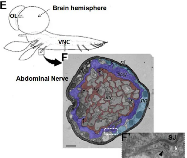
Fig 4. Glial layers of the peripheral nervous system in Drosophila larvae. (E)
Cartoon of the CNS dissected out from Drosophila third larval instar and oriented laterally. OL (optic lobe), VNC (Ventral nerve cord). (F) Cross-section of larval peripheral abdominal nerve observed in electron. Three glial cell layers are present in the larval nerve: axons (ax) are ensheathed by wrapping glial (wg) cells (shown in pink) and the overlying subperineurial glia (spg) which appears as a flattened layer (in purple). SPG itself is surrounded by the perineurial glia (pg) in green, ensheathed by the neural lamella which covers the nervous system. (F’) Close up of the white boxed area in F, showing a part of the adjacent subperineurial and perineurial glia. septate junctions (SJ) appears only in the intercellular space between subperineurial glial cells (white arrowhead), whereas they are missed between perineurial glial cells (black arrowhead).
Fig adapted from (Stork et al., 2008).
SJ level. The implication of the SJ in the barrier function has also been confirmed in
Drosophila by morphological analysis of mutants in which intercellular septa are missing and
that consistently present defective barriers (Baumgartner et al., 1996; Lamb et al., 1998). Interestingly, the ultrastructural features of the SJ seem well conserved in the multiple species in which their presence has been reported, which range from chordates (Rosenbluth, 1995; Banerjee et al., 2006) to porifera, where SJs have been observed between sclerocyte cells that secrete the spicules of the calcareous sponge Sycon ciliatum (Ledger et al., 1975).
However, some structural heterogeneity exists. In insects, Flower and Filshie (Flower and Filshie, 1975) distinguished two different subtypes of septate junctions, based on their characteristic appearance in tangential views: the “pleated” and the “smooth” septate junctions. The pleated SJ, found in ectoderm derived epithelia and in glial sheets, form the typical ladders like electron-dense septa described above (Tepass et al., 2001). In contrast, the smooth SJs are only found in endodermal derivatives, like the midgut and lack the regular arrays of septae (Green et al., 1983).
In invertebrates and particularly in insects, SJ can be easily recognized in most ectodermally derived epithelia. However, very similar adhesion structures have also been observed in the insect nervous system, where they contribute to the formation of a sealing barrier that protects and isolates the neurons from their surrounding environment.
More in detail, a series of electron microscopy observations performed on the nervous system of Drosophila have shown that septate junctions are present at the cell contacts between specialized types of glial cells, the so-called subperineurial glial (spg) cells, which surround and ensheath axon fascicles of central and peripheral nervous system (Stork et al., 2008) (Fig 4). These connections have an essential role for the maintenance of the Blood Brain barrier (BBB) organization, a physiological barrier protecting the nervous system from the high potassium (K+) concentration present in the hemolymph but also regulating the entry of other molecules inside the nervous system (Bainton et al., 2005; Schwabe et al., 2005; Stork et al., 2008).
c) Paranodal septate junctions
Interestingly, structures morphologically very similar to the invertebrate SJ have also been observed in the nervous tissues of vertebrate organisms: the so-called paranodal septate
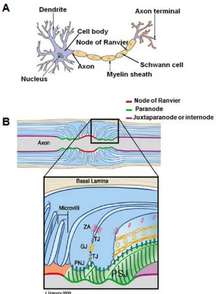
Fig 5. Membrane domain organization of the vertebrate myelinated axons. (A)
Cartoon of a myelinated neuron. Myelinating Schwann cells ensheath all axon surfaces excepting nodes of Ranvier to induce the “saltatory conduction” of action potentials. (B) Schematic representation of myelinated axon shows three distinct compartments or axon domains: the node of Ranvier (in red), the paranode (in green) and the juxtaparanode or internode (in purple). In nodes, axons are in contact with Schwann cellmicrovilli of glial cells, but they are in contact with paranodal myelinated loops in paranodal domain (fig B, close up), whereas internodal domains are ensheated with myelinated axons. Paranodal septate junctions PSJ (presented by the hatched green lines) are formed between paranodal loops and junctional axon domain. Paranodal loop cells are polarized presenting tight junctions (TJ) that provide paracellular barrier between the periaxonal space and the loops, gap junctions (GJ) permit direct communication between loops, and adherens junctions (AJ) that promote attachment between adjacent loops. Such organization of paranodal myelinated loop cells is very similar to that of polarized epithelial cells. Fig adapted
from: (Salzer, 2003).
junctions (Brophy, 2001). This denomination is due to their presence in the paranodal axon domains of the myelinated nerves, at the interface between glial myelinated Schwann cell loops and axonal membranes (Fig 5, B).
It has been recognized that myelinated axons of the central and peripheral nervous systems (Fig 5, A) are compartmentalized into three functionally distinct domains: the nodes of Ranvier, the paranodes, and the juxtaparanodes or internodes (Fig 5, B) (reviewed by Salzer et al., 2003).
Paranodes, located on both sides of Ranvier nodes are the site of attachment of the axonal membrane to the terminal loops of myelinating glial cells, which spiral around the axon, forming a series of septate-like junctions (Fig 5, B). This axo-glial paranodal junction plays three important physiological roles. First, it provides electrical insulation, allowing saltatory conduction of the nerve impulses from one node of Ranvier to the next node. Second, it restricts the lateral mobility of axonal membrane proteins and channels, organizing a fence within the axonal membrane that separates Na+ channels present at the unmyelinated node of Ranvier from K+ channels present under the glial cells, in the juxtaparanode (Bhat, 2003; Bhat et al., 2001). Third, it also provides adhesion and putative intercellular communication between axons and Schwann cells at the level of the axon-glia contact domains.
Thus, paranodal septate junctions establish a physical Blood Brain Barrier (BBB) between the neuron and the ensheathing glial cells by preventing the unregulated exit into the blood of neurotransmitters and other substances emanating from nerve cells, and at the same time blocking the passage of blood material into the nerves (Bellen et al., 1998). They are also likely to contribute to a bi-directional signalling between axons and glial cells and seem to play important roles in the process of myelination, as defects in the organization of paranodal septate junctions have been associated with several neuropathies and dysmyelinating disorders (Griffiths et al., 1996).
We have seen that both epithelial septate junctions and paranodal septate junctions play an equivalent role to that of tight junctions, but differ strikingly in morphological terms. Another remarkable difference between SJ and TJ, as far as epithelial tissues are concerned, is that the position along the lateral cell membrane of these structures is different. In fact, SJ are placed just below the adherens junctions and not above, as TJ are (Fig 6), suggesting that these adhesion structures are completely different, both in composition and in evolutive origin. In fact, for a long time, they have been envisaged as analogous structures rather than homologous ones.
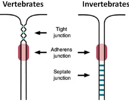
Fig 6. Localization of tight and septate junctions, respectively in vertebrate and invertebrate epithelial cells. In vertebrates, tight
junction (TJ) structures, where adjacent cell membranes join together in a specific sites (shown in green), are localized in the most apical part of epithelial cells, just above the adherens junctions (in pink, left hand panel). Whereas in invertebrates, tight junctions analogous, called septate junction (SJ), are situated just below adherens junctions (in pink, right hand panel) and are characterized by a ladder-like septa (in green) spanning the intercellular epithelial space. Ref. Fig: (Tepass, 2003). 27
However, the detailed characterization of their multiple components has shown that these structures share more similarities than previously thought, a feature that also applies to the paranodal septate junctions. So, how are these junctional complexes organized at the molecular level?
IV. Molecular organization of the sealing junctions
a) Molecular organization of TJ
After the first electron microscopy observations, cell biologists began to focus in the study of TJ, to unmask the nature of its molecular components. Contrary to what was initially thought, the nature of the TJ strands is not predominantly lipidic, but made of protein complexes arranged like beads that span the paracellular space. In fact, at the level of the TJ, the adjacent plasma membranes are hold together by rows of transmembrane junctional proteins. On the one hand, the extracellular domains of these proteins directly interact with one another to occlude the intercellular space and create a seal (Schneeberger and Lynch, 1992; Gumbiner, 1993; Anderson and van Itallie, 1995). On the other hand, the intracellular part of these transmembrane proteins associates with a set of cytosolic components that anchor the strands to the actin cytoskeleton. In this way, the tight junctions allow the cytoskeletons of adjacent cells to join together. At the molecular level, the different components of the tight junctions can be broadly separated in three different groups:
- The Claudins: tetraspan membrane proteins, like Occludin, which are allegedly responsible for the selective barrier function (Fig 7 A, B).
- A large set of different single-pass transmembrane proteins that can be collectively designed as junctional adhesion molecules (JAM) (Fig 7, C).
- Multiple cytoplasmic adaptor proteins, that form the so called tight junction plaque and include the Zonula Occludens ZO-1, ZO-2 and ZO-3, all belonging to the MAGUKs (Membrane-Associated Guanylate Kinase) family and other proteins containing a PDZ domain (PSD-95/Discs large/ZO-1) which interact specifically with the
Fig 7. Topology of tight junction integral membrane proteins. (A) Occludin has four
transmembrane domains with two extracellular loops, one short intracellular loop with amino and carboxy terminal cytoplasmic domains. (B) Claudin-1 also has four transmembrane domains, but does not display sequence similarity to Occludin. Note that the cytoplasmic tail of claudin-1 is shorter than that of Occludin. Different Claudin members show variability in the aminoacid composition of their extracellular loops. (C) Junctional adhesion molecule 1 (JAM-1) also known by JAM-A has a single transmembrane domain; its extracellular domain contains two immunoglobulin-like loops that are formed by disulphide bonds. The first amino terminal loop is known to mediate homologous interaction with JAM-1 molecule, present in the adjacent cell membrane. Molecular weight, isoform and aminoacid number of each molecule are also mentioned.
Ref. Fig: (Schneeberger and Lynch, 2004).
cytoplasmic domains of Occludin, Claudins and JAM and form a scaffold capable of recruiting other signalling proteins and cytoskeleton components to the TJs.
Occludin derives its name from the latin word ‘‘occludere’’, to close, and represents the first
example of integral membrane proteins specifically found in TJ strand (Furuse et al., 1993; Ando-Akatsuka et al., 1996). Occludin encodes for a membrane protein with four transmembrane domains, a topology that generates two extracellular loops, one intracellular loop and two amino- and carboxy-terminal cytoplasmic domains (Fig 7, A). Although Occludin is specifically localized at the tight junctions, its physiological function is still unclear. In fact, epithelial tissues deficient for Occludin do not show obvious defects at the level of the TJ and the paracellular diffusion barriers seem functional (Saitou et al., 1998). However, Occludin knock-out mutant mice display clear phenotypes, such as growth retardation, male sterility, and a tendency to develop gastritis, suggesting that the digestive apparatus barrier function could be impaired (Saitou et al., 2000). The presence of functional tight junctions in these mutants has stimulated the study of other membrane proteins present in the TJ complex, leading to the discovery of the key role played by Claudins in paracellular barriers.
Claudins, also named from the latin “claudere”, to close, have emerged as key components of
the TJs and are thought to have a direct function in barrier and tight junction strand formation (Inai et al., 1999; McCarthy et al., 2000). They form a large multigene family with approximately 24 members in human and mice (Van Itallie and Anderson, 2006; Furuse and Tsukita, 2006; Angelow et al., 2008), but recently they have also been identified in insects (Wu et al., 2004). Claudins are also tetraspan proteins, sharing a similar topology to that of Occludin (Fig 7, B). Claudins are known to interact in a homo-and heterophilic way in the plane of the membrane (Furuse et al., 1999; Blasig et al., 2006), but also with the Claudins of the adjacentcells (Furuse et al., 1999), thereby sealing the cell junctions. Ectopic expression of Claudins in fibroblasts results in the formation of tight junction-like structures, indicating that Claudin expression has a capital role in driving tight junction formation. Interestingly, the observed profusion of Claudin paralogues seems to provide a molecular basis for the different selective properties exhibited by different epithelial barriers. Indeed, manipulations altering the type of Claudin expressed in a tissue seem to have a direct impact on paracellular ion and/or size selectivity (Van Itallie et al., 2001; Nitta et al., 2003). It seems that these selective properties reside in the extracellular loops of Claudins that contain several electrically
Fig 8. Schematic representation of the basic molecular components of tight junctions in epithelial cells. Claudins,
Occludin and the junctional adhesion molecules (JAMs) are the most prominent components. Within the cytoplasm many first-order adaptor proteins, including the scaffolding proteins Zonula Occludens 1, 2 and 3 (ZO-1–3) bind to the cytoplasmic tail of intramembrane proteins and provide a direct link to the actin-based cytoskeleton. Among the second-order adaptor molecules, Cingulin is shown. Signaling and regulatory proteins include multi-PDZ-protein 1 (MUPP1) and MAGI (membrane-associated guanylate kinase with inverted orientation of protein– protein interaction domains).
31
charged residues. In fact, it is known that different members of the Claudin family exhibit a large variability of isoelectric points and, for instance, point mutations altering the charge in the first extracellular loop of Claudin-15, expressed in the mammalian polarized Madin-Darby canine kidney cells (MDCK), result in changes of barrier ion specificity(Colegio et al., 2002). Thus it is now widely accepted that barrier specificity is largely due to the type of claudin(s) present at the tight junctions (Anderson et al., 2004; Furuse and Tsukita, 2006).
JAMs or junctional adhesion molecules are the second type of integral membrane proteins
localized at TJ. They belong to a family of single-span transmembrane proteins characterized by the presence of immunoglobulin extracellular domains (Fig 7, C) (Martin-Padura et al., 1998; Ebnet et al., 2004). For instance, in humans, the family consists of four closely related molecules called JAM-A, -B, -C and JAM-4 (Ebnet et al., 2004). Differing from Claudins, their expression in fibroblasts does not induce the formation of tight junctional strands, suggesting that they may play a subsidiary role in TJ assembly. As their name implies, JAM’s main contribution seems to be mediating cell adhesion. In fact, they are supposed to hold together the two opposing membranes of the tight junction thanks to their capacity to interact in an homophilic and heterophilic way (Keiper et al., 2005). However, it seems that this is not their only function, as they also mediate interactions with a wide range of cytoplasmic proteins. In particular, they are thought to play a role in the regulation of the cell polarity, as JAM-C is necessary for the recruitment of the cell polarity complexes PAR6, Cdc42, PKCl and PATJ during mammalian spermatid differentiation (Gliki et al., 2004).
Scaffolding proteins: The incorporation and association of the transmembrane proteins
Occludin, Claudins, and JAMs in tight junctional strands requires the local clustering of these proteins in a particular membrane region. Although direct interactions between Occludin, Claudins, and JAMs may contribute to their clustering, this process mainly relies on the scaffolding properties of their cytoplasmic binding partners.
An important group of tight junctional scaffolding molecules are the zonula occludens proteins ZO- 1, ZO-2 and ZO-3 (Fig 8). These proteins belong to the MAGUK family and are characterized by the presence of three N-terminal PDZ domains, an SH3 domain followed by a catalytically active guanylate kinase domain. These proteins can interact directly with Occludin, Claudins and JAMs via their PDZ domains (Furuse et al., 1994; Haskins et al., 1998; Itoh et al., 1999; Ebnet et al., 2000), whereas their C-terminus can associate with filamentous actin, thus providing a direct link with the actin cortex (Fig 8) (Fanning et al.,
1998; Wittchen et al., 1999). In fact, their interaction with actin could be essential for their localization at the level of the TJ, as it has been shown for the ZO-1 protein (Fanning et al., 2002). In addition, ZO-1 has been shown to form homodimers and also heterodimers with either ZO-2 or ZO-3, a property that could contribute decisively to the clustering of the TJ components. Along this line, it has been shown recently that both ZO-1 and ZO-2 are essential for Claudin clustering, strand formation and barrier maintenance (Umeda et al., 2006).
The ZO proteins are not the only PDZ-motif adaptors present in TJ. Several proteins, such the Multi-PDZ domain protein 1 (MUPP1) and the membrane-associated guanylate kinase with inverted domain orientation (MAGI) proteins have been shown to interact with one or more integral membrane TJ components (Fig 8) (Schneeberger and Lynch, 2004). However, it is unclear if these molecules are part of a structural core essential for tight junctions stability or if they serve a subsidiary regulatory function, as TJ are dynamic structures whose properties change in different cellular and physiological situations. For instance, Cingulin, a non-PDZ tight junctional plaque protein, also interacts with ZOs, JAMs, and actin via its head domain, whereas its central domain is required for homodimerization and can interact with myosin. As such, this protein may be an important regulator of tight junctional dynamics during actomyosin contraction (Clayburgh et al., 2005).
As we have seen, the general molecular organization of tight junctions stands on a group of transmembrane proteins (Occludin, various Claudins and JAMs) whose C-terminal cytoplasmic sequences present high affinity for scaffolding proteins, mostly containing one or more PDZ domains. The study of invertebrate septate junctions has shown that their structural logic is very similar to that of TJ, and that both complexes are formed by similar types of molecules.
b) Molecular organization of SJ
Basically, the most detailed molecular dissection of invertebrate septate junction (SJ) has been carried out in Drosophila. In this organism, many SJ components have been identified and it has been found that some of them play equivalent molecular functions to known vertebrate TJ proteins. Moreover, in many cases the septate and tight junction components appear to be clear homologous proteins that share the same organization and domain composition.
As in vertebrates, the internal logic of SJ allows to classify its components in three groups.
First, the Drosophila SJs present transmembrane proteins like the Claudins, supposed to maintain the barrier function of these structures. For instance, three fly Claudins displaying the characteristic topology of these proteins have been described: Megatrachea (Mega) (Behr et al., 2003), Sinuous (Sinu) (Wu et al., 2004) and Kune Kune (Kune Kune) (Nelson et al., 2010). However, Occludin seems to be absent in this insect.
Second, the Drosophila SJ complex is also composed by a group of cell adhesion molecules that includes both transmembrane proteins, such as Neurexin IV (Baumgartner et al., 1996), Gliotactin (Genova and Fehon, 2003; Schulte et al., 2003), Fasciclin III (Woods et al., 1997), Neuroglian (Hortsch et al., 1995; Genova and Fehon, 2003) and the Na+/K+ ATPase pump (Paul et al., 2003)), and also Glycosylphosphatidylinositol (GPI) anchored cell membrane proteins, such as Lachesin (Llimargas et al., 2004), Contactin (Faivre-Sarrailh et al., 2004) and Melanotranferrin (Tiklová et al., 2010).
The third type of SJ proteins consists of scaffolding adaptor molecules found at the cytoplasmic side of the membrane. These proteins include ZO homologous proteins with different PDZ proteins, like Dlg (Woods et al., 1991) and Varicose (Wu et al., 2007), founding members of the MAGUK family. Many other fly scaffolding components, notably Coracle, a cytoskeletal linker belonging to the 4.1, Ezrin, Radixin, Moesin (FERM) protein domain family (Fehon et al., 1994; Lamb et al., 1998) and Scribble (Bilder et Perrimon, 2000), a protein with leucine-rich repeats (LRRs) and PDZ domains, known to regulate cell polarity, are also associated with vertebrate tight junctions (D’Atri et al., 2002), thus pointing on the molecular and functional conservation existing between tight and septate junction structures.
In Drosophila polarized cells, the different SJ components appear clustered in a membrane region placed below the adherens junctions. Multiple observations have shown that in mutant background for one of the SJ components, the other components appear systematically mislocalized and found uniformly distributed along the lateral membrane (Genova and Fehon, 2003; Faivre Sarrailh et al., 2004; Llimargas et al., 2004; Moyer et al., 2008; Tiklova et al., 2010 …). These observations have lead to the notion that the different components of SJ are
Fig 9. Schematic representation of septate junction molecular complex between
Drosophila epithelial cells. Different protein interactions are shown between septate junction components. The cell adhesion molecules NeurexinIV, D-Contactin and Neuroglian interact together to form a tripartite complex, also present in vertebrate PSJ. Coracle and NeurexinIV also form an interdependant complex with Neuroglian and the Na+/K+ ATPase pump. Other hypothetical interactions are supposed to occur between the cytoplasmic PDZ-binding domains of Neurexin IV with the PDZ domains of Scribble or Discs large, as well as interactions between Coracle and the 4.1-protein-binding domain of Discs large, but also between Gliotactin and the PDZ domains of Scribble and between the PDZ domains of Scribble and Discs large. These interactions are indicated by broken arrows; however they still need to be demonstrated. These molecules, with the exception of the homophilic cell adhesion molecule Fasciclin III and the Drosophila ankyrins, have now been demonstrated to be essential for the function of septate junctions in the epithelia and nervous system of Drosophila. Ref. adapted from: (Hortsch M and Margolis B, 2003).
interdependent for their clustered localization and therefore for the organization of the whole SJ complex.
Moreover, genetic and biochemical studies have unveiled a complex network of interactions between some of the SJ components (Fig 9). For example, it has been shown that the FERM protein Coracle (Cor) interacts with the cytoplasmic tail of Nrx IV (illustrated in Fig 9) and, accordingly, the clustered localization of Cor is lost in NrxIV mutants (Ward et al., 1998). Further studies based on immunoprecipitation experiments have also indicated that Cor and Nrx IV are found in an interdependent complex with the Na+/K+ ATPase pump and the transmembrane protein Nrg (Genova and Fehon, 2003). In addition, it has been shown that the cytoplasmic tail of Nrx IV binds to the PDZ domain of the scaffolding MAGUK protein Varicose (Wu et al., 2007). Finally, biochemical experiments indicate that MTf, a conserved transferrin family of GPI anchored iron-binding protein, also interacts with Nrx IV, Cont and Nrg complex (Tiklová et al., 2010).
However, even though more and more interactions between septate junction components become apparent, further studies will be required to understand how the SJ complex is assembled and how its integrity is maintained during development. In particular, the dynamic of the interactions established between SJ components has hardly been explored. For instance, it has been shown that in mutants for Nrx IV, Contactin protein seems unable to reach the plasma membrane and is seen instead accumulating in intracellular vesicles (Faivre-Sarrailh et al., 2004). This finding suggests that SJ complexes may preassemble en route to the membrane, but we still know little about how the trafficking of the different septate junction components is organized and how these proteins are addressed to a particular region of the cell membrane. These questions have just begun to be analyzed in a seminal study focusing on the MTf SJ component (Tiklová et al., 2010). This work has shown that before SJ formation, the MTf protein is uniformly distributed along the lateral membrane, then enters different endosomal compartments (Rab5 positive early endosomes and Rab11 positive recycling endosomes), and is finally reshipped to the apical part of the lateral membrane where it forms a cluster at the level of the SJ. This suggests that membrane recycling plays an active role in the initial clustering of the SJ components, but it could also participate in SJ maintenance during development and/or in physiological regulation of paracellular barrier activity. Indeed, the endocytosis of SJ components could be a recurrent way to regulate the properties of barriers, as many studies in the vertebrate TJ have demonstrated (Utech et al., 2010).
Figure 10. Schematic model describing molecular interactions at the paranodal region of myelinated axons. A cis complex of adhesion molecules Caspr
(contactin-associated protein) and Contactin, present in the lipid raft domain of the axon, are interacting together. Caspr binds in its cytoplasmic region to protein 4.1B, a member of the 4.1 family of cytoskeletal and cytoplasmic adaptor proteins. Caspr/Contactin complex interacts with the glial protein, NF155 (Neurofascin 155), anchored to lipid rafts of the myelinated terminal loops membrane. Fig. adapted from: (Labasque and Faivre-Sarrailh,
2010) and (Salzer, 2003). 37
c) Molecular organization of paranodal septate junctions
Septate junctions display extraordinary similarities with the vertebrate paranodal septate junctions (PSJ) not only at the morphological level, but also at the molecular one, as they share many different membrane adhesion molecules and cytoplasmic adaptors.
The structural core of the PSJ is composed by members of the Immunoglobulin cell adhesion molecules (Ig L1-CAM) family, including Caspr/Paranodin (NCP1), F3/Contactin and Neurofascin 155 (NF-155), which are the respective homologues of Drosophila Neurexin IV, D-Contactin and Neuroglian. These proteins are thought to be associated with lipid rafts in the glial membrane domains, and their insertion in these membrane microdomains seems important for the function of paranodal junctions (Fig 10) (Maier et al., 2007), also reviewed by (Labasque and Faivre-Sarrailh, 2010). It is known that the Neurexin-type Caspr/Paranodin protein interacts with GPI-anchored Contactin in a cis-configuration (Peles et al., 1997). In paranodal junctions, this complex binds by its cytoplasmic tail to the scaffolding 4.1B protein (Denisenko-Nehrbass et al., 2003), also present in invertebrate SJ and that acts as an important linker between the membrane proteins and the cytoskeletal network. Finally, this complex mediates cell adhesion by a trans interaction with the Neurofascin-155 present in the opposite membrane (Fig 10) (Charles et al., 2002). The interaction between these PSJ proteins is required for the organization of the proper axon functions and the nerve potential action conduction (Bhat et al., 2001; Boyle et al., 2001). In Paranodal region, Claudins play an intriguing role, as they are also present, but appear associated with the formation of TJ between myelinated loops surrounding axons (Poliak et al., 2002).
Despite recent advances, we are still far from having understood the cellular and molecular mechanisms controlling the assembly and maintenance of the PSJ, and their specific functions in the vertebrate nervous system. However, the picture emerging from the studies comparing TJ, SJ and PSJ at the molecular level indicates that strong parallelisms exist between invertebrate and vertebrates at the level of their structural components (see Table 1), pleading for an ancient common origin for all of the extant sealing junctions.
This consideration also implies that studies focusing on the insect septate junctions could be specially informative to shed light on the molecular complexity, the assembly mode and the interactions existing between the different components of paranodal septate junctions.