TH `
ESE
TH `
ESE
En vue de l’obtention du
DOCTORAT DE L’UNIVERSIT´E DE
TOULOUSE
D´elivr´e par : l’Universit´e Toulouse 3 Paul Sabatier (UT3 Paul Sabatier)
Pr´esent´ee et soutenue le 14 D´ecembre 2018 par :
Marine Cambon
Heterogeneity within infections: the case of the
vector-borne insect pathogen, Xenorhabdus nematophila
JURY
Claire Valiente-Moro Maˆıtre de conf´erence Rapporteuse
Dieter Ebert Professeur d’universit´e Rapporteur
Franck Dedeine Maˆıtre de conf´erence Membre du Jury
Mathieu Sicard Professeur d’universit´e Membre du Jury
Sophie Gaudriault Charg´ee de Recherche Directrice
Jean-Baptiste Ferdy Professeur d’universit´e Directeur
´Ecole doctorale et sp´ecialit´e :
SEVAB : ´Ecologie, biodiversit´e et ´evolution Unit´e de Recherche :
´Evolution et Diversit´e Biologique, UMR UPS-CNRS-IRD, Toulouse
Diversit´e, G´enomes et Interactions Microorganismes-Insectes, UMR UM-INRA, Montpellier
Directeur(s) de Th`ese :
Jean-Baptiste Ferdy et Sophie Gaudriault Rapporteurs :
` A Mutti
Remerciements
First of all, I would like to thankClaire Valiente-Moro, Dieter Ebert, and Guillaume
Mitta, who kindly agreed to evaluate this manuscript. I would like also to thank Ma-thieu Sicard and Franck Dedeine, who followed this work since the first year, and
provided me some very useful comments and ideas. Then of course, I need to thank all the people who participated to this adventure, starting with my two awesome supervisors.
Jean-Baptiste Ferdy, j’ai du mal `a r´ealiser que ce s´ejour sous ton aile touche `a sa
fin. L’id´ee d’essayer de trouver les mots pour te dire toute ma reconnaissance me parait assez sotte, la pudeur et le tact n’ayant jamais ´et´e mon fort. Mais apr`es tout, ¸ca fait longtemps que je ne me suis rien cass´e... Je pense qu’il faut simplement que je te dise toute l’admiration que j’ai toujours eue pour toi, depuis mon premier cours de stats en L2, jusqu’`a nos derni`eres aventures de code R. `A coups de randomisation, de fonction log, de mod`eles, de Linux, de LATEX, d’injections, de musique, de principes. De toujours plus de r´eplicats, de toujours plus d’exigence, de toujours plus de compr´ehension et de rires. Si on devait m’ˆoter ce que tu m’as appris et transmis, bien sˆur il ne resterait plus grand chose de ma science, mais on m’amputerait aussi d’une grosse partie de ma personnalit´e, de ce que je suis devenue pendant ces 5 ans, ou presque. J’aime beaucoup l’id´ee que quand je serai vieille, je pourrai raconter avec ´emotion que j’ai eu un mentor dans ma jeunesse. Je raconterai que ce mentor ´etait une personne exceptionnelle tant par son intellect que par sa p´edagogie, sa franchise, son humour, sa culture, et ses quelques particularit´es. Je raconterai tout ce que je lui dois. J’en ferai probablement des caisses, gonfl´ee de fiert´e d’avoir ´et´e son ´el`eve. D’ici l`a, j’esp`ere r´eussir `a me faire pousser une barbe `a ton image, bien que mes tentatives aient ´et´e infructueuses jusqu’`a pr´esent.
Mais cette aventure de th`ese, c’est aussi la d´ecouverte d’une civilisation qui m’´etait jusqu’alors inconnue. Un grand ´ecart rendu possible grˆace `a toiSophie Gaudriault, qui m’as fait rentrer incognito chez les microbiologistes, sans montrer pˆate blanche ! Tu as eu la patience de m’apprendre les bases d’un nouveau langage et m’as prise par la main tout au long de mes premiers pas dans la vie de chercheur. Ta volont´e d’aller au fond des choses m’a aussi pouss´ee `a r´efl´echir et prendre du recul sur ce que je savais d´ej`a, ou pensais savoir. Tu as pris le temps de toujours m’expliquer avec d´etails et pr´ecision le pourquoi du comment, r´eussissant la d´elicate tˆache d’ˆetre `a la fois exigeante et rassurante. Mais c’est aussi en dehors du labo que tu m’as beaucoup apport´e, au cours de nos longues discussions
parfois bien ´eloign´ee des bact´eries. Je garderai un excellent souvenir des moments pass´es chez toi, que ce soit `a jouer du saxophone ou avec ta famille. Mon estomac (ou peut-ˆetre mon microbiote intestinal ?) me pousse aussi `a mentionner que j’ai toujours tr`es bien mang´e avec toi, et ¸ca, ¸ca compte beaucoup pour moi !
Nathalie Parthuisot, tu resteras sans doute ma super-h´ero¨ıne pr´ef´er´ee. Je ne
com-prends d’ailleurs pas pourquoi personne n’a encore ´ecrit de BD sur toi. Ton ´energie, ta rigueur, ta capacit´e `a abattre des quantit´es de boulot incroyables n’ont pas fini de m’im-pressionner. Tu as ´et´e un pilier indispensable `a cette th`ese que je n’aurais jamais pu terminer sans toi, ce qui a malheureusement failli arriver plus d’une fois ! Tu m’as appris une bonne partie de ce que je sais (plus ou moins bien) faire avec mes dix doigts dans un labo, et tu as ´et´e d’une patience infinie pour r´epondre aux questions bˆetes et incessantes que je suis venue te poser pendant mes manips. Tu as aussi ´et´e une oreille attentive, et j’ai ador´e tous les moments pass´es en ta compagnie. Si quelqu’un arrive un jour `a sauver le monde, ¸ca ne m’´etonnerait pas que ce soit toi. T’aurais pas une petite id´ee de manip avec 10000 r´eplicats et 80 contrˆoles n´egatifs, randomis´es et r´epartis en 105 plaques 96 puits pour arrˆeter le r´echauffement climatique ? Y’aura peut ˆetre quelques comptages `a faire apr`es, mais c’est un d´etail !
Si je dois `a Nathalie une bonne partie de mes comp´etences de labo, je dois l’autre partie Jean-Claude Ogier, Sylvie Pag`es, Anne Lanois qui m’ont appris certaines techniques, mais qui ont aussi r´ealis´e certaines parties de mes manips quand je me trouvais de l’autre cˆot´e de l’Occitanie. Merci beaucoup pour votre temps et votre gentillesse. Merci ´
egalementMarie Frayssinet, d’avoir organis´e mes deux sorties sur le terrain. Merci pour ton agr´eable compagnie et ton aide pour mes manips (`a la frontale).
Pour finir en beaut´e les remerciements adress´es aux personnes m’ayant aid´ee pour ex´ecuter des centaines d’insectes et assister `a la naissance de milliards de n´ematodes, mes pens´ees sont bien sˆur pour vous Adrian Bach et Pierre Lafont, les deux meilleurs stagiaires que cette terre ait connue. Merci mille fois pour votre aide, pour votre enthou-siasme en d´epit du caract`ere extrˆemement r´ebarbatif de certaines de vos tˆaches, merci de m’avoir ´ecout´ee plus d’une fois raconter passionn´ement la grˆace du n´ematode et la fougue de la bact´erie. Merci d’avoir surtout ´et´e des amis. Adrian, merci pour ces quelques godets, ces moult concerts, ces partages. Pierre, merci pour ces discussions `a coeur ouvert et pour ces critiques (qui a dit mauvaise foi ?) sur le foutu monde dans lequel on vit.
Mˆeme sans forc´ement mettre les mains dans le cambouis (ou dans le cadavre d’insecte), certains justiciers de l’ombre m’ont apport´e leur aide pr´ecieuse pendant cette th`ese. Merci
David Duneau pour nos innombrables discussions, merci pour tes id´ees, tes critiques,
ton soutient infaillible depuis le jour o`u tu m’as dit “Tu vas l’avoir ce concours SEVAB”. Merci d’avoir sans cesse cherch´e les mots pour me rassurer. Merci d’avoir ´et´e franc et juste. Merci ´egalementLucie Zinger pour ton aide dans la folle entreprise d’apprendre `a traiter des donn´ees metabarcoding, alors que je n’avais qu’une vague id´ee de ce que voulait
dire le mot s´equen¸cage. Merci pour toutes les ressources que tu as mises `a disposition de tous et que nous suivons comme les dix commandements, merci pour ta patience et le temps que tu as pass´e `a r´epondre `a mes questions techniques et moins techniques.
Et bien sˆur, vient maintenant le temps des remerciements pour ceux qui n’ont pas activement particip´e `a ma th`ese, mais qui ont rendu ma vie tellement g´eniale pendant ces trois ans, `a m’en empˆecher de dormir. Tout d’abord, mes acolytes de bagne, F´elix
Pellerin et Sebastien Cally avec qui j’ai partag´e rires et confidences pendant ces 3/4
ans. F´elix, merci pour toutes ces aventures, que j’´eviterai de conter ici, mais qui resteront grav´ees un bon bout de temps dans ma m´emoire. Rien que d’y penser j’me marre encore. Seb, merci pour ton immense amour, merci d’avoir pris soin de moi comme tu pouvais ces derniers mois. Je suppose que mon mutisme humide n’a pas ´et´e facile pour toi, surtout connaissant ta grande empathie (h´e oui, il y un coeur sous ces muscles). Mais je te remercie d’avoir ´et´e le parfait compagnon de bureau, merci pour ton soutien et pour toute les fa¸cons que tu as eues de me faire rire et de me nourrir quelques fois.
Merci `a mes autres acolytes de bureau, le fameux Quartet compos´e d’Isabelle Mar´echaux,
Arthur Kocher et C´eline Van de Paer. Apr`es ma th`ese, je suis dispo pour une tourn´ee
mondiale. J’en profite pour remercier par la mˆeme occasionBoris Delahaie, qui au mˆeme titre qu’Isa, m’a accueillie dans son palace `a Montpellier. J’ai hˆate de connaˆıtre le prochain ´
episode sur vos voisins.
Merci ´Eglantine Mathieu-B´egn´e, la nouvelle recrue du bureau 26, heureuse maman elle aussi de 15 phasmes tout mignons. Reviens vite du terrain, ¸ca commence `a bien faire. Merci `a toute la joyeuse compagnie d’EDB, Isabelle Cantera pour ta joie, ta mal-adresse et tes taches qui font qu’on est deux vilains petits canards, et `a deux c’est va-chement plus cool. Merci Luana Bourgeaud pour ta douceur apaisante entrecoup´ee de r´epliques cinglantes et bien marrantes, merci Ma¨eva Gabrielli pour ton ´energie et tes gags, et merci `a toute les deux d’ˆetre les Tic et Tac dont on ne saurait se passer. Merci
Maxime Pineaux pour ta cuisine, pour nos ´echanges de BD et pour toutes ces vannes
et ces “C¸ a biche ?”. Merci Thibaut Rota (ah merde t’es Ecolab toi...) pour ces moments pass´es ensemble `a vider nos sacs. Merci `a Lucie Kuzinsky, d’avoir tent´e de m’int´egrer dans les soir´ees de filles (j’y ´etais presque), merciK´evin Cilleros d’avoir ´et´e mon sauveur dans tous les moments de panique aussi bien administratives qu’existentielles. Merci pour tout ce chocolat, et pour mon cˆalin hebdomadaire. MerciJade Bruxaux d’avoir ´et´e ma sauveuse plus d’une fois, au mˆeme titre que K´evin, et d’ˆetre quelqu’un sur qui on peut compter en toutes circonstances. Merci Fabian Fisher pour tes blagues (elle ´etait facile celle l`a !) et ton cerveau toujours en alerte, merci Nicolas Labri`ere (alias buddy) pour tous ces moments qu’on a pass´es sous l’eau, merci de m’avoir r´ecemment sauv´e la vie une 50aine de fois. Merci Julian Lionel Donald de d´egager autant de choses positives. Pour une raison que j’ignore, d`es que je m’approche de toi, je me sens bien. Et en plus on peut parler de la Reine et manger du pudding. Merci Juan Carvajal pour les cˆalins et les
fiestas, merci Jordi Salmona d’ˆetre Jordi Salmona (je n’ai pas assez de mots dans mon vocabulaire pour te d´ecrire !). Merci Jessica Cˆote d’Escalquens de m’avoir fait rire et de m’avoir nourrie plusieurs fois (enfin mˆeme si c’est c’est Antho qui fait les galettes), merci d’ˆetre aussi jeune dans ta tˆete (hehe tu sais que je t’aime hein ?). Merci Josselin
Cornuault pour toutes ces s´eances d’escalades o`u tu m’as pouss´ee `a me surpasser. Enfin,
disons plutˆot `a monter sur le mur sans trop rˆaler.
MerciAntoine Fouquet, Anthony Bouetard, Arthur Kocher et Pascal Marrot
pour ces soir´ees musicales (et arros´ees) qu’on a pass´ees ensemble. Vous m’avez offert des moments de bonheur simples et complets, qui m’ont fait un bien fou. Antoine, merci `a toi qui m’a nourrie et accueillie `a bras ouverts plus d’une fois, me tendant une ´epaule sur laquelle pleurnicher, tout en me servant une bi`ere, sur un fond de blague salace. Merci d’avoir en toi cette douceur aux gros sabots qui m’a donn´e envie de te parler de tout (mˆeme si on finissait souvent par parler de notre sujet de pr´edilection).
Merci ´egalementAmaury Payelleville, tu m’as permis de vite m’int´egrer `a Montpel-lier, tu m’as guid´ee lorsque j’aurais pu me perdre `a jamais, en allant au RU par exemple. Tu m’as fait rencontrer une bande de joyeux lurons, h´eberg´ee, invit´ee `a festoyer, bref tu as rendu mes s´ejours `a Montpellier plus qu’agr´eables.
Merci `aCatherine Caunes, Dominique Pantalacci et Linda Jalabert de m’avoir matern´ee pendant ces trois ans, d´enouant avec patience et bienveillance mes noeuds ad-ministratifs.
Merci `aOlivia Charrier et Mathilde Malbreil qui m’ont, dans mes jeunes ann´ees acad´emiques, beaucoup inspir´ee et donn´e envie de faire cette th`ese.
Si j’ai pu aller au bout de cette th`ese, c’est aussi parce que j’avais un contrepoids de taille : la musique. Merci `a Julien Marty d’avoir rendu l’apprentissage du saxophone passionnant, voir indispensable pour moi. Merci au Big Band de Musique et Ondes etAlain Guelfi avec qui j’ai ador´e jouer pendant deux ans. Et bien sˆur O2 Jazz sextet pour tous ces moments g´eniaux d’´echange pass´es avec vous, en r´ep´etition et sur sc`ene.
Merci `a tous les musiciens de la sc`ene Toulousaine que j’ai inlassablement arpent´ee pendant ces trois ans, avec bien sˆur un souvenir ´emu des jams de la Maison Blanche qui manquent cruellement `a ma vie. Merci Matt´eo, Tristan, Matthias, Georges, Joris pour ces bouff´ees d’oxyg`ene bimensuelles.
Enfin, merciAchkar et Laur`ene, d’avoir ´et´e jusque l`a et pour longtemps encore, des amis sans faille. Merci Manu d’avoir ´et´e `a mes cˆot´es pendant un bon bout de temps et d’avoir eu cette place sp´eciale dans ma vie. Merci Lucie ma cousine g´enialissime d’ˆetre mon partenaire de symbiose mutualiste.
Et bien sˆur, merci `a toute mon incroyable famille,Sourzat et Cambon. Merci `a mes
parents et mon fr`ere d’avoir suivi avec attention mes aventures, merci d’ˆetre tout ce
dont un enfant et une soeur pourraient rˆever. Ma plus grande motivation depuis mon premier dessin jusqu’`a cette th`ese, aura ´et´e de vous rendre fiers.
Contents
Introduction 1
1 Infectious diseases and Pasteur-Koch heritage . . . 1
2 Heterogeneity in infections and consequences from the pathogen point of view . . . 3
2.1 Heterogeneity in the biotic environment of the pathogen, or inter-specific heterogeneity . . . 3
2.2 Heterogeneity in the pathogen population during the course of in-fection, or intra-specific heterogeneity . . . 5
2.3 The specific case of vector-borne diseases . . . 9
3 Xenorhabdus-Steinernema complexes: combination of multiple heterogene-ity levels . . . 10
3.1 Entomopathogenic nematodes-bacteria pairs . . . 10
3.2 Xenorhabdus-Steinernema life cycle . . . 10
3.3 Xenorhabdus nematophila, a model species . . . 12
3.4 Heterogeneity in Xenorhabdus-Steinernema infections . . . 14
4 Objectives of the present thesis . . . 16
1 A role of phenotypic heterogeneity in Xenorhabdus nematophila infec-tions? 19 Prolonged culture and long lasting infections select for poorly transmitted bac-terial variants . . . 21
Mixtures of phenotypic variants inside infections by X. nematiphila . . . 45
Interlude 57 2 Microbial communities during Xenorhabdus nematophila infections 59 Changes in rearing conditions rapidly modify gut microbiota structure in Tene-brio molitor larvae . . . 61
Bacterial community profile after lethal infection of Steinernema-Xenorhabdus pairs into Tenebrio molitor larvae . . . 87
Impact of nematode and bacteria doses on bacterial community profile in insect cadavers . . . 107
General conclusions and perspectives 117
1 Phenotypic heterogeneity in X. nematophila . . . 117 1.1 A comparison of Photorhabdus luminescens and
X. nematophila variants . . . 117
1.2 Selection of variants in X. nematophila: with- and between-hosts selective pressures . . . 118 1.3 What’s next? . . . 119 2 The potentially underestimated role of host and vector microbiota in
Xenorhab-dus life cycle . . . 122
2.1 Xenorhabdus nematophila does not dominate the bacterial
community inside insect cadavers . . . 122 2.2 Alcaligenes faecalis, an artificial symbiont? . . . 122 2.3 What’s next? . . . 123
Appendices 137
Supplementary material for Chapter 1: “Prolonged culture and long lasting infections select for poorly transmitted bacterial variants” 139 Supplementary material for Chapter 2: “Changes in rearing conditions
Introduction
1 Infectious diseases and Pasteur-Koch heritage
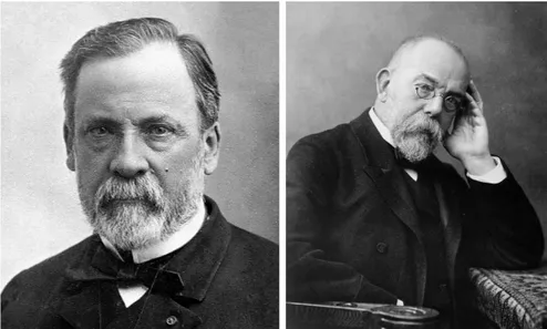
In the late XIXth century, scientists discovered that microbes can be the cause of diseases. The establishment of this “germ theory of diseases”1, opposed to the “spontaneous generation theory” took an important turn in 1857 with the work of the French chemist Louis Pasteur. Pasteur discovered that, while yeasts were respon-sible for alcohol production during fermentation, wine and beer could be turned sour by another type microorganism (Pasteur 1857). Pasteur was convinced that these living mi-croorganisms were the cause of liquid putrefaction, and not arising from putrefaction as taught by the “spontaneous generation theory”. He also had the intuition that the same reasoning could apply to human, animal and plant diseases. Using a now famous experi-mental design (see Figure 1), he demonstrated in 1861 that the microbes responsible for liquid putrefaction were present in the air and that they could be killed by heating the liquid (Pasteur 1861). These findings stroke a fatal blow to the spontaneous generation
Figure 1 – Copy of Pasteur’s drawings of “swan-necked” flasks. These flasks were made by Pasteur to refute the spontaneous generation theory. Pasteur boiled some broth into these flasks to sterilize it. The swan neck allowed air exchanges between the outside and the inside of the flask, but prevented the entrance of dust and airborne microorganisms which deposited by gravity in the lower section of the swan neck. Using this design, Pasteur showed that the broth remains intact until the swan neck is broken. Then, airborne microorganisms can enter the flask and colonize the broth. This experiment demonstrated that life cannot arise by itself inside the broth.
theory, and participated to the initiation of reflections about the role of microorganisms in diseases. Later on, Pasteur conducted some work on silkworms, showing that p´ebrine disease, causing at that time disasters in the french silk industry was in fact due to a microorganism that could transmit from diseased to uninfected silkworms.
Figure 2 – Louis Pasteur (left) and Robert Koch (right) Photography by Paul Nadar
and Wilhelm Fechner. Public domain, via Wikimedia Commons https://commons.wikimedia.org/w/ index.php?curid=422990 https://commons.wikimedia.org/wiki/File:Robert_Koch.jpg
In the meanwhile, the German physician Robert Koch worked on the cause of anthrax in cattle and sheep. In 1876, he isolated a bacteria from infected animals, and established for the first time the link between a specific microorganism and a specific disease (Koch 1876). Later on, Koch discovered the microbes that cause tuberculosis and cholera (Koch 1882). Now deeply convinced that some diseases were caused by microbes, he defined in 1882 four rules that he suggested to be strictly followed to demonstrate that a mi-croorganism was the cause of a disease: (1) The organism must be present in every case of the disease. (2) The organism must be isolated from a host with the corresponding disease and grown in pure culture. (3) Samples of the organism removed from the pure culture must cause the corresponding disease when inoculated into a healthy, susceptible laboratory animal. (4) The organism must be isolated from the inoculated animal and identified as being identical to the original organisms isolated from the initial, diseased host (Lerner et al. 2006b; Lerner et al. 2006a).
The Pasteur and Koch heritage, together with the work of many other scientist from the XIXth century, led to a dogma, associating one microorganism to one disease. It allowed a huge jump in the understanding of infectious diseases, their epidemiology, as well as medical care and prevention.
2 Heterogeneity in infections and consequences from
the pathogen point of view
The “germ theory of diseases” and Pasteur’s and Koch’s work basically described in-fections as interactions between one host and a single pathogen. This classical model of single-species infection was criticised in the early years of the XXth century (Evans 1976). Scientists realised that infections could also be the consequence of several (sometimes many) different microorganisms and that some diseases could not be understood without taking into account complex multi-species interactions. I will use the term “heterogeneous infections” to designate these complex situations where a community of microbes all to-gether contribute to the exploitation of a single host. Heterogeneous infections can arise in several distinct ecological situations discussed below, and in all cases, pathogens that are embarked in these communities may interact with many different microbes. Their pathogenicity, their transmission, and many of the traits that make them pathogens are most probably affected by these interactions. This is the phenomenon this thesis aims at surveying.
The understanding of heterogeneity in infections can be critical from a clinical point of view, as it can increase or moderate the detrimental effect of pathogens on their hosts. However, since the focus of my work is the pathogen itself rather than the host, I will
try to describe in the following lines the several forms of heterogeneity in infections and their consequences from the pathogen point of view. I will
mostly focus on what happens during the course of one infection. The term “pathogen populations” will thus refer to the populations of pathogens inside one host, during an infection, and not the population of pathogen at the ecosystem level.
2.1 Heterogeneity in the biotic environment of the pathogen, or
inter-specific heterogeneity
Multiple infections: several pathogenic species or strains can infect a given host
The most intuitive way by which infections can be heterogeneous is when a host is infected by several pathogenic species at the same time, producing some inter-specific heterogeneity. A nice example of such multiple infections has been described in the field vole, Microtus agrestis. Telfer et al. (2010) showed that at least four different pathogens, including virus, protozoan and bacteria, can co-infect a host and interact with each other. For instance, they demonstrate that individuals previously infected by the bacterium
Anaplasma phagocytophilum are more susceptible to infections by the protozoan Babesia microti. On the other hand, individuals that are previously infected by the bacterium
Bartonella spp. are less susceptible to Babesia microti infections. This mixture of
facilita-tion and protecfacilita-tion effects most probably result from direct or indirect (e.g. mediated by host immune defenses) interactions between pathogens inside the host body. The ability of a given pathogenic species to be transmitted cannot be understood here without taking into account the presence or absence of other pathogenic species inside the host.
Multiple infections may also involve different strains of a single pathogen species, rather than different species as in the previous example. In this case, the coexistence of closely related pathogens (strains or isolates), probably sharing the same ecological niche, implies potentially strong competitive interactions. This is documented, for in-stance, for the agent of the rodent malaria, Plasmodium chabaudi, where a single host can be infected by several parasite strains at the same time. These strains, with different levels of virulence, have been shown to compete with each other inside their host, as they all exploit the same resources: red blood cells (Bell et al. 2006).
Multiple infections can have several evolutionary consequences for pathogens. The most commonly assumed is that it should select for higher virulence in pathogen species or strains (Alizon et al. 2013). In the example ofPlasmodium chabaudi cited above, strains that were able to more efficiently exploit red blood cells (i.e. with a higher virulence), reached higher densities when co-infecting a host with a less virulent strain (Bell et al. 2006). A positive selective pressure should thus act on these more virulent strains, while less virulent ones are counter selected as they are less efficient during multiple in-fections. The selective pressure that result from competitive interactions between pathogens are predicted to lead to the evolution of various strategies, such as niche di-vergence or production of interference molecules to prevent competition (see Mideo 2009, for a review).
Interactions with the host microbiota
Pathogens may not only encounter other pathogenic species during host infection but also a potentially diverse microbial community. Indeed, each single host individual shel-ters a huge amount of microorganisms, essentially non pathogenic microbes, called the microbiota. Pathogens that infect a host therefore do not colonize an empty environ-ment, and their interactions with the host microbiota need to be taken into account to understand infectious processes and the evolution of pathogens (Beliz´ario et al. 2015; Stecher et al. 2008).
The most complex and studied part of microbiota is that of the digestive tract of the hosts. Consequently, most studies of interactions between a pathogen and the microbiota of its host concern pathogens that pass through their host gut during their life cycle. For example, the human enteric pathogen Salmonella enterica serovar Typhimurium (S. Tm) needs to outcompete its host microbiota in order to invade the digestive tract. In the lab, mice with their native microbiota are resistant to colonization byS. Tm. Contrarily, mice
that have a reduced intestinal flora are susceptible to colonization. Using a metagenome analysis, Brugiroux et al. (2016) identified the functions fulfilled by the members of the mouse native microbiota that were essential for protection against S. Tm colonization. Doing so, they were able to reconstruct a minimal bacterial community that provides protection once established in germ-free mice. Here, the protective effect of the gut microbiota against pathogens arises from a wide range of functions fulfilled by multiple microorganisms.
In some cases however, specific members of the host gut microbiota can have a direct negative effect on pathogens through the production of deleterious molecules. It is the case for example forEnterococcus mundtii, a bacteria found in the gut of the lepidopteran
Spodoptera littoralis. The symbiont E. mundtii secretes an antimicrobial peptide into
its host gut lumen. This peptide selectively kills opportunistic pathogens such as E.
faecalis, but does not affect the resident gut flora (Shao et al. 2017). The specificity
of this antimicrobial molecule toward a pathogen could suggests some strong selective pressure from pathogens on members of the host microbiota.
On the other hand, the host microbiota can have a beneficial impact on pathogens. For instance, the virulent potential of the entomopathogenic bacteria B. thuringiensis have been highly debated (see for example Raymond et al. 2010). B. thuringiensis produces a toxin that, when ingested by an insect, is responsible for intestinal wall perforation and subsequent insect death. The actual cause of host death have been thought to be the gut paralysis and associated feeding cessation that are observed in insects after toxin ingestion. However, a recent study showed that when insects do not control the gut microbiota (RNAi-mediated silencing of an immune gene), the lesions induced by B.
thuringiensis allow the passage of bacteria through the intestinal barrier associated with
a significant enhancement of host larvae mortality (Caccia et al. 2016). This septicemia enhanced by the host microbiota is thought to be in part the cause of insect death. Here, the host microbiota may have influenced the evolution ofB. thuringiensis virulence factors that can be costly, and potentially useless in this case.
2.2 Heterogeneity in the pathogen population during the course
of infection, or intra-specific heterogeneity
Within-host evolution
Heterogeneity can arise within the pathogen population itself, transforming a clonal infection into an assemblage of several pathogenic sub-populations (see Didelot et al. 2016, for a review). The simplest mechanism that cause this intra-specific heterogeneity is the accumulation of mutations while pathogens multiply in their host. These mutations, can allow adaptive evolution of pathogens during infection. An example of such host adaptation have been shown in Burkholderia dolosa, which can opportunistically infect
humans suffering from lung fibrosis. Lieberman et al. (2011) showed thatB. dolosa strains acquire within their host, and after the initial infection event, mutations in genes involved in resistance to antibiotics, which are usually administrated to patients suffering from lung fibrosis. They moreover found evidences of positive selection acting on genes that may undergo oxygen-dependent regulation, which could be crucial for the adaptation of free-living bacteria to the cystic fibrosis lung where oxygen availability is reduced. These findings suggest pathogens adaptation to a new ecological niche during infection. In some cases, within-host adaptation can be accelerated by a global increase of mutation rate, through the loss of functionality in DNA repair systems. For example, in some strains of B. dolosa, mutations into genes involved in DNA repair are associated to an excess of mutation, in the case of infection of a host with lung fibrosis (Lieberman et al. 2014).
Within host evolution can also be accelerated by horizontal gene transfer events, which allows exchanges of complete genes and functions between bacteria. For example, Stecher et al. (2012) showed that infection by S. enterica serovar Typhimurium promotes some bloom of Escherichia coli in the host gut, and that these blooms are as-sociated with an increase of horizontal gene transfers via cell conjugation between S. Tm and E. coli. Here, infection can potentially promote the spread of virulence or antibiotic-resistance factors from pathogens to commensal bacteria. In any case, mutations and horizontal gene transfers initially bring some genetic diversity in the pathogen popula-tion, but the persistence of heterogeneity on the long run will depend on selection.
Phenotypic heterogeneity and immune evasion
While mutation events produce random variations at the scale of the whole genome, some mechanisms produce variation on specific genomic regions. One of them, quite com-mon in bacteria, is phase variation. Phase variation is defined as a genetic or epigenetic mechanism resulting in a high rate of reversible phenotypic variation during the course of infection (van der Woude et al. 2004). One example of such phase variation is described in
Borrelia burgdorferi, the agent of Lyme disease. The bacterium possesses two plasmids
re-sponsible of gene conversion events on genes encoding for antigenic motifs. These conversion events, associated to duplicated genes transpositions produce a large number of distinct antigenic motifs, probably through modifications of surface proteins (Embers et al. 2004; Labandeira-Rey et al. 2003). This specific case of phase variation, called antigenic variation, allows the pathogen to escape its host immune system by producing a large diversity of antigenic motifs.
This escape of immune system through phase variation can also be mediated by epige-netic mechanisms. In Salmonella enterica, the modification of the O-antigen subunits of lipopolysaccharides present at the cell surface is involved in intestinal persistence in the mouse model. Indeed, modifying this O-antigen by the addition of some sugars allows to
escape the immune system. These additions of sugars are mediated by the gtr operon, which expression can be switched ON and OFF by DNA methylation (Garcia-Pastor et al. 2018).
Phenotypic heterogeneity, division of labour and bet-hedging
In many bacterial species, phase variation does not allow immune evasion, but rather produces phenotypic forms which lack key pathogenic functions (van der Woude et al. 2004; van der Woude 2011; van der Woude 2006). The fact that a sub-population of the pathogen which cannot accomplish the infection by their own are generated during the course of an infection can seem paradoxical but is well understood in the human pathogen Salmonella enterica serovar Typhimurium. This bacterium infects the digestive tract and breaches the gut wall of its host. It then colonizes lymphoid tissues, establishes into phagocytotic cells that carry them to the spleen and other systemic tissues. During its life cycle, S. Typhimurium exhibits heterogeneity in the expression of some genes (as described above). These differences in expression are stochastic (i.e. not triggered by environmental cues) and produce phenotypic heterogeneity.
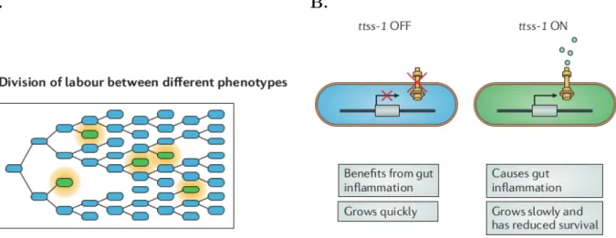
A. B.
Figure 3 – Phenotypic heterogeneity can lead to the division of labour in clonal
groups. This function of phenotypic heterogeneity manifests in infections with Salmonella
enterica subsp. enterica serovar Typhimurium, used as an example here. A. Phenotypic
het-erogeneity can lead to interactions and the division of labour within clonal populations. For a genotype that expresses two different phenotypes (blue and green cells), individuals that express the green phenotype do not continue to grow but do produce a resource (orange) that promotes the growth of the blue phenotype. B. S. Typhimurium exhibits phenotypic heterogeneity in the expression of the virulence locus type three secretion system 1 (ttss-1 ), which encodes a multi-protein secretion apparatus. This leads to a division of labour between ttss-1 OFF and ttss-1 ON subpopulations. The ttss-1 ON subpopulation invades host tissue and causes inflammation but suffers a reduction in growth and survival. The ttss-1 OFF subpopulation benefits from the inflammation and proliferates. Figure and legend adapted from from Ackermann et. al (2015)
In the gut of the host, this heterogeneity consists mainly in the existence of two sub-populations: a slow-growing subpopulation of S. Typhimurium that expresses a
viru-lence gene (ttss-1 ON) coexists with a fast-growing subpopulation that is phenotypically avirulent (ttss-1 OFF). Cells with virulence functions induce inflammation mechanisms, which contributes to kill commensal bacteria in the digestive tract of the host (Kaiser et al. 2012). The avirulent form of S. Typhimurium, which do not pay the cost of ex-pressing virulence factors, multiply without suffering from competitive interactions with the commensal bacteria, thanks to the virulence factors produced by the avirulent form (Figure 3a). Phenotypic heterogeneity allows a form division of labours where two subpopulations of the pathogen fulfill different functions (Figure 3b). The functional spe-cialization of these subpopulations provides a competitive advantage to S. Typhimurium against other bacteria inside the host.
A. B.
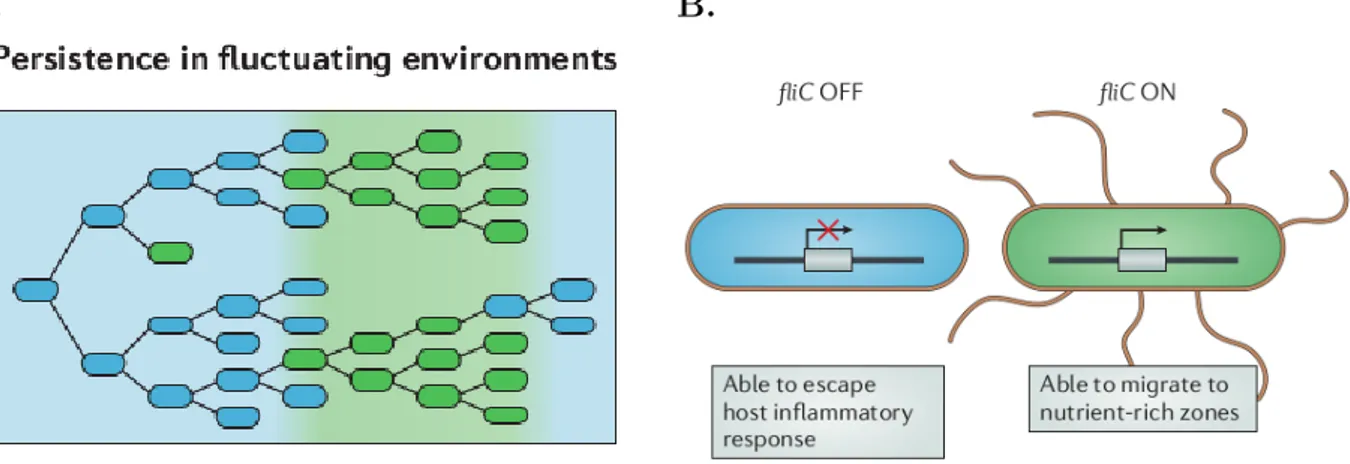
Figure 4 – Phenotypic heterogeneity can promote persistence in fluctuating
envi-ronments. These function of phenotypic heterogeneity manifests in infections with Salmonella
enterica subsp. enterica serovar Typhimurium, used as an example here. A. A genotype that
expresses two different phenotypes (blue and green cells) can persist in an environment that fluc-tuates between two states (light blue and light green) in which only individuals that express the matching phenotype can survive. B. S. Typhimurium exhibits phenotypic heterogeneity in the expression of the flagellin gene fliC, and this allows this organism to persist in an environment that fluctuates between favouring flagellation (fliC ON) and selecting against it (fliC OFF). Here, fliC regulation allows to escape caspase-1 inflammatory response that is only effective in some of the host compartments Figure and legend adapted from Ackermann et. al (2015)
A second form of heterogeneity is described in S. Typhimurium after it has crossed the intestinal barrier of its host. It switches ON or OFF the expression of the flagellin gene fliC with a low probability and independently from environmental cues (Ackermann 2015; Stewart et al. 2011). This results in mixed population of flagellate and unflagellate cells. Flagellate cells are able to migrate toward nutrient sources but are suppressed by the host immune system in some compartments. Thus they only perform well in the tissues where these defenses are down. On the contrary, unflagellate cells can resist the host defenses but cannot migrate to reach nutrient sources (Figure 4B). Unflagellate cells are advantageous when the pathogen reach specific compartments. S. Typhimurium thus need to cope with a fluctuating and unpredictable environment as it migrates through different tissues of the host. The ON or OFF switching of the expression of the flagellin
gene fliC with a low probability and independently from environmental cues (Ackermann 2015; Stewart et al. 2011) can thus be interpreted as a bet-hedging strategy. This strategy consists in simultaneously producing different phenotypic forms, each adapted to a different type of environment. Doing so increases the chance that one of the phenotypic forms performs well in the current environment (Figure 4A).
2.3 The specific case of vector-borne diseases
In the above lines, we saw that contrarily to what was assumed by Pasteur and Koch, infections are often interactions between a host and a complex assemblage of organisms inside this host, potentially including non pathogenic microorganisms, several species and strains of pathogens, and several phenotypic forms of a given pathogenic strain. To make the picture even more complex, we must pay attention to the particular case of vector-borne diseases. These pathogens interact with another protagonist, the vector, which brings another source of heterogeneity in infections. Here again, I will not focus on differences that can exist between several vectors individuals, but rather discuss the implication of microbial community heterogeneity in the specific case of vector-borne diseases.
From the pathogen point of view, vectors can usually be seen as obligatory intermediate hosts. Consequently, all forms of heterogeneity described above can potentially apply in the vector environment. To my knowledge, few studies have investigated the role of phenotypic heterogeneity and within-vector evolution in vector-borne diseases. However, an increasing number of studies concern the role of vector microbiota in the transmission of human diseases. It has been shown for instance, that mosquito microbiota influences vector competence to the transmission of the human parasite Plasmodium falciparum. In particular, members of the mosquito microbiota could reduce the vector colonisation by P. falciparum through competition for resources, immune priming, and production of secondary metabolites (Cirimotich et al. 2011; Weiss et al. 2011; Dennison et al. 2014). On the other hand, some bacterial taxa from the vector microbiota could have a positive effect on P. falciparum colonization, as showed for Enterobacteriaceae in Boissi`ere et al. (2012)
The same kind of observations hold for the agent of Lyme disease,Borrelia burgdorferi and its tick vector, Ixodes scapularis. Narasimhan et al. (2014) showed that the alter-ation of the tick gut microbiota altered in return its intestinal peritrophic matrix. This peritrophic matrix being essential for B. burgdorferi colonisation, dysbiosed ticks are less colonized by the pathogen. Here again, the vector microbiota plays a crucial role for pathogen transmission.
In the two examples cited above, members of the vector microbiota can probably be transmitted together with the pathogen. They can potentially play a role in the infectious
process, and interact with the pathogen. Although studies on the role of vector microbiota on pathogen transmission are increasingly conducted, very few have focused on role of vector microbiota in the infectious process within the host.
3 Xenorhabdus-Steinernema complexes: combination
of multiple heterogeneity levels
3.1 Entomopathogenic nematodes-bacteria pairs
Xenorhabdus and Photorhabdus, two closely related bacterial genera, belonging to
the Enterobacteriaceae family (γ-Proteobacteria), are mutualistically associated with ne-matodes from Heterorhabditis and Steinernema genera, respectively. These nematodes, which have a very similar life cycle (see section 3.2), are found in soil where they infect a wide variety of insect larvae (Peters 1996). Due to their large distribution around the world and their potential interest in pest control, entomopathogenic nematodes, and their bacterial symbiont have been the object of many studies.
This work is focused on the Xenorhabdus bacterial genus, which includes several species, associated with several Steinernema nematode species. A tight co-evolution be-tweenXenorhabdus and Steinernema species is supported by the fact that the
Xenorhabdus-Steinernema symbiosis is very specific: nematodes of a given species only ”allow” one
species ofXenorhabdus to colonize their gut. Some close relative species from the
Xenorhab-dus genus can be artificially re-associated to non-native Steinernema species, but the
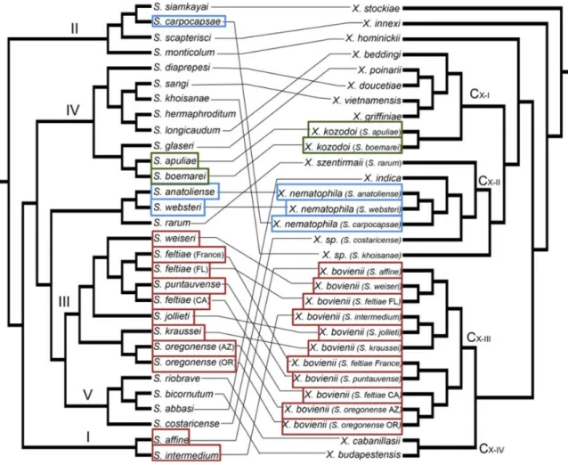
ben-efit (see section 3.2 for details) they bring to the nematode is usually much lower than that brought by the native symbiont, and they cannot be transmitted by the nematode descendants (Sicard et al. 2004b; Chapuis et al. 2009). This specificity is permitted by a complex molecular dialogue between the two parts of the symbiosis which as been well described in X. nematophila-S.carpocapsae (Heungens et al. 2002; Goodrich-Blair 2007; Chaston et al. 2013). Surprisingly, this specificity of association does not seem to result from co-speciation events, as suggested by the low relatedness of the two taxonomic struc-tures of Xenorhabdus genera and their host nematodes (Figure 5 Boemare (2002) and Lee et al. (2010)).
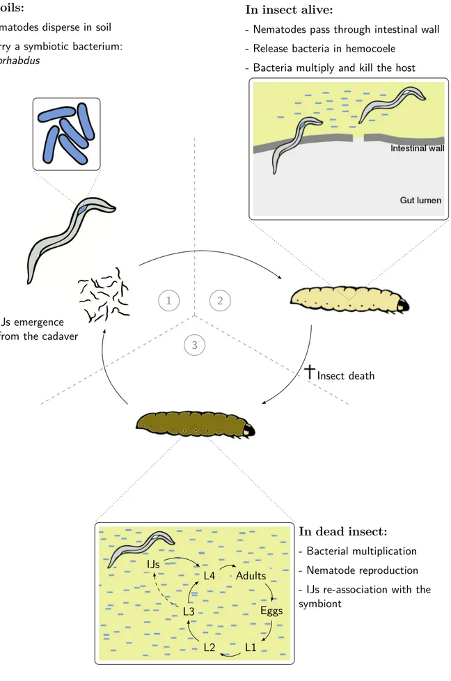
3.2 Xenorhabdus-Steinernema life cycle
Only differentiated L3 larval stages of nematodes disperse in the soil. These infective juveniles (IJs) are protected by both L3 and L2 cuticles, the later obstructing their mouth and anus which prevent them from feeding, but allow them to persist inside the soil (Sicard et al. 2004a). These IJs are colonized by an almost clonal population of Xenorhabdus in an intestinal receptacle (Martens et al. 2003). The IJs disperse in soil until they find a
Figure 5 – Multi-locus cophylogeny of nematode species from the Steinernema genus, and their symbiont from the Xenorhabdus genus. Coloured squares represent different strains of the same Xenorhabdus species. Each Steinernema species are linked to their native symbiont strain. Nematode phylogeny is based on one nuclear, 28S rRNA, and two mitochondrial genes, COI and 12S rRNA. Bacteria phylogeny is based on the 16S ribosomal gene, and two housekeeping genes,serC and recA. Figure from Lee et al. (2010)
larval insect host to invade (Figure 6.1). They usually penetrate the insect larvae through natural openings like mouth and anus. Within the larvae of the host insect (e.g. Galleria
mellonella or Spodoptera sp. in lab conditions), nematodes perforate the intestinal wall
and releases their Xenorhabdus symbionts into the insect hemolymph (Figure 6.2) (Sicard et al. 2004a; Snyder et al. 2007). The bacteria grow to high densities into the extracellular matrix of the host tissues (Sicard et al. 2004a), reduce the host immune response through haemolysin production, which targets the host hemocytes (Vigneux et al. 2007), inhibi-tion of antimicrobial peptides producinhibi-tion (Park et al. 2007) and phenol-oxidase activity (Crawford et al. 2012). The insect host dies of septicemia in approximately 24h (Dowds et al. 2002; Richards et al. 2009). Xenorhabdus then produces enzymes which degrade insect tissues to provide nutrients for the nematodes (Caldas et al. 2002; Chen et al. 1996). Nematodes perform several cycles of reproduction within the insect cadaver (Figure 6.3).
When nematode number becomes high and nutrients are limiting, Steinernema juveniles re-associate with Xenorhabdus and differentiate into non-feeding IJs. The IJs finally dis-perse into the soil in the search of a new host (Figure 6.1). For simplicity purposes, IJs of Steinernema will be designate as Xenorhabdus vector, although they also have a role to play in killing the host.
3.3 Xenorhabdus nematophila, a model species
Among these bacteria associated with entomopathogenic nematodes,Xenorhabdus
ne-matophila have been extensively studied because of its particularly high virulence toward
many model insects (McMullen et al. 2017; Ogier et al. 2014; Kim et al. 2017). More-over, Steinernema carpocapsae, its nematode vector, can be experimentally deprived of its symbiont (Sicard et al. 2003). These aposymbiotic nematodes are of great interest to perform re-associations with other strains or species of symbionts, and investigate evolu-tionary questions. For example, the interaction betweenX. nematophila and its vector S.
carpocapsae can be considered as mutualistic because, first, the bacteria cannot survive
more than a few days in the soil unless they are carried by a nematode (Morgan et al. 1997) and, second, symbiotic S. carpocapsae (i.e colonized by X. nematophila) have a much higher reproductive rate than aposymbiotic nematodes (Sicard et al. 2003). This later observation might be attributed to (1) a more effective inhibition of the insect im-mune system, (2) a better nutrition of the nematode in the insect when associated with
X. nematophila (Richards et al. 2009) and (3) a protection against ’enemies’, by
pro-ducing antimicrobial molecules that could eliminate microbial competitors encountered in soils or in insect cadavers (Singh et al. 2015), or dissuading insects from feeding on its insect-host cadaver (Zhou et al. 2002). On the other hand, some studies have shown that the survival rate of the IJs decreases when they are associated to X. nematophila (Emelianoff et al. 2007; Emelianoff et al. 2008a). It suggests that nematodes experience a survival-reproduction trade-off which is induced by the symbiosis, and which probably has shaped the co-evolution between the two partners (Chapuis et al. 2012).
The fact that we can produce aposymbiotic IJs of S. carpocapsae, that we can repro-duce the whole life cycle of S. carpocapsae-X. nematophila pairs in lab conditions, and that the whole genome of X. nematophila F1 strain has been sequenced (Lanois et al. 2013) makes X. nematophila a very convenient model species. Moreover, several aspects of its biology that I will describe further make it very interesting to study heterogeneity in infections.
1 In soils:
- Nematodes disperse in soil - Carry a symbiotic bacterium:
Xenorhabdus
2
In insect alive:
- Nematodes pass through intestinal wall - Release bacteria in hemocoele
- Bacteria multiply and kill the host
3
Insect death IJs emergence
from the cadaver
Adults Eggs L1 L2 L3 L4 IJs In dead insect: - Bacterial multiplication - Nematode reproduction - IJs re-association with the symbiont
Figure 6 – Life cycle of the Xenorhabdus bacterium carried by its nematode vector
Stein-ernema and infecting the insect host Galleria mellonella larvae usually used in laboratory
3.4 Heterogeneity in Xenorhabdus-Steinernema infections
Intra-specific heterogeneity
X. nematophila isolated from the symbiotic nematodes found in soil has the
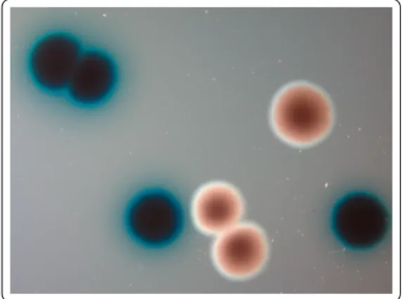
particular-ity to adsorb bromothymol blue. Therefore, it forms blue colonies when it grows on agar containing this dye (Figure 7). When these blue colony-forming cells, referred as group 1, are grown in in vitro culture, a second form of X. nematophila called group 2 may appear after several days of incubation (Figure 7). This group 2 form produces red colonies on the same culture medium (Akhurst 1980). Group 2 cells also present many phenotypic differ-ences with group 1 cells: they have a reduced antimicrobial (Akhurst 1980), proteolytic and lipolytic activity (Thaler et al. 1998), and a reduced production of flagellar filaments (Volgyi et al. 1998; Givaudan et al. 1996; Givaudan et al. 1995). Although group 2 are usually obtained inin vitro cultures, Akhurst (1980) reported that these phenotypic vari-ants ofX. nematophila could also arise during insect infection. These phenotypic variants have been known and studied for the past four decades, yet the molecular mechanisms responsible for the production of heterogeneity, as well as their implication during host infection and re-association with the vector, are not fully understood. I will try in the first chapter of this thesis to bring some answers to these questions.
Figure 7 – Group 1 (blue) and group 2 (red) colonies ofXenorhabdus nematophila obtained by plating a stationary phase culture on solid medium containing bromothymol blue
Another type of intra-specific heterogeneity that will not be further developed in this thesis but deserves to be mentioned, is the fact that several strains of X. nematophila can infect a host at the same time. Indeed, multiple nematodes should enter the in-sect, as Steinernema reproduction is sexual. To cope with this intra-specific competition within hosts, Xenorhabdus species produce some toxins that can kill unrelated strains of the same species (Hawlena et al. 2010a). In several species of Xenorhabdus, including
X. nematophila, these spiteful interactions between unrelated strains due to bacteriocin
production benefit kin, i.e. closely related cells, but reduce the combined virulence of com-peting strains (Vigneux et al. 2008; Bashey et al. 2012). Moreover, these anti-competitor
abilities have been shown to trade-off against nematode reproductive success (Bertoloni Meli et al. 2018). This trade-off, together with the fact that in nature, non-inhibiting isolates can be found in high frequency, suggest that there may be two different strategies for pathogen success: one competitive strategy, which would be favored in competition with sensitive strains, and one reproductive strategy, favored in a non-competitive con-text (Bertoloni Meli et al. 2018). This concon-text-dependant fitness supports the idea that intra-specific competition, associated with an heterogeneous environment, may maintain diversity in the pathogen population2, and highlights the role of intra-specific competition in mutualism evolution.
Inter-specific heterogeneity
Similarly, several species ofXenorhabdus can infect one host at the same time. Bashey et al. (2013) showed that these inter-specific interaction of Xenorhabdus can favor higher virulence, or stains that are able to produce bacteriocin as described above. However,
Xenorhabdus is also able to produce a large number of antibiotics that affect bacterial
competitors from other genus (Singh et al. 2008; Singh et al. 2015; Park et al. 2009). This suggests that interactions with other bacteria may be a strong seclective pressure for Xenorhabdus. Indeed, the bacteria have to multiply and stay for around ten days inside an insect cadaver, were multiple competitors can develop. These competitors can potentially originate from the soil, but also from the insect microbiota, and from the nematode microbiota. It have been commonly assumed that producing these antibiotics was a way for Xenorhabdus to dominate the bacterial community inside insect cadavers. In the second chapter of this thesis, I focused on the bacterial communities that can be found in insect cadavers.
2. Here I mean the population of pathogens in a given environment, opposed to the term “pathogen population” within one host
4 Objectives of the present thesis
Chapter 1: A role of phenotypic heterogeneity in Xenorhabdus
nematophila
infections?
In the first chapter of this manuscript, I investigated the potential role of the generation of phenotypic variants ofX. nematophila during infec-tion. The first part brings some answers about the genetic mechanisms that generate these phenotypic variants. I then investigated the potential adaptive value of these phenotypic variants during X. nematophila life cycle.Chapter 2: Microbial communities during X. nematophila
infec-tions
In the second chapter, I investigated the composition of microbial communities during X. nematophila infections. First, we developed an experimental procedure that allows to modify the gut microbiota of lab-reared insect in order to mimic soil-dwelling insects. We then used these insects colonized by a relevant set of microorganisms to de-scribe the microbial community composition inside insect cadavers at the time-point of the re-association between X. nematophila and its vector.Glossary
Adaptation: A trait that has been fixed by adaptive evolution, i.e. because it conferred greater fitness to its carrier.
Adaptive evolution: Changes of a trait frequency in a population due to natural selection (i.e. different from evolution due to genetic drift). We will use the phrase ”adaptive evolution” as an equivalent to Darwinian evolution.
Antigenic motifs: Any external structure of a cell which can be recognised by a host immune system.
Bet-hedging: Evolutionary strategy where different phenotypes are produced indepen-dently of environmental cues, which increases the chance that at least one phenotype will perform well in one state of a fluctuating environment.
Division of labours: Evolutionary strategy where individuals within a group specialize in certain tasks to benefit the whole group.
Ecological niche: The set of biotic and abiotic conditions that allows the persistence of a given species.
Fitness: The average number of gene copies transmitted to the next generation.
Gene conversion: Homogenisation of homologous regions from two DNA strands that initially contained different alleles. As a result, the recipient strand carry the same allele as the donor strand.
Germ theory of diseases: A XIXth century theory claiming that microbes can be the cause of some diseases, and not their consequence, as taught by the spontaneous generation theory.
Horizontal gene transfer: Exchange of genomic material between two bacterial cells from different strains/species.
Host: Here refers to any multi-cellular organism that can be colonized by microorgan-isms.
Metagenome: The set of all the genomes of the cells from a bacterial population. Microbiota: All microorganisms associated to a host at one time point.
Pathogen: Here refers to microorganisms (virus, bacteria, protozoans) which have a detrimental effect on a host.
Phase variation: A stochastic and reversible change of phenotype in bacteria, caused by a genetic or epigenetic mechanism.
Selective pressure: Any cause for an organism with a particular trait to have a fitness advantage (positive selective pressure) or disadvantage (negative selective pressure).
Spontaneous generation theory: Theory according to which living beings could arise from inanimate matter.
Symbiont: Microorganism living in close association (positive, negative or neutral) with a macroorganism.
Trade-off: Constrains (e.g. physical or physiological) that make it impossible to invest resources in two life-history traits at the same time. Trade-offs usually result in a negative relation between those traits. From an evolutionary perspective, they can lead to a compromise that consists in intermediate values of traits, or conversely to specialization, with the possibility that specialists coexist in populations.
1
A role of phenotypic heterogeneity in
Xenorhabdus nematophila
infections?
Cells ofXenorhabdus nematophila that are isolated from nematodes usually form blue colonies when plated on agar containing bromothymol blue. After a prolonged in vitro culture, or injection in insects of these blue colony-forming cells, some cells forming red colonies appear. Compared to blue cells (primary variants) red cells (secondary variants) lack motility, and have a reduced production of enzymes and antimicrobial compound (Boemare et al. 1988; Givaudan et al. 1995; Boemare et al. 1997; Volgyi et al. 1998; Forst et al. 2002). I will call group 1 and group 2 the cohort of variants whose phenotypes correspond to that of primary and secondary variants historically described.
Several terms have been used so far to name this production of different phenotypes: phase variation, phenotypic variation, phenotypic switching, phenotypic heterogeneity. Most often, different scientific communities use different words. For the present work, which is at the intersection of microbiology and evolutionary biology, I chose to use the term phenotypic switching to designate the production of different contrasted phe-notypes, called phenotypic variants, which results in phenotypic heterogeneity in a bacterial population, without any prior on the mechanisms involved.
In fact, inXenorhabdus, the mechanisms responsible for the production of phenotypic variants have not been fully understood, although some clues about the implication of the lrp gene have been showed. Lrp (leucine-responsive regulatory protein) is a global regulator of expression inX. nematophila, and lrp mutants exhibit a phenotype similar to that of group 2 phenotypic variants (Cowles et al. 2007). Moreover, a constant expression of lrp suppresses phenotypic switching (Hussa et al. 2015). Although lrp is pointed out in the production of phenotypic variants, the exact molecular mechanism remains to be elucidated.
Apart from the molecular mechanism that allows the emergence of these variants, their potential role in X. nematophila life cycle also have to be clarified. It is probably not a way for X. nematophila to escape the host immune system. Most of the genes whose expression differs between group 1 and 2 cells are indeed expressed long after host
death (Jubelin et al. 2013). Moreover, both group 1 and group 2 cells are able to kill their host when injected into the insect (Volgyi et al. 1998). The reduced activity of some genes in group 2 variants have been shown to be detrimental to the nematode, however group 1 and group 2 are both able to re-associate with nematodes and group 2 have been shown to be preferentially carried by nematodes when a mixture of the two groups was injected into an insect (Sicard et al. 2005; Richards et al. 2008). All these findings are somehow contradictory with the fact that nematodes freshly sampled from the wild have never been reported to carry group 2 variants. This paradox could come from the fact that some conditions favor the multiplication and transmission of group 1 variants, while other favor that of group 2 variants but are rarely encountered in nature. In any case, the interactions between group 2 variants and nematodes still need to be examined in details to better understand whether or not this phenotypic switching could be adaptive in the entomopathogenic symbiosis.
The aim of this work was to bring some answers to the following questions: Which
molecular mechanisms are responsible for the production of phenotypic het-erogeneity in X. nematophila populations? Are these mechanisms adaptive?
We constituted a collection of 34 variants which we then characterized, both phenotypi-cally and genetiphenotypi-cally. This was made collectively with teams from EDB lab in Toulouse, and DGIMI lab in Montpellier. My contribution to this work was to measure the viru-lence of the 34 variant when directly injected into insects, and test their adaptive nature
Prolonged culture and long lasting infections select for
poorly transmitted bacterial variants
Marine Cambon
∗†1, Nathalie Parthuisot
∗1, Sylvie Pag`es
2, Anne Lanois
2,
Alain Givaudan
2and Jean-Baptiste Ferdy
11
Evolution et Diversit´e Biologique - UMR 5174 CNRS-UPS Universit´e
´
Paul Sabatier - F-31062 Toulouse cedex 9
2
DGIMI - UMR1333 INRA -Universit´e Montpellier - F-34095 Montpellier
cedex 5
February 13, 2019
The authors declare that they have no competing financial interests in relation to the work described in this paper.
∗The first two authors equally contributed to this work †cambonmarine@gmail.com
Abstract
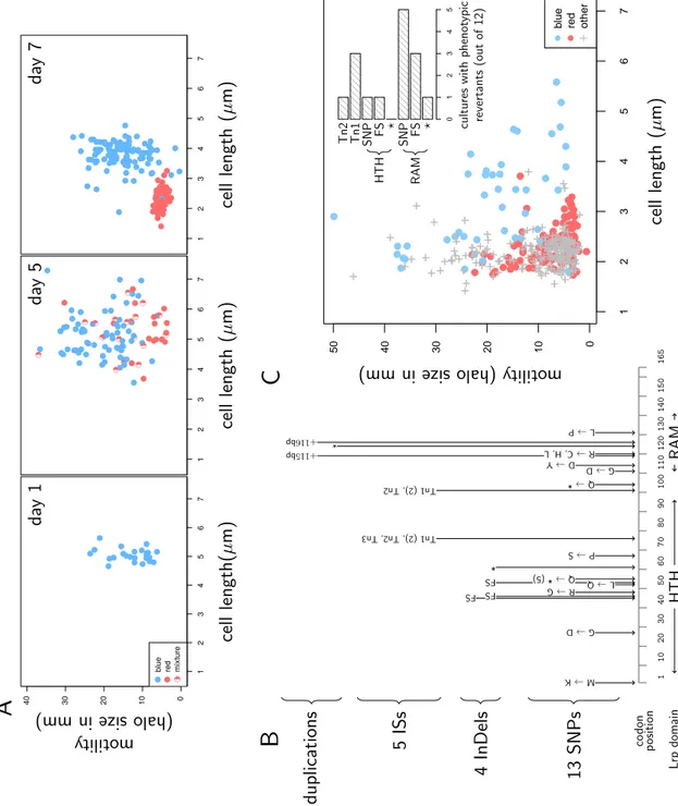
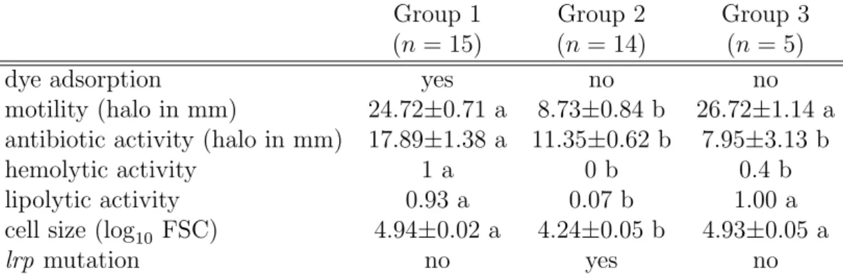
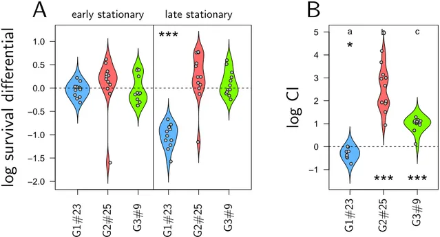
Bacterial infections are often composed of cells with distinct phenotypes, that can be produced by genetic or epigenetic mechanisms. Heterogeneity in the course of infection has proved to be important in many pathogens, because it can alter both pathogenicity and transmission. We studied a large cohort of phenotypic variants of the insect pathogen Xenorhabdus nematophila. This bacterium kills insects and multiplies in the cadaver before being transmitted by the soil nematode vector Steinernema carpocapsae. We found that, based on several phenotypes, variants are clustered in three groups, one of which corresponding to mutations in the gene encoding the major regulator Lrp. These Lrp defective mutants survive better and reach high loads during prolonged stationary phase, which probably explains why they increase in frequency during infections. The third group of variants is less frequent, bears no mutation in lrp but is also advantaged in late stationary phase. Both group 2 and group 3 variants thus have a Growth Advantage in Stationary Phase (GASP phenotype), but we found evidences that this advantage trades off against transmission by nematodes: the highest load a variant reaches in insects, the least it is transmitted by nematode vectors.
Introduction
Bacterial infections, even started from a single pathogenic strain, often end up being composed of cells with different phenotypes. In cases where groups of cells with distinct phenotype interact to better exploit their host (Diard et al. 2013), the molecular mechanisms that produce this diversity can be considered as adaptations, which ultimately increase pathogens transmission. To demonstrate this theory, it is necessary to understand both what these mechanisms are, and how they impact the success of the infection.
Mutation is probably the most obvious mechanism that produces diversity dur-ing infection. Its importance is increasdur-ingly admitted (Didelot et al. 2016), notably because cases are now accumulating where it fuels the evolution of pathogens in-side the host, in particular in human diseases (e.g. Markussen et al. 2014; Young et al. 2017). While mutation generally occurs throughout the whole genome, other mechanisms exist that impact a restricted set of genes. These can be epigenetic alterations, where clonal populations of bacteria modify their phenotype by chang-ing their regulatory state (van der Woude 2006; van der Woude 2011). These can also be genetic alterations, as in phase variation where a specific high rate mutation mechanism produces reversible changes in one or a few genes (van der Woude et al. 2004; Bayliss 2009). Phase variation is described as the basic mechanism that makes antigenic variation a successful instrument for some pathogens to escape their host immune system (e.g. Zhang et al. 1997).
adaptive nature of mechanisms that control phenotypic variation inside an infection. This pathogen indeed kills insects and proliferates in their cadaver for one or two weeks, before it is transmitted by the nematode vector Steinernema (Goodrich-Blair 2007). During this long period, Xenorhabdus maintains high densities inside the insect and, therefore, potentially accumulates phenotypic variation. As the complete life cycle of Xenorhabdus can be experimentally reproduced (Chapuis et al. 2012), it is possible to quantify how this variation impacts each of its different stages.
Xenorhabdus isolated from the wild typically are in a form described as pri-mary, but in culture media they convert to another, secondary, form when reaching long-term stationary-phase (Boemare et al. 1988). In its seminal paper, Akhurst (1980) showed that secondary forms of Xenorhabdus also appear during infection, and that this occurs at a rate that greatly varies among Xenorhabdus strains. Al-though the phenotypic differences between the two forms can also vary depending on strain and species, the primary but not secondary form cells are able to bind bromothymol blue dye, are motile, agglutinate red blood cells and produce fimbriae, haemolysins, proteases, antimicrobials and crystalline inclusion bodies (Boemare et al. 1988; Givaudan et al. 1995; Boemare et al. 1997; Volgyi et al. 1998; Forst et al. 2002). The alternation between primary and secondary forms has so far been in-terpreted as a case of phase variation (Boemare et al. 1988). As the phenotype of secondary forms matches that of lrp defective mutants (Cowles et al. 2007; Hussa et al. 2015; Cao et al. 2017; Casanova-Torres et al. 2017; Engel et al. 2017), it has also been proposed that the Lrp master regulator could control the production of secondary variants in X. nematophila (Cowles et al. 2007). It is not demonstrated, though, that lrp is systematically involved when this phenomenon occurs during Xenorhabdus infections. Interestingly, genes with differential expression between primary and secondary forms have also been shown to play a role in the interac-tion between X. nematophila, its nematode vector and its insect target (Richards et al. 2009), demonstrating that the production of secondary forms should impact X. nematophila interactions with their invertebrate hosts. However, despite reduced production of virulence factors in secondary forms, all forms of X. nematophila are capable to kill in insects (Volgyi et al. 1998). In addition, Sicard et al. (2004a) showed that both primary and secondary forms could be transmitted by the ne-matode S. carpocapsae. The goal of this paper is to better understand first, the molecular mechanisms that are responsible for the production of phenotypic vari-ants, and second, the impact of these variants on the transmission by the vector. To do so, we investigated a large collection of X. nematophila isolates with vari-ous phenotypic forms. We found that secondary forms are not phase variants but rather plain lrp mutants, and that at least a third phenotypic form exists in X. ne-matophila which is not a lrp mutant. All these variants have a Growth Advantage in Stationary Phase (GASP, Finkel 2006) which probably explains why they reach
higher loads than primary forms during late infection. We then quantified how these variants are transmitted by the nematode vector S. carpocapsae and found that isolates that reach the highest densities in insects are the least transmitted by nematodes. X. nematophila therefore seem to experience a trade-off between traits that are favored during late infection and traits that increase transmission.
Materials and Methods
Obtention of X. nematophila isolates
Xenorhabdus nematophila isolates were obtained from static cultures of the GFP-labelled strain F1D3 (Sicard et al. 2004a). Samples of ten independent LB cultures of F1D3 were streaked onto NBTA plates (Akhurst 1980) after 3, 7 and 13 days of incubation at 28 ◦C. Primary forms form blue colonies on NBTA while secondary variants form red colonies (Boemare et al. 1988; Givaudan et al. 1995; Boemare et al. 1997). Each time a red colony was observed, a blue colony from the same Petri dish was also sampled. Overall, we obtained 34 distinct isolates (see Supp. Mat. 1) which we stored in 20 % glycerol at -80◦C.
lrp sequencing
X. nematophila colonies were lysed in sterile miliQ water after several freezing-thawing cycles. lrp gene was amplified using the high-performance GoTaq G2 DNA Polymerase (Promega, France) with primers lrp-L (5’-CATATTGCGGATTTAGGG-ATTG-3’) and lrp-R (5’-GGGACTGCATAGGCAAGAATAC-3’). PCR products were sequenced by GenoScreen, France and mutations were determined by compar-ing aligned lrp sequences to that of the X. nematophila F1 reference genome (Lanois et al. 2013).
Measuring colony phenotypes
For each isolate, we measured four phenotypic traits that differ among Xenorhab-dus variants. Swimming motility was measured as the diameter of a halo formed by motile bacteria on 0.35 % agar culture medium (Givaudan et al. 1995; Boemare et al. 1997). Antibiotic activity was quantified by measuring the diameter of inhibition ha-los, using Micrococcus luteus as a target strain (Givaudan et al. 2000). Extracellular lipolytic activity was assessed by the presence of precipitated material surrounding the colony cultured on Tween 20 agar (Givaudan et al. 2000). Haemolytic activity was quantified by the presence of a clearing surrounding bacteria grown on standard sheep blood agar plates (Vigneux et al. 2007).