Pépite | Le rôle des vésicules extracellulaires dérivées des microglies dans les processus inflammatoires et tumoraux du SNC : étude in vitro
138
0
0
Texte intégral
(2) Thèse de Adriana-Natalia Murgoci, Université de Lille, 2018. University of Veterinary Medicine and Pharmacy in Košice (Slovakia) Université de Lille1 - Sciences et Technologies (France) École Doctorale Biologie-Santé de Lille PhD thesis G 292/18 In fulfilment of the requirements for the degree of Doctor in Science and Philosophy from the University of Veterinary Medicine and Pharmacy in Kosice and the Université de Lille1 - Science et Technologies Presented by. Adriana-Natalia Murgoci. The role of microglia derived extracellular vesicles in inflammatory and tumorous processes of the CNS: in vitro study International co-supervised PhD thesis under the direction of Prof. Michel Salzet, PhD. and Assoc. prof. MVDr. Dasa Cizkova, DrSc. Presented in Kosice, Slovakia, 26th September 2018 Committee members:. © 2018 Tous droits réservés.. Prof. MVDr. Jan Motlík, DrSc.. Reporter. Assoc. Prof. Stefania Mondello, PhD.. Reporter. Prof. MVDr. Juraj Koppel, DrSc.. Examiner. Prof. Isabelle Fournier, PhD.. Examiner. Prof. Serge Nataf, PhD.. Examiner. Assoc. Prof. Mangesh Bhide, PhD.. Examiner. Assoc. Prof. MVDr. Daša Čížková, DrSc.. Co-Director. Prof. Michel Salzet, PhD.. Co-Director. lilliad.univ-lille.fr.
(3) Thèse de Adriana-Natalia Murgoci, Université de Lille, 2018 The role of microglia derived extracellular vesicles in inflammatory and tumorous processes of the CNS: in vitro study – Adriana-Natalia Murgoci. Concluding work assignment Name and surname, student`s titles:. Adriana-Natalia Murgoci, Mgr.. Title of the concluding work:. The role of microglia derived extracellular vesicles in inflammatory and tumorous processes of the CNS: in vitro study. Type of the concluding work :. PhD thesis. Name, surname and titles of the supervisor: Name, surname tutor/consultant:. and. titles. of. the. Michel Salzet, Prof. PhD. Daša Čížková, MVDr. Assoc. Prof. DrSc. University Lille1, France. Place of the studies (Department/Institute):. PRISM Laboratory INSERM U1192, Lille, France University of Veterinary Medicine and Pharmacy in Košice, Slovakia Institute of Neuroimmunology, Slovak Academy of Sciences, Bratislava, Slovakia. Name, surname and titles of the head of the Michel Salzet, Prof., PhD. Department/Institute: Michal Novák, MVDr., Prof. DSc., Dr.h.c. Annotation of the concluding work:. Isolation, characterisation of microglia extracellular vesicles and analyses of their effect on glioma 3D spheroid model.. Language of the work:. English. Date of approval of the assignment:. 7.09.2015. ........................................... signature of the supervisor. © 2018 Tous droits réservés.. .......................................... signature of the supervisor. ........................................ signature of the student. lilliad.univ-lille.fr.
(4) Thèse de Adriana-Natalia Murgoci, Université de Lille, 2018 The role of microglia derived extracellular vesicles in inflammatory and tumorous processes of the CNS: in vitro study – Adriana-Natalia Murgoci. Zadanie záverečnej práce Meno a priezvisko, tituly študenta:. Adriana-Natalia Murgoci, Mgr.. Názov záverečnej práce:. The role of microglia derived extracellular vesicles in inflammatory and tumorous processes of the CNS: in vitro study. Typ záverečnej práce:. PhD thesis Michel Salzet, Prof., PhD.. Meno, priezvisko a tituly školiteľa: Meno, priezvisko školiteľa/konzultanta:. a tituly. Daša Čížková, MVDr. Assoc. Prof., DrSc. externého. University Lille1, France. Školiace pracovisko (katedra/ústav):. PRISM Laboratory INSERM U1192, Lille, France University of Veterinary Medicine and Pharmacy in Košice, Slovakia Institute of Neuroimmunology, Slovak Academy of Sciences, Bratislava, Slovakia. Meno, priezvisko pracoviska. a tituly. vedúceho Michel Salzet, Prof., PhD. Michal Novák, MVDr., Prof., DSc., Dr.h.c. Izolácia, charakterizácia mikrogliových. Anotácia záverečnej práce:. extracelulárnych vezikúl a analýza ich účinku na 3D sféroidný model gliómu.. Jazyk, v ktorom sa práca vypracuje:. Angličtina. Dátum schválenia zadania:. 7.09.2015. ........................................... podpis školiteľa záverečnej práce. © 2018 Tous droits réservés.. ......................................... podpis školiteľa záverečnej práce. ............................... podpis študenta. lilliad.univ-lille.fr.
(5) Thèse de Adriana-Natalia Murgoci, Université de Lille, 2018 The role of microglia derived extracellular vesicles in inflammatory and tumorous processes of the CNS: in vitro study – Adriana-Natalia Murgoci. " Învaţă de la ape să ai statornic drum,. Learn from the waters never to back down,. Învaţă de la flăcări că toate-s numai scrum,. Learn from the flames that everything’s just ashes,. Învață de la umbră să taci și să veghezi,. Learn from the shadow how to shut up and listen,. Învață de la stâncă cum neclintit să crezi.. Learn from the rock how to watch without blinking,. Învață de la soare cum trebuie s-apui,. Learn from the sun how you should talk,. Învață de la piatră cât trebuie să spui,. Learn from the rock how you should say it,. Învață de la vântul ce adie pe poteci. Learn from the wind which bows through the path,. Cum trebuie prin lume, de liniștit să treci.. How you, through life should quietly pass. Învață de la toate, căci toate sunt surori,. Learn from all, because all are connected. Cum treci frumos prin viață, cum poți frumos să. How you should go through life, how you should. mori. die. Învață de la vierme, că nimeni nu-i uitat. Learn from the worm that no one’s insignificant,. Învață de la nufăr să fii mereu curat.. Learn from the water lily, always to be clean,. Învață de la vultur, când umerii ţi-s grei. Learn from the vultures when you are overwhelmed,. Și du-te la furnică să vezi povara ei. And go to the ant and see her arduous work,. Învață de la greier, când singur ești să cânți,. Learn from the moon not to be afraid,. Învață de la lună să nu te înspăimânți.. Learn from the birds to always be on the go,. Învață de la păsări să fii mai mult în zbor,. Learn from everything, that everything passes,. Învață de la toate, că totu-i trecător,. Behold, the son of sacrifice, through the world you. Ia seamă, fiu al jertfei prin lumea-n care treci, Să înveți din tot ce piere, tu să trăiești în veci!”. pass through To learn from what dies, to live forever.. (from folklore lyrics). © 2018 Tous droits réservés.. lilliad.univ-lille.fr.
(6) Thèse de Adriana-Natalia Murgoci, Université de Lille, 2018 The role of microglia derived extracellular vesicles in inflammatory and tumorous processes of the CNS: in vitro study – Adriana-Natalia Murgoci. Acknowledgement To move the boundaries of knowledge, we need to work together, to ask good questions and to find the best answers. This thesis represents not only my work, it is a team work of people from two research laboratories. Therefore, I would like to thank all those who have made the realization and writing of this PhD thesis possible. First of all I want to thank to my supervisors, that together lead me through this exciting experience. “Ďakujem vám veľmi pekne” to Assoc. Prof. MVDr. Dasa Cizkova DrSc. my thesis supervisor in Slovakia, which welcomed me to this beautiful country, for her endless support, her guidance, enthusiasm and motivation. She showed me that being researcher do not mean to be a machine, that there is possible to have a nice career and also a personal life. “Merci beaucoup” to Prof. Michel Salzet, my thesis supervisor in France, who had trust in me from beginning and open me the opportunities that I had in the last five years in the world of Science. It is an honour and a professional challenge for me to start my research work under led of a world-renowned and respected scientist who has dedicated his entire life to Science. Thank to my thesis reporters Prof. MVDr. Jan Motlík, DrSc., Assoc. Prof. Stefania Mondello, PhD. and Prof. MUDr. Andrea Čalkovská, DrSc., for having kindly accepted to evaluate my work. Also thanks to my thesis examiners Prof. MVDr. Juraj Koppel, DrSc., Assoc. Prof. Mangesh Bhide, PhD., Prof. Isabelle Fournier, PhD. and Prof. Serge Nataf, PhD., for accepting to judge my work. Special thanks for Prof. MVDr. Michal Novák, DrSc., Dr.h.c., who allowed me to work at the Institute of Neuroimmunology, Slovak Academy of Sciences in Bratislava. Important help came from DVM. Veronika Cubínková, PhD. from the CDVP facility who was taking care of animals, important component during my researches. I thank to all my colleagues for PRISM laboratory, especially to Jusal Quanico, PhD., and from NiU laboratories, especially to MSc. Petra Majerová, PhD., that have helped me all the time on research work and outside the laboratories too. I would like to thank to my teachers and professors from University of Veterinary Medicine and Pharmacy in Kosice and University Lille 1 for allowing me to study, consult, and pass all exams required for PhD defence. Thanks to all my friends around the world, who showed me that distances are just numbers. We are the sum of all the interactions that we have with the people around us. Thank you all for support, ideas, motivation, criticism. Each of you has influenced me to become the person I am today. Last, but not least “mulțumesc” to my family. To my mom, my dad, my brother Cezar, for unconditioned support and motivation to be better every day. And I also thank Andrei for his patience and support to accomplish this work.. © 2018 Tous droits réservés.. lilliad.univ-lille.fr.
(7) Thèse de Adriana-Natalia Murgoci, Université de Lille, 2018 The role of microglia derived extracellular vesicles in inflammatory and tumorous processes of the CNS: in vitro study – Adriana-Natalia Murgoci. Abstract One of the most important discoveries of the past 30 years, is the detection of extracellular vesicles (EVs) released by cells leading to a new inter-cellular communication. Research teams are studying these vesicles in order to define their origin, molecular compositions and function. In general, they are considered as key players in cell interconnections. Exosomes are a subpopulation of EVs, with a very small diameter (about 100 nm), with endocytic origin. They carry a variety of bioactive molecules such as proteins, lipids, miRNAs and DNA. So far, it has been proven that all cell types analysed are able to release exosomes in physiological and pathological processes. Our interest was to study the involvement of microglial EVs, in particular exosomes, in tumor processes that develop within the central nervous system. Microglia are immune cells of the brain and exosomes of microglia may be important factors involved in the overall functions of these glioma cells. In our studies, we have described a reproducible and highly efficient method for yielding purified primary microglia cells, followed by EVs isolation and their characterization. The results show that there are no morphological differences between microglial cells from different tissue origins e.g. brain cortex and spinal cord of rat. On the other hand, using a large-scale proteomic platform, we demonstrate that microglial cells derived from the brain cortex and spinal cord express different phenotypes under both control and inflammatory conditions. This difference has been confirmed also at the level of the exosomes that microglia secrete. In vitro bioassays demonstrate that microglia-derived exosomes tested on 3D spheroids of rat gliomas were able to inhibit tumor invasion. These results made it possible to demonstrate that exosomes derived from microglia could be used as nano-therapeutic agents vis-à-vis gliomas. Gliomas are heterogeneous and highly invasive primary brain tumors, characterized by a high infiltration of microglial cells and macrophages. The immunosuppressive tumor environment is known to orient immune cells towards a pro-tumoral and anti-inflammatory phenotype, so the current challenge for cancer therapy is to find a way to reorient macrophages toward an anti-tumoral phenotype. Based on previews studies obtained in the laboratory, we have shown the major role of prohormone convertase PC1/3 in macrophages. Its inhibition makes it possible to produce anti-tumor factors and to activate the Th1 pathway when cells are stimulated by LPS. Since it is difficult to treat patients with LPS, we searched for a sterile ligand of TLR4 common to LPS and we used the antitumor drug Paclitaxel. Our studies have shown that Paclitaxel activates the TLR4-dependent MyD88 signaling pathway and mimics the action of LPS. Thus, in our study, we showed that EVs isolated from KD PC1/3 macrophages treated with this drug inhibited the growth of the rat glioma C6 line. We isolated the exosomes and our results highlight the potential of a therapeutic strategy combining Paclitaxel and PC1/3 inhibition to switch macrophages to an anti-tumor immunophenotype and to use of the exosomes produced by these cells as therapeutic agents.. © 2018 Tous droits réservés.. lilliad.univ-lille.fr.
(8) Thèse de Adriana-Natalia Murgoci, Université de Lille, 2018 The role of microglia derived extracellular vesicles in inflammatory and tumorous processes of the CNS: in vitro study – Adriana-Natalia Murgoci. Abstrakt Jedným z najdôležitejších objavov za posledných 30 rokov je detekcia extracelulárnych vezikúl (EVs) uvoľnených bunkami, ktoré slúžia ku medzibunkovej komunikácii. Výskumné tímy študujú tieto vezikuly, aby určili ich pôvod, zloženie a funkcie. Vo všeobecnosti sa považujú za kľúčových hráčov pre medzibunkové interakcie. Exozómy predstavujú subpopuláciu EVs s veľmi malým priemerom (približne 100 nm), endocytového pôvodu. Obsahujú rôzne bioaktívne molekuly ako proteíny, lipidy, miRNA a DNA. Bolo dokázané, že všetky bunkové typy sú schopné uvoľňovať exozómy počas fyziologických a patologických podmienkach. Našim cieľom bolo preskúmať úlohu extracelulárnych vezikúl vylúčených z mikroglií, najmä exozómov, v nádorových procesoch centrálneho nervového systému. Mikroglia predstavuje imunitné bunky v mozgu a ich exozómy môžu ovplyvniť funkciu gliómových buniek. V našich štúdiách sme využili reprodukovateľnú a vysoko účinnú metódu na získanie purifikovaných primárnych mikrogliových buniek s následnou izoláciou exozómov a ich charakterizáciou. Výsledky naznačujú, že medzi mikrogliou mozgu a miechy nie sú morfologické rozdiely. Na druhej strane, použitím rozsiahlej proteomickej platformy sme zistili, že mikroglie z mozgovej kôry a miechy sa líšia a predstavujú rôzne fenotypy v kontrolných aj zápalových podmienkach. Tento rozdiel bol potvrdený aj na úrovni exozómov, ktoré mikroglia vylučuje. Biologické testy in vitro potvrdili, že exozómy vylúčené mikrogliou, testované na 3D sféroidoch (C6 gliómov) boli schopné inhibovať inváziu nádorov. Tieto výsledky naznačili možné použitie exozómov z mikroglií pre nano-terapeutickú liečbu gliómu. Malígne gliómy sú heterogénne, vysoko invazívne primárne nádory mozgu, charakterizované vysokou infiltráciou mikrogliálnych buniek a makrofágov. Je známe, že imunosupresívne nádorové prostredie orientuje imunitné bunky na pro-tumorálny a protizápalový fenotyp. Súčasnou výzvou pre terapiu nádorových ochorení je nájsť spôsob, ako preorientovať makrofágy na protinádorový fenotyp. V našich štúdiách sme potvrdili hlavnú úlohu prohormónu konvertázy PC1/3 v makrofágoch. Jeho inhibícia umožňuje tvoriť protinádorové faktory a aktivovať dráhu Th1 (LPS stimulácia, TLR4). Použili sme protinádorový liek Paclitaxel, ktorý aktivuje signalizačnú dráhu MyD88 závislú od TLR4 a napodobňuje účinok LPS. Potvrdili sme, že EV izolované z KD PC1/3 makrofágov (knock down PC1/3), ktoré boli ovplyvnené Paclitaxelom inhibovali rast línie C6 potkanieho gliómu. Tieto výsledky naznačujú na účinnosť kombinovanej terapie, t.j inhibície PCl/3 a Paclitaxelu, a prepnutie na makrofágy s protinádorovým imunofenotypom a produkciou exozómov s protinádorovým účinkom.. © 2018 Tous droits réservés.. lilliad.univ-lille.fr.
(9) Thèse de Adriana-Natalia Murgoci, Université de Lille, 2018 The role of microglia derived extracellular vesicles in inflammatory and tumorous processes of the CNS: in vitro study – Adriana-Natalia Murgoci. Abstract L'une des découvertes les plus importantes de ces 30 dernières années est la détection de vésicules extracellulaires (EVs) libérées par les cellules comme moyen de communication intercellulaire. Les chercheurs et médecins commencent à s'intéresser de plus en plus à ces vésicules afin de définir leurs origines, leurs compositions moléculaires et leurs fonctions. En général, elles sont considérées comme des acteurs clés dans les connexions cellulaires. Les exosomes sont une souspopulation des EVs, de très petit diamètre (environ 100 nM), d'origine endocytaire et qui portent une variété de molécules bioactives telles que les protéines, les lipides, les miARN et l'ADN. Jusqu'à présent, il a été prouvé que tous les types cellulaires analysées sont capables de libérer des exosomes dans les processus physiologiques et pathologiques. Notre intérêt était d'étudier l'implication des EVs issues des microglies, en particulier les exosomes, et ceux-ci dans les processus tumoraux qui se développent place au sein système nerveux central. Les microglies sont les cellules immunitaires du cerveau et les exosomes des microglies pourraient être des facteurs importants impliqués dans l’ensemble les fonctions de ces cellules. Au cours de nos études, nous avons décrit une méthode reproductible et très efficace pour isoler à partir de cellules microgliales issues de cultures primaires leurs EVs et les caractériser. Les résultats montrent qu'il n'y a pas de différences morphologiques entre les cellules microgliales issues d'origines tissulaires différentes e.g. cortex et moelle épinière. D'autre part, en utilisant une plateforme de protéomique à grande échelle, nous démontrons que les cellules microgliales dérivées du cortex et de la moelle épinière des rats expriment des phénotypes différents tant dans les conditions physiologiques normales ou inflammatoires. Cette différence a été confirmée également au niveau des exosomes qu’elles sécrètent. Des essais biologiques in vitro démontrent que les exosomes dérivées de microglies testées sur des sphéroïdes 3D de gliomes de rat étaient capables d'inhiber l'invasion tumorale. Ces résultats ont permis de mettre en évidence que des exosomes dérivées de la microglie pouvaient être utilisés comme agents nano thérapeutiques vis-à-vis des gliomes. Les gliomes sont des tumeurs cérébrales primaires hétérogènes et hautement invasives, caractérisées par une forte infiltration de cellules microgliales et de macrophages. L'environnement tumoral immunosuppresseur est connu pour orienter les cellules immunitaires vers un phénotype pro-tumoral et anti-inflammatoire, de sorte que le défi actuel pour la thérapie contre les gliomes est de trouver un moyen de réorienter les macrophages vers un phénotype anti-tumoral. Sur la base des études précédentes conduites au laboratoire, on a montré le rôle majeur de prohormone convertase PC1/3 au niveau des macrophages. Son inhibition permet de produire des facteurs anti-tumoraux et d’activer la voie Th1 quand ceux-ci sont stimulés par du LPS. Étant donné, qu’il est difficile de traiter des patients avec du LPS, nous avons recherché un ligand stérile du TLR4 commun au LPS et nous nous sommes focalisés sur le médicament antitumoral Paclitaxel. Nos études ont permis de mettre en évidence que le Paclitaxel active la voie de signalisation MyD88 dépendant du TLR4 et imite l'action du LPS. Ainsi, dans notre étude, nous avons montré que les EVs isolées à partir de cellules KD PC1 / 3 traitées avec ce médicament inhibaient la croissance de la lignée C6 de gliome de rat. Nous avons isolé les exosomes et nos résultats mettent en valeur le potentiel d'une stratégie thérapeutique combinant Paclitaxel et inhibition de PC1/3 et utilisation des exosomes produits par ces cellules comme agents thérapeutiques.. © 2018 Tous droits réservés.. lilliad.univ-lille.fr.
(10) Thèse de Adriana-Natalia Murgoci, Université de Lille, 2018 The role of microglia derived extracellular vesicles in inflammatory and tumorous processes of the CNS: in vitro study – Adriana-Natalia Murgoci. Publications. Publications 1. Murgoci, A.-N., Cizkova, D., Majerova, P., Petrovova, E., Medvecky, L., Fournier, I., and Salzet, M. Brain Cortex Microglia Derived Exosomes: Nanoparticles for Glioma Therapy. ChemPhysChem, 19(10), 1205–1214. https://doi.org/10.1002/cphc.201701198 (2018). 2. Murgoci A.-N., Mallah K., Aboulouard S., Lefebvre C., Kobeissy F., Fournier I., Cizek M., Cizkova D., Salzet M., The origin of microglia determines the content of exosomes and biological function (submitted, 2018). 3. Murgoci A.-N., Aboulouard S., Cizkova D., Salzet M., Trypsin/lysine C digestion step in EVs isolation: application to glioma and macrophages exosomes (submitted, 2018). 4. Duhamel, M., Rose, M., Rodet, F., Murgoci, A.-N., Zografidou, L., RégnierVigouroux, A., Vanden Abeele, F., Kobeissy, F., Nataf, S., Pays, L., et al. Paclitaxel treatment and PC1/3 knockdown in macrophages is a promising anti-glioma strategy as revealed by proteomics and cytotoxicity studies. Mol. Cell. Proteomics MCP, mcp.RA117.000443, https://doi.org/10.1074/mcp.RA117.000443 (2018). 5. Cizkova, D., Quanico, J., Kanoub, M.-A., Zahiri, F., Rodet, F., Murgoci, A.-N., Cubinkova, V., Fournier, I., and Salzet, M. Shedding New Light on Spinal Cord Injury via a Spatio- Temporal Proteomic and Physiological Approaches. Ann. Trauma Acute Care Volume 2, Article 1007, (2018). 6. Duhamel, M., Rodet, F., Murgoci, A.-N., Desjardins, R., Gagnon, H., Wisztorski, M., Fournier, I., Day, R., and Salzet, M. The proprotein convertase PC1/3 regulates TLR9 trafficking and the associated signaling pathways. Sci. Rep. 6, 19360, (2016). 7. Duhamel, M., Rodet, F., Murgoci, A.-N., Wisztorski, M., Day, R., Fournier, I., and Salzet, M. Proprotein convertase 1/3 inhibited macrophages: A novel therapeutic based on drone macrophages. EuPA Open Proteomics 11, 20–22, (2016). https://orcid.org/0000-0002-4492-6804. Book chapters 1. Cizkova, D., Murgoci, A.-N., Kresakova, L., Vdoviakova, K., Cizek, M., Smolek, T., Cubinkova, V., Quanico, J., Fournier, I., and Salzet, M. Understanding Molecular Pathology Along Injured Spinal Cord Axis: Moving Frontiers toward Effective Neuroprotection and Regeneration. In Essentials of Spinal Cord Injury, IntechOpen: IntechOpen, http://dx.doi.org/10.5772/intechopen.72118 (2017).. © 2018 Tous droits réservés.. lilliad.univ-lille.fr.
(11) Thèse de Adriana-Natalia Murgoci, Université de Lille, 2018 The role of microglia derived extracellular vesicles in inflammatory and tumorous processes of the CNS: in vitro study – Adriana-Natalia Murgoci. Oral communications 1. Murgoci A.-N., Salzet M., Cizkova D., The content and biological functions of microglia exosomes are determined by origin of cells that release them, Advances in Slovak Experimental Neuroimmunology 2018, 09-11.05.2018, Smolenice, Slovakia. 2. Murgoci A.-N., Medvecky L., Cubínková V., Gimeno J.-P., Cizkova D., Salzet M., New direction in CNS research: microglia derived exosomes, Euron PhD Days, 2526.10.2017, Maastricht, Holland. 3. Murgoci A.-N., Medvecky L., Gimeno J.-P., Majerová P., Salzet M., Cizkova D., Proteomic and morphological features of rat microglia derived exosomes, The 11th Central and Eastern European Proteomic Conference, 27-29.09.2017, Kosice, Slovakia. 4. Murgoci A.-N., Medvecky L., Gimeno J.-P., Majerová P., Salzet M., Cizkova D., Characterization of exosomes derived from cortical and spinal cord primary microglia, Current Status of Experimental Neuro-Immunology in Slovakia, 2426.05.2017, Smolenice, Slovakia. 5. Murgoci A.-N., Medvecky L., Cizkova D., Salzet M., Characterization of exosomes purified from primary cortical and spinal cord rat microglia, Euron PhD days, 1314.10.2016, Lille, France. 6. Murgoci A.-N., Cizkova D., Salzet M., Characterization of exosomes purified from primary cortical and spinal cord rat microglia, Club Jeunes SFEAP, 10-12.10.2016, Chambery, France. 7. Murgoci A.-N., Medvecky L., Salzet M. and Cizkova D., Identification of exosomes purified from primary cortical and spinal cord rat microglia, Club Jeunes SFEAP, 25-27.05.2016, Lille, France.. Posters 1. Murgoci A.-N., Cizkova D., Majerová P., Cubínková V., Petrovova E., Danko J., Isabelle Fournier I., Salzet M., New direction in CNS therapy: Cortex Microglia Derived Exosomes, 11th FENS Forum of Neuroscience, 07-11.07.2018, Berlin, Germany. 2. Murgoci A.-N., Salzet M., Cizkova D., Microglia cells localization determine the content of released exosomes and their biological function, 13th EFIS-EJI Tatra Immunology Conference, 09-13.06.2018, Strbské Pleso, Slovakia. 3. Murgoci A.-N., Medvecky L., Gimeno J.-P., Cizkova D., Salzet M., Characterisation of extracellular vesicles released by cells involved in glioblastoma microenvironment, 1st meeting of the French Society of Extracellular Vesicles, 0607.11.2017, Paris, France.. © 2018 Tous droits réservés.. lilliad.univ-lille.fr.
(12) Thèse de Adriana-Natalia Murgoci, Université de Lille, 2018 The role of microglia derived extracellular vesicles in inflammatory and tumorous processes of the CNS: in vitro study – Adriana-Natalia Murgoci. 4. Murgoci A.-N., Medvecky L., Gimeno J.-P., Majerová P., Petrovova E., Salzet M., Cizkova D., Isolation of primary cortical and spinal cord rat microglia followed by characterization of microglia derived exosomes, FENS Regional Meeting, 2023.09.2017, Pecs, Hungary. 5. Murgoci A.-N., Medvecky L., Gimeno J.-P., Majerová P., Cizkova D., Salzet M., Microglia derived exosomes: morphology and cargo analyses, Journée André Verbert, 14.09.2017 Lille, France (Award for Best Poster Presentation). 6. Murgoci A.-N., Duhamel M., Rodet F., Desjardins R., Gagnon H., Wisztorski M., Fournier I., Day R., Cizkova D., Salzet M., The role of proprotein convertase PC1/3 in regulation of TLR9 trafficking and signalling pathways in macrophage and microglia cells, 10th FENS Forum of Neuroscience, 02-06.07.2016, Copenhagen, Denmark. 7. Murgoci A.-N., Medvecky L., Salzet M. and Cizkova D., Identification of exosomes purified from primary cortical and spinal cord rat microglia, Extracellular vesicles: friends and foes, 07-09.06.2016, Rehovot, Israel. 8. Murgoci A.-N., Duhamel M., Rodet F., Desjardins R., Gagnon H., Wisztorski M., Fournier I., Day R., Salzet M., The proprotein convertase PC1/3 regulates TLR9 trafficking and the associated signaling pathways in macrophage and microglia cells, Réunion Annuelle du Club Français de NeuroImmunologie, 06.11.2015, Paris, France. 9. Murgoci A.-N., Duhamel M., Rodet F., Desjardins R., Gagnon H., Wisztorski M., Fournier I., Day R., Salzet M., The proprotein convertase PC1/3 regulates TLR9 trafficking and the associated signaling pathways in macrophage and microglia cells, Euron PhD days, 05.10.2015, Maastricht, Holland.. © 2018 Tous droits réservés.. lilliad.univ-lille.fr.
(13) Thèse de Adriana-Natalia Murgoci, Université de Lille, 2018 The role of microglia derived extracellular vesicles in inflammatory and tumorous processes of the CNS: in vitro study – Adriana-Natalia Murgoci. Table of content. 1. INTRODUCTION ..................................................................................................... 18 1.1 CENTRAL NERVOUS SYSTEM ..................................................................................... 18 1.2 TUMOR OF CENTRAL NERVOUS SYSTEM: GLIOBLASTOMA ......................................... 19 1.3 IMMUNE CELLS INVOLVED IN TUMOR ENVIRONMENT ................................................ 22 1.4 EXTRACELLULAR VESICLES ...................................................................................... 27 1.4.1. Exosomes ......................................................................................................... 28. 2. AIM AND OBJECTIVES ......................................................................................... 40. 3. MATERIALS AND METHODS .............................................................................. 42 3.1 CHEMICALS AND EQUIPMENT .................................................................................... 42 3.2 ANIMALS .................................................................................................................. 42 3.3 CELLS AND CULTURE CONDITIONS ............................................................................ 43 3.3.1. Primary microglia cells .................................................................................... 43. 3.3.2. C6 glioma cell .................................................................................................. 44. 3.3.3. Dorsal root ganglia........................................................................................... 45. 3.4 IMMUNOCYTOCHEMISTRY OF MICROGLIA CELLS ....................................................... 45 3.5 EXOSOMES ANALYSES............................................................................................... 46 3.5.1. Exosomes purification protocol ....................................................................... 46. 3.5.2. Exosomes PKH-labelling ................................................................................. 46. 3.5.3. Scanning electron microscopy (SEM) ............................................................. 47. 3.5.4. Nanoparticle tracking analysis (NTA) ............................................................. 47. 3.6 FUNCTIONAL ANALYSES ........................................................................................... 48 3.6.1. Spheroid generation and embedding in collagen matrix ................................. 48. 3.6.2. Co-culture of microglia exosomes with C6 spheroids ..................................... 48. 3.6.3. Co-culture of microglia exosomes with DRGs ................................................ 49. 3.7 MICROGLIA CELLS: PROTEIN ISOLATION AND IDENTIFICATION.................................. 49. © 2018 Tous droits réservés.. lilliad.univ-lille.fr.
(14) Thèse de Adriana-Natalia Murgoci, Université de Lille, 2018 The role of microglia derived extracellular vesicles in inflammatory and tumorous processes of the CNS: in vitro study – Adriana-Natalia Murgoci. 3.7.1. FASP protocol.................................................................................................. 49. 3.7.2. MS data acquisition ......................................................................................... 50. 3.7.3. Data processing ................................................................................................ 50. 3.8 MICROGLIA DERIVED EXOSOMES: PROTEIN ISOLATION AND IDENTIFICATION ........... 51. 4. 3.8.1. Mass spectrometry analysis ............................................................................. 51. 3.8.2. LC MS/MS analysis ......................................................................................... 51. 3.8.3. Data analyses ................................................................................................... 51. RESULTS ................................................................................................................... 53 4.1 PRIMARY RAT MICROGLIA ISOLATION ....................................................................... 53 4.1.1. Efficacity of primary rat microglia isolation ................................................... 54. 4.1.2. No morphological differences between cortex and spinal cord microglia ...... 55. 4.2 EXOSOMES CHARACTERISATION ............................................................................... 56 4.2.1. Microglia exosomes isolation .......................................................................... 56. 4.2.2. Microglia exosomes labelling with PKH67 ..................................................... 56. 4.2.3. Exosomes characterisation using scan electron microscopy ........................... 57. 4.2.4. Nanoparticle Tracking Analysis for microglia exosomes................................ 58. 4.3 PROTEOMIC ANALYSES ............................................................................................. 59 4.3.1. Proteomic studies for cortex and spinal cord microglia cells .......................... 59. 4.3.2. Proteomic analyses of exosomes released by microglia cells.......................... 64. 4.4 BIOLOGICAL ACTIVITIES OF MICROGLIA EXOSOMES .................................................. 68 4.4.1. Microglia exosomes effect on glioma cells ..................................................... 68. 4.4.2. Microglia exosomes effect on DRG ................................................................ 70. 4.5 ENZYMATIC DIGESTION STEP USED DURING EXOSOMES ISOLATION ........................... 73. © 2018 Tous droits réservés.. 4.5.1. Rat C6 Glioma Quick Step exosomes purification .......................................... 73. 4.5.2. Protein analyses of glioma C6 released exosomes .......................................... 74. 4.5.3. Biological effect of exosomes treated with trypsin ......................................... 76. lilliad.univ-lille.fr.
(15) Thèse de Adriana-Natalia Murgoci, Université de Lille, 2018 The role of microglia derived extracellular vesicles in inflammatory and tumorous processes of the CNS: in vitro study – Adriana-Natalia Murgoci. 4.6 EXOSOMES AS THERAPY FOR GLIOBLASTOMA ........................................................... 77 4.6.1. Extracellular vesicles released by PC1/3 KD macrophages after Paclitaxel. treatment repress more efficiently the growth and invasion of C6 spheroids ............. 78 5. DISCUSSION ............................................................................................................. 79. 6. CONCLUSIONS ........................................................................................................ 84. 7. RESUMÉ: ................................................................................................................... 86 7.1 ÚVOD ........................................................................................................................ 86 7.2 CIEĽ PRÁCE ............................................................................................................... 87 7.3 MATERIÁL A METÓDY ............................................................................................... 88 7.3.1. Primárne kultúry mikroglie .............................................................................. 88. 7.3.2. Proces purifikácie a charakterizácie exozómov ............................................... 88. 7.3.3. Funkčné analýzy .............................................................................................. 89. 7.3.4. Proteomické analýzy ........................................................................................ 89. 7.4 VÝSLEDKY ................................................................................................................ 90 7.4.1. Izolácia primárnej mikroglie a LPS stimulácia ................................................ 90. 7.4.2. Izolácia a charakterizácia exozómov ............................................................... 90. 7.4.3. Proteomické analýzy ........................................................................................ 91. 7.4.4. Biologická aktivita exozómov izolovaných z mikroglie ................................. 93. 7.4.5. Izolácia exozómov pomocou trypsínu (trypsín/ lyzín C)................................. 94. 7.5 DISKUSIA .................................................................................................................. 95 7.6 ZÁVER ...................................................................................................................... 99 8. REFERENCES......................................................................................................... 101. ANNEX ............................................................................................................................. 110. © 2018 Tous droits réservés.. lilliad.univ-lille.fr.
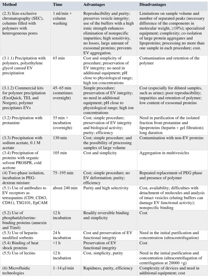
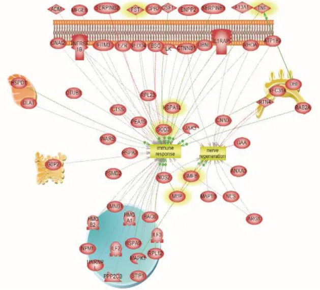
(16) Thèse de Adriana-Natalia Murgoci, Université de Lille, 2018 The role of microglia derived extracellular vesicles in inflammatory and tumorous processes of the CNS: in vitro study – Adriana-Natalia Murgoci. List of tables and figures. Table 1, Various subpopulation of extracellular vesicles .................................................... 28 Table 2, Human proteins often identified during experiments ............................................ 31 Table 3, Comparison of different methods currently used for EVs isolation ...................... 34 Table 4, LFQ value of the identified proteins involved in immune response or neurogenesis from microglia issued from two different territories (cortex or spinal cord)....................... 63. Figure 1, Schematic overview of central nervous system ................................................... 18 Figure 2, Distribution of malignant primary brain tumors .................................................. 19 Figure 3, Glioblastoma multiforme overview...................................................................... 20 Figure 4, Annual glioblastoma cases by regions ................................................................. 21 Figure 5, Microglia and macrophages have distinct cellular origins ................................... 23 Figure 6, Microglial cells morphology during activation process ....................................... 24 Figure 7, Macrophages origin and phenotype paradigm ..................................................... 25 Figure 8, Glioblastoma tumor microenvironment ............................................................... 26 Figure 9, Schematic representation of the major populations of extracellular vesicles ...... 27 Figure 10, Schematic presentation of exosomes biogenesis ................................................ 29 Figure 11, Exosomes cargo .................................................................................................. 30 Figure 12, Technics used for exosomes characterisation .................................................... 38 Figure 13, Overview of methods approaches for isolation and characterisation of EVs..... 39 Figure 14, Glioblastoma environment ................................................................................. 40 Figure 15, Protocol scheme of experiments......................................................................... 44 Figure 16, Protocol scheme of exosomes isolation.............................................................. 46 Figure 17, Microglia cells and NSCs in culture................................................................... 53 Figure 18, Double immunocytochemistry for cortex microglia cells .................................. 54 Figure 19, Double immunocytochemistry for rat neural stem cells .................................... 55 Figure 20, Morphological aspects of microglia cells ........................................................... 55 Figure 21, Fluorescent microscope analyses of exosomes .................................................. 57 Figure 22, SEM characterisation of microglia exosomes .................................................... 58 Figure 23, Nanoparticle tracking analysis size distribution profiles ................................... 59 Figure 24, Venn diagram of the unique protein of microglia cells ...................................... 60. © 2018 Tous droits réservés.. lilliad.univ-lille.fr.
(17) Thèse de Adriana-Natalia Murgoci, Université de Lille, 2018 The role of microglia derived extracellular vesicles in inflammatory and tumorous processes of the CNS: in vitro study – Adriana-Natalia Murgoci. Figure 25, Pathway analyses of specific proteins isolated from microglia cells treated with LPS....................................................................................................................................... 61 Figure 26, Heat map of the common proteins identified for microglia cells ....................... 62 Figure 27, Venn diagram of proteins of microglia exosomes.............................................. 64 Figure 28, Pathway analyses of proteins from exosomes of microglia ............................... 65 Figure 29. Pathway analyses of protein from exosomes of cortex and spinal cord microglia treated with LPS................................................................................................................... 66 Figure 30, Heatmap from shotgun proteomic analysis of microglia exosomes................... 67 Figure 31, Microglia exosomes effect under glioma spheroids ........................................... 69 Figure 32, The effect of microglia exosomes and conditioned medium on DRG ............... 71 Figure 33, DRG neurites length growth after treatment with microglia exosomes ............. 72 Figure 34, Experiment overview for exosomes treated with trypsin ................................... 73 Figure 35, Heatmap from shotgun proteomic analysis of glioma exosomes ....................... 74 Figure 36, Protein composition of glioma exosome ............................................................ 75 Figure 37, Treated macrophage exosomes effect on glioma spheroids ............................... 76 Figure 38, Macrophages exosomes effect on growth and invasion of C6 spheroids........... 78 Figure 39, General schema for biological functions of exosomes released by microglia isolated from two sources, cortex and spinal cord ............................................................... 83. © 2018 Tous droits réservés.. lilliad.univ-lille.fr.
(18) Thèse de Adriana-Natalia Murgoci, Université de Lille, 2018 The role of microglia derived extracellular vesicles in inflammatory and tumorous processes of the CNS: in vitro study – Adriana-Natalia Murgoci. Abbreviations ABs BBB BSA CAT cDNA CM CNS CSF CT DCs DLS EM ESCRT FBS GBM GSCs LPS miRNA MRI mRNA MS MVB MVs ncRNA NK NTA ssDNA TAMs Th. Apoptotic bodies Blood-brain barrier Bovine serum albumin Computed axial tomography Complementary deoxyribonucleic acid Conditioned media Central nervous system cerebrospinal fluid Computed tomography Dendritic cells Dynamic Light Scattering Electron microscopy Endosomal sorting complex required for transport Fetal bovine serum Glioblastoma multiforme Glioma stem cells Bacterial lipopolysaccharides Micro ribonucleic acid Magnetic resonance imaging Messenger ribonucleic acid Mass spectrometry Multivesicular bodies Microvesicles Non-coding ribonucleic acid Natural killers Nanoparticle tracking analysis single-stranded deoxyribonucleic acid Tumor-associated macrophages T helper cells. -17© 2018 Tous droits réservés.. lilliad.univ-lille.fr.
(19) Thèse de Adriana-Natalia Murgoci, Université de Lille, 2018 The role of microglia derived extracellular vesicles in inflammatory and tumorous processes of the CNS: in vitro study – Adriana-Natalia Murgoci. 1. Introduction. 1.1 Central nervous system Central nervous system (CNS) has evolved over the last 600 million years, being the most complex living system known. Mammalian CNS (Figure 1) is composed of the brain and spinal cord (Nowinski, 2011). The main function of the brain is to control body functions, interprets information received from receptors and project the essence of the mind and soul. Intelligence, creativity, emotion, and memory are also controlled by the brain. Protected within the skull, it is composed of the cerebrum, cerebellum, and brainstem (Rughani and Penar, 2015). Figure 1, Schematic overview of central nervous system. Source: Adriana-Natalia Murgoci (a) CNS components: brain and spinal cord. (b) CNS cells environment: neurons, astrocytes, oligodendrocytes, ependymal cells and microglia cells. (Figure was created by using Servier Medical Art).. Central nervous system presents a complex cellular structure represented by neurons and glial cells: astrocytes, oligodendrocytes, ependymal cells and microglia cells (Figure 1b). Neurons, the effector cells, interact with other cells to support their metabolic requests and respond to environmental stimuli. They communicate with each other to transmit signals through synapses. At the same time, they interact with astrocytes that sense and respond to neuronal activity and participate to the re-uptake of neurotransmitters (Basso and Bonetto, -18© 2018 Tous droits réservés.. lilliad.univ-lille.fr.
(20) Thèse de Adriana-Natalia Murgoci, Université de Lille, 2018 The role of microglia derived extracellular vesicles in inflammatory and tumorous processes of the CNS: in vitro study – Adriana-Natalia Murgoci. 2016). Astrocytes also form end-foot processes, which constitute the blood-brain barrier, and regulate nutrients delivery based on neuronal activity. Through the capillaries, astrocytes sense external stimuli and can participate to the inflammatory response upon activation of microglia (Verkhratsky et al., 2010). Oligodendrocytes are involved in the repair and regeneration of the CNS following a lesion, by secreting the myelin sheath around the axons. Microglial cells, on the other hand, are the resident immune cells in this CNS (Gallo and Deneen, 2014; Luo et al., 2010).. 1.2 Tumor of central nervous system: glioblastoma Glioblastoma multiforme (GBM), or glioblastoma, is one of the most common type of brain tumor in both children and adults, which can arise in the brain “de novo,” or evolve from lower-grade astrocytomas or oligodendrogliomas. This is the most invasive type of glial tumors, rapidly growing and commonly spreading into nearby brain tissue, often referred to as a grade IV astrocytoma (Ostrom et al., 2014). It occurs most often in the cerebral hemispheres, especially in the frontal (23.6%, and temporal lobes 17.4%) of the brain (Figure 2). Figure 2, Distribution of malignant primary brain tumors. Source: a) Zhang et al., 2013; b) Statistical Report of 2009-2013 by CBTRUS (a) Magnetic resonance image (MRI) of a 44‑ year‑ old female with grade IV glioblastoma. MRI image shows a mass (red circle) infiltrating through the left frontal lobe into the cortex, with mass effect on the ventricles. (b) Distribution* of all primary brain and other CNS tumors. *Percentages may not add up to 100% due to rounding.. -19© 2018 Tous droits réservés.. lilliad.univ-lille.fr.
(21) Thèse de Adriana-Natalia Murgoci, Université de Lille, 2018 The role of microglia derived extracellular vesicles in inflammatory and tumorous processes of the CNS: in vitro study – Adriana-Natalia Murgoci. According to World Health Organization, GBM is a devastating brain cancer that typically results in death in the first 15 months after diagnosis. Malignant gliomas are primary brain tumors, heterogeneous and highly invasive (Figure 3a). The most commonly occurring types of brain and other CNS tumors are: meningioma (36.6%), pituitary tumors (15.9%), gliomas (14.9%) and nerve sheath tumors (8.2%) (Figure 3b). The 32% of primary brain and other CNS tumors are malignant and 68% are non-malignant (Ostrom et al., 2016). Figure 3, Glioblastoma multiforme overview. Source: a) Wikimedia.org Credit: Sbrandner. b) Statistical Report of 2009-2013 by CBTRUS (a) Transversal section through a human brain showing a large tumor (red circle) in one of the temporal lobes. (b) Distribution of malignant primary brain and other CNS tumors by CBTRUS Histology Groupings and Histology.. The survival rate is very low, once diagnosed, most of the patients die within one year and only 5% survive more than 5 years, despite the aggressive therapies tested. Over the world, the incidence rate of brain cancers and central nervous system tumors is worldwide about 5.47 cases per 100,000 population (Ostrom et al., 2016). Every year there are diagnosed around 8,000 to 10,000 cases of glioblastoma multiforme. It has a high incidence all over the world and in Europe, there are 27 000 new cases of malignant astrocytic tumors diagnosed per year. Incidence is significantly higher in males than in females, the adjusted rates being 5.4 and 3.6, respectively. Frequency increases with age, from 0.9 in children to 12.1 in the elderly (aged 65+ years). However, the distribution of GBM incidence is different throughout the continent (Figure 4), with the highest rates in the North of Europe, UK and Ireland (5.7) and the lowest in the South and East of Europe (3.9 and 3.7, respectively) (Gatta et al., 2016).. -20© 2018 Tous droits réservés.. lilliad.univ-lille.fr.
(22) Thèse de Adriana-Natalia Murgoci, Université de Lille, 2018 The role of microglia derived extracellular vesicles in inflammatory and tumorous processes of the CNS: in vitro study – Adriana-Natalia Murgoci. Figure 4, Annual glioblastoma cases by regions. Incidence per 100000 persons EUROPE AVERAGE. 3,45. UK (FEMALE). 2,7. UK (MALE). 4,05. SPAIN. 3,12. FRANCE. 2,38. CROATIA. 4,8. AUSTRIA. 3,64. US. 3,05 0. 1. 2. 3. 4. 5. 6. Source: Brain and Other Nervous System Tumors. National Cancer Institute SEER Database For Europe and US, the average incidence of GBM is similar approximately 3.5 cases per 100 000 persons, with the highest rate in UK and Ireland (5.1 cases per 100.000) and the lowest (3.1) in eastern Europe.. Till this moment there is not clearly stablished what causes GBM, but there are more risk factors that may increase a person's chance of developing glioblastoma such as: lifestyle habits, environmental and occupational exposures to toxic agents, genetic factors and infections. Brain tumors are associated with several familial cancer predisposition syndromes, such as: Li-Fraumeni syndrome, neurofibromatosis, tuberous sclerosis and Turcot's syndrome (Ostrom et al., 2014; Reilly, 2009). Early diagnostic of brain tumor is difficult because there are non-specific symptoms for this pathology. Most of patient complain about headaches, nausea, vomiting, and seizures. Symptoms are variable and depend on the size and location of the tumor. For example, a patient with a temporal lobe tumor could experience hearing and vision problems, while a patient with a frontal lobe tumor could experience personality changes (Porter, 2012). Glioma diagnosis involves a medical history and physical examination, followed by a neurological exam. To confirm GBM it is demanded a magnetic resonance imaging (MRI) or/and computed tomography (CT or CAT scan), which use computers to create detailed images of the brain. Also, it is recommended to perform a biopsy of tissues suspected to be malignant. This procedure depend on the location of the tumor, and the biopsy and removal of the tumor may be performed at the same time (American Brain Tumor Association). -21© 2018 Tous droits réservés.. lilliad.univ-lille.fr.
(23) Thèse de Adriana-Natalia Murgoci, Université de Lille, 2018 The role of microglia derived extracellular vesicles in inflammatory and tumorous processes of the CNS: in vitro study – Adriana-Natalia Murgoci. To develop a new non-invasive diagnosis method based on detection of biomarkers specifically for GBM and to improve current diagnosis, prognosis and to guide therapeutic decisions, it is very important to know and to understand glioma environment. Cancer cells have the capacity to manipulate the immunity cells to optimize conditions for growth and metastasis in multiple ways (Schorey and Bhatnagar, 2008). For long time, it is known that cells communicate via direct cell-to-cell contact and through secretion of soluble proteinbased factors such as cytokines and growth factors. However, a novel mechanism that promote tumor progression have recently emerged. It was demonstrated that components secreted by cancer cells are involved in tumor progression. Between these components are the exosomes, small vesicles with endocytic origin that carry a variety of bioactive molecules. Cancer cell-derived exosomes have been shown to participate in crucial steps of metastatic spread of a primary tumor (Lobb et al., 2017). For example the colorectal cancer cells-derived exosomes are capable to induce tumor-like morphological changes in colonic mesenchymal stromal cells from the bone marrow (Lugini et al., 2016).. 1.3 Immune cells involved in tumor environment Microglia, glial cell types of CNS, are an important integral component of neuroglial cell network. They are distributed in brain and spinal cord, account for 5–20% of the total glial cell population and have two main roles, the immune defence and CNS maintenance (Ginhoux et al., 2013). Microglia cells have haematopoietic origin. During embryonic development, in primary haematopoiesis, the progenitor cells of microglial cells are produced at the level of the vitelline vesicle (Figure 5). They colonize the brain during foetal development. In adulthood, during certain inflammatory conditions, the recruitment of macrophages can supplement the microglial population.. -22© 2018 Tous droits réservés.. lilliad.univ-lille.fr.
(24) Thèse de Adriana-Natalia Murgoci, Université de Lille, 2018 The role of microglia derived extracellular vesicles in inflammatory and tumorous processes of the CNS: in vitro study – Adriana-Natalia Murgoci. Figure 5, Microglia and macrophages have distinct cellular origins. Source: adapted from Saijo and Glass, 2011 Microglia cell progenitors colonize the neuroepithelium from E9.5 to transform into microglia. Blood brain barrier (BBB) forms from E13.5 and isolate CNS from other monocyte cells. In adulthood during CNS pathology, macrophage can cross BBB and become integrated in the microglial network.. Macrophages belong to the mononuclear phagocytes cell family, distributed in all tissues where they play roles in development, tissue repair, homeostasis and immune responses. Macrophages are differentiated monocytes and in case of an injury or infection by a pathogen of CNS, circulating monocytes are recruited from monocytic reservoirs such as blood, spleen or bone marrow, they are differentiating into macrophages, crossing blood brain barrier (BBB) and infiltrating the CNS, establishing and performing same functions as microglia cells (Cortez-Retamozo et al., 2012; Kettenmann et al., 2011; Saijo and Glass, 2011). Microglia cells are implicated in different physiological and pathological processes. In CNS these cells interact with neurons and other glial cells by production of biologically active substances such as growth factors, cytokines, and other factors. Under physiological conditions, microglial cells have a small cell body and fine extensions, called resting or ramified state (Figure 6).. -23© 2018 Tous droits réservés.. lilliad.univ-lille.fr.
(25) Thèse de Adriana-Natalia Murgoci, Université de Lille, 2018 The role of microglia derived extracellular vesicles in inflammatory and tumorous processes of the CNS: in vitro study – Adriana-Natalia Murgoci. Figure 6, Microglial cells morphology during activation process. Source: Adriana-Natalia Murgoci Microglia are morphologically and functionally dynamic cells able to change form from highly ramified, during resting and surveillance state, to completely lacking processes, having an ameboid morphology during phagocytic state.. Because of their activation in response to danger signal linked to a pathogen or lesion, they evolve towards less branched forms with a larger cell body or typically amoeboid forms. This microglial activation will induce production and release of pro- and anti-inflammatory factors, will promote cells ability to move, to phagocyte cell debris and to proliferate (Walker et al., 2014). Macrophages are cells with high plasticity as well. Similarly, as in case of microglia cells, the phenotype of these cells depends on the physiological and/or pathological environment. Macrophages can promote inflammation or to turn the inflammatory response off, if it is not necessary. According to inflammatory phase, pro- or anti-inflammatory one, macrophages can have two states of polarization, analogy with the classification of Th1 and Th2 lymphocytes (Ostuni et al., 2015) (Figure 7). Macrophages called "M1" produce proinflammatory cytokines, participate in antigen presentation, and have an anti-tumor role. They can be activated either by interferon-γ or by bacterial lipopolysaccharides (LPS) and can secrete cytokines such as TNF-α, IL12, IL23 and IL6 in large quantities, the latter will stimulate Th1 lymphocytes to eliminate the pathogens. Some markers are specific for the M1 phenotype such as nitric oxide (NO) or reactive oxygen intermediates (ROI). In contrast, so-called "M2" macrophages produce anti-inflammatory cytokines and have pro-tumor functions. They are involved in a Th2 lymphocyte response. They can be activated by -24© 2018 Tous droits réservés.. lilliad.univ-lille.fr.
(26) Thèse de Adriana-Natalia Murgoci, Université de Lille, 2018 The role of microglia derived extracellular vesicles in inflammatory and tumorous processes of the CNS: in vitro study – Adriana-Natalia Murgoci. various inhibitory cytokines such as IL4 and IL10. The M2 markers are for example, ARG1 and CD206 (Figure 7). This classification of macrophages phenotypes is very simplistic and does not represent the complexity of macrophages activities (Martinez and Gordon, 2014). Figure 7, Macrophages origin and phenotype paradigm. Source: Adriana-Natalia Murgoci Macrophages differentiate from monocytes. Their phenotype is influenced by the microenvironment. In presence of LPS or IFRγ macrophages have a “M1” phenotype, pro-inflammatory one, that have anti-tumoral activities, but in presence of IL4 or IL10 macrophages turn to “M2” phenotype, they produce anti-inflammatory cytokines and have pro-tumor activity. (Figure was created by using Servier Medical Art).. Microglia cells and macrophages are immune effector cells. However, it was showed that in tumors there is a large population of macrophages that are associated with pathological states. In this case, they are called tumor-associated macrophages (TAMs). They can represent up to 50% of the tumor mass. In the case of GBM, 30% to 50% of the cells present in the glioma cells microenvironment are microglial cells or macrophages (Morantz et al., 1979). Glioma cells release chemoattractant factors as MCP-1, CSF-1, CXCL12, POSTN, VEGF, which attract microglia and macrophages to tumor tissue, causing them to adopt the M2 phenotype, to reduce anti-tumoral functions and to promote glioma growth and migration (Hambardzumyan et al., 2016; Kennedy et al., 2013; Quail and Joyce, 2017; Shi et al., 2015), Studies show that GBM progression is not determined just by interaction between tumor cells and TAM (Figure 8). The rest of cells that form tumor microenvironment have -25© 2018 Tous droits réservés.. lilliad.univ-lille.fr.
(27) Thèse de Adriana-Natalia Murgoci, Université de Lille, 2018 The role of microglia derived extracellular vesicles in inflammatory and tumorous processes of the CNS: in vitro study – Adriana-Natalia Murgoci. strong connection with tumor proliferation, spreading and drug resistance (Quail and Joyce, 2017). Astrocytes are one of the most abundant cell type in CNS. These cells regulate cell survival pathways and promote glioma invasion via the gap junction protein connexin43 (Cx43), IL-6 and through upregulation of cytomembrane MMP14 (Chen et al., 2016; Sin et al., 2016). Oligodendrocyte progenitor cells play an important role in promoting neovascularisation of glioma by disrupting the BBB via regulation of platelet-derived growth factor receptor-α (Huang et al., 2014). Figure 8, Glioblastoma tumor microenvironment. Source: Adriana-Natalia Murgoci Tumor cells develop interspersed within the normal brain parenchyma. Glioma microenvironment is complex, which in addition to tumor cells includes neurons, astrocytes, oligodendrocytes, microglia cells and infiltrated macrophages, which became TAM. (Figure was created by using Servier Medical Art).. Over recent decades, a new type of cell-cell interaction was discovered. It seems that cells can release in extracellular space vesicles with very small diameter which incorporates a mixt cargo represented by proteins, lipids, mRNA, microRNA or DNA. These are called extracellular vesicles (EVs) and are assumed as key players in physiological and pathological processes via their cargo transferred between different cells. All cell types. -26© 2018 Tous droits réservés.. lilliad.univ-lille.fr.
(28) Thèse de Adriana-Natalia Murgoci, Université de Lille, 2018 The role of microglia derived extracellular vesicles in inflammatory and tumorous processes of the CNS: in vitro study – Adriana-Natalia Murgoci. examined so far release EVs, including tumor cells. Tumor-derived EVs affect the phenotype and transcriptome of recipient cells and allow tumor promotion.. 1.4 Extracellular vesicles During last decades it was proved that eukaryotic and prokaryotic cells release small phospholipid-enclosed vesicles into extracellular space. These extracellular vesicles (EVs) can be isolated from all body fluids like: blood, breast milk, saliva, urine, amniotic fluid, cerebrospinal fluid, bile, ascites, or semen. The nomenclature and the classification for all types of EVs is still a matter of debate. The origin, and nature of these vesicles can be sours of classification. EVs can also be distinguished in their size, cellular origin, or their functions (Colombo et al., 2014). It is suggested to classify EVs into three broad classes: exosomes, formed by inward budding of the endosomal membrane; microvesicles (MVs) or ectosomes, formed by direct budding from the plasma membrane; and apoptotic bodies (ABs), formed in cells undergoing programmed cell death (Figure 9) (Kanada et al., 2016). Figure 9, Schematic representation of the major populations of extracellular vesicles. Source: Adriana-Natalia Murgoci Mainly EVs are grouped in three subtypes: exosomes released by exocytosis, ectosomes are secreted by outward budding of the plasma membrane and apoptotic bodies that are released by programmed cell death during the later stages of apoptosis. (Figure was created by using Servier Medical Art).. -27© 2018 Tous droits réservés.. lilliad.univ-lille.fr.
(29) Thèse de Adriana-Natalia Murgoci, Université de Lille, 2018 The role of microglia derived extracellular vesicles in inflammatory and tumorous processes of the CNS: in vitro study – Adriana-Natalia Murgoci. EVs subpopulations continue to be redefined by their size, composition and function (Table 1). Exosomes are approximately 30–100 nm in diameter, MVs are in the range of 100–1000 nm. In addition to these two classes, apoptotic bodies are a type of MVs but produced from cells undergoing apoptosis, and they are approximately 500–5000 nm in diameter (Théry et al., 2009).. Table 1, Various subpopulation of extracellular vesicles. Origin. Exosomes. Size. Function. Endocytic. 30-150. Intercellular. pathway. nm. communication. Markers. Contents. Alix, Tsg101,. Proteins and nucleic. CD81, CD63, CD9, flotillin. Microvesicles. Apoptotic Bodies. Plasma membrane. Plasma membrane. 501,000 nm 5002,000 nm. acids (mRNA, miRNA and other non-coding RNAs) Proteins and nucleic. Intercellular. Integrins, selectins,. acids (mRNA, miRNA. communication. CD40. and other non-coding RNAs). Facilitate. Annexin V,. Nuclear fractions, cell. phagocytosis. phosphatidylserine. organelles. Source: www. EVpedia.com. 1.4.1. Exosomes First time the exosomes were described in 1984 by studies in JCB (Harding et al.,. 1983) and in Cell (Pan and Johnstone, 1983), published within a week of each other. Authors reported that in reticulocytes, transferrin receptors associated with small ∼50 nm vesicles are literally jettisoned from maturing blood reticulocytes into the extracellular space. The name “exosome” for these extracellular vesicles was coined a few years later by Rose Johnstone (Harding et al., 2013). Exosome are small (30–150 nm) EVs of endosomal origin that are released as a consequence of the fusion of multivesicular endosomes or multivesicular bodies (MVBs) with the plasma membrane (Figure 10) (Pan and Johnstone, 1983). The molecular -28© 2018 Tous droits réservés.. lilliad.univ-lille.fr.
(30) Thèse de Adriana-Natalia Murgoci, Université de Lille, 2018 The role of microglia derived extracellular vesicles in inflammatory and tumorous processes of the CNS: in vitro study – Adriana-Natalia Murgoci. machineries implicated in the biogenesis and secretion of exosomes is the endosomal sorting complex required for transport (ESCRT). The ESCRT machinery is predominantly involved in binding, sorting, and clustering of ubiquitinylated proteins and receptors (Colombo et al., 2013). Figure 10, Schematic presentation of exosomes biogenesis. Source: Murgoci et al., 2018 The exosomes are secreted into the extracellular environment through fusion of multivesicular bodies (MVB) with the cell surface. Exosomes cargo is represented by proteins, lipids, mRNA, miRNA, DNA.. 1.4.1.1 Exosomal composition The exosomes bear proteins, lipids, and RNAs derived from the donor cell (Figure 11). These vesicles mediate intercellular communication between different cell types in the body, affecting normal and pathological conditions. Exosome specific protein markers include endosome associated proteins, proteins involved in MVB formation (e.g., small Rab family GTPases, annexins and flotillin), proteins involved in exosome biogenesis (e.g., Alix, Tsg101 and ESCRT complex), tetraspanins (e.g., CD9, CD37, CD53, CD63, CD81 and. -29© 2018 Tous droits réservés.. lilliad.univ-lille.fr.
(31) Thèse de Adriana-Natalia Murgoci, Université de Lille, 2018 The role of microglia derived extracellular vesicles in inflammatory and tumorous processes of the CNS: in vitro study – Adriana-Natalia Murgoci. CD82), heat shock proteins (Hsp70, Hsp90) and epithelial cell adhesion molecules (EpCam) and lipid-related (Figure 11). The 10 most often identified human proteins during experiments are presented in Table 2 (ExoCarta: Home - Exosome database, n.d.; Keerthikumar et al., 2016; Simpson et al., 2012). In addition to common set of proteins, involved in exosome biogenesis, there have shown that cells release exosomes that carry cell type-specific signature, reason why exosomes are good candidates for diseases biomarkers (Mathivanan et al., 2010). Figure 11, Exosomes cargo. Source: adapted from Théry et al., 2009 Schematic presentation for composition of a typical exosome.. Exosomes contain beside proteins, lipids like ceramide, cholesterol, and sphingolipids (Figure 11). The exosome lipid membrane contains detergent-resistant like lipid rafts. These are enriched in lipids, and various proteins such as flotillin. On exosomes surface were identified polysaccharide and glycan signatures, predominantly comprising of mannose, sialic acids or complex N-linked glycan (Kalra et al., 2016).. -30© 2018 Tous droits réservés.. lilliad.univ-lille.fr.
(32) Thèse de Adriana-Natalia Murgoci, Université de Lille, 2018 The role of microglia derived extracellular vesicles in inflammatory and tumorous processes of the CNS: in vitro study – Adriana-Natalia Murgoci. Table 2, Human proteins often identified during experiments. UniProt accession. Protein name. Gene symbol. Identification number. 244444. Glyceraldehyde-3-phosphate dehydrogenase (GAPDH) (EC 1.2.1.12) (Peptidyl-cysteine Snitrosylase GAPDH) (EC 2.6.99.-). GAPDH GAPD CDABP0047 OK/SW-cl.12. 345. P06733. Alpha-enolase (EC 4.2.1.11) (2-phospho-Dglycerate hydro-lyase) (C-myc promoter-binding protein) (Enolase 1) (MBP-1) (MPB-1) (Nonneural enolase) (NNE) (Phosphopyruvate hydratase) (Plasminogen-binding protein). ENO1 ENO1L1 MBPB1 MPB1. 344. P14618. Pyruvate kinase PKM (EC 2.7.1.40) (Cytosolic thyroid hormone-binding protein) (CTHBP) (Opainteracting protein 3) (OIP-3) (Pyruvate kinase 2/3) (Pyruvate kinase muscle isozyme) (Thyroid hormone-binding protein 1) (THBP1) (Tumor M2PK) (p58). PKM OIP3 PK2 PK3 PKM2. 330. P11142. Heat shock cognate 71 kDa protein (Heat shock 70 kDa protein 8) (Lipopolysaccharide-associated protein 1) (LAP-1) (LPS-associated protein 1). HSPA8 HSC70 HSP73 HSPA10. 328. P07355. Annexin A2 (Annexin II) (Annexin-2) (Calpactin I heavy chain) (Calpactin-1 heavy chain) (Chromobindin-8) (Lipocortin II) (Placental anticoagulant protein IV) (PAP-IV) (Protein I) (p36). ANXA2 ANX2 ANX2L4 CAL1H LPC2D. 318. P63104. 14-3-3 protein zeta/delta (Protein kinase C inhibitor protein 1) (KCIP-1). YWHAZ. 313. P62937. Peptidyl-prolyl cis-trans isomerase A (PPIase A) (EC 5.2.1.8) (Cyclophilin A) (Cyclosporin Abinding protein) (Rotamase A) [Cleaved into: Peptidyl-prolyl cis-trans isomerase A, N-terminally processed]. PPIA CYPA. 313. P62258. 14-3-3 protein epsilon (14-3-3E). YWHAE. 308. P00338. L-lactate dehydrogenase A chain (LDH-A) (EC 1.1.1.27) (Cell proliferation-inducing gene 19 protein) (LDH muscle subunit) (LDH-M) (Renal carcinoma antigen NY-REN-59). LDHA PIG19. 307. P07737. Profilin-1 (Epididymis tissue protein Li 184a) (Profilin I). PFN1. 307. Source: Kim et al., 2013. -31© 2018 Tous droits réservés.. lilliad.univ-lille.fr.
(33) Thèse de Adriana-Natalia Murgoci, Université de Lille, 2018 The role of microglia derived extracellular vesicles in inflammatory and tumorous processes of the CNS: in vitro study – Adriana-Natalia Murgoci. The attention to EVs increased after having been reported, that between specific protein and lipid composition, exosomes carry a select set of functional mRNAs, microRNAs (miRNAs), and other non-coding RNAs (ncRNAs) (Valadi et al., 2007). The exosomal RNAs can be transferred between close cells or distant cells and can subsequently modulate recipient cells. MiRNAs are a class of small non-coding RNA molecule (containing about 22 nucleotides) which function as posttranscriptional regulators of gene expression (Bartel, 2004). These molecules are biologically active, being involved in cell proliferation, differentiation, migration and even pathological processes. It was reported that are classes of miRNAs that are preferentially sorted into exosomes, like miR-320, distributed in exosomes derived from normal tissue and tumors, and miR-150, highly expressed in exosomes derived from the HEK293T cell line, peripheral blood of tumor patients, colonystimulating factor 1 (CSF-1)-induced bone marrow-derived macrophages, and the serum of colon cancer patients (Zhang et al., 2015). Results show that exosomes miRNA analyses could serve as a potential diagnostic biomarker for multiple pathologies as example miR-165p, miRNA-302d-3p, miR-574-5p could be good biomarker candidates for type 1 diabetes mellitus (T1DM), or expression of miR-497 and miR-125b could be an indicator between low- and high-grade glioma (Banelli et al., 2017; Bell and Taylor, 2017; Garcia-Contreras et al., 2017). Recent studies report that single-stranded DNA (ssDNA), mitochondrial DNA, and oncogene amplifications (i.e., c-myc) can represent cargo of exosomes (Figure 11). David Lyden and collaborators demonstrate that the majority of DNA associated with tumor exosomes is double-stranded and (Kahlert et al., 2014) demonstrate that exosome-derived DNA carry mutations identical to their parental cancer cells or tumors. Research data relieve that exosome-derived DNA can be a novel biomarker source in cancer detection (Thakur et al., 2014).. 1.4.1.2 The exosomes function The first role assigned to exosomes was to mediate intercellular communication. These vesicles can regulate the bioactivities of recipient cells due to the transported cargo in the extracellular space. With the capability to transfer nucleic acids, proteins and lipids, exosomes are able to influence physiological and pathological processes of both donor and recipient cells (Gourlay et al., 2017). It was shown that exosomes play important roles in -32© 2018 Tous droits réservés.. lilliad.univ-lille.fr.
Figure
Documents relatifs