OATAO is an open access repository that collects the work of Toulouse researchers and makes it freely available over the web where possible
Any correspondence concerning this service should be sent
This is an author’s version published in: http://oatao.univ-toulouse.fr/ 25332
To cite this version:
Rondeau, Alexia . Assessment of the risk of spread of peste des
petits ruminants in South Africa through use of spatial multi-criteria decision analysis. Thèse d'exercice, Médecine vétérinaire, Ecole
ANNEE 2017 THESE : 2017 – TOU 3 – 4117
ASSESSMENT OF THE RISK OF SPREAD
OF PESTE DES PETITS RUMINANTS IN
SOUTH AFRICA THROUGH USE OF
SPATIAL MULTI-CRITERIA DECISION
ANALYSIS
THESE
pour obtenir le grade de DOCTEUR VETERINAIRE
DIPLOME D’ETAT
présentée et soutenue publiquement devant l’Université Paul-Sabatier de Toulouse
par
RONDEAU Alexia
Née, le 13 juin 1991 à TOURS (37)_
Directeur de thèse : Mme Mathilde PAUL
_
JURY
PRESIDENT : M. Pierre DELOBEL ASSESSEURS : Mme Mathilde PAUL
Mme Agnès WARET-SZKUTA
Professeur à l’Université Paul-Sabatier de TOULOUSE
Maître de Conférences à l’Ecole Nationale Vétérinaire de TOULOUSE Maître de Conférences à l’Ecole Nationale Vétérinaire de TOULOUSE
Répartition des Enseignants-Chercheurs par Département.
Mise à jour : 03/11/2017
DIRECTRICE:
I
SABELLE CHMITELINELEVAGEETPRODUITS/SANTE PUBLIQUEVETERINAIRE
SCIENCESBIOLOGIQUESET FONCTIONNELLES
SCIENCESCLINIQUESDESANIMAUX DECOMPAGNIE,DESPORTETDE
LOISIRS
Responsable : M. SANS
ALIMENTATION ANIMALE :
M. ENJALBERT Francis, PR Mme PRIYMENKO Nathalie, MC Mme MEYNADIER Annabelle, MC
EPIDEMIOLOGIE : Mathilde PAUL, MC PARASITOLOGIE-ZOOLOGIE : M. FRANC Michel, PR M. JACQUIET Philippe, PR M. LIENARD Emmanuel, MC Mme BOUHSIRA Emilie, MC
HYGIENE ET INDUSTRIE DES ALIMENTS :
M. BRUGERE Hubert, PR M. BAILLY Jean-Denis, PR Mme BIBBAL Delphine, MC Mme COSTES Laura, AERC Mme DAVID Laure, MCC
PATHOLOGIE DE LA REPRODUCTION : M. BERTHELOT Xavier, PR
M. BERGONIER Dominique, MC Mme CHASTANT-MAILLARD Sylvie, PR Mme HAGEN-PICARD Nicole, PR M NOUVEL Laurent-Xavier, MC Mme MILA Hanna, MC
PATHOLOGIE DES RUMINANTS : M. SCHELCHER François, PR M. FOUCRAS Gilles, PR M CORBIERE Fabien, MC M. MAILLARD Renaud, PR M. M. MEYER Gilles, PR
PRODUCTION ET PATHOLOGIE AVIAIRE ET PORCINE :
Mme WARET-SZKUTA Agnès, MC M. JOUGLAR Jean-Yves, MC M. GUERIN Jean-Luc, PR M. LE LOC’H Guillaume, MC
PRODUCTIONS ANIMALES
AMELIORATION GENETIQUE ECONOMIE :
M. DUCOS Alain, PR M. SANS Pierre, PR M. RABOISSON Didier, MC
Responsable : Mme GAYRARD
ANATOMIE :
M. MOGICATO Giovanni, MC M. LIGNEREUX Yves, PR Mme DEVIERS Alexandra, MC
ANATOMIE PATHOLOGIQUE - HISTOLOGIE : M. DELVERDIER Maxence, PR
Mme LETRON-RAYMOND Isabelle, PR Mme BOURGES-ABELLA Nathalie, PR Mme LACROUX Caroline, PR
M GAIDE Nicolas, AERC
BIOLOGIE MOLECULAIRE :
Mme BOUCLAINVILLE-CAMUS Christelle, MC
MICROBIOLOGIE – IMMUNOLOGIE - MALADIES INFECTIEUSES :
M. MILON Alain, PR
M. BERTAGNOLI Stéphane, PR M. VOLMER Romain, MC Mme BOULLIER Séverine, MC Mme DANIELS Hélène, MC
BIOSTATISTIQUES :
M. CONCORDET Didier, PR M. LYAZRHI Faouzi, MC
PHARMACIE-TOXICOLOGIE :
M. PETIT Claude, PR Mme CLAUW Martine, PR M. GUERRE Philippe, PR M. JAEG Philippe, MC
PHYSIOLOGIE –PHARMACOLOGIE THERAPEUTIQUE :
M. BOUSQUET-MELOU Alain, PR Mme GAYRARD-TROY Véronique, PR Mme FERRAN Aude, MC
M. LEFEBVRE Hervé, PR
BIOCHIMIE. :
Mme BENNIS-BRET Lydie, MC
ANGLAIS :
M. SEVERAC Benoît, PLPA Mme MICHAUD Françoise, PCEA
Responsable : Mme CADIERGUES
ANESTHESIOLOGIE M. VERWAERDE Patrick, MC CHIRURGIE : M. AUTEFAGE André, PR M. ASIMUS Erik, MC M. MATHON Didier, MC
Mme MEYNAUD-COLLARD Patricia, MC Mme PALIERNE Sophie, MC
MEDECINE INTERNE :
Mme DIQUELOU Armelle, MC M. DOSSIN Olivier, MC Mme LAVOUE Rachel, MC
Mme GAILLARD-THOMAS Elodie, MCC
OPHTALMOLOGIE :
M. DOUET Jean-Yves, MC
DERMATOLOGIE :
Mme CADIERGUES Marie-Christine, PR
IMAGERIE MEDICALE
M. CONCHOU Fabrice, MC
BIOLOGIE MOLECULAIRE. : Mme TRUMEL Catherine, PR
PATHOLOGIE DES EQUIDES :
M. CUEVAS RAMOS Gabriel, MC Mme LALLEMAND Elodie, AERC
REMERCIEMENTS
A Monsieur le Professeur Pierre DELOBEL,
Chef du Service des Maladies Infectieuses et Tropicales Centre Hospitalier Universitaire de Toulouse
Qui nous fait l’honneur d’accepter la présidence de ce jury de thèse. Hommages respectueux.
A Madame Mathilde PAUL,
Maitre de Conférences à l’Ecole Nationale Vétérinaire de Toulouse Epidémiologie, gestion de la santé des élevages avicoles et porcins
Pour avoir accepté la direction de cette thèse, pour votre patience et vos conseils avisés. Tous mes remerciements.
A Madame Agnès WARET-SZKUTA,
Maitre de Conférences à l’Ecole Nationale Vétérinaire de Toulouse Production et pathologie porcine
Pour m’avoir dirigée vers le CIRAD quand je cherchais un sujet, et pour avoir accepté l’assessorat de cette thèse.
ACKNOWLEDGEMENTS
To Dr. Mathilde Paul (ENVT, CIRAD) and Pr. Eric Etter (UP, CIRAD) for their supervision both in France and in South Africa,
To Dr. Daouda Kassié (CIRAD) and Dr. Annelise Tran (CIRAD) for their technical assistance with ARCGIS and the MCDA approach,
To the DAFF veterinarians and animal health technicians who accepted to assist us with data collection, and especially to Dr. Lesley Van Helden, Dr. Krpasha Govindasamy and Dr. Keith Perrett for the time they dedicated to answer my numerous questions,
To the experts who kindly accepted to answer our expert opinion survey part of the MCDA approach and made the experimental part of this study achievable,
To Professors and Master students of the Faculty of Veterinary Medicine of the University of Onderstepoort, South Africa, who welcomed me in their Department of Production Animal Studies,
To France Veterinaire International (FVI) for their financial support,
TABLE OF CONTENTS
REMERCIEMENTS ... 3 ACKNOWLEDGEMENTS ... 4 TABLE OF CONTENTS ... 5 LIST OF ABBREVIATIONS ... 7 LIST OF FIGURES ... 8 LIST OF TABLES ... 10RESUME LONG EN FRANÇAIS ... 12
1. Répartition géographique et importance économique ... 12
2. Agent étiologique et tableau clinique ... 13
3. Diagnostic et prévention ... 14
4. Epidémiologie ... 14
5. Analyse du risque de transmission de la PPR à l’aide de la méthode spatialisée d’aide décisionnelle multicritère ... 16
6. Améliorations du modèle ... 18
INTRODUCTION ... 20
PART 1: BIBLIOGRAPHICAL BACKGROUND ... 21
Chapter 1: Review of Peste des Petits Ruminants ... 22
7. Causative agent : Peste des Petits Ruminants Virus (PPRV) ... 22
8. Clinical symptoms of Peste des petits ruminants ... 26
9. Post-mortem findings ... 30
10. Diagnosis... 32
11. Vaccines ... 35
Chapter 2: Epidemiology of Peste des Petits Ruminants ... 38
1. Epidemiological patterns ... 38
2. Sources of virus ... 39
3. Host range ... 39
4. Risk factors ... 41
5. Origin of the disease ... 43
6. Global distribution ... 44
7. Molecular epidemiology ... 47
Chapter 3: Peste des Petits Ruminants targeted as the next eradicated disease ... 50
1. Importance of Peste des Petits Ruminants ... 50
2. Eradicating Peste des Petits Ruminants: Advantages and challenges ... 51
3. The PPR Global Control and Eradication Strategy ... 53
4. The SADC Control Strategy for Peste des Petits Ruminants ... 56
PART 2: APPLICATION OF SPATIAL-MCDA TO ASSESS THE RISK OF SPREAD OF PPR
IN SOUTH AFRICA ... 60
Chapter 1: Introduction ... 61
1. South African context ... 61
2. The interest of the MCDA approach ... 67
Chapter 2: Materials and Methods ... 69
1. Definition of the problem ... 69
2. Identification of risk factors ... 69
3. Data collection ... 74
4. Geo-processing of PPR risk factor layers ... 76
5. Expert opinion survey ... 76
6. Factor standardization ... 80
7. Generation of weights for each risk factor ... 81
8. Combination of the risk factors into suitability maps ... 82
9. Production of two additional suitability maps at provincial scale ... 84
Chapter 3: Results ... 85
1. Weights attributed by experts ... 85
2. Output of the Suitability maps ... 85
Chapter 4: Discussion ... 89
Chapter 5: Conclusion ... 100
References ... 102
Appendix 1: Risk factor datasets ... 121
Appendix 2: Expert opinion survey ... 129
Appendix 3: Scripts for standardization in R ... 135
Appendix 4: Provincial small ruminant density datasets ... 138
LIST OF ABBREVIATIONS
PPR : Peste des Petits RuminantsPPRV : Peste des Petits Ruminants Virus RNA : Ribonucleic acid
RNP : Ribonucleoprotein IFN : Interferon
FAO : Food and Agriculture Organization of the United Nations CCPP : Contagious Caprine Pleuropneumonia
RT-PCR : Reverse Transcriptase Polymerase Chain Reaction ELISA : Enzyme-Linked Immunosorbent Assay
PCR : Polymerase Chain Reaction
qRT-PCR : quantitative Reverse Transcriptase Polymerase Chain Reaction MAb : Monoclonal antibody
LAMP : Loop-mediated isothermal amplification VNT : Virus Neutralization Test
OIE : Office International des Epizooties
C-ELISA : Competitive Enzyme-Linked Immunosorbent Assay ICE-ELISA : In-Cell Enzyme-Linked Immunosorbent Assay RPV : Rinderpest Virus
DIVA : Differentiation of infected and vaccinated animals DRC : Democratic Republic of Congo
UN COMTRADE : United Nations International Trade Statistics Database SADC : Southern African Development Community
TADs : Transboundary Animal Diseases MCDA : Multi Criteria Decision Analysis SAPS : South African Police Service
DAFF : Department of Agriculture, Forestry and Fisheries VS : Veterinary Services
AHT : Animal Health Technician
OIE PVS : Office International des Epizooties ‘s Performances of Veterinary Services FMD : Foot and Mouth Disease
MCE : Multi Criteria Evaluation GIS : Geographic Information System HPAI : Highly Pathogenic Avian Influenza SANPARKS : South African National Parks
CIRAD : Centre de cooperation Internationale en Recherche Agronomique pour le Développement UP : University of Onderstepoort
ENVT : Ecole Nationale Vétérinaire de Toulouse
GAP KZN : Goat Agrobusiness Project of Kwazulu Natal CR : Consistency Ratio
CI : Consistency Index RI : Random Index
WLC : Weighted Linear Combination SI : Suitability Index
CSIR: Council for Scientific and Industrial Research ASF: African Swine Fever
LIST OF FIGURES
Figure 1: Un-rooted neighbour-joining tree showing the relationships between morbilliviruses. Cited from (Parida et al. 2015) ... 22 Figure 2: Schematic diagram of Peste des Petits Ruminants virion structure (adapted from (Banyard et al. 2010)). The PPRV glycoproteins (F and H) form spikes and are set inside the envelope. The M protein coats the inner surface of the envelope. The RNP is composed of N, P and L proteins in association with the genome. ... 24 Figure 3: Apathy and prostration in a goat suffering from Peste des Petits Ruminants. Photo Courtesy of D.P. Kshirsagar (Sharma et al. 2015). ... 26 Figure 4: Ulcers on the tongue and gum (A) and on the lips (B) which can eventually lead to a coat of dying cells forming a cheesy deposit (C: photo of W.P. Taylor). Photographs Courtesy: (A) : G. Misinzo (Torsson et al. 2016); (B): G. Misingo(Kgotlele et al. 2014)) and (C): M.D. Baron (Baron 2011). ... 27 Figure 5: Upper and lower lips covered with scabs (A). Mucopurulent nasal discharge (B) (photo by P.L. Roeder). Obstructed nose and matted eyelids due to dry oculonasal discharge (C). Photo Courtesy of : (A):D.P. Kshirsagar (Sharma et al. 2015), (B): M.D. Baron (Baron 2011) and (C): G. Misinzo (Torsson et al. 2016). ... 28 Figure 6: Respiratory distress in a goat suffering from Peste des Petits Ruminants. Photo Courtesy of D.P. Kshirsagar (Sharma et al. 2015). ... 28 Figure 7: Diarrhea soiling the perineum in a male goat suffering from Peste des Petits Ruminants. Photograph Courtesy of G. Misinzo (Torsson et al. 2016). ... 29 Figure 8: Nodular lesions on the neck of a goat suffering from PPR. Note the severe ulcers on face, nostril and lips and the matted eyelids. Photograph Courtesy of E.A. Muse (Muse, Matondo, et al. 2012). ... 29 Figure 9: Sunken eyeballs, lips covered with scabs and congestion of the ocular mucosa. Photo: W.P. Taylor, Courtesy of M.D. Baron (Baron 2011). ... 30 Figure 10: Congestion of the intestines (A) and pneumonia (B) in a goat confirmed with PPR in Ngorongoro, Tanzania. Photo Courtesy G. Misinzo (Torsson et al. 2016). ... 31 Figure 11: Hemorrhagic lymph nodes in the gastrointestinal (A) and respiratory (B) system of PPR-suspected animals (see arrow heads) in Tanzania. Photographs Courtesy of G. Misinzo (Kgotlele et al. 2014). ... 31 Figure 12: Spread of PPRV throughout the world. (a) From 1942 to 1982; (b) From 1983 to 1987; (c) From 1988 to 2003. Adapted from (OIE 2017; Parida et al. 2015). ... 44 Figure 13: Countries infected by PPRV from 1988 to May 2017, according to OIE. Some countries did not report PPRV presence to OIE yet. Adapted from (OIE 2017; Parida et al. 2015) ... 46 Figure 14: Distribution of PPRV lineages. Adapted from (Parida et al. 2015; Banyard et al. 2010; Libeau, Diallo, Parida 2014; M. Muniraju et al. 2014). ... 48
Figure 15: The step-wise approach of the PPR Global Control and Eradication Strategy (FAO, OIE
2015). ... 55
Figure 16: SADC Situation for PPR infection in 2012 (a) and in 2017 (b). Adapted from (SADC 2012). ... 57
Figure 17: Geographical situation of South Africa. ... 61
Figure 18: Membership functions used in the expert opinion survey ... 78
Figure 19: Best-fitted membership functions and the choice of thresholds (example) ... 79
Figure 20: Example of a filled-in pair-wise comparison matrix ... 79
Figure 21: Simplified Saaty Scale used for comparing risk factors in the analytical hierarchy process (Saaty 1980). ... 80
Figure 22: Conversion of the pair-wise comparison matrix into a numerical matrix (example for HPAI H5N1). ... 81
Figure 23: Example of a numerical matrix filled-in by an expert, with risk factors’ weights for HPAI H5N1 risk of spread. ... 81
Figure 24: Output suitability map for a given risk in Europe, obtained by the WLC method (Tran 2017) ... 83
Figure 25: Suitability map at the national scale ... 86
Figure 26: Suitability map for Northern Cape Province ... 87
LIST OF TABLES
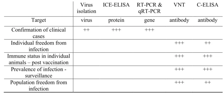
Table 1: Main test methods available for the diagnosis of Peste des Petits Ruminants and their purpose. Key: +++: recommended method; ++: suitable method. Adapted from (Baron et al. 2016; OIE 2013) ... 35 Table 2: Risk factors for PPR identified in the literature published until May 2017. Updated from (Waret-Szkuta 2011; Tran 2013) ... 70 Table 3: Risk factors associated with spread of PPR in small ruminant populations, selected for South Africa and associated hypothesis for PPR. Risk factors for which geographical data was available are highlighted in blue. *A proxy is a variable assumed to resemble the risk factor. ... 73 Table 4: Geographical datasets collected and the associated sources ... 75 Table 5: Geographical datasets collected for the provincial maps ... 84 Table 6: Weights attributed by the experts. Minimum and Maximum values are indicated in brackets. ... 85 Table 7: Comparison of the weights and rank of the risk factors used in PPR risk mapping using MCDA approach. ... 90
RESUME LONG EN FRANÇAIS
Dans un contexte où le virus de la peste des petits ruminants (PPR) se propage en direction de l’Afrique Australe, et considérant la présence d’importantes populations de moutons et de chèvres en Afrique du Sud, un intérêt croissant est porté aux approches d’analyse de risque avec pour objectif de déterminer les zones du pays les plus à risque pour la transmission de la maladie. Notre travail porte sur une analyse spatiale conduite en appliquant la méthode d’aide décisionnelle multicritère. Dans une première partie, nous présentons les données bibliographiques concernant le virus et la maladie, avant d’appliquer la méthode dans la seconde partie – expérimentale - et produire les premières cartes de risque sur la PPR en Afrique du Sud.
1. RÉPARTITION GÉOGRAPHIQUE ET IMPORTANCE ÉCONOMIQUE
La peste des petits ruminants est une maladie hautement contagieuse due à un virus appartenant au genre Morbillivirus. Ce genre regroupe par ailleurs d’autres virus d’intérêt vétérinaire et médical, comme le virus de la peste bovine (éradiqué en 2011), le virus de la rougeole chez les primates et le virus de la maladie de carré chez les canidés.La maladie fut décrite pour la première fois en 1942 en Côte d’Ivoire (Gargadennec, Lalanne 1942). Pendant plusieurs décennies, on pensait la maladie limitée à Afrique de l’Ouest. Ce n’est qu’après l’identification du virus responsable en 1979 (Gibbs et al. 1979) que les scientifiques ont détecté la présence de la maladie dans de nombreux pays d’Afrique, d’Asie, du Moyen et du Proche-Orient. En Afrique, la peste des petits ruminants était initialement observée autour de la ceinture sahélienne, puis s’est propagée au Maghreb, en Afrique de l’Est et en direction de l’Afrique Australe. En Asie, la maladie s’est récemment propagée en Asie Centrale (OIE 2017).
Quatre lignées du virus ont été définies (I, II, III, IV) sur la base des séquences de gènes du virus. Ces lignées ont été nommées selon l’ordre apparent de propagation du virus. Ainsi, les lignées I et II sont essentiellement retrouvées en Afrique de l’Ouest, la lignée III en Afrique de l’Est et la lignée IV en Asie et au Moyen-Orient. Actuellement, la lignée IV est retrouvée dans la plupart des infections récentes en Afrique et en Asie, ce qui peut suggérer une virulence accrue pour cette lignée (Kwiatek et al. 2011).
Environ 330 millions de personnes à travers l’Afrique, le Moyen-Orient et l’Asie élèvent du bétail, dont des moutons et des chèvres (FAO, OIE 2015). Ces derniers représentent une source de lait, de viande, de laine et sont considérés comme de la trésorerie qui peut être vendue pour couvrir les frais de santé ou de scolarité. La peste des petits ruminants affecte aujourd’hui presque 70 pays qui ensemble abritent plus de 80% des populations mondiales de petits ruminants. L’impact financier annuel de la peste des petits ruminants est estimé entre 1.45 et 2.1 milliards de dollars par an (FAO, OIE 2015).
Son impact sanitaire et financier important a conduit l’OIE et la FAO à mettre en place la Stratégie Mondiale pour l’Eradication et le Contrôle de la PPR. Cette stratégie vise l’éradication de la peste des petits ruminants d’ici 2030. A l’échelle de l’Afrique du Sud, la Communauté de Développement d’Afrique Australe (SADC) a mis en place une stratégie de contrôle régionale en 2012 suite à l’incursion du virus en Tanzanie et à l’épidémie dévastatrice en République Démocratique du Congo (tous deux membres de la SADC).
2. AGENT ÉTIOLOGIQUE ET TABLEAU CLINIQUE
Les virions sont des particules de forme généralement sphérique. Le génome viral est un monobrin d’ARN négatif d’une longueur de 15,498 nucléotides (Bailey et al. 2005). Ce génome code pour 8 protéines : 2 glycoprotéines de surface, une protéine matricielle, 3 protéines composant le complexe de réplication (Bailey 2007), et 2 protéines non-structurales dont le rôle précis est actuellement encore discuté (Sanz Bernardo 2017).
Le virus de la peste des petits ruminants est enveloppé, particulièrement sensible dans le milieu extérieur. Le virus est facilement détruit par les désinfectants les plus communs, et possède une demi-vie à 37°C de 2 heures (Kumar et al. 2014). Son mode de transmission est donc essentiellement direct.
Le virus induit une immunosuppression majeure qui peut durer plusieurs semaines (Jagtap et al. 2012). Une fois pris en charge au niveau de la muqueuse respiratoire, le virus se réplique une première fois au sein des nœuds lymphatiques régionaux, puis dissémine dans tout l’organisme via des lymphocytes infectés. L’infection induit une réponse immunitaire cellulaire et humorale. Chez les nouveaux nés de mères infectées ou immunisées, les anticorps maternels sont protecteurs pendant 3 à 4 mois (Libeau et al. 1992). De plus, les animaux qui survivent à l’infection développement une immunité qui les protège pour toute la durée de leur vie (Cosby, Chieko, Yamaguchi 2006).
La forme clinique la plus couramment rencontrée est la forme aigue (Lefèvre, Diallo 1990). Les symptômes ressemblent à ceux de la peste bovine, mais chez les moutons et chèvres. La période d’incubation est de 4 à 6 jours. Les premiers symptômes, rencontrés également dans le tableau clinique de la forme suraiguë, sont une hyperthermie sévère (41-42°C), une prostration, des écoulements nasaux et oculaires, parfois accompagnés d’une diarrhée profuse. S’en suivent une congestion des muqueuses buccales et oculaires associée à des zones d’ulcération et d’érosion qui évoluent en zone de nécrose épithéliale, essentiellement dans la cavité buccale (FAO 1999). Les écoulements finissent par devenir muco-purulents et former des croûtes (FAO 1999). Les complications bactériennes sont fréquentes, notamment en région pulmonaire. A l’examen nécropsique, on retrouve essentiellement des lésions de gastro-entérite (avec congestion de la muqueuse), pneumonie, stomatite ulcéreuse (FAO 1999).
3. DIAGNOSTIC ET PRÉVENTION
La peste des petits ruminants étant une maladie listée du Code sanitaire pour les animaux
terrestres de l’Office International des Epizooties (OIE), sa déclaration est obligatoire, et le
diagnostic de certitude passe par un diagnostic de laboratoire qui doit utiliser une technique reconnue par l’OIE.
Le diagnostic direct porte soit sur la détection du virus (avec le test de neutralisation virale), soit sur la détection de protéines virales (ICE ELISA) soit la détection d’acides nucléiques (RT-PCR). Le diagnostic indirect se fait essentiellement en utilisant la technique ELISA. Les tissus à échantillonner pour le diagnostic sont des écouvillons sur les muqueuses nasales, buccales ou conjonctivales. Il est également possible de prélever du sang. Sur carcasse, il est possible de prélever les nœuds lymphatiques mésentériques, bronchiques, ainsi que la rate, les poumons et la muqueuse intestinale (OIE 2013).
Quant aux mesures prophylactiques, la vaccination est la principale mesure efficace et disponible. En effet, dans les pays endémiques de PPR, la restriction des mouvements d’animaux, la désinfection approfondie et l’abattage sanitaire peuvent être difficiles à mettre en place. Autrefois interdite pour ne pas interférer avec le programme d’éradication de la peste bovine, la vaccination contre la peste des petits ruminants est aujourd’hui disponible. En effet, les petits ruminants étaient autrefois vaccinés contre la PPR avec le vaccin contre la peste bovine (vaccin hétérologue). En l’absence de vaccin DIVA disponible, la vaccination des petits ruminants avait alors été interrompue pour surveiller l’efficacité du programme d’éradication de la peste bovine. Quatre vaccins vivants atténués ont été développés, et tous protègent efficacement contre les différentes lignées du virus de la peste des petits ruminants et empêchent la transmission du virus d’un animal infecté à un autre. L’immunité protective est induite dès la première administration. Le protocole vaccinal actuel préconise une vaccination tous les 3 ans (Diallo et al. 2007; Saravanan et al. 2010). Cependant, comme les chèvres et moutons élevés dans les pays endémiques de peste des petits ruminants ne sont généralement pas gardés plus de 3 ans, une injection suffit pour la durée de vie économique de l’animal. Les principaux défis à relever concernant les vaccins vivants atténués sont leur sensibilité à la chaleur et leur incapacité à différencier les animaux vaccinés des animaux infectés. Les recherches actuelles portent sur ces deux points d’amélioration, ainsi que sur le développement de vaccins multivalents pour optimiser les dépenses liées à la vaccination dans les pays du Sud.
4. EPIDÉMIOLOGIE
Deux formes épidémiologiques peuvent être observées. La forme enzootique se rencontre essentiellement au Sahel et en Asie du Sud et se traduit par des baisses de productivité et une sensibilité accrue aux surinfections. La forme épizootique se rencontre dans des troupeaux à faible séroprévalence avec une cyclicité tous les 4-6 ans, quand le renouvellement du troupeau est suffisamment avancé pour avoir une majorité d’individus naïfs. En cas d’épizootie, les taux de morbidité et de mortalité peuvent alors atteindre 80%-90% et 50-80%, respectivement (Lefèvre, Diallo 1990). Cette forme s’observe en Afrique de l’Ouest et dans les régions récemment infectées telles que l’Afrique de l’Est.
Les sources virales sont essentiellement les individus infectés, via les sécrétions orales, nasales, lacrymales et fécales (Abubakar et al. 2012). Aucun stade de porteur sain n’a été décrit (Hamdy et al. 1976). L’excrétion de matériel viral a été détecté dès 3 jours post-infection (Couacy-Hymann, S.C. Bodjo, et al. 2007) et jusqu’à 16 semaines post-post-infection (Wasee Ullah et al. 2016).
La maladie de la peste des petits ruminants est avant tout une maladie du mouton et de la chèvre. A ce jour, ces deux espèces sont considérées comme les seules capables d’excréter et transmettre le virus. Des séroprévalences ont été détectées chez les bovins, les camélidés et les buffles domestiques. Des épidémies ont été observées chez des populations de petits ruminants sauvages, la plupart du temps vivant dans des conditions de semi-captivité. A l’heure actuelle, les différentes publications mettent en avant l’hypothèse pour laquelle les petits ruminants sauvages sont sensibles à la peste des petits ruminants mais incapables de transmettre le virus au sein de leurs populations (Couacy-Hymann et al. 2005).
Plusieurs facteurs de risque ont été identifiés dans la littérature. Le manuel sur la peste des petits ruminants de l’OIE décrit une sensibilité accrue des moutons en Asie de l’Ouest et du Sud, tandis qu’en Afrique les chèvres sont décrites comme plus fréquemment atteintes (FAO 1999). Les animaux âgés entre 4 et 12 mois sont considérés comme plus à risque (Diallo 2003a; Gopilo 2005). Les races guinéennes de chèvres ont été rapportées plus sensibles que les races sahéliennes (Lefèvre, Diallo 1990). Le sexe a également été décrit comme un facteur de risque avec des observations de séroprévalences plus élevées chez les femelles que chez les mâles (Waret-Szkuta et al. 2008; Aziz-ul-Rahman et al. 2016; Kihu et al. 2015). Cependant les femelles sont souvent gardées plus longtemps dans les troupeaux, ce qui peut expliquer une plus grande séroprévalence au sein de ce sexe.
Les systèmes de production avec transhumance et le pastoralisme en particulier ont été décrits comme facteurs de risque à plusieurs reprises (Abubakar et al. 2009; M. Abubakar et al. 2011; Bett et al. 2009; Singh et al. 2004; Megersa et al. 2011; Mahajan et al. 2012; Shankar 1998; Nanda 1996). Les fortes densités en animaux (Al-Majali et al. 2008; Khan 2008; Ozkul et al. 2002; Kardjadj et al. 2015) et l’élevage mixte de chèvres et moutons (Al-Majali et al. 2008; Anderson, McKay 1994; Kardjadj et al. 2015) peuvent augmenter le risque de transmission de la peste des petits ruminants. Enfin, l’introduction d’animaux achetés sur les marchés (Abubakar et al. 2009; M. Abubakar et al. 2011; Singh et al. 2004; Mbyuzi et al. 2014) et le partage des pâtures et points d’abreuvement entre différents troupeaux (Mbyuzi et al. 2014; Lefèvre 2003) ont également été rapportés comme des pratiques à risque.
Les pratiques traditionnelles (Bazarghani 2006) et religieuses (Bonniwell 1980) impliquant le commerce de petits ruminants vivants (telles que les mariages, funérailles) peuvent favoriser l’introduction d’individus infectés dans des zones indemnes. Quant à l’influence de la saison, certains auteurs rapportent une sensibilité accrue pendant la saison humide et chaude (Rony et al. 2017; Mondal 2014; M. Abubakar et al. 2011; Taylor, Barrett 1990), et d’autres pendant la saison sèche et fraîche (Obi et al. 1983; Singh et al. 2004; Abubakar et al. 2009). Enfin, la qualité de la surveillance et l’accès aux services vétérinaires influencent également la propagation de la PPR (Al-Majali et al. 2008; Bett et al. 2009; Bazarghani 2006).
5. ANALYSE DU RISQUE DE TRANSMISSION DE LA PPR A L’AIDE DE
LA METHODE SPATIALISEE D’AIDE DECISIONNELLE MULTICRITERE
Alors que le virus de la peste des petits ruminants se propage vers le sud du continent Africain, l’Afrique du Sud apparaît comme un pays à risque pour la propagation de la peste des petits ruminants, et ce pour plusieurs raisons. Tout d’abord, environ 67% des chèvres et 12% des moutons élevés en Afrique du Sud appartiennent à des éleveurs de subsistance dans des zones communales (NERPO 2013). Ce mode d’élevage peut être considéré comme une pratique à risque car d’une manière générale, les éleveurs en zone communale souffrent d’infrastructures défectueuses, d’un manque d’accès aux services vétérinaires, ou encore de niveaux d’éducation inférieurs aux éleveurs professionnels. De plus, les animaux élevés en zones communales sont rarement identifiés et souvent laissés libres de vagabonder dans les villages et d’ainsi partager des zones d’abreuvement et de pâtures avec d’autres individus issus de troupeaux différents.De plus, l’Afrique du Sud souffre d’un manque de traçabilité quant aux mouvements d’animaux au sein de son territoire et avec les pays voisins. En effet, un important marché illégal est observé, en particulier à l’occasion de fêtes traditionnelles et religieuses.
Ainsi, l’Afrique du Sud se présente comme le candidat idéal pour une analyse de risque afin de déterminer les zones les plus à risque pour la transmission du virus dans le cas où il aurait été introduit sur le territoire. Cependant, étant donné que l’Afrique du Sud est indemne de PPR, nous ne disposons pas des données de foyers nécessaires pour conduire une analyse spatialisée quantitative. Dans ce type de cas, il nous est possible d’utiliser des méthodes se basant sur la connaissance, et non sur les données. Dans ce travail, nous avons appliqué la méthode d’aide décisionnelle multicritère (MCDA) spatialisée. Cette méthode associe connaissances scientifiques et avis d’experts pour déterminer les zones géographiques les plus adaptées à la propagation du virus, et donc les plus à risque. Le résultat de ce travail est sous forme de cartes géographiques, qui apporteront un support visuel aux autorités compétentes pour déterminer où concentrer les mesures de contrôle et de surveillance tout en optimisant les ressources financières et logistiques.
La méthode MCDA spatialisée a déjà été utilisée pour identifier les zones à risque pour la Fièvre de la Vallée du Risque en Afrique (Tran et al. 2016; Clements, Pfeiffer, Martin 2006) et en Europe (Sánchez-Vizcaíno, Martínez-López, Sánchez-Vizcaíno 2013; Tran et al. 2013), pour l’Influenza Aviaire hautement pathogène H5N1 en Asie (Stevens 2013; Paul et al. 2016). Pour la PPR, la méthode a déjà été utilisée à l’échelle du continent Africain (Waret-Szkuta 2011) et à l’échelle de l’Afrique de l’Est (Tran 2013). Cependant, aucune analyse de risque concernant la PPR en Afrique du Sud n’a encore jamais été portée à notre connaissance. Ce travail constitue donc une première étape d’analyse de risque spatialisée utilisant la méthode d’aide décisionnelle multicritère.
La première étape de la méthode consiste en l’identification des facteurs de risque. Une revue bibliographique a été menée pour actualiser les facteurs de risque identifiés dans la littérature lors de précédents travaux (Waret-Szkuta 2011; Tran 2013). Ces facteurs ont ensuite été confrontés à la situation de l’Afrique du Sud et adaptés. Nous avons retenu les facteurs de risque suivants :
- La densité de chèvres - La densité de moutons
- La proximité aux parcs nationaux et réserves
- La proximité aux rivières, considérées comme les points d’abreuvement principaux pour les animaux élevés en zones communales
- La proximité aux marchés légaux et illégaux. Le commerce illégal de petits ruminants en Afrique du Sud ayant essentiellement lieu au sein des stations de taxi.
- La proximité aux routes : qui sont la voie de transport majeure pour le commerce légal et illégal de petits ruminants
- La distance aux bureaux des services vétérinaires
La deuxième étape de la méthode consiste en l’acquisition de données géographiques correspondant aux sept facteurs de risque sélectionnés. Les données concernant les densités d’animaux ont été celles issues du modèle « Gridded Livestock of the World » (GLW) de la FAO. Les cartes représentant les parcs nationaux et les rivières d’Afrique du Sud sont issues de sites internet gouvernementaux. Les localisations des marchés officiels de petits ruminants ont été répertoriées à l’aide de magazines professionnels destinés aux éleveurs. La carte des routes ainsi que les localisations des stations de taxi (marchés illégaux) sont issues de la banque de données OpenStreetMap. Les localisations des services vétérinaires ont été repérées sur une carte en s’appuyant sur les sites internet des services vétérinaires des neuf provinces.
Chacun des sept facteurs de risque a été représenté sur une carte propre. Les données ont été visualisées et travaillées à l’aide de logiciels de géomatique, afin de produire une carte par facteur de risque sous le format raster et avec une résolution de 1km2 (celle imposée par le jeu de données du modèle GLW).
La troisième étape fait appel aux avis d’experts. 16 experts ont été contactés parmi des chercheurs et des membres des services vétérinaires sud-africains. Un questionnaire en deux parties a été conçu sur Word et envoyé par email. La première partie a pour but de déterminer la relation entre chaque facteur de risque et le risque de transmission de la PPR. Les experts avaient le choix entre 4 relations (linéaire, sigmoïdale, quadratique, trapézoïdale) et devaient éventuellement décider de valeurs seuil. La deuxième partie du questionnaire, concernant le poids des facteurs, est présentée sous forme de matrice de comparaison. Chaque expert comparait les facteurs de risque deux à deux et accordait un poids à chaque comparaison. Huit experts ont renvoyé le questionnaire complété. Les données de quatre experts ont été conservées pour la suite de la méthode car ces experts ont montré un indice de cohérence (ou consistency ratio) satisfaisant (CR<0.13).
Les facteurs de risque n’étant pas exprimés dans les mêmes unités (par exemple, la densité est en nombre d’individus/km2, et les proximités indiquées en mètres), ils ont été standardisés pour pouvoir les combiner et produire une carte finale. Cette standardisation utilise l’approche « fuzzy » sur les données de la première partie des questionnaires. En effet, chaque relation correspond à une fonction mathématique que l’on applique aux données brutes pour obtenir une valeur standardisée (entre 0 et 1). Ainsi, pour chaque facteur de risque identifié, on obtient une carte où chaque pixel possède une valeur allant de 0 (risque minimal) à 1 (risque maximal).
A partir des données des quatre experts retenus pour la modélisation, un poids moyen a été calculé pour chaque facteur de risque. Chacun de ses poids a été attribué à la carte du facteur de risque correspondant, en appliquant la méthode de combinaison linéaire pondérée. Le résultat de cette combinaison est une carte de risque finale, où chaque pixel possède une valeur pouvant aller de 0 (risque minimal de transmission) à 1 (risque maximal de transmission).
La carte finale établie à l’échelle nationale révèle que l’aptitude de l’Afrique du Sud à transmettre le virus dans le cas où il aurait été introduit varie de 0.12 à 0.89 en fonction des zones géographiques (avec une aptitude moyenne nationale de 0.27).
La réalisation de deux cartes additionnelles, à l’échelle de la province du Cap Occidental et de la province du Cap du Nord, a été rendue possible grâce à la collecte de données de recensement des chèvres et moutons, mises à disposition par les services vétérinaires de ces provinces. Pour la province du Cap Occidental, l’aptitude à transmettre le virus varie de 0.11 à 0.66 (avec une moyenne sur la province de 0.25). Pour la province du Cap du Nord, l’aptitude varie de 0.1 à 0.71 (avec une moyenne sur la province à 0.22). Lors de la pondération des facteurs de risque, les experts ont classé la densité de chèvres, la proximité aux marchés, la densité de moutons et la distance aux services vétérinaires comme les facteurs les plus importants, avec des poids respectifs de 0.335, 0.174, 0.169, et 0.137.
6. AMÉLIORATIONS DU MODÈLE
La méthode spatialisée d’aide décisionnelle multicritère présente de nombreux atouts, comme la capacité à produire des cartes de risque dans des régions indemnes ou lorsque les données épidémiologiques sont rares ou indisponibles. Elle permet d’apporter un support visuel relativement instinctif d’aide à la décision quant aux zones à cibler en matière de surveillance et de contrôle.
Un autre atout est que cette méthode autorise l’intégration a posteriori de données cartographiables qui viendraient à devenir disponibles, ou disponibles avec une plus grande précision. Cependant, le modèle a également le défaut de ses qualités : il repose sur une succession de choix discutables ; d’abord pour la sélection des facteurs de risque, puis pour les relations, pour les valeurs seuil et les poids des facteurs.
Une limite importante à prendre en compte est la dépendance du modèle à la qualité des données utilisées. C’est d’ailleurs sur ce point que sont axées nos principales voies d’amélioration. En effet, les données utilisées dans ce travail concernant les densités d’animaux sont issues d’une modélisation basée sur des données de recensement fournies à l’échelle de la province. Leur précision est donc discutable.
La difficulté de la collecte de données de recensement plus précises réside dans le fait que les services vétérinaires eux-mêmes ont des difficultés pour recenser avec rigueur et précision les animaux sur leur territoire, notamment à cause des mouvements incontrôlés au sein du pays et à travers les frontières.
De plus, la méthode utilisée dans ce travail nécessite de ne considérer que les facteurs de risque qui peuvent être cartographiés, alors que certains comme les mouvements d’animaux lors des fêtes traditionnelles par exemple peuvent constituer un facteur de risque très important mais difficilement cartographiable.
Enfin, le faible nombre d’experts ayant répondu et ayant été retenu (25% de l’échantillon initial) nous contraint à interpréter nos cartes avec précaution.
La dernière étape de la méthode d’aide décisionnelle multicritère consiste à évaluer quantitativement la sensibilité et l’incertitude du modèle, afin de vérifier que la carte finale est suffisamment indépendante des choix des experts pour être interprétable. Nous recommandons que cette analyse quantitative soit considérée comme un prérequis avant d’utiliser nos cartes en tant qu’aide à la décision.
INTRODUCTION
Peste des Petits Ruminants (PPR) is a highly contagious viral disease of sheep and goats, first discovered in 1942 in the Ivory Coast, and today spread throughout Western, Central and Eastern Africa, the Middle East and Asia. With morbidity and fatality rates up to 100 % and 90 % respectively (FAO, OIE 2015), PPR represents a major threat to food security for the 330 millions of people keeping livestock in these developing regions. Its high economic importance brought the OIE and FAO to target PPR as the next disease to be eradicated by 2030.
In Africa, PPR is progressively spreading towards Southern Africa, with first OIE notifications in Kenya (2006), Uganda (2007), Tanzania (2008) and Angola (2012) and evidence of PPRV antibodies in Zambia in 2015. This apparently rapid spread of the virus can be explained by high small ruminant densities in most of endemic countries, uncontrolled trading and transhumant movements, sharing grazing areas and watering points, etc. and that often without consideration of national boundaries.
As PPR is spreading southwards, there is a need to anticipate further PPR introduction and transmission in Southern African countries so that new infection cases can be prevented or rapidly controlled. To provide the most accurate risk prediction, epidemiological data currently known on PPR must be adapted to each considered country or region, mainly because of differences among small ruminant farming systems, livestock trade, etc.
This study focuses on assessing the risk of PPR spread within South Africa, located at the southern tip of the African continent. Our work aims at identifying high risk areas for PPR spread in South Africa through spatial risk analysis, in the case where PPRV would have been introduced on the territory. Priority areas for prevention and control of PPR were assessed through a Geographical Information System (GIS)-based risk model: spatial Multi Criteria Decision Analysis (MCDA). This “knowledge-driven” approach consists in risk factors identification and collection of the corresponding geographical data, expert opinion survey on these risk factors and how they impact on the suitability of the disease spread, and geo-processing of the collected data with a GIS software. The final output of our study is a suitability map that highlights the most likely areas in South Africa for PPR spread, providing support for prioritizing and implementing early detection and control strategies.
In a first part, we browsed literature to review the current knowledge on PPR, including etiology, medical aspects and epidemiology. In a second part, we applied the spatial-MCDA methodology to map the most suitable areas in South Africa for PPR spread. Finally, results of our work were discussed and some improvements were suggested to enhance accuracy and validity of our spatial model in a near future.
PART 1: BIBLIOGRAPHICAL BACKGROUND
CHAPTER 1: REVIEW OF PESTE DES PETITS
RUMINANTS
Peste des Petits Ruminants (PPR) is also known as “goat plague”, “Kata”, “syndrome of stomatitis-pneumoenteritis” or “ovine rinderpest”. This highly infectious disease is due to a morbillivirus and causes rinderpest-like symptoms, mainly in sheep and goats.
7. CAUSATIVE AGENT : PESTE DES PETITS RUMINANTS VIRUS (PPRV)
7.1.
Taxonomy of PPRV
Peste des Petits Ruminants virus (PPRV) belongs to genus Morbillivirus, sub-family Paramyxovirinae, family Paramyxoviridae, order Mononegavirales. There are various morbilliviruses of medical and veterinary importance, and one of the characteristics of morbilliviruses is their limited ability to infect different species. We find Measles virus in primates, Canine distemper virus in canids, Phocine distemper virus in pinnipeds, and several cetacean morbilliviruses that are found in marine mammals. Rinderpest virus has been globally eradicated in 2011. Recently, a new morbillivirus (Feline morbillivirus) has been described in cats (Woo et al. 2012). These morbillivirus species can be divided into monophyletic lineages following genetic analysis (figure 1).
Figure 1: Un-rooted neighbour-joining tree showing the relationships between morbilliviruses. The phylogenetic tree was built using partial N gene sequences of 230 nucleotides (accession nos. NC_006383, Peste des petits ruminants virus; NC_001498, Measles virus; AB547189, Rinderpest virus; NC_001921, Canine distemper virus; KC802221, Phocine distemper virus; JQ411016, Feline morbillivirus; AY949833, Porpoise morbillivirus; NC_005283, Dolphin morbillivirus; AF200818, Pilot whale morbillivirus) with 1000 bootstrap replicates and Kimura 2-parameter model in MEGA 5.2. The scale bar indicates nucleotide substitutions per site. Cited from (Parida et al. 2015)
7.2.
Virus morphology and genome structure
PPRV virions are pleomorphic – but generally spheric – enveloped particles. The virus particle size has been determined to be between 400 and 500 nm (Gibbs et al. 1979). The PPR virus genome is a non-segmented, single stranded, negative-sens RNA molecule, that consists of 15,948 nucleotides (Bailey et al. 2005) and six genes that encode eight proteins.
The genome is encapsidated by the N protein, forming a helicoidal nucleocapsid. As the genome is simple negative RNA, it cannot be translated directly into proteins; it has to be transcribed into messenger RNA. This transcription stage is done by a RNA-dependent RNA polymerase complex, formed by two other nucleocapsid proteins: the large polymerase protein (L) and the co-factor phosphoprotein (P). P, N and L proteins form the functional replication complex (Bailey 2007). The N protein is a major viral protein, produced in highest amount in Morbilliviruses (Diallo et al. 1987). It is also a strong immunostimulatory protein. The L protein is the largest viral protein and is expressed in the smallest amount of the infected cells (Kumar et al. 2014).
The nucleocapsid is surrounded by a lipoproteic envelope that is derived from infected cell membrane. This envelope contains two glycoproteins: the viral fusion protein (F) and haemagglutinin protein (H). These proteins form spikes of about 10 nm (Bourdin, Laurent-Vautier 1967) on the surface of the viral envelope, and play crucial roles during the initial steps of the virus replication. Based on what we know about Measles virus proteins, the H protein is responsible for attachment of the virion to the host cell. Then, via the F protein, the viral and the cell membranes are fused, allowing the release of the viral nucleocapsid into the cell cytoplasm (Moll, Klenk, Maisner 2002). The PPRV F protein is also responsible for the syncytium formation, a cytopathic effect common for all Paramyxoviruses. The syncytium formation consists in the fusion of the membrane of an infected cell with the membrane of an adjoining cell. PPRV F protein also has haemolysin property (Devireddy et al. 1999). As for the PPRV H protein, it has both haemagglutinin and neuraminidase activities and is thus sometimes named as Haemagglutinin-neuraminidase (HN) protein.
The inner surface of the viral envelope is coated by the matrix protein (M). This protein is thought to link the RNP and the cytoplasmic tails of the surface glycoproteins (H and F) and to play an important role in virus formation. The organization of the PPRV genome and proteins is represented in figure 2.
Figure 2: Schematic diagram of Peste des Petits Ruminants virion structure (adapted from (Banyard et al. 2010)). The PPRV glycoproteins (F and H) form spikes and are set inside the envelope. The M protein coats the inner surface of the envelope. The RNP is composed of N, P and L proteins in association with the genome.
Besides these six structural proteins, two non-structural proteins (C and V) are also produced from an alternative reading frame of the P protein. Their precise molecular functions for PPRV still need to be studied, but several studies on Rinderpest and Measles viruses demonstrated the role of C protein in regulation of virus replication (Escoffier et al. 1999) and RNA synthesis (Baron, Barrett 2000), virulence determination (Patterson et al. 2000), modulation of Large polymerase activity (Ito et al. 2013) and blocking of induction of type I interferon (Boxer, Nanda, Baron 2009). The V protein is thought to regulate RNA synthesis (Sweetman, Miskin, Baron 2001), and a recent study has shown that PPRV V protein blocks IFN action at multiple points in the pathway (Sanz Bernardo 2017). Finally, it appears that the C protein is thought to have an even more crucial role in blocking the induction of IFN during the initial stages of PPRV infection (Sanz Bernardo 2017).
7.3.
Resistance to physical and chemical action
As an enveloped virus, PPRV is particularly easily destroyed outside the host. The half-life of PPRV is about 2 hours at 37°C and it can be completely inactivated at 50°C within 60 minutes. The virus is quite stable between pH 5.8-10.0 but inactivated in pH<4.0 or >11.0. PPRV is susceptible to the most common disinfectants such as alcohol, ether, phenol and sodium hydroxide. The virus can survive for long periods in chilled and frozen tissues (Coetzer, Tustin 2004).
7.4.
Pathogenesis
The pathogenesis of PPRV is still under study. Most of the knowledge is based on comparison with related morbilliviruses, mostly Rinderpest and Measles viruses. During infection with PPRV, the virus is initially taken up by antigen presenting cells present in the intraepithelial space and lamina propria of the respiratory mucosa (naso-pharyngeal/respiratory epithelium) (Borrow, Oldstone 1995; Yanagi, Takeda, Ohno 2006). From this site of inoculation, the virus reaches the local draining lymphoid tissues, where the primary virus replication takes place. Then, the virus spreads throughout the body via infected lymphocytes, thanks to both lymphatic and vascular systems (Pope et al. 2013; Esolen et al. 1993; Osunkoya et al. 1990). Viral antigens have been observed by immunohistochemical staining in the lymphoid organs, facial epithelia and gastrointestinal tract (Kumar et al. 2004; Pope et al. 2013). PPRV is thus considered as a lymphotropic and epitheliotropic virus. PPRV infection leads to extensive necrosis in lymphoid organs (Peyer’s patches, spleen, thymus, lymph nodes).
The PPRV causes transient but major immunosuppression. This feature is due to virus multiplication in lymphoid tissues and in peripheral leucocytes, but also due to its interference with IFNs production. A study reported that the proportion of circulating WC1+ γ/δ T-cells and CD14+monocyte/macrophage cells did not change after PPRV infection, while the CD4+
cell sub population was the only one that decreases in number from 4 days post-infection (dpi) (Herbert et al. 2014). This immunosuppression can last for weeks and hence impacts the extent and severity of the disease (Jagtap et al. 2012) with higher susceptibility to secondary infections.
7.5.
Immunological features
The PPRV infection induces both cellular and humoral immune responses. These immune responses are mainly directed against the N proteins and the two surface glycoproteins (F and H) (Sinnathamby et al. 2001). However, immunisation with H and/or F induces protective humoral immunity whilst immunisation with the N protein does not (Sinnathamby et al. 2001; Diallo et al. 2007).
In new-born animals from infected and/or immunized mothers, maternal antibodies remain able to neutralise the virus for 3-4 months (Libeau et al. 1992). Finally, as for other morbilliviruses, animals that recover from PPR usually develop a life-long immunity to re-infection (Cosby, Chieko, Yamaguchi 2006).
8. CLINICAL SYMPTOMS OF PESTE DES PETITS RUMINANTS
Three main clinical pictures of PPR can be observed (Lefèvre, Diallo 1990; FAO 1999):
8.1.
The peracute form
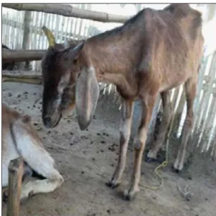
This form is mainly found in goats. The incubation period is about two days. The first clinical sign is a high hyperthermia (41-42°C) followed by prostration, apathy, harsh coat and anorexia. Animals present nasal discharge, lacrimation, and sometimes profuse diarrhea following a short constipation stage. The evolution is rapid, lasting for 5-6 days after the onset of hyperthermia, and death occurs before the onset of any other striking symptom.
8.2.
The acute form
This is the most characteristic form of the disease. The symptoms resemble those of Rinderpest, but in small ruminants. The incubation period is 4-6 days (ranging from 3 to 14 days). The first symptoms are those of the peracute form (pyrexia lasting 3-5 days, anorexia, prostration, etc.) (Figure 3).
Figure 3: Apathy and prostration in a goat suffering from Peste des Petits Ruminants. Photo Courtesy of D.P. Kshirsagar (Sharma et al. 2015).
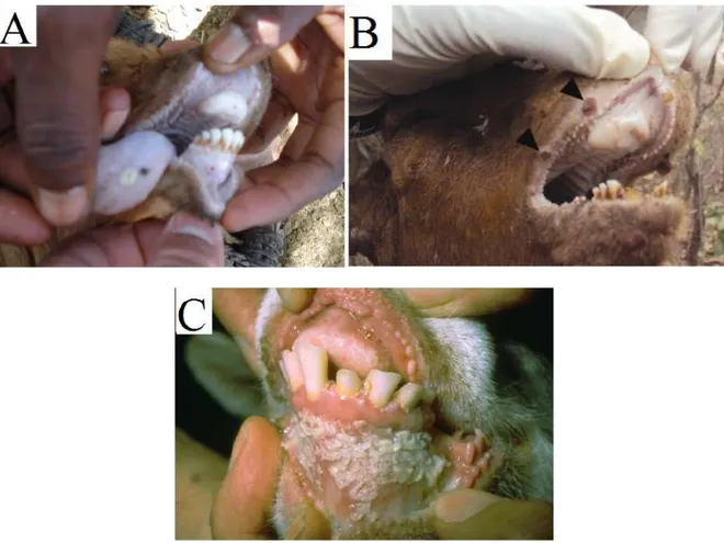
One or two days after the setting of pyrexia, the mucosa of the mouth and eyes become congestive. That congestion is followed by epithelial necrosis on gums, dental pad, palate, lips, inner aspects of the cheeks and upper surface of the tongue. Initially forming pin-point areas (figure 4A), these necrosis areas enlarged and join together. These erosive and ulcerative lesions become coated with dying cells forming a thick cheesy and malodorous deposit (figure 4B).
Figure 4: Ulcers on the tongue and gum (A) and on the lips (B) which can eventually lead to a coat of dying cells forming a cheesy deposit (C: photo of W.P. Taylor). Photographs Courtesy: (A) : G. Misinzo (Torsson et al. 2016); (B): G. Misingo(Kgotlele et al. 2014)) and (C): M.D. Baron (Baron 2011).
The lips tend to be covered with scabs (figure 5A). The ruminant shows excessive salivation and refuses to open his mouth because of the pain. Similar necrosis process can also be found in the mucous membranes of the nose, the vulva and the vagina. Oculonasal discharge is observed, and can last 14 days. First seromucous, it becomes mucopurulent (figure 5A) as a result of secondary bacterial infections, and tends to crust, matting the eyelids together and obstructing the nose (figure 5C).
Figure 5: Upper and lower lips covered with scabs (A). Mucopurulent nasal discharge (B) (photo by P.L. Roeder). Obstructed nose and matted eyelids due to dry oculonasal discharge (C). Photo Courtesy of : (A):D.P. Kshirsagar (Sharma et al. 2015), (B): M.D. Baron (Baron 2011) and (C): G. Misinzo (Torsson et al. 2016).
Signs of pneumonia are usually observed in a later stage, with animals coughing, having pleural rales and abdominal breathing. Severely affected animals show marked dyspnea; extension of the head and neck, dilatation of the nostrils, protrusion of the tongue and soft painful coughs (figure 6).
Figure 6: Respiratory distress in a goat suffering from Peste des Petits Ruminants. Photo Courtesy of D.P. Kshirsagar (Sharma et al. 2015).
Diarrhea is commonly observed two to three days after the onset of the fever. The feces are initially soft, then watery, foul-smelling and may contain blood stains and pieces of dead gut tissue. This may not be obvious in early or mild cases.
Figure 7: Diarrhea soiling the perineum in a male goat suffering from Peste des Petits Ruminants. Photograph Courtesy of G. Misinzo (Torsson et al. 2016).
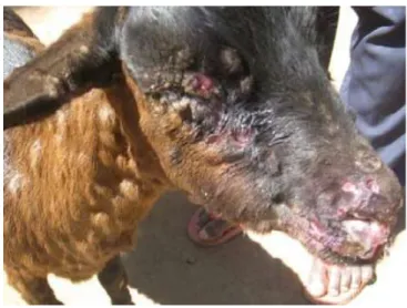
In the later stages of the disease, small nodular lesions are commonly found on the outside of the lips around the muzzle and on the neck (figure 8). The cause is not exactly known but is thought to be Dermatophilus infection or reactivation of a latent contagious ecthyma infection, causing confusion for differential diagnosis.
Figure 8: Nodular lesions on the neck of a goat suffering from PPR. Note the severe ulcers on face, nostril and lips and the matted eyelids. Photograph Courtesy of E.A. Muse (Muse, Matondo, et al. 2012).
In absence of complications, illness may last about 8-10 days, leading in either death or in recovery with long-lasting immunity.
8.3.
The subacute form
Subclinical form seems to be particularly prevalent in some endemic areas where some local breeds are thought to have innate resistance. The disease lasts for about 10 to 15 days, with inconsistent symptoms (prostration, serous nasal discharge, diarrhea), and sometimes papules or pustules at a later stage.
8.4.
Complications
The commonest complications are:
- Pneumonia or bronchopneumonia with bacterial secondary infection, particularly
Pasteurella haemolytica or Pasteurella multocida type A;
- Reactivation of a latent parasitosis (coccidiosis, piroplasmosis, trypanosomiasis) due to immunodepression
- Abortion
Factors such as co-infection with pre-existing parasites, virus or bacteria, and the nutritional status of the animals may also contribute to the disease severity and impact on the outcome (Couacy-Hymann, C. Bodjo, et al. 2007; Ugochukwu, Agwu 1991).
9. POST-MORTEM FINDINGS
The victim shows high degree of dehydration, with sunken eyeballs (figure 9) and watery feces soiling the hindquarters. We find the erosive, congestive, and necrotic lesions already observed in mouth, lips, and nose of live animals.
Figure 9: Sunken eyeballs, lips covered with scabs and congestion of the ocular mucosa. Photo: W.P. Taylor, Courtesy of M.D. Baron (Baron 2011).
Besides these external lesions, the lesions that we can observe are those of conjunctivitis, rhinotracheitis, ulcerative stomatitis, gastroenteritis and pneumonia. The erosive lesions observed in the mouth may extend on the esophagus until the reticulo-rumen junction.
The mucosa of the abomasums, small intestines and large intestines are congested (figure 10A), hemorrhagic, and sometimes zebra stripes (congestion lines) are observed on the mucosa of large intestine and rectum. The congestion is particularly observed in the ileo-caecal junction.
Figure 10: Congestion of the intestines (A) and pneumonia (B) in a goat confirmed with PPR in Ngorongoro, Tanzania. Photo Courtesy G. Misinzo (Torsson et al. 2016).
Peyer’s patches may be necrotic, as the spleen and the liver. We can observe enlargement and congestion of the lymph nodes associated with the intestines (figure 11A) and the lungs (figure 11B). The lesions on the lungs are those of interstitial pneumonia. The anterior and cardiac lobes of the lungs are firm to the touch, dark red coloured (figure 10B).
Figure 11: Hemorrhagic lymph nodes in the gastrointestinal (A) and respiratory (B) system of PPR-suspected animals (see arrow heads) in Tanzania. Photographs Courtesy of G. Misinzo (Kgotlele et al. 2014).
Histopathologically, PPRV form syncytia in the lymph nodes, splenic white pulp and gastrointestinal sub-mucosal lymphoid tissue as soon as 5-7 dpi. These syncytia eventually lead to necrosis/apoptosis. Squamous epithelial syncytia can also be observed in digestive tract epithelium and tonsillar and facial tissues (Pope et al. 2013).
10. DIAGNOSIS
There are some events that can help the farmer or the veterinarian to suspect Peste des petits ruminants (FAO 1999):
- The recent movement or gathering of small ruminants of different ages; - The introduction of recently purchased animals;
- The return of unsold animals from the market;
- The onset of rainy or dry cold season, changing the housing and feeding; - Or a change in husbandry such as intensification of livestock.
10.1. Differential diagnosis
Based on mouth lesions, it is possible for farmers, veterinarians and veterinary technicians to misdiagnose with rinderpest, foot and mouth disease, bluetongue, or contagious ecthyma. The difficulty in breathing can be found in clinical pictures of pneumonic pasteurellosis, or contagious caprine pleuropneumonia (CCPP). The diarrhea can be due to coccidiosis, or gastrointestinal helminth infestations. None of these diseases cause the full clinical picture of PPR (respiratory, diarrhea, necrotic stomatitis), but animals may not present all of the clinical signs. The diagnosis of PPR may also be complicated by secondary bacterial infections. Therefore, laboratory confirmation of PPR is necessary for definitive diagnosis.
10.2. Laboratory diagnosis
A comprehensive review of the available laboratory techniques and the promising techniques has recently been published (Santhamani, Singh, Njeumi 2016). In this section, we will focus on the main laboratory techniques and their application.
10.2.1. Collection of samples
Depending on what is under investigation (i.e. viral excretion or seroprevalence), the best moment for sampling will be different. Recently, very sensitive methods like RT-PCR or immunocapture ELISA techniques have been developed. Such methods can detect viral shedding 1-3 days before the onset of clinical signs (Couacy-Hymann, S.C. Bodjo, et al. 2007; Couacy-Hymann et al. 2009).
Generally speaking, sample collection for virological investigation must be done on animals showing clinical signs (mainly from 4 to 17 dpi). For retrospective serology, sampling can be made at any time within 3 years after seroconversion of the animals (Zahur 2015).
In live animals, swabs can be made of the conjunctival discharges and from nasal and buccal mucosa, ideally during the erosive mucosal phase of the disease. The use of swab is more and more common; as it is safe, simple and painless for animals, breeders are usually more compliant for sample collection with this technique.
At necropsy, samples can be collected aseptically on fresh carcasses from mesenteric and/or bronchial nodes, lungs, spleen and intestinal mucosa, ideally on 2-3 animals. The samples for histopathology must be places in 10% neutral buffered formalin solution. Biopsies can also be done on live animals during the febrile stage of the disease, although rarely achievable on the field.
Blood collection is always valuable, either at the early stage of the disease for virus isolation, PCR or hematology (whole blood collection on anticoagulant, ideally heparin) or at a later stage for serological diagnosis.
Samples for virus isolation must be kept chilled but not frozen from the collection site to the laboratory. Indeed, the efficiency of laboratory diagnosis is greatly influence by the integrity of the sample, thus care must be taken for the conditions of collection and transportation. The laboratory diagnosis techniques can be sorted in two categories: the techniques detecting antibody response, and the techniques detecting PPRV presence (viral antigen, viral nucleic acid or virus isolation).
10.2.2. Virus isolation
Virus isolation in cell culture is considered as the gold standard technique for virus identification. The cell culture is most commonly done on Vero cells. However, this technique is time-consuming and requires cell culture facilities. Results are obtained in 10-12 days. Thus, virus isolation cannot be used routinely or as a trigger alert technique.
10.2.3. Antigen detection
Agar gel immunodiffusion and counter immunoelectrophoresis were developed in the 1980’s. They detect antigens from swabs and tissues of infected animals, using PPRV-specific antibodies. As their sensitivity is low, these techniques cannot be used for mild forms of the disease nor for early stages of infection due to the low quantity of viral antigen that is excreted. Immunohistochemistry techniques have also been developed: the fluorescent antibody test (requiring fluorescent microscope and technical expertise) and the immunoperoxidase test (requiring only a light microscope, more accessible for less well-equipped laboratories). A haemagglutination test is also available, and based on the haemmagglutination property of the PPRV H protein. Although simple to perform, this test lacks specificity as many virus families have haemagglutination properties as well.
The two following methods (ELISA and immunochromatography) are the most employed are promising antigen detection methods. Immunocapture ELISA is a test using monoclonal antibodies (MAbs) against PPRV N protein. A sandwich ELISA test is also available and both immunocapture and sandwich ELISAs are available in a commercial kit. The major benefit of such tests is that they have desirable sensitivity and specificity levels and they are user-friendly. Results are obtained in 2 hours. In addition, an immunochromatographic lateral flow device has been recently developed as a pen-side test. This technique is using H protein-specific MAb (Brüning-Richardson et al. 2011). This method is considered cost effective and easy to perform, giving results in a matter of minutes. It has recently been validated under field conditions for early diagnosis (from 4 dpi, so before the onset of the severe clinical sign)