Open Archive TOULOUSE Archive Ouverte (OATAO)
OATAO is an open access repository that collects the work of Toulouse researchers and
makes it freely available over the web where possible.
This is an author-deposited version published in :
http://oatao.univ-toulouse.fr/
Eprints ID : 18533
To link to this article : DOI:10.1016/j.otsr.2016.06.014
URL :
https://doi.org/10.1016/j.otsr.2016.06.014
To cite this version : Thévenin-Lemoine, Camille and Vial, Julie and
Labbé, Jean-Louis and Lepage, Benoît and Ilharreborde, Brice and
Accadbled, Franck MRI of acute osteomyelitis in long bones of
children: pathophysiology study.
(2016) Orthopaedics &
Traumatology: Surgery & Research, vol. 102 (n° 7). pp. 831-837.
ISSN 1877-0568
Any correspondence concerning this service should be sent to the repository
administrator:
[email protected]
MRI of acute osteomyelitis in long bones of children: Pathophysiology
study
C. Thévenin-Lemoine
a,∗, J. Vial
b, J.L. Labbé
c, B. Lepage
d, B. Ilharreborde
e,
F. Accadbled
a, la SOFOP
aService d’orthopédie pédiatrique, hôpital des Enfants, Toulouse, France bService d’imagerie médicale pédiatrique, hôpital des Enfants, Toulouse, France
cService de chirurgie orthopédique, centre hospitalier térritorial de Nouvelle Calédonie, Nouméa, New Caledonia dService d’épidémiologie médicale, hôpital Purpan, Toulouse, France
eService d’orthopédie pédiatrique, hôpital Robert-Debré, Paris, France
Keywords: Acute osteomyelitis Child
MRI
a b s t r a c t
Introduction: The classic pathophysiology of acute osteomyelitis in children described by Trueta has a metaphyseal infection as the starting point. This hypothesis was recently brought into question by Labbé’s study, which suggested a periosteal origin. Thus, we wanted to study this disease’s pathophysiology through early MRI examinations and to look for prognostic factors based on abnormal findings. Material and methods: This was a prospective, multicentre study that included cases of long bone osteomyelitis in children who underwent an MRI examination within 7 days of the start of symptoms and within 24 hours of the initiation of antibiotic therapy. We also collected clinical, laboratory and treatment-related data.
Results: Twenty patients were included, including one with a bifocal condition. The lower limb was involved in most cases (19/21). Staphylococcus aureus was found most frequently. Metaphyseal involve-ment was present in all cases. No isolated periosteal involveinvolve-ment was found in any of the cases. No prognostic factors were identified based on the various abnormal findings on MRI.
Conclusion: Our study supports the metaphyseal origin of acute osteomyelitis in children. Level of evidence: II.
1. Introduction
Acute osteomyelitis (AOM) is a bone infection that spreads hematogenously. The incidence is 1 case per 5000 to 10,000 peo-ple per year in France, with the lower limb involved in 70% of cases[1]. Imaging has a predominant role in the diagnosis of AOM. Although bone scan imaging has long been the gold standard, it has been replaced by magnetic resonance imaging (MRI) because of the possibility of detailed analysis of tissue involvement and its non-irradiating character. It has excellent sensitivity (98%) and specificity (92%)[2].
The pathophysiology of acute osteomyelitis in children, as described by Trueta[3]more than 60 years ago has metaphyseal inoculation as the primary cause, from which an intra-osseous abscess forms that can spread to form a subperiosteal abscess.
This pathophysiological mechanism was recently questioned by Labbé et al.[4]; their ultrasonography study suggested that the primary cause was actually periosteal inoculation. This hypoth-esis was recently fuelled by the publication of a case report[5]. But to our knowledge, no study up to now has investigated AOM pathophysiology using modern means.
Hence, we carried out a study in which the primary objective was to evaluate whether performing MRI early on would help to clarify the pathophysiology of AOM. The secondary objective was to bring to light, based on the abnormal MRI findings, any relationships with age, bone location, causative bacterium, initial laboratory findings, progression with treatment and occurrence of complications.
2. Material and methods
This was a prospective, observational, multicenter, non-randomised study under the auspices of the French Paediatric Orthopaedic Society (SOFOP). Six sites participated in the
Table 1
Results.
Patient Gender Age (years) Duration symptoms (d) Bone CRP (ng/mL) Blood culture Bone biopsy Secondary surgery Clinical regression (day 3) CRP day 3 (ng/mL) Clinical regression (day 45) CRP day 45 (ng/mL) X-rays day 45
1 M 8.5 3 Distal femur 155.8 MSSA No Yes 32 Yes 5 Periosteal apposition 2 F 3 1 Distal femur 88 MSSA No Yes 68.3 Normal
3 F 11.5 5 Proximal tibia 157.9 MSSA Abscess No 248 Yes 1.02 Lysis and periosteal apposition 4 F 9 3 Proximal tibia 63.7 Negative No Yes 20.9 Yes 2 Normal 5 M 6.1 1 Distal tibia 187 MRSA Abscess Yes 53 Yes 10 Normal 6 M 1.4 Proximal radius 36 Negative No Yes 10
7 M 6.6 3 Distal femur 221 MSSA Abscess No 103 Yes 10 Periosteal apposition 8 F 1.1 3 Proximal femur 9 Negative No Yes Yes 10 Normal
9 M 12 6 Proximal femur 97 MSSA MSSA Abscess No 137.8 Yes 1 Normal 10 M 13 1 Proximal tibia 26.1 MSSA MSSA Abscess Yes 24.8 Yes 1 Normal
11 M 9 2 Distal tibia 78 Negative No No 180 Yes 1 Periosteal apposition 12 M 12 2 Proximal femur 90 MSSA No No 125 Yes 1.4 Periosteal apposition 13 M 7 1 Proximal tibia 140 CoNS MSSA Abscess Yes 45.5 Yes 3.2 Lysis and periosteal
apposition 14 F 7 4 Distal tibia 151 Negative No No 176 Yes 13 Periosteal apposition 15 M 4.5 1 Distal fibula 37 Negative MSSA No Yes 24 Yes 2 Normal
16 M 13.3 1 Distal femur and proximal tibia
76 MSSA MSSA No No Yes 5 Lysis 17 M 6 4 Distal tibia 18 MSSA No Yes 24 Yes 5 Lysis 18 M 2.5 1 Distal ulna 16 Negative MSSA No Yes 2 Yes 2 Lysis 19 M 5.3 5 Proximal tibia 45 MSSA MSSA No Yes 11 Yes 10 Normal 20 F 3.3 3 Proximal fibula 151 Negative S. pyogenes No Yes 103 Yes 6 Lysis
Mean 7 2.6 92 77 5
Table 2
Abnormal findings on MRI.
Patient Bone Time to MRI (days) Metaphysis Diaphysis (mm) Physis (mm) Epiphysis (mm) SP abscess (mm) ST extension ST abscess Metaphyseal abscess Joint involvement Devascularization (mm)
1 Distal femur 4 Yes No 30 36 40 Yes No No Yes 34 2 Distal femur 3 Yes No 14 No 38 Yes No No Yes 22 3 Proximal tibia 6 Yes 100 20 53 24 Yes No No No 10 4 Proximal tibia 4 Yes No 14 25 No Yes No No No 12 5 Distal tibia 2 Yes No 9,5 No 97 Yes No No Yes 14 6 Proximal radius 7 Yes 67 No No 21 Yes No No No No 7 Distal femur 2 Yes 80 40 13 80 Yes 10 10 Yes 63 8 Proximal femur 7 Yes No No No No Yes No No No No 9 Proximal femur 6 Yes 140 No No 22 Yes No No No 66 10 Proximal tibia 2 Yes No No No No Yes No No No No 11 Distal tibia 5 Yes No No No No No No No No No 12 Proximal femur 4 Yes No No No No Yes No No No No 13 Proximal tibia 2 Yes No 12 No 40 Yes No No Yes No 14 Distal tibia 5 Yes No 9 6 No Yes No No No No 15 Distal fibula 3 Yes No 11 12 25 Yes No No Yes 9 16 Distal femur and
proximal tibia 2 Yes No 8 11 12 18 No Yes No No Yes 5 17 17 Distal tibia 5 Yes 75 7.5 17 No Yes No No Yes 20 18 Distal ulna 2 Yes 80 No No No Yes 21 Yes Yes 14 19 Proximal tibia 5 Yes No 6.5 10 No Yes No No No 9.5 20 Proximal fibula 3 Yes No No No No Yes No No No 6
Mean 4 90 15 20 43 16 10 22
study (Fort-de-France, Genève, Lille, Nouméa, Robert-Debré and Toulouse). The study was approved beforehand by the French Advi-sory Committee for Data Processing in Health Research (CCTIRS).
Over the 1-year inclusion period, any child between 1 and 15 years of age with AOM of the long bones, diagnosed by increased uptake on bone scan or by bone or periosteal inflammatory signals on MRI, in the absence of trauma or signs of tumour proliferation, was included. Microbiological confirmation was not mandatory. An MRI was performed at the latest 7 days after the appearance of clin-ical signs using a standardised protocol established by the French Paediatric and Prenatal Imaging Society (SFIPP): T1 and STIR (short tau inversion recovery) sequences in the long axis of the limb; axial T2 sequence and biplanar T1 sequences with fat-saturation (Fat-Sat) and intravenous injection of gadolinium chelate, one of which was in the long axis of the limb. Sub-acute and chronic (defined by presence of radiographic changes) osteomyelitis cases were excluded, as were those in whom the antibiotic therapy had been started more than 24 hours before the MRI. The MRI check-list captured the following data: location of the infection (which bone, epiphysis-metaphysis-diaphysis location, proximal or dis-tal), extent of diaphyseal involvement, extension to growth plate or epiphysis, presence of subperiosteal abscess, soft tissue exten-sion, joint involvement and devacularized area. For the quantitative parameters, the largest dimension was measured in millimetres. A devascularized area was defined as the presence of a hypointense signal in an intra-osseous area in all the sequences that did not have increased gadolinium uptake. It was differentiated from an intra-osseous abscess by the absence of a fluid centre and periph-eral gadolinium uptake. All the MRI sequences were read by a senior radiologist (JV). The following data were also collected at enrolment: age, presence of fever, local inflammatory signs and lab-oratory findings (CBC, SR, CRP and blood cultures). At day 3 (± 1 day) of the hospital stay, clinical signs, CRP levels and data related to the antibiotics (molecule, dosage and start date) were collected. At the day 45 follow-up visit (± 5 days), clinical signs, CRP lev-els, radiographic findings (normal, osteolysis, periosteal new bone formation) and the end date of antibiotics were recorded.
Clinical and imaging data were collected for all the children and lesions. The need for secondary surgery, clinical regression and CRP levels on day 3, along with the presence of abnormal radiological findings, and the CRP levels on day 45 were compared based on
features found on MRI (Fisher’s exact test). An alpha of 5% was used with each test. This analysis was performed using Stata SE 11.2 software.
3. Results
The study included 14 boys and 6 girls, with an average age of 7.1 years (1.1–13.3) (Tables 1 and 2). The average duration of symptoms at the time of the MRI was 4 days (2–7). The infec-tion was located in the tibia in 10 cases, femur in 7 cases, fibula in 2 cases, radius in 1 case and ulna in 1 case. One patient had two foci of infection (femur and tibia). At the time of the diagno-sis, the average body temperature was 38.7◦C (± 1), the average CRP was 92 ng/mL (9–221) and the average white blood cell count was 11,400/mm3 (4600–31,920). Blood cultures were positive in
10 cases (50%), all for Staphylococcus aureus; three cultures secreted toxins (panton–valentine leukocidin [PVL] in two cases and toxic shock syndrome toxin-1 [TSST-1] in one case). One positive S. epi-dermidis sample was considered cross contaminated. A bone biopsy was performed in nine cases (45%), with 100% positive rate. Cross-referencing of blood cultures with local sampling led to bacterial identification in 15 cases (75%): 14 S. aureus and 1 Streptococcus pyogenes.
Ultrasonography was performed in eight cases, with three showing evidence of a subperiosteal abscess. Bone scan was per-formed in four cases, all of which showed increased uptake. MRI revealed metaphyseal involved in all cases, characterised by an inflammatory signal with increased uptake after injection (Figs. 1 and 2). The diaphysis was involved in 6 cases (29%), meta-physis in 13 cases (62%) and epimeta-physis in 10 cases (48%) (Fig. 3). A devascularized area (Fig. 4) was identified in 14 cases (67%) and joint involvement in 10 cases (48%). A subperiosteal abscess (Fig. 5) was found in nine cases (43%) and a metaphyseal abscess (Fig. 6) in two cases (10%). Soft tissue extension was found in 20 cases (95%) with an abscess (Fig. 5b) in two cases (10%).
Surgery to drain the subperiosteal abscess was done in six cases (29%). The initial course of antibiotics was given through the peripheral intravenous route. Monotherapy was used in 12 cases and bitherapy in 8 cases. The main antibiotic was penicillin in 11 cases, cephalosporin in 8 cases and vancomycin in 1 case. The sec-ond antibiotic was gentamicin in seven cases and clindamycin in
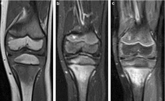
Fig. 1. Acute osteomyelitis of the proximal tibia with an area of intramedullary oedema in the metaphysis with T1 hypointensity (a), STIR hyperintensity (b) and increased
Fig. 2. Punctiform hyperintensity on T1 sequences in the medullary canal and
sub-periosteal space suggestive of acute osteomyelitis of the proximal tibia.
one case. The average duration of intravenous antibiotic therapy was 7.4 days (2–18). The average duration of the oral continuation therapy was 34.9 days (14–75). The oral antibiotics were mainly clindamycin (9 cases) or amoxicillin–clavulanic acid (6 cases). At day 45, all of the abnormal clinical and laboratory findings were completely normal in every case. Radiological analysis at day 45 found periosteal new bone formation in five cases, osteolysis in four cases and both in two cases.
We found no link between the duration of symptoms and the various abnormal MRI findings described. There was a statistically significant relationship between the presence of a subperiosteal abscess and surgery being performed (55.6% vs. 9.1%; P = 0.05). However, there was no relationship with any of the other abnor-mal MRI findings described. We found no statistical relationship between the various MRI criteria and the regression of clini-cal signs and CRP levels on days 3 and 45. We also found no
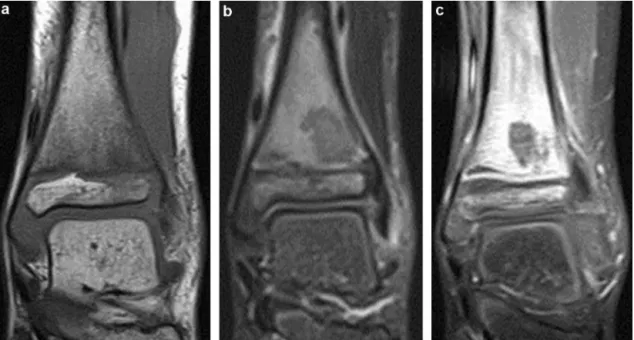
Fig. 3. Lesion with STIR hyperintensity in the metaphysis and epiphysis junction
of the distal tibia with hypointense sclerotic margins and hyperintense peripheral oedema.
statistical relationship between the various MRI criteria and abnor-mal radiological changes on day 45.
4. Discussion
The goal of this study was to test the hypothesis that osteomyeli-tis had a periosteal origin by performing MRI early on in the disease course. We could not confirm this hypothesis, as metaphyseal involvement was present in every case and no case had an isolated subperiosteal abscess. Looking back at the published studies sug-gesting a periosteal origin, the diagnosis in the Labbé study[4]was based on ultrasonography or computed tomography, which may have been unable to detect an underlying intra-osseous infection. In the case report by Weenders et al.[5], although there was predom-inant periosteal involvement, there also appears to be metaphyseal involvement in the published images.
The main limitation of our study is its lack of statistical power due to the small sample size. This can be explained by the
Fig. 5. Subperiosteal abscess of the distal femur with fluid centre based on STIR hyperintensity (a) with increased uptake in the peripheral shell (b), with small soft tissue
abscess in contact with the periosteal one.
tive inclusion and exclusion criteria, particularly the requirement to carry out the MRI before or within 24 hours of the initiation of antibiotics. In fact, the availability of MRI is still limited[6]and we did not want it to delay the initiation of antibiotics for the purpose of this study.
If we compare our study to other published French studies[7,8]
(comparable in terms of the bacterial ecology), it is interesting to note that our selection criteria induced a selection bias (Table 3). Performing an MRI is more complicated in smaller children, some-times requiring general anaesthesia. As a consequence, the average age of our study population was relatively high (7.1 years). The selection of an older sub-population explains the predominance of S. aureus, high initial CRP levels (95 ng/mL) and high number of pos-itive blood cultures (60%). This means that Kingella kingae infections were excluded as they are only present in children under 4 years of age[8]. The fact that we focussed solely on the long bones may also have contributed to excluding Kingella infections, as small bone involvement is characteristic of this bacteria[9].
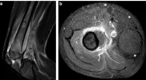
Fig. 6. Ulnar distal metaphyseal intra-osseous abscess in a 2-year-old child with
oval liquid centre in T1 hypointensity (a), STIR hyperintensity, (b) without increase gadolinium uptake (c). The injected sequences help to clearly see the ring-shaped uptake in the wall of the abscess (c).
Table 3
Comparison of published French studies on acute osteomyelitis.
Doit et al.[7] Ferroni et al.[8] Current study
Age (years) 2.3 3.4 7.1
CRP (ng/mL) 45 98
Blood culture (%) 9.7 60
Staphylococcus aureus (%) 67 57 93
Kingella kingae (%) 5 30 0
In our study, the total duration of antibiotic therapy was long: the mean duration of intravenous treatment was 7 days (2–18) and the oral treatment was 35 days (14–75). This is much longer than recent recommendations of 2–4 days of intravenous treatment and 21 days of total treatment[10,11]. In reality, because of the strong inflammatory syndrome, the treatment had to be extended until the clinical and laboratory signs had normalized.
We also could not identify prognostic factors for abnormal MRI findings. This could also have been due to the study’s low statistical power, but also to the antibiotic therapy’s good efficacy, with a 100% healing rate at day 45. There also was no correlation between MRI lesions and the duration of symptoms. This made it impossible to establish a chronology for the appearance of these lesions. We also found no link between abnormal MRI findings and the type of microorganism due of the predominance of S. aureus in our study. MRI made it possible to diagnose more subperiosteal abscesses (9 cases) than ultrasonography (3 cases). This may have led to more surgical procedures being performed in our study than ultrasono-graphy only (7 cases, 35%). This leads us to wonder whether certain abscesses could have resolved with antibiotics only and if there is a critical abscess size that should be drained surgically.
Thus, it seems essential to extend this type of study, since a larger population will be needed to define prognostic factors. It is also possible that one day we will better modulate the type and duration of the antibiotic therapy based on these factors, in a man-ner that optimises antibiotic usage.
5. Conclusion
Our study supports a metaphyseal origin for osteomyelitis but can not eliminate the possibility of forms with a periosteal origin. Various abnormal changes were found on MRI, but no prognostic value could be identified. The implication of these findings from a therapeutic viewpoint needs to be defined with a larger study population.
Disclosure of interest
The authors declare that they have no competing interest.
References
[1]Steer AC, Carapetis JR. Acute hematogenous osteomyelitis in children: recog-nition and management. Paediatr Drugs 2004;6:333–46.
[2]Mazur JM, Ross G, Cummings J, Hahn GA, McCluskey WP. Usefulness of mag-netic resonance imaging for the diagnosis of acute musculoskeletal infections in children. J Pediatr Orthop 1995;15:144–7.
[3]Trueta J. Treatment of acute osteomyelitis. Lancet Lond Engl 1948;2(6515):68.
[4]Labbé J-L, Peres O, Leclair O, Goulon R, Scemama P, Jourdel F, et al. Acute osteomyelitis in children: the pathogenesis revisited? Orthop Traumatol Surg Res 2010;96:268–75.
[5]Weenders SGM, Janssen NE, Landman GWD, van den Berg FP. Subperiosteal abscess in a child. Trueta’s osteomyelitis hypothesis undermined? Orthop Trau-matol Surg Res 2015;101:763–5.
[6]Jaramillo D. Infection: musculoskeletal. Pediatr Radiol 2011;41(Suppl. 1): S127–34.
[7]Doit C, Bonacorsi S, Mariani P, Lorrot M, Mazda K, Pennec¸ot G, et al. COL7-01 Épidémiologie des infections ostéo-articulaires (IOA) chez l’enfant: étude rétrospective de 177 cas documentés entre 2000 à 2008. Med Mal Infect 2009;39(Suppl. 1):S12.
[8]Ferroni A, Al Khoury H, Dana C, Quesne G, Berche P, Glorion C, et al. Prospec-tive survey of acute osteoarticular infections in a French paediatric orthopedic surgery unit. Clin Microbiol Infect 2013;19:822–8.
[9]Kanavaki A, Ceroni D, Tchernin D, Hanquinet S, Merlini L. Can early MRI dis-tinguish between Kingella kingae and Gram-positive cocci in osteoarticular infections in young children? Pediatr Radiol 2012;42:57–62.
[10]Jagodzinski NA, Kanwar R, Graham K, Bache CE. Prospective evaluation of a shortened regimen of treatment for acute osteomyelitis and septic arthritis in children. J Pediatr Orthop 2009;29:518–25.
[11]Pääkkönen M, Peltola H. Antibiotic treatment for acute haematogenous osteomyelitis of childhood: moving towards shorter courses and oral admin-istration. Int J Antimicrob Agents 2011;38:273–80.