O
pen
A
rchive
T
oulouse
A
rchive
O
uverte
(OATAO)
OATAO is an open access repository that collects the work of some Toulouse
researchers and makes it freely available over the web where possible.
This is
an author'sversion published in:
https://oatao.univ-toulouse.fr/23079Official URL :
https://doi.org/10.1016/j.diii.2017.09.003
To cite this version :
Any correspondence concerning this service should be sent to the repository administrator: tech-oatao@listes-diff.inp-toulouse.fr
Faruch Bilfeld, Marie and Lapègue, Franck and Sans, Nicolas and Chiavassa Gandois,
Hélène and Laumonerie, Pierre and Larbi, Ahmed Ultrasonography study of the
suprascapular nerve. (2017) Diagnostic and Interventional Imaging, 98 (12). 873-879. ISSN
2211-5684
OATAO
Ultrasonography
study of the suprascapular
nerve
M. Faruch Bilfeld
a,∗, F. Lapègue
a, N. Sans
a,
H. Chiavassa Gandois
a, P. Laumonerie
b, A. Larbi
caRadiology Department, CHU Toulouse-Purpan, place du Docteur-Baylac, 31059 Toulouse cedex 9, France
bAnatomy Laboratory, Toulouse Rangueil Faculty of Medicine, 133, route de Narbonne, 31062 Toulouse, France
cRadiology Department, hôpital universitaire Carémeau, place du Pr.-Robert Debré, 30029 Nîmes, France KEYWORDS Suprascapular nerve; Ultrasonography; Cadaver study; Anatomical study; Healthy volunteers Abstract
Purpose: The aim of the study was to evaluate the assessability of the suprascapular nerve (SSN) by ultrasonography in cadavers and healthy volunteers.
Materials and methods: With ultrasonography guidance, needles were placed at origin of the SSN of four cadavers and evaluated by dissection. Two blinded radiologists performed 60 ultra-sonography scans in 30 healthy volunteers to study the entire SSN at five anatomical landmarks.
Results: Dissection revealed that the needles were correctly located at the nerve’s origin. There were no significant differences between the two radiologists’ measurements of nerve size and depth. The interobserver correlation for the description of the nerve at the five predefined anatomical landmarks was very good (ICC = 0.7—1).
Conclusion: Five anatomical landmarks were used to analyze the SSN with ultrasonography. Its supraclavicular portion was easier to describe than its scapular portion; a segment of the SSN was not visible between these two portions.
Abbreviations: ASM, anterior scalene muscle; BMI, body mass index; BP, brachial plexus; C5, C5 root; ICC, intraclass correlation coefficient; ISM, infraspinatus muscle; MSM, middle scalene muscle; OHM, omohyoid muscle; R1, first rib; SD, standard deviation; SSA, suprascapular artery; SSM, supraspinatus muscle; SSN, suprascapular nerve; STpd, posterior division of superior trunk; STad, anterior division of superior trunk; TCA, transverse cervical artery.
∗Corresponding author.
Introduction
Suprascapular nerve (SSN) neuropathies are usually caused by direct compression due to a labral or mucoid cyst. SSN is also the nerve most often implicated in neuralgic amyotro-phy[1,2]. Magnetic resonance imaging (MRI) is currently the gold standard for the investigation of SSN neuropathy[3]. As it is difficult to view the SSN with MRI in most patients because the nerve is too small, MRI is used to look for indi-rect signs of SSN damage such as the presence of denervation and to look for the cause of compression[4]. Knowledge of ultrasonography anatomy of the SSN could help in the diag-nosis and understanding of SSN neuropathies, in order to identify direct signs of nerve damage.
The SSN arises from the superior trunk of the brachial plexus (BP). It originates in the ventral rami of the C5 and C6 cervical nerves and occasionally C4 (Fig. 1) [5]. It arises about 3 cm above the clavicle then descends laterally into the supraclavicular fossa and dives under the omohy-oid muscle (OHM)[6]. It then adopts a lateral and posterior course and penetrates the supraspinatus fossa by making its way in an osteofibrous canal between the superior trans-verse ligament and the suprascapular notch (Fig. 1). It goes towards the spinoglenoid notch where its terminal branches enter the infraspinatus muscle (ISM)[6]. The SSN is a mixed nerve. It provides motor innervation to the supraspinatus (SSM) and infraspinatus muscles, and sensory innervation to the acromioclavicular joint, subacromial bursa, and gleno-humeral joint. The distribution of the cutaneous sensory territory varies[7].
The development of SSN blocks has improved our knowl-edge of the ultrasonographic anatomy of the SSN; however, published studies have only described isolated anatomical landmarks that can be used to view the nerve in discrete locations[8—10]. Martinoli et al. used ultrasonography to describe several cases of suprascapular neuropathy, how-ever the damaged SSN was not viewed directly[11].
The aim of the study was to evaluate the assessability of the SSN by ultrasonography in cadavers and healthy volun-teers.
Materials and methods
Ultrasonography in cadavers
The brachial plexuses of four fresh cadavers (82- and 79-year-old males, 83- and 80-79-year-old females) (eight BPs in all) were evaluated to determine how well ultrasonography can pinpoint the origin of the SSN in the brachial plexus. For this portion of the study, a portable ultrasound machine (Sonosite®, General Electric-Healthcare , Milwaukee, WI, USA) with a 12-MHz superficial probe was used.
Under ultrasonography guidance, a radiologist with 20 years’ experience in musculoskeletal imaging (F.L.) located the origin of the SSN in the left and right limbs, and then placed a 21G needle using an in-plane approach, at the point where the nerve arises from the brachial plexus. The BP was then dissected by an orthopedic surgeon with 5 years’ experience (P.L.) through the supraclavicular route along the needle track to confirm that the needle was correctly positioned at the origin of the SSN.
Ultrasonography in healthy volunteers
This portion of the study was approved by our local research ethics board and each volunteer provided written consent for participation. The volunteers had no history of trauma, medical condition or surgery in the cervical area or shoulder girdle.
All ultrasonography scans were performed bilaterally (60 total scans of the cervical area) by two radiologists with 20 and 12 years’ experience in musculoskeletal imaging (F. L, M. F.-B.) who were blinded to each other’s findings.
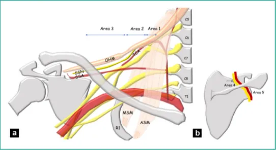
Figure 1. a: topography of the SSN in the suprascapular region. Area 1: origin of SSN on the superior trunk of the BP in the interscalene
triangle delimited anteriorly by the ASM and posteriorly by the MSM. Area 2: SSN across from the first rib (R1). It crosses the TCA. Area 3: SSN in the lateral supraclavicular fossa below the OHM. It is accompanied by the SSA; b: topography of the SSN in the scapular region. Area 4: the SSN (yellow) heads into the supraspinatus fossa after passing in the suprascapular notch (arrow) appended to the SSA (red). Area 5: the SSN in the spinoglenoid notch.
A 14—18 MHz probe was used (Aplio®500, Toshiba, San Jose, California, United States).
The ultrasonography examination was performed using the following protocol:
• for the ultrasonography analysis of the cervical portion of the SSN, the patient lay on the examination table, with the arm along the body and head in neutral position. The axial plane of the BP was scanned[12,13]. In the intersca-lene triangle, the most superficial C5 root was identified [14]. Using the ‘‘elevator technique’’, the C5 root and then the superior trunk were followed until the origin of the SSN was located. The elevator technique consists of follow a tube-like structure (nerve or tendon) along is minor axis with ultrasound. The SSN was then analyzed and followed with the ‘‘elevator technique’’ in the axial plane until it dove deep into the supraspinatus fossa; • to assess the scapular portion of the SSN, the patient
sat across the examiner with the arm hanging alongside the body in neutral position. The SSN was analyzed suc-cessively in the supraspinatus fossa and the spinoglenoid notch.
Five ultrasonographic landmark areas were analyzed along the SSN course:
• area 1: the nerve’s origin at the interscalene triangle. The nerve’s smallest diameter and distance from the skin were measured. We determined whether the nerve originated at C5, the superior trunk or a division of the superior trunk (Fig. 2a);
• area 2: at the first rib across from the transverse cer-vical artery (TCA) [10]. The nerve’s smallest diameter and distance from the skin were measured in centimeters (Fig. 2b);
• area 3: in the lateral supraclavicular fossa below the OHM. The nerve’s smallest diameter and distance from the lat-eral edge of the subclavian artery to where the nerve was no longer visible were measured (Fig. 2c);
• area 4: in the supraspinatus fossa. The exposure of the nerve was graded on a 3-point scale: 0 - suprascapular neurovascular bundle is not visible, 1 - neurovascular bun-dle is visible but not distinguishable from the SSN, 2 - SSN is visible (Fig. 3a);
• area 5: in the spinoglenoid notch. The exposure of the nerve was graded on a 3-point scale: 0 - suprascapular neurovascular bundle is not visible, 1 - neurovascular bun-dle visible but not distinguishable from the SSN, 2 - SSN is visible (Fig. 3b).
Statistical analysis
The measurements made by the most experienced radi-ologist (FL) were used in the descriptive analysis. Those of the second radiologist (MFB) were used to assess mea-surement reproducibility. The descriptive analysis consisted of calculating mean and standard deviation values (SD). To evaluate the reproducibility of the findings, the inter-observer agreement was determined by calculating the intraclass correlation coefficient (ICC) and with Student’s
t-test. Reproducibility of 0.81 < ICC < 1 was considered very
good; 0.61 < ICC < 0.80 good, 0.41 < ICC < 0.60 moderate and ICC < 0.40 poor. Statistical tests where the P-value was below
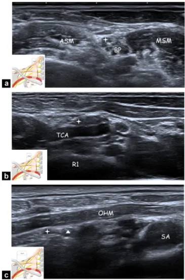
Figure 2. Ultrasonography of the cervical portion of the SSN; a:
area 1: axial ultrasonography view showing the SSN (star) where it arises from the BP at the level of the interscalene triangle between ASM and MSM; b: area 2: axial ultrasonography view showing the SSN (star) over the first rib (R1) crossing the TCA; c: area 3: axial ultrasonography view showing SSN (star) and the SSA (triangle) in the lateral supraclavicular fossa going under the OHM. SA corresponds to the subclavian artery.
0.05 were considered statistically significant. The statistical tests were carried out with MedCalc® software.
Results
Ultrasonography in cadavers
The needles at the origin of the SSN were visible in all four cadavers after dissection of the musculocutaneous layers; it was located a mean of 1 mm (SD: 0.8 mm) from the origin. In three cadavers, the SSN arose directly from the superior trunk of the brachial plexus (BP); in one cadaver, it arose from the posterior branch of the superior trunk (Fig. 4a, b).
Ultrasonography in healthy volunteers
Thirty healthy volunteers (12 men, 18 women) ranging from 25 to 56 years of age (mean age: 33.4 years, mean weight:
Figure 3. Ultrasonography of the scapular portion of the SSN; a: area 4: axial ultrasonography view showing the SSN (star) and the SSA (triangle) in the supraspinatus fossa, under the supraspinatus muscle (SSM); b: area 5: axial ultrasonography view showing the SSN (star) and the SSA (triangle) in the spinoglenoid notch (small arrow), under the infraspinatus muscle (ISM).
Figure 4. Anterosuperior view of the right supraclavicular region
in a cadaver; a: photo: superior view of the right supraclavicular region showing the needle positioned with ultrasonography guid-ance at the origin of the SSN (star) on the superior trunk (ST) after dissection of the skin layers; b: photo: right lateral oblique view showing the origin of the SSN. In this case, the SSN originates after the ST division on the posterior division of the superior trunk (STpd). Here, STad corresponds to anterior division of superior trunk, C5 corresponds to C5 root, and OHM corresponds to omohyoid muscle.
65.9 kg, mean height: 1.71 m and mean body mass index [BMI]: 22.4 kg/m2) participated in this portion of the study.
The SSN was visible over its cervical course by ultrasono-graphy in 100% of cases (ICC = 1) where it originates in the interscalene triangle, 100% of cases (ICC = 1) over the first rib and 100% of cases (ICC = 1) in the lateral cervical area. The diameter of the SSN and depth of the SSN relative to the skin plane are given inTable 1. The measurements made by the two radiologists did not differ significantly. Student’s t-test values are also listed inTable 1. The TCA was visible at the level of the first rib in 93.3% of cases (56/60) for observer 1 and in 90% of cases (54/60) for observer 2 (ICC = 0.89; 95% CI: 0.84—0.92).
The nerve was not visible when it passed under the trapezium muscle, between the lateral supraclavicular fossa and the supraspinatus fossa. Both observers could not fol-lowed it in all cases with the elevator technique in the lateral aspect of the lateral subclavicular fossa (area 3), as the SSN changes it orientation and dives under the trapez-ius muscle. During its deep course under the trapeztrapez-ius, the nerve could not be viewed in the axial plane (100% of cases, 60/60) using the elevator technique, for both observers. The SSN was visible again when it emerges in the supraspinatus fossa (area 4).
The exposure of the SSN in the supraspinatus fossa and in the spinoglenoid notch are given inTable 2.
The SSN was found to arise from the superior trunk in 88% of cases (53/60) and from a division of the superior trunk in 13% (7/60) for observer 1, from the superior trunk in 81.5% of cases (49/60) and from a division of the superior trunk in 18.5% (11/60) for observer 2 (ICC = 0.71; 95% CI: 0.56—0.81).
Discussion
Based on this study’s findings, five anatomical landmarks can be used to analyze the SSN with ultrasonography. SSN can be followed using the ‘‘elevator technique’’ from its origin until
Table 1 Diameter and depth of the SSN in its supraclavicular course. Mean in mm (SD, min—max) Student’s t-test (P value) Observer 1 Observer 2 Area 1 Interscalene triangle Nerve diameter 1.3 (± 0.2; 1—1.9) 1,4 (± 0.2; 1.1—1.9) 0.18 Depth relative to skin 5.1 (± 1.7; 2.7—10.6) 5 (± 1.5; 2.7—11.1) 0.21
Area 2
Costoclavicular outlet
Nerve diameter 1.4 (± 0.2; 0.9—2.3) 1.3 (± 0.2; 1—2.1) 0.17 Depth relative to skin 5.9 (± 1.2; 2.8—12.8) 6.1 (± 1.4; 2.6—13.2) 0.24
Area 3
Lateral supraclavicular fossa
Nerve diameter 1.3 (± 0.2; 1—2.2) 1.3 (± 0.3; 1.1—1.9) 0.74 Distance to lateral edge of subclavian artery 18.4 (± 7.1; 5—39) 19.2 (± 7.5; 6—36) 0.12
Table 2 Exposure of the SSN in its scapular course.
Observer 1 Observer 2 ICC
Supraspinatus fossa 0.81 (95% CI: 0.70—0.88)
Grade 0 26.7% (16/60) 28.5% (17/60)
Grade 1 58.3% (35/60) 60% (36/60)
Grade 2 15% (9/60) 11.5% (7/60)
Spinoglenoid notch 0.91 (95% CI: 0.85—0.94)
Grade 0 8.5% (5/60) 8.5% (5/60)
Grade 1 60% (36/60) 63.5% (38/60)
Grade 2 31.5% (19/60) 28% (17/60)
it exits the lateral supraclavicular fossa where it changes orientation and dives under the trapezius muscles.
Because the nerve is superficial during its supraclavi-cular course (mean depth of 5.1 mm at origin and 5.9 mm at first rib), it can be analyzed accurately with a high-frequency probe. Ultrasonography is more effective for direct viewing of the SSN in the cervical region than in the scapular region: it is visible in 100% of cases in the cervi-cal region for both observers. Based on our findings, it is more difficult to view the SSN directly in the scapular area. Nevertheless, the neurovascular bundle (without specifically distinguishing the nerve) was found in 58% of cases in the supraspinatus fossa and 60% of cases in the spinoglenoid notch. Thus, either the SSN or the neurovascular bundle was visible in the supraspinatus fossa in 73% of cases and in the spinoglenoid notch in 91% of cases. This leads us to believe that ultrasonography is a good imaging modality for rul-ing out extrinsic compression of the SSN durrul-ing its scapular course, but that it would be unreliable for analyzing changes in the nerve’s structure. This ultrasonography technique is reproductible. Our assessment of interobserver repro-ducibility found no significant differences between the two radiologists in the diameter or depth measurements in the cervical area. The nerve can be viewed reproducibly over its scapular course in the supraspinatus fossa (ICC = 0.81; 95% CI: 0.70—0.88) and spinoglenoid notch (ICC = 0.91; 95% CI: 0.85—0.94).
This was the first study to describe a method for analyzing the SSN from its origin to its terminus with ultrasonogra-phy using five anatomical landmarks. Viewing the SSN’s origin with ultrasonography has been already described by Martinoli et al. [11]. In a study of the vascularity of the supraclavicular fossa, Mulhy et al. also described the TCA ultrasonography-based anatomy, but did not describe the relationship between the SSN and TCA [10]. Anesthesiolo-gists use OHM as anatomical landmark to carry out SSN blocks by the anterior approach[9,15]. Martinoli et al. described the SSN scapular course with ultrasonography in patients with nerve compression by mucoid cysts[11]. Being familiar with several anatomical landmarks over the nerve’s course (C5 root, TCA and OHM) makes it easier to recognize the SSN even when faced with anatomical variations such as a bifid or short OHM or a missing TCA[10,16].
We found that the nerve originated in the superior trunk in 88% of cases (53/60) and in a division of the superior trunk in 13% of cases (7/60). However, the interobserver reproducibility (ICC = 0.71; 95% CI: 0.56—0.81) showed this analysis to be challenging. In a cadaver study with 100 spec-imens, Arad et al. described 61 cases in which the origin was after the division of the superior trunk, 29 cases at the divi-sion, 6 cases at the superior trunk, and 4 cases at C5[17]. The differences with our study could be explained by the nerve fascicles originating upstream of the ‘‘anatomical’’ division of the sheaths. This left us wondering whether
ultrasonography was showing the nerve itself surrounded by the epineurium or showing its fascicles and whether the ori-gin of the fascicles was more proximal than the oriori-gin of the nerve itself[18]. A histological study will be needed to answers these questions[19].
There are two major clinical benefits to being able to see the SSN with ultrasonography. The first is that directly seeing the SSN makes it possible to carry out an ultrasound-guided suprascapular block. Anesthetic blocks of the SSN are done for chronic shoulder pain such as adhesive capsulitis or chronic rheumatic pain[20,21]. SSN blocks are also done intraoperatively or postoperatively, alone or in combination with axillary nerve block, partic-ularly for rotator cuff surgery[22,23]. Two approaches have been described in the literature for performing ultrasound-guided SSN block: posterior approach aiming at the nerve in the spinoglenoid notch and a supraclavicular approach [9,15,24]. There are no published studies comparing these two approaches. Our study shows that the SSN is easier to view in the supraclavicular region; consequently, we recommend doing ultrasound-guided SSN blocks through a supraclavicular approach. It is important to know the anatomical relationships of the SSN with the transverse cer-vical artery in order to evaluate the iatrogenic risks related to injection of an anesthetic [15,25,26]. The second clin-ical benefit revolves around exploration of suprascapular neuropathies. With imaging, the diagnosis of suprascapular neuropathies is based on finding direct signs (pathological changes in the SSN) and indirect signs (muscle denerva-tion edema) of nerve damage [27—30]. MRI is currently the gold standard for exploring SSN neuropathy by high-lighting denervation-related hypersignal in the SSM and ISM [4,31,32]. With technical advancements in MRI improving spatial resolution, two small cases studies have reported the SSN was larger and hyperintense over its scapular course [3,33]. However, it is difficult to view the SSN with MRI because the nerve is so small [4]. Ultrasound has excel-lent spatial resolution[34]. In our study, the SSN could be located in 100% of healthy volunteers over its supraclavicular course, leading us to recommend using US to study the SSN in the context of suprascapular neuropathy. Ultrasonogra-phy does not allow to explore the SSN under the trapezium muscle whereas MRI should do. To our knowledge, there is no published case describing specific lesions in this area. Given its low cost and ease of access, it could be used along-side MRI when the latter cannot discern the etiology of the neuropathy.
Our study has certain limitations. The pilot cadaver study included only four specimens (8 plexuses) and dissection was needed to confirm the needle position at the origin of the SSN on the plexus. Nevertheless, the origin of the nerve is the most important anatomical landmark; once this land-mark is identified, the nerve is followed over its course by ultrasonography with an axial translation of the probe. Our population included young subjects with a BMI between 18 and 25 kg/m2, which made the ultrasonography easier
to carry out. There was no gold standard for the evalu-ation on volunteers. It would have been more precise to measure both largest and smallest diameters. We did not evaluate our technique in obese subjects or patients with muscle denervation; the results may have been different in these cases. Since US is operator-dependent, the findings
may be less robust when the examination is performed by less-experienced radiologists.
The SSN can be analyzed with ultrasonography from its origin on the BP to its terminus in the infraspinatus fossa using five anatomical landmarks over its course. Over its supraclavicular course, ultrasonography provides an excel-lent view of the SSN in a population of healthy volunteers. The SSN is harder to see in the scapular area. The nerve cannot be seen with US between the supraclavicular and infraclavicular regions.
Acknowledgments
The authors wish to thank Joanne Archambault, PhD, for the editorial assistance provided during the preparation of this manuscript.
Disclosure of interest
The authors declare that they have no competing interest.
References
[1]Zehetgruber H, Noske H, Lang T, Wurnig C. Suprascapular nerve
entrapment. A meta-analysis. Int Orthop 2002;26:339—43.
[2]Gaskin CM, Helms CA. Parsonage—Turner syndrome: MR
imag-ing findimag-ings and clinical information of 27 patients. Radiology 2006;240:501—7.
[3]Ahlawat S, Wadhwa V, Belzberg AJ, Batra K, Chhabra A.
Spec-trum of suprascapular nerve lesions: normal and abnormal neuromuscular imaging appearances on 3-T MR neurography. Am J Roentgenol 2015;204:589—601.
[4]Blum A, Lecocq S, Louis M, Wassel J, Moisei A, Teixeira P. The
nerves around the shoulder. Eur J Radiol 2013;82:2—16.
[5]Shin C, Lee S-E, Yu K-H, Chae H-K, Lee K-S. Spinal root origins
and innervations of the suprascapular nerve. Surg Radiol Anat 2010;32:235—8.
[6]Warner JP, Krushell RJ, Masquelet A, Gerber C. Anatomy
and relationships of the suprascapular nerve: anatomical con-straints to mobilization of the supraspinatus and infraspinatus muscles in the management of massive rotator-cuff tears. J Bone Joint Surg Am 1992;74:36—45.
[7]Ajmani ML. The cutaneous branch of the human suprascapular
nerve. J Anat 1994;185:439—42.
[8]Chan C, Peng PWH. Suprascapular nerve block: a narrative
review. Reg Anesth Pain Med 2011;36:358—73.
[9]Battaglia PJ, Haun DW, Dooley K, Kettner NW. Sonographic
measurement of the normal suprascapular nerve and omohyoid muscle. Man Ther 2014;19:165—8.
[10]Muhly WT, Orebaugh SL. Sonoanatomy of the vasculature at the
supraclavicular and interscalene regions relevant for brachial plexus block. Acta Anaesthesiol Scand 2011;55:1247—53.
[11]Martinoli C, Gandolfo N, Perez MM, Klauser A, Palmieri F, Padua
L, et al. Brachial plexus and nerves about the shoulder. Semin Musculoskelet Radiol 2010;14:523—46.
[12]Martinoli C, Bianchi S, Santacroce E, Pugliese F, Graif M, Derchi
LE. Brachial plexus sonography: a technique for assessing the root level. Am J Roentgenol 2002;179:699—702.
[13]Demondion X, Herbinet P, Boutry N, Fontaine C, Francke J-P,
Cotten A. Sonographic mapping of the normal brachial plexus. Am J Neuroradiol 2003;24:1303—9.
[14] Lapegue F, Faruch-Bilfeld M, Demondion X, Apredoaei C, Bayol MA, Artico H, et al. Ultrasonography of the brachial plexus, normal appearance and practical applications. Diagn Interv Imaging 2014;95:259—75.
[15] Siegenthaler A, Moriggl B, Mlekusch S, Schliessbach J, Haug
M, Curatolo M, et al. Ultrasound-guided suprascapular nerve block, description of a novel supraclavicular approach. Reg Anesth Pain Med 2012;37:325—8.
[16] Rai R, Ranade A, Nayak S, Vadgaonkar R, Mangala P,
Krishna-murthy A. A study of anatomical variability of the omohyoid muscle and its clinical relevance. Clin Sao Paulo Braz 2008;63:521—4.
[17] Arad E, Li Z, Sitzman TJ, Agur AM, Clarke HM. Anatomic sites of
origin of the suprascapular and lateral pectoral nerves within the brachial plexus. Plast Reconstr Surg 2014;133:20e—7e.
[18] Reina MA, Villanueva MC, Machés F, Carrera A, López A, De
Andrés JA. The ultrastructure of the human spinal nerve root cuff in the lumbar spine. Anesth Analg 2008;106:339—44.
[19] Siqueira MG, Foroni LHL, Martins RS, Chadi G, Malessy MJA.
Fascicular topography of the suprascapular nerve in the C5 root and upper trunk of the brachial plexus: a microanatomic study from a nerve surgeon’s perspective. Neurosurgery 2010;67:402—6.
[20] Checcucci G, Allegra A, Bigazzi P, Gianesello L, Ceruso M, Gritti
G. A new technique for regional anesthesia for arthroscopic shoulder surgery based on a suprascapular nerve block and an axillary nerve block: an evaluation of the first results. Arthrosc J Arthrosc Relat Surg 2008;24:689—96.
[21] Shanahan EM, Ahern M, Smith M, Wetherall M, Bresnihan B,
FitzGerald O. Suprascapular nerve block (using bupivacaine and methylprednisolone acetate) in chronic shoulder pain. Ann Rheum Dis 2003;62:400—6.
[22] Lee JJ, Kim D-Y, Hwang J-T, Lee S-S, Hwang SM, Kim GH, et al.
Effect of ultrasonographically guided axillary nerve block com-bined with suprascapular nerve block in arthroscopic rotator cuff repair: a randomized controlled trial. Arthrosc J Arthrosc Relat Surg 2014;30:906—14.
[23] Bennett DL, Cronin AM, Palmer WE, Kattapuram SV, Huang
AJ. Optimization and standardization of technique for
fluoroscopically guided suprascapular nerve blocks. Am J Roentgenol 2014;202:576—84.
[24]Harmon D, Hearty C. Ultrasound-guided suprascapular nerve
block technique. Pain Physician 2007;10:743—6.
[25]Rothe C, Steen-Hansen C, Lund J, Jenstrup MT, Lange KHW.
Ultrasound-guided block of the suprascapular nerve — a vol-unteer study of a new proximal approach. Acta Anaesthesiol Scand 2014;58:1228—32.
[26]Hackworth RJ. A new and simplified approach to target
the suprascapular nerve with ultrasound. J Clin Anesth 2013;25:347—8.
[27]Scalf RE, Wenger DE, Frick MA, Mandrekar JN, Adkins MC. MRI
findings of 26 patients with Parsonage—Turner syndrome. Am J Roentgenol 2007;189:W39—44.
[28]Pottecher P, Flageul B, Sibileau E, Laredo J-D, Bousson V.
Peripheral hypertrophic neuropathy due to leprosy: ultrasound and MR imaging findings. Diagn Interv Imaging 2016;97:471—3.
[29]Deniel A, Causeret A, Moser T, Rolland Y, Dréano T,
Guillin R. Entrapment and traumatic neuropathies of the elbow and hand: an imaging approach. Diagn Interv Imaging 2015;96:1261—78.
[30]Dikici AS, Bakan S, Kandemirli SG, Sonmez S, Ersen E,
Comunoglu N, et al. Suprascapular nerve compression due to rib osteochondroma: MR imaging features. Diagn Interv Imaging 2016;97:109—11.
[31]Kim S-J, Hong SH, Jun WS, Choi J-Y, Myung JS, Jacobson
JA, et al. MR imaging mapping of skeletal muscle denerva-tion in entrapment and compressive neuropathies. Radiogr Rev 2011;31:319—32.
[32]Ludig T, Walter F, Chapuis D, Molé D, Roland J, Blum A. MR
imag-ing evaluation of suprascapular nerve entrapment. Eur Radiol 2001;11:2161—9.
[33]Sneag DB, Saltzman EB, Meister DW, Feinberg JH, Lee SK,
Wolfe SW. The MRI bullseye sign: an indicator of periph-eral nerve constriction in Parsonage—Turner syndrome. Muscle Nerve 2017;56:99—106.
[34]Nazarian LN. The top 10 reasons musculoskeletal sonography is
an important complementary or alternative technique to MRI. Am J Roentgenol 2008;190:1621—6.