/ /63s
Université de Montréal
Genetically engineering the mouse genome to smdy the raie of the
Phosphotyrosyl Phosphatase Activator (PTPA) gene in the response to oxidative stress
Par Cyril Sabbah
Programme de Biologie Moléculaire
Faculté des études supérieures
Mémoire présenté à la faculté des études supérieures en vue de l’obtention du grade de Maître ès science (M.Sc.)
en biologie moléculaire Août 2005 Grade confere . . àcompterdu ‘ 2OO5[; 06 ©, Cyril Sabbah, 2005 It
(s»1
o
u5i
•1
o
Direction des bibliothèques
AVIS
L’auteur a autorisé l’Université de Montréal à reproduire et diffuser, en totalité ou en partie, par quelque moyen que ce soit et sur quelque support que ce soit, et exclusivement à des fins non lucratives d’enseignement et de recherche, des copies de ce mémoire ou de cette thèse.
L’auteur et les coauteurs le cas échéant conservent la propriété du droit d’auteur et des droits moraux qui protègent ce document. Ni la thèse ou le mémoire, ni des extraits substantiels de ce document, ne doivent être imprimés ou autrement reproduits sans l’autorisation de l’auteur.
Afin de se conformer à la Loi canadienne sur la protection des renseignements personnels, quelques formulaires secondaires, coordonnées ou signatures intégrées au texte ont pu être enlevés de ce document. Bien que cela ait pu affecter la pagination, il n’y a aucun contenu manquant. NOTICE
The author of this thesis or dissertation has granted a nonexclusive license allowing Université de Montréal to reproduce and publish the document, in part or in whole, and in any format, solely for noncommercial educational and research purposes.
The author and co-authors if applicable retain copyright ownership and moral rights in this document. Neither the whole thesis or dissertation, nor substantial extracts from it, may be printed or otherwise reproduced without the author’s permission.
In compliance with the Canadian Privacy Act some supporting forms, contact information or signatures may have been removed from the document. While this may affect the document page count, it does flot represent any Ioss of content from the document.
Ce mémoire intitulé:
Genetically engineering the mouse genome to study the role ofthe
Phosphotyrosyl Phosphatase Activator (PTPA) gene in the response to oxidative stress
Présenté par: Cyril Sabbah
a été évalué par unjurycomposé des personnes suivantes
Eric Milot Président Rapporteur
Dindial Ramotar et Elliot Directeurs de
Drobetsky Recherche
Résumé
L’ oxygène est indispensable à la vie aérobique. Cependant, certains métabolites de cette molécule peuvent devenir des espèces oxygénées activées (EOA), des radicaux libres extrêmement réactifs pouvant causer des dommages à plusieurs composants cellulaires tels que les lipides, les protéines et l’ADN. Les EOA sont une source de stresse inévitable et permanent puisqu’elles sont générées par des processus endogènes; ces molécules sont des sous-produits de la respiration cellulaire et sont générées par des cellules immunitaires pour combattre l’infection de corps étrangers. De plus, ils sont produits à la suite de l’exposition à plusieurs agents exogènes tels que certaines longueurs d’onde émises par le soleil ainsi que de nombreux métaux et drogues. Par conséquent, les cellules ont développés plusieurs mécanismes qui permettent d’empêcher ou de réparer les dommages dus aux EOA. Notamment, la réparation par excision des bases est la voie principale de réparation de l’ADN endommagé par ces radicaux libres, permettant d’éviter à ce que les lésions oxydatives deviennent des mutations permanentes. Cependant, si ces mécanismes sont altérés, les cellules peuvent devenir particulièrement vu]nerables face aux dommages causés par les EOA. En effet, de nombreuses pathologies sont associées au stress oxydatif, tels que certaines maladies cardiovasculaires, neurodégénératives, le diabète et le cancer. Une meilleure connaissance des gènes impliqués dans la réponse cellulaire face aux stresses oxydatifs pourrait nous permettre de mieux comprendre ces maladies sur le plan biologique.
Lors d’un criblage de souches de levures mutantes, l’homologue du gène Phosphotyrosyl Phosphatas e Activator (PIPA) humain, RRD 1, à été identifié comme jouant un rôle important dans le maintient de l’intégrité de l’ADN exposé aux EAO. Quoique le mécanisme d’action et la fonction précise de PTPA ne soient pas bien caractérisés, plusieurs évidences suggèrent que ce gène joue un rôle dans plusieurs processus biologiques, dont certains étant liés à la carcinogenèse. Etant donné l’importance du stresse oxydatif dans l’initiation de certains types de cancer et du rôle de PTPA dans le maintien de l’intégrité l’ADN face à ce stresse, nous avons chercher à étudier ce gène chez les mammifères en produisant de souris ayant soit une délétion, soit une surexpression de la protéine Ptpa. Nous avons préparé la construction d’ADN en vue de créer des souries knockout «conditionnelle» pour ce gène, et avons générer des
colonie de souris ayant intégré une copie supplémentaire du gène PIPA fusionné avec une protéine dérivée de la protéine fluorescente verte sans, pour autant, exprimer le transgene. Une fois que ces souris manipulées génétiquement seront établies, elles pourront être exposées a divers sources de stresse oxydatif. Ceci permettrait l’étude du fonctionnement de PIPA dans la réponse aux EOA.
Mots clés: PIPA, RRD1, stress oxydatif, UVA, dommages à l’ADN, souris transgénique, souris knockout, cre-loxP, protéine verte fluorescente
Abstract
Oxygen molecules are essential for aerobic life. However, certain free radical metabolites of oxygen molecules, Reactive Oxygen Species (ROS), can cause significant damage to cellular lipids, proteins and DNA. ROS are a constant threat to celiular integrity as they are produced by endogenous processes; they are normal byproducts of ceilular respiration and are produced by ceils of the immune system to combat foreign organisms. In addition, exogenous agents such as a subset of solar waveiengths and a number of drugs and metals also generate ROS. Consequently, celis have deveioped a number of methods of containing the damaged caused by ROS, including the Base Excision Repafr pathway, the primary une of defense against oxidative DNA damage, which prevents the accumulation of irreversibie mutations. Therefore, celis in which these mechanisms are altered have increased susceptibiiity to ROS-induced damage. Indeed, a variety of conditions and diseases, such as cardiovascular and neurodegenerative diseases, diabetes, and cancer have been associated to oxidative stress. Therefore, the study of genes involved in the cellular response to ROS may achieve a greater understanding ofthe biology ofthese diseases.
In searching for genes that are involved in the response to oxidative stress, our laboratories have previously identified the yeast homologue of the human Phosphotyrosyl Phosphatase Activator (PIPA) gene as playing an important role in maintaining the integrity of DNA in response to oxidative stress. Although studies have flot yet revealed its specific function or mechanism of action, data suggest that PIPA may have a number of important cellular functions, many of which are consistent with a function of PIPA in pathways related to carcinogenesis. Given the ciinical importance of oxidative stress in certain types of cancer as well as PIPA’ s involvement in maintaining DNA integrity, our laboratories sought to elucidate this gene’s function in mammals by generating PTPA knockout and transgenic mice. Recent advances that aliow for a high degree of “tailoring” of the mouse genome have prompted us to design a targeting vector that wiil enable the generation of “conditionai” knockout mice. In addition, we have produced animais having integrated a DNA constmct which was anticipated to express the Ptpa protein fused with a variant of the Green Fluorescent Protein within thefr genome, although these did not express the transgene per se. When mouse mes with the Ptpa
deletion and overexpression wiil be estabiished, these animais can be exposed to oxidative stress-causing agents. It is expected that these experiments wiiI further oui
understanding ofthis gene’s foie in the response to oxidative stress.
Keywords: PTPA, RRD1, oxidative stress, UVA, DNA damage, transgenic mice, knockout mice, cre-loxP, green fluorescent protein
Table of Contents
List of figures X
List of abbreviations XII
Acknowledgments XIII
1. Introduction 2
1.1. Oxidative stress and disease 2
1.1.1. The Oxygen Paradox 2
1.1.2. Cellular Ioxicity ofROS 2
1.1.3. Sources ofROS 3
1.1.4. Cellular Defence Against ROS Insuit 4
1.1.5. Mutagenic effect of ROS and Melanoma 6
1.2. The Involvement ofthe Phosphotyrosyl Phosphatase Activator Gene in the
Response to Oxidative Stress 7
1.2.1. Identification of Genes with a Role in the Response to Oxidative Stress 7 1.2.2. PIPA Involvement in Maintaining DNA Integrity upon Exposure to
Oxidative Stress 8
1.2.3. Complexity of PTPA Functions 2
1.2.3.1. PP2A Proteins are Multifunctional Phosphatases Involved in
Carcinogenesis 9
1.2.3.2. PIPA: A Survey ofthe Literature 11
1.2.3.3. BiochemicalFunctions ofPtpa 11
1.2.3.4. Ptpa Protein is Conserved in Organisms 12
1.2.3.5. Genetic Features of PIPA 14
1.2.3.6. PTPA’s Role in Response to Oxidative Stress and in Pathways Related to
Carcinogenesis 14
1.2.3.7. Phenotypes Associated with PIPA 16
1.3. Manipulating the Mouse Genome to Study Gene Function and Disease 17
1.3.1 Genetically Modified Mice 17
1.3.2 Conditional Knockout Mice 18
1.3.3 Ihe Use of Green Fluorescent Protein Variants in Mice 21 1.3.4. Genetically Engineering the Mouse Genome to Study Genes Involved in the
Response to Oxidative Stress 21
1.4. Rationale 24
2. Materials and methods 25
2.1. Cloning Iechniques 25
2.1.1. Preparation of Competent Celis for Cloning 25 2.1.2 Transformation of Competent Celis with Plasmid DNA 25 2.1.3. Mini-and Maxi-Preparation Methods for Plasmid Purification 25 2.1.4. Restriction Digestions and DNA Modification for Cloning 26 2.1.5. Polymerase Chain Reaction to Generated Clonable Fragments 26
2.1.6. Fragment Purification for Cloning 27
2.1.7. Ligation Reactions for Cloning ofDNA Fragments 27
2.1.8. General Cloning Strategies 27
2.2. Generating Iransgenic Mice 28
2.2.2 Screening for Transgenic Mice.28
2.2.2.1. Extraction ofMouse Tau Genomic DNA 28
2.2.2.2. Restriction-Digestion and Treatment of Genomic DNA for Southem
Blotting 29
2.2.2.3. Transfer of DNA to the Nitrocellulose Membrane 29
2.2.2.4. Preparation ofthe Radioactive Probe 30
2.2.2.5. Hybridization of Radioactive Probe to DNA on Nitrocellulose Membrane 30
2.2.2.6. Membrane Washes and Exposition 30
2.2.3. Dissections and Preservation oflndividual Organs for Protein Extractions... 31
2.2.4. Western bloffing 31
2.2.4.1. Protein SD$-PAGE Electrophoresis 31
2.2.4.2. Transfer of Proteins to the Nitrocellulose Membrane 31 2.2.4.3. Membrane Blocking, Blotting and Exposure 32
2.2.5. Macroscopic EYFP Detection 32
2.6. Media, Buffers, Antibiotics and Solutions 33
2.6.1. Media 33
2.6.2. Antibiotics 34
2.6.3. Oligonucleotides Used as Primers for PCR Reactions 34
2.6.4. Buffers 35
2.6.5. Solutions 35
3. Resuits 38
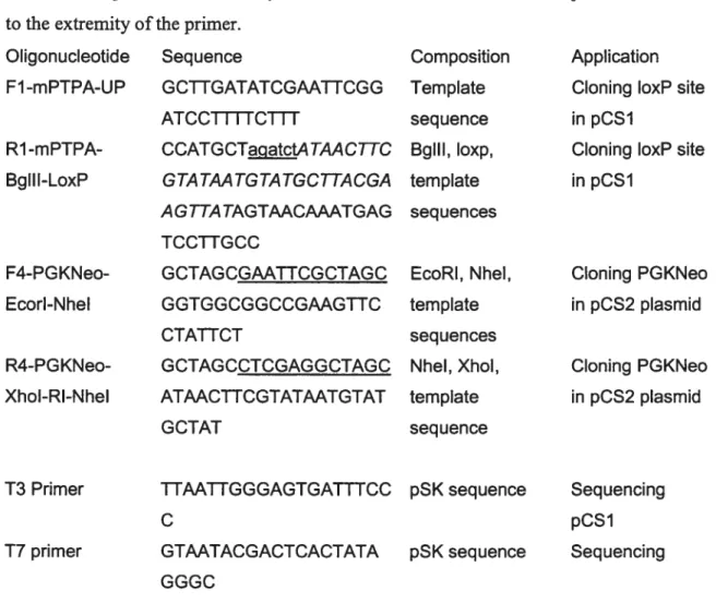
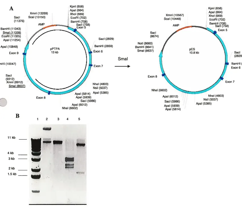
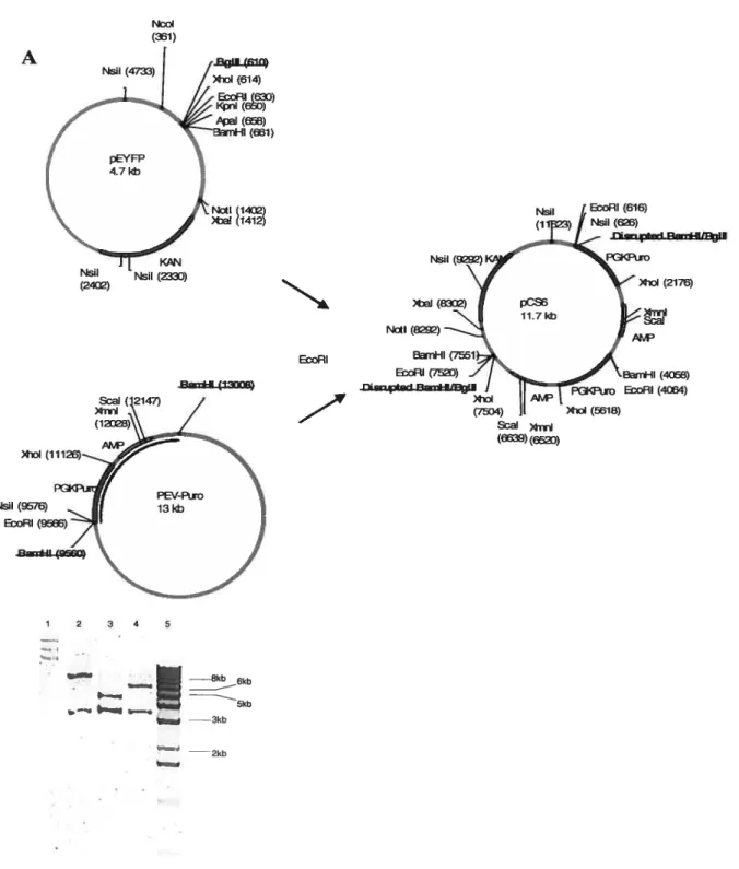
3.1. Cloning of PTPA-Neo-loxP 38
3.1.1. General Cloning Considerations 38
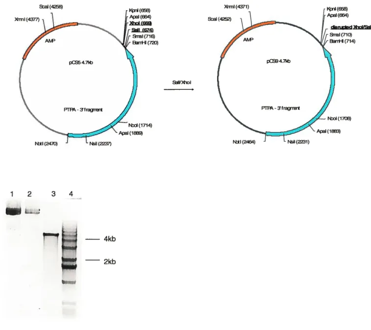
3.1.2. Cloning ofpCS 39 3.1.3. Cloning ofpCSl 41 3.1.4. Cloning ofpCS2 42 3.1.5. Cloning ofpC$4 44 3.1.6. Cloning ofpCS5 46 3.1.7. Cloning ofpCS9 48 3.1.8. Cloning ofpCS6 50 3.1.8. Cloning ofpCS7 52 3.1.9. Cloning ofpCS8 52
3.1.10. Future Cloning ofpCSlO 54
3.2 Transgenic mice 56
3.2.1. Screening for Transgenic Mice by Southem Blot 56 3.2.2. Assessing Transgene Expression by Western Blotting 61 3.2.3. Macroscopic Observation ofEYFP to Assess Transgene Expression 62
4. Discussion 64
4.1. The Creation of Conditional PIPA Knockout Mice Targeting Vector 64 4.1.1. Targeting Vector Must be Isogenic with Mouse ES Strain 65
4.1.2. Utilizing Selection Markers 65
4.1.3. Use ofthe Frt/Flp System 66
4.1.4. Length of the Targeting Construct 67
4.1.5. Incorporation of loxP Sites 68
4.2. The Creation ofMice Overexpressing PIPA and Fused to EYFP 70 4.2.1. Choice of Green Fluorescent Protein Variant 71
4.2.2 Detection ofTransgene Expression 73
4.2.3. Possible Reasons for the Absence ofTransgene Expression 73 4.2.4. Proposed Modifications for Future Transgenic PTPA-GFP Studies 74 4.3. Future Directions for PTPA Conditional Knockout and Transgenic Mice 74
5. Conclusion 76
6.Appendix 79
List of figures
Figure 1: Base Excision Repair pathway 5
figure 2: Ptpa is conserved in organisms 13
Figure 3: Homologous recombination between the targeting vector and ifie endogenous
gene 19
figure 4: The Cre recombinase mediates recombination between loxP sites 20
figure 5: Cloning ofpCS 40
Figure 6: Cloning ofpC$1 41
Figure 7: Cloning ofpCS2 43
Figure 8: Cloning ofpCS4 44
Figure 9: Cloning ofpCSS 47
Figure 10: Cloning ofpCS9 49
Figure 11: CloningofpC$6 51
figure 12: Cloning ofpCS7 53
figure 13: Cloning ofpCS8 54
figure 14: Future cloning ofpCSlO 58
Figure 15: Southem blots for offspring oftransgenic mice 6, 7, 16 59 Figure 16: Western blotting for detection oftransgene expression using (A) anti-PTPA
and(B) 62
Figure 17: Generating PTPA-loxP mice 69
List of tables
Table 1. Oligonucleotide primers used for PCR amplification of fragments for cloning or
List of abbreviations ES: embryonic stem
HR: homologous recombination DNA: deoxyribose nucleic acid
PTPA: Phosphotyrosyl Phosphatase Activator BER: Base Excision Repair
NER: Nucleotide Excision Repafr
dNTP: deoxyribonucleoside triphosphate dCTP: Deoxycytidine 5 ‘-triphosphate PCR: polymerase chain reaction
Neo: neomycin gene conferring resistance to G41 $ Puro: puromycin gene conferring resistance to puromycin Amp: gene conferring resistance to ampicillin
Kan: gene conferring resistance to kanamycin UV: ultraviolet
Ex: Excitation maximum Em: Emission maximum pSK: Bluescript cloning vector
Acknowledgments
Ffrst and foremost, I would like express my gratitude my research dfrectors, Dr. Dindial Ramotar and Dr. Elliot Drobetsky for having granted me the chance to embark on this ambitious and challenging project, as well as for their patience throughout my studies. This has been a fruly worthwhile leaming experience.
In addition, I owe a debt of gratitude to the members of the labs. The moral and technical support that they have provided throughout my studies was greatly appreciated. In particular, I thank Julie Douville for her constant support, advice and laborious efforts. Many friendships have been fostered through this project.
Last but certainly flot least, I would like to thank my family for their encouragement and guidance. Finally, a special thanks to my wife and daughter who patiently tolerated my absence “outside of standard working hours,” and have endured the occasional work-related mood swings, seemingly inherent to scientific research.
1. Introduction
1.1. Oxidative stress and
disease
1.1.1.
The Oxygen ParadoxOxygen is a mixed blessing for aerobic life. On the one hand, it is essential for cellular respiration and energy metabolism. Yet this molecule can become a potent agent capable of causing significant damage to various macromolecules. Denham Harman (1956) first developed the notion that oxygen can be detrimental to the ceil, hypothesizing that oxygen free radicals are involved in general cellular damage, mutagenesis, cancer and aging. Since then, this hypothesis lias been extensively researched (Droge, 2000; Behrend et al., 2003), and many other conditions are cunently associated with oxygen cytotoxicity, including neurodegenerative and heart diseases (Lebovitz et aï., 1996; Norata et al., 2003) and diabetes (Nath et al., 1984).
Oxygen molecules become toxic when thefr metabolites are activated, via a series of intercellular chemical reactions, into free radical reactive oxygen species (ROS), or molecules with an unpafred electron in their outer valence sheil. ROS can be very unstable and highly reactive, “attacking” neiglibouring, generally non-radical, molecules in seeking to fil their valence sheli. In the process, reactive oxygen species damage the “donor” molecules, effectively transforming these into ftee radicals, which in turn, will also seek to fr11 their outer valence shell. Thus, a single free radical can cause a chain reaction, potentially damaging multiple cellular components (Turrens et al., 2003).
1.1.2. CellularToxicity of ROS
Reactive oxygen species cause damage to cellular lipids, proteins and DNA. ROS mediated lipid peroxidation could dismpt the fluidity and permeability of the cytoplasmic and mitochondrial membranes, interfering with the proper functioning of membrane bound proteins (Van der Vliet and Bast, 1992). Reactive oxygen species can also disrupt protein flmction by causing site-specific amino acid modifications, altering proteic electrical charge or triggering peptide fragmentation (Grune et al., 1997). Finaliy, ROS can produce a variety of DNA lesions, inciuding modified bases (Téouie and Cadet,
1978), oxidized apurinic/apyrimidinic sites (Mitra et al., 2002) and strand breaks resulting from damage to the sugar moiety of DNA (Dizdaroglu et al., 1975). These damages can eventually give rise to permanent mutations (Loeb and Preston, 1986). One frequent oxidative DNA lesion is the 7,8-dihydro-8-oxoguanine (8-oxoG) (Groilman and Moriya, 1993). Lefi unrepaired, this premutagenic lesion can become an irreversible OEC—*TA transversion (Shibutani et al., 1991; Cheng et al., 1992). Interestingly, such transversions are ofien present in cancerous ceils, particularly in oncogenes and tumour suppressor genes (Aguilar et al., 1993; Jones et al., 2004; Yokota and Kohno, 2004). Another example ofthe mutagenic effects ofROS, is the deamination of normal cytosine nucleic acids into the premutagenic 5-hydroxyuracil and uracil glycol lesions, which can lead to a CG—*TA transition if left unrepaired (Kreutzer et al., 1992).
Oxygen free radicals are ofien so reactive that they do not diffuse more than a few molecular diameters before reacting with another molecule (Pryor et al., 1996). Thus, the damage localized within the nucleus is probably the result of free radicals produced later in the free radical chain reaction. In fact, quantitatively speaking, it has been estimated that DNA may be the least significant target of ROS (Mamett, 2000). Nonetheless, it is estimated that one human cell produces up to 10,000-20,000 oxidative DNA lesions in a 24-hour period (Ames et al., 1993; Jackson and Loeb, 2001). In addition, damage incurred by DNA is qualitatively of great significance because; whereas other cellular components can be newly synthesized when damaged, unrepaired DNA can Jead to permanent and irreversible mutations.
1.1.3. Sources 0f ROS
It would be expected that organisms would seek to minimize their exposure to reactive oxygen species. However, the formation of ROS is neither abnormal nor rare. As mentioned, ROS produced by mitochondria are natural by-products of cellular respiration, continuously and inevitably endangering celis (Cadenas and Davie, 2000). Further, higher eukaryotes actively produce oxygen free radicals. Neutrophils and macrophages exploit free radical toxicity by generating ROS as a means of destroying foreign bodies during inflammation (Chong et al., 1989; Roos et aÏ., 2003).
In addition, humans are exposed to a variety of exogenous sources of oxidative stress, including ionizing radiation (Ward, 1989), cigarette smoke (Churg, 2003), trace metals found in industrial waste, such as lead, mercury and cadmium (Ercal et al., 2001), and various therapeutic and experimental drugs such bleomycin and adriamycin (Kappus, 1987) and the model carcinogen, 4-NQO (Ramotar et al., 1998). Oxidative stress is also generated upon exposure to the ultraviolet (UV) subset of solar radiation. The sun emits 1.3V light of ail wavelengths, commonly categorized as UVA (400 nm - 320 nm), UVB (320 run - 290 nm), UVC (290 nm - 100 nm) Although UVC is extremely genotoxic, it is
not a major public health concem because wavelengths of less than 300 nm are largely absorbed by ozone in the stratosphere before it reaches the Earth (Latarjet, 1935). Consversely, UVA and UVB wavelengths reach the Earth. These generate chemically distinct types of DNA damage these wavebands. However, both UVA and, to a lesser extent, UVB, can generate R0$ (Cadet et al., 2005). Thus, these exogenous sources of ROS are compounded to the endogenous “load” constantly challenging the integrity of the celi.
1.1.4. Cellular Defence Against ROS Insuit
Organisms have adapted to the continuous and unavoidable exposure to oxygen free radicals by developing several strategies to attenuate ROS cytotoxicity. Various superoxide dismutase (SOD), catalase and glutathione peroxidase enzymes constitute an important me of defence. Briefly, SOD can convert two superoxide radicals (02) into less reactive hydrogen peroxide (H202) (Noor et al., 2002), which is subsequently, converted into water and oxygen in a process mediated by catalase and glutathione peroxidase (de Haan, 1995). In addition, higher eukaryotes produce antioxidants, molecules that provide free radicals with an electron to fil their outer valence sheil (Jacob and Burri, 1996). Together, these mechanisms allow the celi to contain the effects of oxygen free radicals.
However, preventative measures are insufficient to protect the integrity of DNA, which camiot be newly re-synthesised once mutations have incurred. Consequently, celis have developed mechanisms to repair oxidative lesions before these become permanent mutations. Most important is the Base Excision Repafr (BER) pathway, in winch a
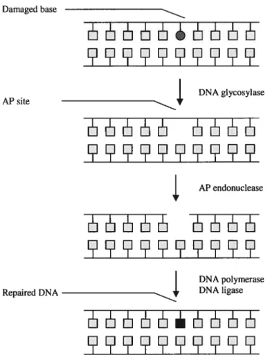
variety of proteins acts sequentially to remove the base and sugar moieties of the damaged nucleic acid, incorporating the appropriate undamaged nucleic acid in its place. Briefty, a DNA glycosylase is recruited to the site of the lesion, disassociating the base from the sugar moiety of the nucleic acid and creating an apurinic/apyrimidic (AP) site (Figure 1). Subsequently, an AP endonuclease disrupts the sugar backbone, leaving bebind a gap in the double stranded structure of the DNA. This gap is filled by a DNA polymerase, and the newly ïncorporated nucleic acid is ligated to the DNA by a ligase (Fortinietal., 2003). Damaged base AP site Repaired DNA DNA glycosylase ]1E —11 AP endonuclease HE5LSL5 E5JJJ JJL1 DNA polymerase DNA ligase
EIL5JJ
Figure 1: Base Excision Repair pathway. This is the most important for the repair mechanism of oxidative DNA lesions
The repafr proteins, such as DNA glycosylases and AP endonucleases, are relatively well conserved in organisms (Barziiay and Hickson, 1995; McCullough et aï., 1999). Studies in yeast and mammalian ceils have demonstrated that ceils with a deficiency in one or several of these genes show an increase in premutagenic lesions and, sometimes, a hypersensitivity to oxidative stress-inducing agents (Waiker et al., 1994, Doetsch et al., 2001, Kim et al., 2002).
Aithough BER is the primary response to oxidative stress-induced DNA lesions, other mechanisms aiso contribute to the repair of these lesions. For example, UVC generates dipyrimidine photoproducts lesions (chemicaily distinct from oxidative lesions) which are primarily repafred by the Nucleotide Excision Repair (NER) pathway. Nonetheless, there is some overlap of these repair pathways; the NER and other repair mechanisms contribute to the repafr of oxidative DNA lesions, albeit to a lesser extent relative to the BER (Scott eÏ al., 1999, Rybanska and Pfrsei, 2003).
1.1.5. Mutagenic effect of ROS and Melanoma
Oxidative damage to DNA may be a significant factor in the dramatic increase of melanoma incidences rates in North America, Europe and Australia in the 1 970s and 1 908s (Arrnstrong and Kricker, 1994, Hall et al. ,1999). The higffly mutagenic UVB component of solar radiation has long been established as the major cause of this disease (de Gruiji, 2000). However, there is a growing consensus that solar UVA plays a more significant role in the development ofmelanoma than was first thought. This is supported by several experiments conducted in murine and human celi mes, whole animais, as well as a number of epidemiologicai studies (Jones et al., 1987, Lundgren et al., 1988, Sterenborg and van der Leun, 1990, Setiow et al. 1993, and Ley, 1994). In addition, various mes of evidence indicate that oxidative stress plays a significant role in cutaneous carcinogenesis. For example, it has been demonstrated that normal melanocytes from melanoma patients have increased sensitivity to peroxidizing agents (Grammatico et al., 1998), that the skin antioxidant defense system is impaired in melanoma celis and normal melanocytes of these patients (Picardo et al., 1996). Further, antioxidant supplementation was found to have a photoprotective effect on ceils (Lopez Torres et al., 1998). Another study demonstrated that compounds that generate oxygen
free radicals increase the effect of malignant conversion of cancerous skin celis (Athar et al., 1989). The authors of ffiis report present the hypothesis that the accumulation of genetic lesions and strand breaks and chromosomal aberrations induced by ROS hasten the process by which celis become malignant.
Thus, although UVB is incontestably more genotoxic than UVA and remains the major factor in the incidence of melanoma, there is growing evidence that UVA-induced reactive oxygen species may play a role in this carcinogenesis that is more significant than was initially thought.
1.2. The Involvement of the Phosphotyrosyl Phosphatase Activator
Gene in the Response to Oxidative Stress
1.2.1. Identification of Genes with a Rote in the Response to Oxidatïve Stress
Given the significant impact of oxidative stress upon human health, our laboratories were interested in identifying novel genes that perform key fimctions in the cellular response to this type of stress, particularly those that play a role in the repair of ROS-induced DNA damage. Jnitially, the budding yeast Saccharomyces cerevisiae was chosen as the organism in which to conduct this search because its genome is well characterized and relatively easy to manipulate. In addition, fundamental processes, including DNA repair pathways, are weIl conserved from yeast to humans. Isolating oxidative stress response genes in yeast could lead to the identification of novel functions ofnew or previously characterized genes in humans.
As described by Ramotar et al. (1998), a wide panel of yeast mutants were subjected to a three-step screen. Initially, mutants showing hypersensitivity to 4-nitroquinoline 1 -oxide (4-NQO) were isolated. This drug generates two different types of DNA damage: genotoxic “bullcy” lesions, primarily repaired by NER, and oxidative DNA lesions, primarily repafred by the BER pathway. Mutants obtained from this initial screen were expected to demonstrate hypersensitivity to 4-NQO due to defects in a number of mechanisms, including the NER and BER pathways. To differentiate the ROS-response mutants from the ones with other defects, the 4-NQO hypersensitive strains were exposed to UVC (254 nm UV light). The mutants showing full cross-sensitivity to the genotoxic
effects of UVC were precluded from further study because these were likely to carry defects in the NER. Conversely, the mutants exhibiting normal sensitivity to UVC were retained. The latter carried mutations in candidate genes having a RO$-specific function. Ihe fmal step consisted of characterizing these yeast strains, identifying the mutated genes and confirming ffieir role in the cell’s response to oxidative stress.
1.2.2. PTPA Involvement in Maintaining DNA Integrity upon Exposure to Oxidative Stress
When the mutants were characterized, it was found that several ROS hypersensitive strains carried a mutation on RRD 1, one of the two yeast homologues of the human Phosphotyrosyl Phosphatase Activator (PIPA). Ihis gene was subsequently deleted in various wild-type yeast backgrounds and exposed to different oxidative stress inducing agents. In addition to the previously observed hypersensitivity to 4-NQO, RRD1-defecient ceils were hypersensitive to other ROS-inducing agents including UVA, UVB, and diamide. Conversely, these strains were not hypersensitive to genotoxic agents that do flot cause oxidative stress, such as 254 nm-UV. further, genetic complementation tests were performed by transforming RRD 1 -deficient yeast celis with vectors driving the constitutive expression of RRD 1. This successfiilly restored normal UVA and 4-NQO resistance. It was also shown that strains lacking RRD1 exhibited a hypermutable phenotype in normal conditions, as well as an increased frequency of chromosomal breakage in comparison to wild-type strains. This phenotype was exacerbated when ceils were treated with 4-NQO. Together, the data strongly suggested that Rrdl plays a role in maintaining the integrity ofDNA in celis challenged with oxidative stress.
1.2.3. Complexity of PTPA Functions
Although PIPA, the mammalian homologue of yeast RRD1, has been the subject of numerous studies since it was identified in 1990, very little is known about its function. The Ptpa protein was found to be an activator of protein phosphatases of type 2A (PP2A), a family of phosphatases that is both structurally complex and multifunctional. Much like PP2A, PIPA seems to 5e involved in a multitude of paffiways. Its complexity has rendered focused and precise studies difficuit to perform.
In addition, two recent reports have showed that this gene activates a broad spectrum of PP2A’s activities (f ellner et aï., 2003; Longin et al., 2004), contrary to what had been assumed for over a decade. Accordingly, hypotheses and speculation of possible roles of PTPA have, until recently, been based on data that was later shown to be false. It is in the context of the complex functions of PP2A and PTPA and erroneous assumptions conceming Ptpa’s biochemical function as a PP2A activator that the ftagmented literature on this gene should be surveyed. Although no clear model of Ptpa action mechanism emerges from the literature, it does not negate the fmding that this gene plays an important role in maintaining DNA integrity in response to ROS insuit. Rather, the sum of Ptpa-related data indicates that this protein is involved in a number of cellular processes, a subset ofwhich is associated with mutagenesis and carcinogenesis.
1.2.3.1. PP2A Proteins are Multifunctional Phosphatases Involved in Carcinogenesïs Protein phosphatases of the family 2A are multifaceted at the biochemical and structural levels. Thefr primary biochemical function is to dephosphorylate phosphorylated serine and threonine residues (Waellcens et al., 1987). However, PP2A also exhibits a low basal level of phosphotyrosine phosphatase activity (Chemoff et al., 1983; Fouilces et al., 1983). Conserved in organisms, PP2A is structurally complex. Minimally, the phosphatase exists as a dimer consisting of a C-catalytic subunit and an A-structural subunit. Both of these subunits can exist in two isoforms in mammals (Mayer et al., 1991; Groves etcil., 1999). This core dimer can associate with a regulatory component, subunit B, to form a trimeric holoenzyme. This subunit, made up of four distinct families (B, B’, B”, B”) and their isoforms (Gentry and Hallberg, 2002), is involved in targeting the PP2A holoenzyme to the appropriate cellular compartment and in providing substrate specificity (Zolnierowicz et al., 1996). In addition to these, a number of subunits that are stmcturally and fimctionally similar to PP2A exist. These PP2A-like proteins include PPP6C, the mammalian homologue of S. cerevisiae SIT4 (Bastians and Ponstingi, 1996, Bastians et al. 1997). Only a few holoenzymes of PP2A have been isolated in humans, but it has been estimated that more than 40 different combinations oftrimeric complexes could be formed (Janssens and Goris, 2001).
The phosphatas es of type 2A in lower organisms share many, but flot ail, of these features. For example, in contrast to mammalian PP2A, S. cerevisiae has numerous C subunits, but these do flot have known functional isoforms. In addition, there are fewer B subunits, and consequentiy there are far fewer possible holoenzyme combinations (Gentry and Haiiberg, 2002).
It appears that the various components of the holoenzymes and thefr isoforms have some partially non-redundant functions. An exampie of this in yeast arises from a genetic study involving the deletions of the S. cerevisiae B-subunits CDC55 and RTS1, which demonstrated that CDC55-deficient strains exhibit defective cytokinesis and abnormal ce!! morphology while RIS 1 -deficient strains are temperature sensitive. It was also shown that the over-expression of one gene did flot compensate for the deletion of the other (Zhao et al., 1997), further indicating that these B-subunits are involved in distinct pathways.
Even isoforms of a given subunit may have non-redundant functions. A striking example ofthis was provided by Gitz et al. (1998), who demonstrated that the
f3
isoform of subunit C could flot compensate for the embryonic lethality phenotype observed in the targeted deletion of the a isoform in mice, despite thefr strong homo!ogy (97% amino acid identity).Given the number of potential holoenzymes and the possibility that these have at least partialiy non-redundant fimctions, it is not surprising that PP2A is associated with a variety of biological processes including the celi cycle (Karaiskou et al., 1999), DNA replication (Lin et al., 1998), transcription (van Zyi et aÏ., 1992), RNA spiicing (Mermoud et al.,1998), translation (Di Como et al., 1996) and morphogenesis (Healy et al.,l 991; Heiler et al. ,1998). In addition, PP2A is an important cellular target of invading toxins and viruses (Garcia et al., 2000).
There are various unes of evidence associating PP2A with cancer. For example, okadaic acid, a strong inhibitor of PP2A activity has been found to promote tumour formation in the skin of mice (Suganuma et al., 1988), and a number of studies have demonstrated loss-of-function mutations in A-subunit isoforms of patients with lung and breast carcinomas and colorectal and colon cancers. (Wang et aÏ.; 1998; Takagi et al.; 2000; Calin et al., 2000; Ruediger et al., 2001). Further, PP2A bas been found to
facilitate the cellular response to oxidative stress by dephosphorylating members of the retinoblastoma (Rb) family of tumour suppressor proteins. These proteins have a variety of fimctions including regulating the E2F transcription factor. This regulation plays an important role in mediating the events leading to the progressionlarrest of the ceil cycle (Muller et al., 2000). Cicchillitti et al. (2003) demonstrated that upon exposure to hydrogen peroxide, a drug that generates significant levels of oxidative stress, PP2A acts to dephosphorylate Rb proteins, causing E2F to arrest ceil growth. This phosphatase is also involved in melanoma; a mutation of the B subunit 56y-l gene was shown to exacerbate the progression of this cancer (Ito et al., 2003). Thus, PP2A is involved in a number of pathways, a subset of which is consistent with a role in carcinogenesis, melanoma progress and the response to oxidative stress.
1.2.3.2. PTPA: A Survey of the Literature
Initially, most research relating to Ptpa sought to elucidate its biochemical function as an activator of PP2A. Later reports focused primarily on genetic and proteomic features of PTPA in different organisms, and more recent studies have principally elaborated on its function at the cellular level. Whereas the biochemical literature has flot led to any real understanding of Ptpa’s role in the ceil, a number of important features and phenotypes have been identffied.
1.2.3.3. Biochemical Funcfions of Ptpa
As mentioned, PP2A can exist as a dimer or trimer, and has two phosphatase activities. It is primarily a Ser/Thr phosphatase, but also exhibits a basal level of tyrosyl phosphatase activity (Chemoff et al., 1983, Fouikes et al., 1983). Initial in vitro assays identified Ptpa as an activator of the tyrosyl phosphatase activity of the dimeric form of PP2A. These assays excluded the possibility of PTPA activating the trimeric holoenzyme (Cayla et al., 1990) or being involved in the SerlThr phosphatase activity of PP2A. Consequently, early studies involved determining the effect of Ptpa on subunits A and C of PP2A only. However, two recent studies indicate that these assumptions were incorrect; PTPA’s main function was found to be the activation of the Ser/Thr phosphatase activity of the PP2A trimeric holoenzyme in vivo (feliner et aï., 2003; Longin et al., 2004). Both reports propose different biochemical models in which Ptpa
regulates the C-subunit’s activity by mediating conformational changes of PP2A. However, these fmdings are flot defmitive and it is unclear if tliey will be instrumental in defming a cogent biological function for PIPA.
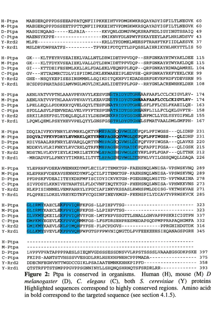
1.2.3.4. Ptpa Protein is Conserved in Organîsms
Homologous Ptpa proteins have been identified in S. cerevisiae (Rrdl and Rrd2), S. pombe (Accession Z98980), Drosophila melanogaster, Xenopus laevis (Van Hoof et al., 199$), as well as many mammals ranging from the rabbit, mouse and dog to humans (Cayla et al., 1990; Cayla et al., 1994; Van Hoof et al., 1994). Mammalian Ptpa appears to be ubiquitous; the protein is present in the brain, liver, kidney, muscle, spleen, pancreas and fat celis of the rabbit and dog (Cayla et al., 1994) as well as various independent ceil unes derived from mice and humans (Cayla et al., 1994; Fellner et al., 2003).
There are some significant differences between the Ptpa proteins of the different organisms. For instance, although mammalian Ptpa proteins are highly homologous to each other (for exampie, 97% amino acid identity is observed between human and rabbit Ptpa), iess similarity is observed between mammalian proteins and those of lower eukaryotes (for example, 50% homology is observed between drosophila and human Ptpa). In addition, S. cerevisiae and drosophila proteins are considerably larger than thefr orthologues in vertebrates; whereas the latter are 37 kDa, the former contain an extended 10 kDa C-terminal tau (Van Hoof et al., 199$). Nonetheless, certain regions are highly conserved in ail organisms (Figure 2). Van Hoof et al. (199$) demonstrated that these regions have fimctional significance by showing that targeted deletions of the conserved areas altered PTPA activity in vitro. Further, genetic complementation studies, in which RRD1-deficient yeast strains were transformed with a vector driving the expression ofthe mammalian Ptpa protein successfully reversed some phenotypes of the mutant strain (Douville et al., 2004).
Ihe presence of Ptpa is crucial to celi survival; yeast strains deficient in both PIPA genes are not viable (Rempola et al., 2001) and ifie inhibition of Ptpa translation by RNA interference causes mammalian celi mes to undergo apoptosis (Feliner et al., 2003).
H- Ptpa MAEGERQPPPDSSEEAPPATQNFI IPKKEIHTVPDMGKWKRSQAYADYIGFILTLNEGVK 60 M- Ptpa MAEGERQPPPDSSEETPPTTQNFI IPKKEIHTVPDMGKWKRSQAYADYIGFILTLNEGVK 60 D-Ptpa MASGINQAAG KLPAIA KKVQNLGDMGVWQKSRAFHDLIGYINGTSSAIQ 49 C-Ptpa MAENSYKPPE KMIIDWFDLNPWYFSKAYEEYLAFLHRLNDSW 43 Y-Rrd2 MLPE KRLLTPDDMKLWEESPTRAHFTKFIIDLAESVK 37 Y-Rrdl MSLDRVDWPHATFS TPVKRIFDTQTTLDFQSSLAIHRIKYHLHKYTTLIS 50 H-Ptpa GK---KLTFEYRVSEAIEKLVALLNTLDRWIDETPPVDQP- -SRFGNKAYRTWYAKLDEE 115
M- Ptpa GK---KLTFDYKVSEAIEKLVALLDTLDRWIDETPPVDQP- -SRFGNKAYRTWYAKLDQE 115
D- Ptpa GI---KTTDEIFESEMLKKLLRLFDALEKLVEQNPPLEQP- -QRFGNKAYRDWAQAI4REL 104
C-Ptpa GV---HTTAMRCTDLVISFIDMLDLEKWAEEIPLEDVSE-QRFGNKAYRKFYEKLCKE 99
Y-Rrd2 GHE- -NSQYKEPISESINSMMNLLSQIKDITQKHPVIKDADSSRFGKVEFRDFYDEVSRN 95 Y-Rrdl HCSDPDPHATASSIAMVNGLMGVLDKLAHLIDETPPLPGP- -RRYGNLACREWHHKLDER 108 H-Ptpa AENLVATWPTHLAAAVPEVAWLKESVG IDYGTG FAAFLCCLCKIGVLRV- 174 M-Ptpa AENLVATWPTHIJAA.AVPEVAVYLKEAVG TRIDYGTG FAAFLCCLCKIGVLRV- 174 D-Ptpa LPELLEQLLPDDKKRYQVELGQYLTESFG TRIDYGTG LSFLFFLCSLFKAEILQE- 163 C-Ptpa SPDLLASVLPENVHDALVELVPYFTESFG TRIDYGSG FLILLFCLQKLGVFTE- 158 Y-Rrd2 SRKILRSEFPSLTDEQLEQLSIYLDESWG IDYGSGH LNFMCLLYGLYSYGIFNLS 155 Y-Rrdl LPQWLQEMLPSEYHEWPELQYYLGNSFGS RLDYGTGHELSFMATVAALDMLGMFPH- 167 H- Ptpa DDQIAIVFIWFNRYLEVMRKLQKTYRNEPAGSQGVWGLD FQFLPFIWGSS---QLIDHP 231
M-Ptpa DDQVAIVFKVFDRYLEVMRKLQKTYRMEPAGSQGVWGLD FQFLPFIWGSS---QLIDHP 231 D-Ptpa RDIVSAALRRFNRYLEVARQLQRTYNNEPAGSQGVWSLD FQFVPFIWGSA---QLAVKS 220
C-Ptpa NDDICVLVLRIFNKYLRVCRHLQTRFIGIEPAGSRGW{AID FQFAPFIFGSA---QLIGSK 215
Y-Rrd2 NDSTNLVLIWFIEYLKIMRILETKY4EPAGSHGVWGL YHFLPFLFGAF---QLTTHK 212
Y-Rrdl -MRGADVFLLFNKYYTIMRRLILTYTfrPAGSHGVWGLD HFHLVYILGSSQWQLLDAQA 226 H-Ptpa YLEPRHFVDEKAVNENHKDYMFLECILFITEMKTGP-FAEHSNQLWNISA-VPSWSKVNQ 289 M-Ptpa HLEPRHFVDEKAVSENHKDYMFLQCILFITEMKTGP- FAEHSNQLWNISA-VPSWSKVNQ 289 D-Ptpa PFDPSKFVDEAIITEYKDHFMFISCIDYICKVKTGH-FGEHSNQLWSITD-VPTWAKINA 278 C- Ptpa SIVPDSYLKIQHVETHAHTSLFLDCVNFINQTKTGP- FHEHSNQLWNISA-VPHWKKVNS 273 Y-Rrd2 HLKPISIHNNELVEMFAHRYLYFGCIAFINKVKSSASLRWHSPMLDDISG-VKTWSIWAE 271 Y-Rrdl PLQPREILDKSLVREYKDTNFYCQGINFINEVKMGP-FEEHSPILYDIAVTVPRWSKVCK 285 H-Ptpa M-Ptpa D-Ptpa C-Ptpa Y-Rrd2 Y-Rrdl H-Ptpa M-Ptpa D- Ptpa LVPPVPVATAPPPPAESLSIEQNVGDSSSESSDNSVVLRPSTSSSSLVAAAEGSGDKPSKE 397 C-Ptpa FKIPS-AANTSTVHSSSWESGDLRRLHSEKHPNEHCPPPMADA 375 Y-Rrd2 DDECNFEGHVHTTWGDCCGIKLPSAIAATEMNKKHHKPIPFD 358 Y-Rrdl QTSTRFPTSTSMPPPGVPPSGNNINYLLSHQNQSHRNQTSFSRDRLRR 393
Figure 2: Ptpa is conserved in organisms. Human (H), mouse (M) D. melanogaster (D), C. elegans (C), both S. cerevisiae (Y) proteins. Highlighted sequences correspond to highly conserved regions. Amino acids
I KAECLE1 I KAECLF QKE IL: F EGEVLI KAEVU SVEVLI FKFGS-LLPIHPVTSG 323 FKFGS-LLPIHPVTSG 323 VYFGE-LMTFEPVSSGTTLSNALLGIWAPPPSKRICIGTPN 337 4MFGS-LFSFDRSEHPRESMEDAPSQDPMPPRAPAQHGMFA 332 FYFSE-FLPCPDGVS PPRGHIHDGTDK 316 FWFGTGFFPWVNIQNGTDLPVFEEKEEESIEQANAGSPGRE 345
The ubiquity of Ptpa expression in mammalian tissues, the presence of the highly conserved regions and the apparent necessity of this protein for ce!! survival in yeast and mammalian celi unes indicate that Ptpa performs vital cellular functions.
1.2.3.5. Genetic Features of PTPA
The coding sequence for the human PTPA gene was found to be localized at chromosome 9q34, spanning 60 kb. The genomic sequence consists of 10 exons and 9 introns. The promoter region lacks the traditional TATA sequence for transcription initiation. However, it was observed that several features of housekeeping genes are present in the PTPA promoter sequence (Van Hoof et aÏ., 1995). For example, many housekeeping genes have a 200-1000 bp unmethylated, typically GC-rich, CpG island spanning the promoter region, frequently extending into the first exon (Bfrd, 1986). Accordingly, the PTPA promoter sequence has an average GC content of more than 70% and an unmethylated CpG island well into ils first exon (from position -159 to +91) (Janssens et al., 1999). Although the promoter sequence was found to have many putative binding sites for a variety of transcription factors (including Myc, NF-cB and Myb), only the Yin Yang 1 (YY1) transcription factor was found to bind PTPA’s promoter sequence to initiate transcription (Janssens et al., 1999). Interestingly, this transcription factor has been associated with cervical and breast cancer (Dong et al.,
1994; Nayak and Das 1999) as well as hepatocarcinogenesis (Parija and Das, 2003). Finally, several alternative spiicing products of Ptpa (transcripts P-ri) were identified, of which only the f3 transcript was detected by western blotting (Janssens et al., 2000). However, in vitro assays revealed that this product did not activate the tyrosyl phosphatase activity of PP2A. It was thus concluded that these products have !imited biological function and are the resuit of spiicing errors. The more recent data indicating that Ptpa activates the Ser/Thr phosphatase activities now casts doubt on this resuit; Ptpa
f3
may well have a functiona! role in vivo.1.2.3.6. PTPA’s Role in Response to Oxidative Stress and in Pathways Related to Carcinogenesis
Douville et al. (2004) have recently confirmed that Rrdl plays a role in the response to oxidative stress and have demonstrated that this function is dependent upon
Rrdl interaction with Sit4, a yeast PP2A-lilce catalytic subunit. Jndeed, yeast strains deficient for Rrdl, Sit4, or both exhibit equal sensitivity to 4-NQO, indicating that these proteins function in a common pathway. Further, these two proteins were found te physicaily interact with each other. Despite this progress, no clear mechanism of action can be assigned to Ptpa. Stili, several unes of evidence support the notion that PTPA is invoived in carcinogenesis.
The p53 tumeur suppressor gene was found te be a target of Ptpa. This gene represses PTPA transcription by binding to its TATA-less minimal promoter, preventing the YY1 transcription factor from binding te the region (Janssens et al., 2000). Indeed, p53 is known te repress the transcription of a number of genes by binding their TATA less promoters (Kley et al., 1992, Sandri et aÏ., 1996).
In addition, numerous studies have aise associated Rrdl te the celi cycle in yeast, aithough its precise foie 5 stiil a subject of controversy. Dr. Gens’ group have pubiished
data suggesting that Rrdl functions in negative regulation through Start (Van Hoof et al., 2000), exciuding involvement in the G2/M transition (Van hoof et al., 2000). Conversely, Mitcheil and Sprague (2001) found that Rrdl does function in the G2IM transition and that it may play a noie in the positive reguiation of Gi. Thus, aithough it is unciear how and at what stage, PIPA appears te be involved in the progression ofthe celi cycle.
Another une of evidence linking PTPA te cancer is a proteomic analysis of a human hepatoceiiuiar carcinoma ceii une HCC-M. The study reveaied a number of preteins that were expressed at abnormai ieveis in these celis, including Ptpa (Ou et ai., 2001). Interestingiy, this proteomic anaiysis aise detected thioredoxin peroxidase (Seow et ai., 2000), an important enzyme in regulating the intraceliular redex envirenment (Gasdaska et ai., 1995). Altheugh preiiminary, given the reiationship of oxidative stress, DNA damage and chronic inflammation with the increased risk ef liver cancer (Wang et ai, 2002; Ichiba et aÏ, 2003), the data is fuliy consistent with PTPA’s putative function in the celiular response te oxidative stress.
In addition, the chromosomal location of the human PTPA gene on chromosome 9q34 may be a corroborating factor. This region contains genes associated with a number ef diseases such as bladder cancer (Kimura et aÏ. 2001), several types of leukemia
(Kaneko et al., 1988; von Lindem et al. 1990; Huntly et al., 2003), as well as amyotrophic lateral scierosis (Chance et al., 1998).
Together with the data on PP2A, the literature supports a role for PIPA in the cellular response to oxidative stress as well as ffie cellular processes associated with carcinogenesis.
1.2.3.7. Phenotypes Associated witli PIPA
Given the broad spectrum of cellular processes in which PP2A is involved, it is not surprising that Ptpa, too, has many functions. lndeed, Ptpa is implicated in a number ofpathways that appear unrelated to its role in maintaining DNA integrity in the response to oxidative stress. For example, yeast ceils deficient in RRDÏ or RRD2 display phenotypes similar to those observed in yeast strains deficient in PP2A subunits. Most notably, deletion of RRD1 causes defects in bud morphology formation that are similar to the ones obtained in strains with a deletion oftwo yeast PP2A catalytic subunits, PPH1 and PPH2. Yeast strains carrying a deletion of the RRD2 gene exhibit altered microtubule dynamics, resembling ifie phenotype obtained upon deletion of a PP2A regulatory B, CDC55 (Van Hoof et ai, 2001). In addition, Rrdl and Rrd2 aiso fimction in the HOG1 kinase pathway (Rempola et al., 2000), which is essential for mediating cellular response to increased extemal osmolarity (Brewster et al., 1993), again unrelated to the response to RO$.
Finally, Ptpa aiso functions in the TOR (Target of Rapamycin) pathway in S. cerevisiae. li yeast, this compiex pathway is important for sensing nutrient availability before initiating cellular division. Thus, the proteins in this pathway regulate translation initiation, ribosome biosynthesis (Powers and Walter, 1999), transcription of numerous enzymes involved metaboiic pathways (Schmelzie and Hall, 2000), and amino acid import (Beck et al., 1999). Rapamycin mimics nutrient deficiency, causing yeast ceils to arrest growth and to behave physioiogically as if they were starved, via the TOR pathway. However, celis lacking RRD 1 are resistant to the starvation mimetic affect of rapamycin (Rempola et al., 2000). This suggests that the Ptpa protein may be centrai to the compiex TOR pathway.
These cellular pathways are, apparently, unrelated to each other. However, the accumulation of unrelated fmdings conceming Ptpa’s fiinctions may eventually help shape an understanding of its broader fimction. For example, Ptpa’s involvement in the TOR pathway may shed light on its fimction within the response to oxidative stress. Di Como and Arndt (1996) have demonstrated that Sit4, ifie PP2A catalytic subunit found to interact with Rrdl to respond to oxidative stress, is also a target of a protein known to be implicated in the TOR pathway, Tap42. In addition, a number of studies have indicated that dysreguiation of the Target of Rapamycin pathway generates “a favorable oncogenic environment” (Huang and Houghton, 2003). Therefore, aithough it is not clear how this network of complex interactions functions, later studies may demonstrate a convergence of the different PTPA-related pathways.
1.3. Manipulating the Mouse Genome to Study Gene Function and
Disease
1.3.1 Genetically Modffled Mice
Genetic manipulation of mammalian celis has proven to be a useffli means of smdying gene function. However, the refmement of moiecular biology techniques has led to a growing trend of studying biological processes in laboratory animais. Most widespread is the manipulation ofthe mouse genome, which enables studies on genes and their impact on animal health relatively rapidly and at a low cost in comparison to research on other mammals. Two of the most common methods used are the targeted deletion and integration of specific genes into the mouse genome.
Creation of these mice consists of four essential steps: (i) the engineering of a DNA construct; (ii) the integration of the construct within the genome of embryonic stem (ES) celis or fertiiized oocytes; (iii) the transfer of these celis to a pseudo-pregnant female who will then give birth to the genetically manipulated mice, and finally; (iv) the strategic pafring of offspring to produce a colony of genetically modified mice.
There are major differences between the production of knockout and transgenic mice. Typicaily, the former involves electroporating the DNA constmct into mouse ES celis. The aim of this step is to replace endogenous DNA sequences with the constmct
via homologous recombination (HR). Since this is a relatively rare event, ES celis must 5e carefulÏy selected before being transferred into a pseudo-pregnant female. Conversely, creating transgenic mice requires the DNA construct to be microinjected directly into a fertilized oocyte. There is no need to grow celis in vitro for selection since the integration of the constmct DNA does flot need to occur at a specific locus (Cheah and Behringer, 2001; Richa, 2001).
Despite these progresses, there is an ever-growing need to develop techniques that will enable a higher degree of “tailoring” of the mouse genome. A number of technologies have recently been developed, refming classical manipulation methods by combining them with strategies that allow for greater versatility, such as conditional targeted deletions and the use ofthe Green fluorescent Protein (GFP).
1.3.2 Conditional Knockout Mice
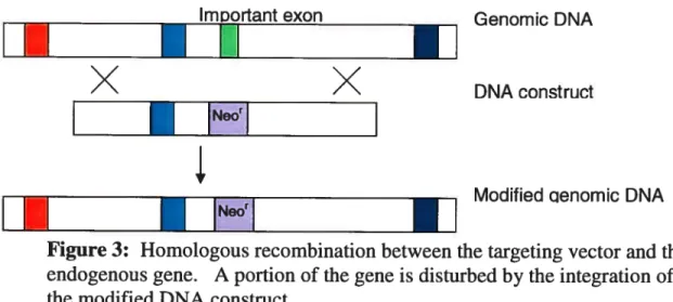
At the molecular level, the strategy used for gene targeting essentially consists of modifying a specific portion of this gene, thought to 5e important for the proper functioning of the resulting protein, via homologous recombination (HR). A crucial step is the design of the targeting vector. The DNA construct must 5e designed to contain two regions of high homology to the targeted gene for HR. These flank a sequence that would disrupt the function of the translated protein. For example, one common approach is to replace an important exon of the targeted gene with a marker for antibiotic resistance. Upon electroporation of embryonic stem celis and homologous recombination, the ES cells will carry an antibiotic resistance gene in lieu of this exon (Figure 3), and will translate truncated, non-functional protein of the targeted gene.
The traditional strategy for targeted deletions has one major disadvantage: the entire organism is defective for the specific gene throughout its development and in all tissues. It may 5e difficult to design and perform precise experiments on animals in which the gene disruption is ubiquitous. Worse, the targeted deletion could lead to embryonic lethality, making it impossible to study gene function during the postnatal period.
ImiDorlant exon
ï
ï
n
X
X
•
Neo’1
Modif leU aenomic DNA
ï
II
Figure 3: Homologous recombination between the targeting vector and the endogenous gene. A portion of the gene is disturbed by the integration of the modified DNA constnict.
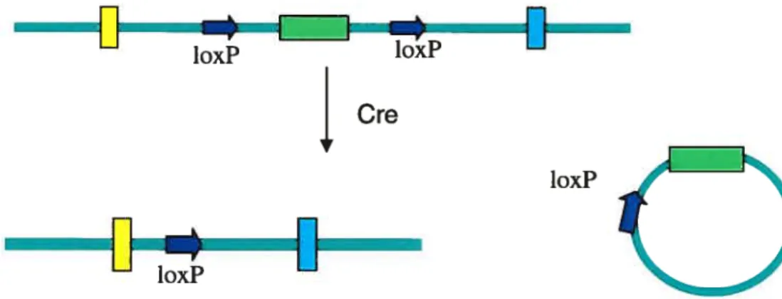
A method of circumventing these challenges is the creation of conditional knockout mice, designed to carry the gene deletion in a subset of tissues andlor at a moment in their lifespan chosen by the experimenter. Thus, temporal and spatial conditional knockouts allow the knockout event to occur post-partum, preventing the problem of embryonic lethality, andlor in specific tissues, enabling a more focused expenmental design. Commonly used tools for developing such conditional knockouts are the Cre/loxP and Flp/frt systems. The Cre/loxP system is based on a recombination that occurs in bacteriophage Pi (Austin et aL, 1981). The Cre recombinase of this bacteriophage recognizes and mediates recombination between two specific, 34 base-long loxP sequences (Figure 4). In knockout studies, the targeting vector is engineered with loxP sites flanking the sequences of the mouse genome that are to be deleted. In contrast to traditional targeted deletions, the purpose of the homologous recombination between the DNA construct and the genomic DNA in ES ceils is not to delete or disrupt sequences in ail tissues, but rather, to incorporate the loxP, which will then facilitate the removal of genomic sequences at a later time andlor in specific celi types. The loxP sequences are engineered within the intron sequence so that these genomic modifications have littie chance of altering the expression or activity of the resulting protein. These loxP-mice are subsequently crossed with mice expressing the Cre recombinase, under either an induci
Genomic DNA
loxP loxP
Cre
loxP F
loxP
Figure 4:
The Cre recombinase mediates recombination between loxP sites. This effectively deletes the loxP-ftanked sequences.ble or a tissue-specific promoter (or both), resulting in mice carrying the loxP sites as well as the Cre recombinase. The offspnng of this cross are wild-type with regards to the targeted gene; only when and where the recombinase is expressed does the recombination between the loxP sites finally occur, deleting the sequence flanked by loxP sites (Orban et al., 1992). Thus, if the Cre recombinase is under the control of an inducible promoter, the knockout event occurs only upon recombinase induction; if the promoter is tissue specific but not temporally controlled, only this tissue type will become deficient for the targeted gene throughout development. Tissues that are flot induced to express the recombinase will express wild-type, full-length protein.
The Flp/frt system is based on S. cerevisiae (Broach and Hicks, 1980) and functions in a manner that is analogous to the Cre/loxP system, the Flp recombinase mediating the recombination between two 48 hp-long frt sites. The FlpIFrt approach is rather efficient and used relatively frequently in cells lines. Although it is less commonly used for generating conditional knockout mice because of certain technical restrictions, these constraints are being gradually overcome (Rodriguez et al. 2000), maldng this a more appealing system.
1.3.3 The Use of Green Fluorescent Protein Variants in Mice
The use of ffie Green Fluorescent Protein (GfP), derived from the jellyfish Aequorea victoria, has heiped create transgenic mice that are more versatile. The main chemical and physical properties ofthe GFP were detailed in the late 1970s (Prendergast et al., 1978) but it was flot for several years that it was used a tool for biological study in other organisms, such as C. elegans (Chaffie et al., 1994), D. meÏanogaster (Wang et al., 1994) and the zebrafish (Amsterdam et al., 1995). Ikawa et al. (1995) produced the first transgenic mouse mes, having integrated the GFP gene in their genome and expressing fluorescence in a variety of tissues.
The original GFPs’ relatively low emission and excitation spectra allow for fluorescence observation upon exposure to a standard handheld UV lamp. In the iast decade, many variants of this protein have been developed, each with slightiy different characteristics. These GFPs can have several applications in mammais. They can be expressed as tags on either the C or N terminais of proteins, and used as non-invasive reporters of gene expression. In addition, the intensity of fluorescence can be used for typing of transgenics (homozygote vs. hemizygote), and as a means for screening for transgenic offspring at birth (Hadjantonakis and Nagy, 2001).
The applications are not limited to fluorescence visualization at the macroscopic level. Indeed, GFPs can be visualized at every stage of animal development. Embryonic and primary celis obtained from tissues expressing fluorescence could be cultured and the fluorescence viewed microscopicaily to determine intracellular protein localization, or used in FACS analyses.
Thus, the transgenic approach, when coupied with GFP technologies, can be an extremely practical and powerful tool for genetic studies.
1.3.4. Genetically Engineering the Mouse Genome to Study Genes Involved in the Response to Oxidative Stress
Mouse studies have been useffil in shaping our understanding of the biological functions of specific genes involved in the response to oxidative stress. For instance, a mouse model for familial amyotrophic lateral sclerosis (ALS) in which mice bear mutations on the SOD1 gene was developed. These animais were found to exhibit
enhanced oxyradical production and lipid peroxidation, as well as several other typical features of oxidative stress (Kruman et al., 1999). Another report shows that SOD2-deficient mice have increased levels of DNA damage and incidences of cancer (Van Remmen et aÏ., 2003). Kim et al. (2004) demonstrated that transgenic mice that overexpress the Sodi protein have increased resistance to oxidative stress induced by kainic acid. These examples highlight how manipulating specific genes provides insight into the genetics and biology ofdisease at the level ofthe entire organism.
Unfortunately, this direct link between gene and health is much less evident in studies examining genes involved in the Base Excision Repair pathway. For instance,
Oggi is a DNA glycosylase, which removes the oxidative lesion $-oxoG in the early
steps of this repair pathway. It was hypothesized that mice lacldng this important glycosylase would accumulate damaged bases and mutations, and therefore, be at a significantly higher risk for free-radical-induced pathology. Surprisingly, OGG1 mice exhibited an increase in lesion frequency, but showed no noticeable health problems (Kiungland et al., 1999, Minowa et al., 2000). Indeed, the phenotype ofhypermutability, with no accompanying increase in disease, was observed in other mouse studies involving targeted knockouts of other DNA glycosylases such as ailcyladenine glyocosyïase (Bevin et al., 1997) and Nthl (Cunningham et al., 2002). Similarly, mice deficient for the DNA polymerase
f3,
the polymerase responsible for fihling the gap lefi by the endonuclease upon excision of the damaged base, exhibit an accumulation of DNA damage whule lacking any noticeable increase in pathological conditions (Cabelof et al., 2002). Thus, it appears that BER proteins have considerably redundant functions.Further, other repair mechanisms are also involved in attenuating the effects of a deficiency in BER genes. For example, the Cockayne syndrome B (CSB) protein is typically associated with both transcription-coupled (TCNER) (Le Page et al., 2000) and global (GNER) Nucleotide Excision Repair pathways (Sunesen et al., 2002). However, OGGF’7CSW’ mice accumulate more premutagenic 8-oxoG lesions than either OGG1 or CSW mice (Osterod et al., 2002), indicating that CSB protein functions in repafring oxidative lesions, albeit to a lesser extent in comparison to OGGÏ.
Interestingly, although genetic studies in mice have flot been successful in establishing a direct correlation between gene and pathology, epidemiological studies in
humans suggest that disruption of ffie BER pathway may increase susceptibility to disease. For example, one noteworthy study indicates that individuals who have low levels of Oggi are at increased risk to lung cancer (Paz-Elizur et al. 20003).
In sum, the relationship between the repafr of oxidative DNA damage and health is complex. As more animal studies are performed, the relative role played by individual genes, and how these genes act in concert to prevent, cause or increase susceptibility to diseases, can be better assessed.
1.4. Rationale
Although vital, oxygen molecules pose a permanent threat to aerobic ceils. When metabolized into free radicals, these can cause significant damage to essential cellular components, including DNA. Generated as by-products of natural biological processes and as the resuit of exposure to various exogenous agents, including the UVA component of solar irradiation, exposure to reactive oxygen species is continuous and unavoidable. Ceils have consequently developed complex prevention mechanisms and DNA repair networks to cope with this paradoxical situation. However, if these mechanisms fail, organisms are at increased risk to oxidative damage and mutagenesis. Jndeed, a number of conditions and diseases are associated with oxidative stress.
The RRD1/PTPA gene was found to play an important role in the cellular response to oxidative stress in yeast. Although it appears to have several distinct functions, several unes of evidence corroborate Ptpa’s putative role in mediating the response to oxidative DNA damage. Further, several features of the PIPA gene suggest it is involved in pathways related to carcinogenesis. Our laboratories aspfred to elucidate this gene’s precise function in the cellular response to oxidative stress and to determine how altering it may affect mammalian health. Genetically manipulated mice are powerful tools to illuminate the function of specific genes and their contribution to overail health. Therefore, we sought to generate two types of genetically manipulated mice to study PIPA: conditional knockout mice, in which this gene could be temporally or spatially disnipted, and transgenic mice that would, express Ptpa ffised to a GFP in addition to the endogenous levels of Ptpa protein.
2. Materials and methods
2.1. Cloning Techniques
2.1.1. Preparation of Competent Celis for Cloning
b prepare competent E. cou DHS-Πcelis, a small amount ofbacteria was scraped
from the frozen stock and allowed to grow in 5mL LB medium with 6mM MgSO4 at 37°C for approximately 18 hours. The 5mL of culture was subsequently added to 200 mL of LB containing 6mM MgSO4. This larger culture was again incubated at 37°C until the optical density was established to be between 480 nm and 550 nm. When this density was reached, the bacteria was alÏowed to cool on ice for 5 minutes, afler which they were placed in 5OmL falcon tubes and centrifuged at low speed (4000 rpm) at 4°C for 20 minutes. Resulting bacterial pellets were obtained and re-suspended in 2OmL of TFB I, incubated on ice for 10 minutes, and centrifuged again at low speed (4000 rpm) at 4°C for 10 minutes. The bacterial pellet was re-suspended in 2mL cold 1FB II, aliquoted in sterile eppendorf tubes (200 i.iL/tube) and stored at -80°C. One tube of prepared competent E. cou was used for one transformation.
2.1.2 Transformation of Competent Celis with Plasmid DNA
An aliquot of competent bacteria was placed on ice for five minutes. Ligated DNA fragments or a circularized plasmid were added to the bacteria and lefi on ice for an additional 30 minutes. The bacteria were then incubated at 42°C for 90 seconds, afier which lmL of LB was added. The celis were allowed to recover from the heat shock and to begin expressing the antibiotic resistance gene for one hour at 37°C , afier which they
were plated on LB agar plates containing 0.1 mg!ml of ampicillin and or 0.5 mg/ml kanamycin (depending on the plasmid) and incubated for 16 - 24 hours at 3 7°C.
2.1.3. Mini-and Maxi-Preparation Methods for Plasmid Purification
Bacterial colonies were picked and inoculated in 3mL of LB medium for 16 - 24
hours at 3 7°C. $mall scale plasmid purification (1 .5mL bacterial culture) was performed as described by Bimboim and Doly (1979). Larger scale purifications (50 - 500mL of
bacterial culture) were performed using the same ratio of solutions. Protein contaminants were eliminated by standard phenollchloroform extractions.
2.1.4. Restriction Digestions and DNA Modification for Clonïng
Afler the plasmid purification, the DNA was digested with restriction enzymes that would enable us to determine if the desired construct was indeed obtained. Usually, for plasmid verification, approximately 250 - 500ng were digested with the appropriate restriction enzymes for 1 - 4 hours with 5 units of the appropriate enzyme and buffer
dilution in a total volume ranging from 15 to 2OjiL, and ifien placed at the appropriate temperature (usually 37°C). Larger scale restriction digestions were performed when the DNA was to be used for cloning purposes; up to l0jig of DNA was used, with 20 units of enzyme and the appropriate buffer dilution in a total reaction volume ranging from 20 to 4OjiL.
When a vector plasmid was linearized with compatible ends, digested DNA was treated with 5 units of Calf Intestinal Ailcaline Phosphatase (ClAP) (Promega) for 45 minutes. The ClAP supplied by Promega is sufficiently versatile to work in virtually all restriction digestion buffers, and was therefore added dfrectly to the restriction digestion reaction.
Although restriction digestion was routinely used as a method of confirming that the appropriate clone was obtained, the integrity of loxP sites, s as well as certain clones were confwmed by automated sequencing using the Research Center’s ABI PRISM 3100 Genetic Analyzer (Applied Biosystems), following the manufacturer’s protocols and procedures.
2.1.5. Polymerase Chain Reaction to Generated Clonable Fragments
The Polymerase Chain Reaction (PCR) was used to amplify fragments, thereby enabling the engineering of convenient restriction sites for cloning. Typically, the program consisted of 30 cycles, each consisting of a denaturing step (95°C for 1 minute), an annealing step (52°C for 1 minute), an elongation step (72°C for 2 minutes/kb to be amplified), and a fmal long elongation step (72°C for 10 minutes). PCR reactions were performed in appropriate reaction buffer dilution (buffer supplied with the enzyme) with
3mM MgSO4, 1 jig of each oligonucleotide, 0.5rnM of each dNTP, 25 - 1 OOng of template
DNA with 1 - 3 units of Pfu polymerase in a total volume of 1 00tL The Pfu was used
because it possesses a proofreading activity, decreasing the chance of errors taldng place during the amplification process.
2.1.6. Fragment Purification for Cloning
Fragments obtained by restriction digestion were resolved and observed on ethidium bromide-stained 1% agarose gels. The specific fragments to be used for cloning were physically removed from the gel using a blade, and purified using Amersham GFX Gel Purification Kit.
2.1.7. Ligation Reactions for Clonïng of DNA Fragments
Relative concentrations of purified DNA fragment were estimated visually on an agarose gel. To maximize cloning efficiency, three ligations were usually performed, each with varying vector:insert ratios ranging from molar ratios of 1:2 to 1:10. Ligations were performed in volumes ranging from lO1iL to 151.tL, using 10 units of Promega or New England Biolabs ligase and the appropriate buffer dilution. The duration and temperature of incubation varied from 1 hour ligation reactions at room temperature, to
16- 24 hour ligation reactions at 16°C.
2.1.8. General Clonïng Strategies
Three strategies were used for the creation of the various constructs. The first was the standard plasmid digestion and fragment purification followed by ligation and transformation in competent bacteria. The second strategy involved amplifying fragments of DNA using specially designed primers to engineer restriction sites for cloning andlor other short sequence within the amplified product. Afler the PCR reaction, the amplified DNA could simply be restriction-digested, purified and ligated as in the first strategy. The third strategy differs from the others in various respects: no phosphatase treatment was performed on linearized plasmids, and fragments were not purffied from agarose gels. Instead, this type of cloning was performed on large fragments that could flot easily be resolved from undigested, supercoiled plasmid on
agarose gel or when only one restriction site was available for cloning in the DNA sequences to be ligated. Thus, plasmids were restriction-digested with a single enzyme, ethanol-precipitated and ligated directly. This method could only be performed when the plasmids carried different resistance genes (ampicillin and kanamycin). Therefore, only constructs containing both plasmids could grow in media containing both antibiotics. The enrichment of positive clones by double selection compensated for the increased chance of obtaining undigested or religated plasmids due to the fact the DNA was flot gel purffied or treated by ClAP.
2.2. Generating Iransgenic Mice
2.2.1. Micorinjection of the Oocyte and Breeding Procedures
The DNA construct microinjected into mouse oocytes was prepared by other members of the laboratory and subsequently implanted in pseudo-pregnant females. The microinjection of the linearized DNA construct into fertilized mouse oocytes and the transfer to pseudo-pregnant females was performed by the Animal facility of the Recherche Guy-Bemier Research Centre according to the protocols approved by the Centre’s Committee for Animal Protection. The pseudo-pregnant females carried these fertilized oocytes to terni. Pups were bom three weeks afier the microinjection and thereafier weaned and crossed with fertile mice CD-l males or females. The pups resulting from this second cross were weaned and screened for transgene integration at the age of 3 weeks or older. When a mouse was established as transgenic, it was crossed with a transgenic sibling, when possible, or with a CD-l mouse. Thus, independent families of transgenic mouse mes were generated. Mice were routinely weaned at the age of three weeks. All matings and screening procedures were performed when mice were at least three weeks of age.
2.2.2 Screening for Transgenic Mice
2.2.2.1. Extraction of Mouse Tau Genomic DNA
Southem blotting is the method of choice for reliable detection of transgene integration. Screenings were performed on DNA purified from mouse tau clippings.