Université de Montréal
The role of frameshifting and transcriptional dysregulation in spinocerebellar ataxia type-3
par
Linh-An C. Tuong
Programmes de Biologie Moléculaire Faculté des Etudes Supérieures (FES)
Mémoire présenté à la Faculté des Études Supérieures en vue de l’obtention d’une Maîtrise en Sciences (M.Sc.)
en Biologie Moléculaire
Juin, 2006
Direction des bibtiothèques
AVIS
L’auteur a autorisé l’Université de Montréal à reproduite et diffuser, en totalité ou en partie, par quelque moyen que ce soit et sur quelque support que ce soit, et exclusivement à des fins non lucratives d’enseignement et de recherche, des copies de ce mémoire ou de cette thèse.
L’auteur et les coauteurs le cas échéant conservent la propriété du droit d’auteur et des droits moraux qui protègent ce document. Ni la thèse ou le mémoire, ni des extraits substantiels de ce document, ne doivent être imprimés ou autrement reproduits sans l’autorisation de l’auteur.
Afin de se conformer à la Loi canadienne sur la protection des renseignements personnels, quelques formulaires secondaires, coordonnées ou signatures intégrées au texte ont pu être enlevés de ce document. Bien que cela ait pu affecter la pagination, il n’y a aucun contenu manquant.
NOTICE
The author of this thesis or dissertation has granted a nonexclusive license allowing Université de Montréal to reproduce and publish the document, in part or in whole, and in any format, solely for noncommercial educationat and research purposes.
The author and co-authors if applicable retain copyright ownership and moral rights in this document. Neither the whote thesis or dissertation, nor substantial extracts from it, may be printed or otherwise reproduced without the author’s permission.
In compliance with the Canadian Privacy Act some supporting forms, contact information or signatures may have been removed from the document. White this may affect the document page count, it does flot represent any loss of content from the document.
Ce mémoire intitulé:
The role ofribosomal frameshiffing and transcriptional dysregulation in spinocerebellar ataxia type-3
présentée par: Linh-An C. Tuong
a été évalué par un jury composé des personnes suivantes:
Edward Bradley président-rapporteur Guy A. Rouleau directeur de recherche Rima Slim membre du jury
Spinocerebellar ataxia type-3 is a late-onset autosomal dominant progressive
neurodegenerative disorder caused by a CAG trinucleotide repeat expansion in the MJD] gene. The pathology of SCA3 is characterized by the presence ofnuclear inclusions (NIs) within neurons of selectively affected brain regions. One of the contributing factors to neuronal toxicity is the production ofpolyalanine proteins through a ribosomal -1 frameshifi within the CAG-repeat during translation. To further investigate ribosomal frameshifting, we established a cellular mode! using three full-length MJD1 constructs based on the cDNA of the MJDJ gene coding for various polyglutamine repeat lengths (CAG9, CAG 100, and CAA96). We confirmed that with these constructs, -1
frameshifting occurs exclusively with the expanded-CAG repeat construct (CAG100), as it appears to be dependent on the energy requirements necessary to unfold secondary mRNA structures ofthe long CAG repeats. The presence oftoxic polyalanine proteins produced by frameshifting most likely contributes to the pathogenesis ofSCA3. Through the use of our newly established cellular mode! and Drosophila model, we also
investigated transcriptional dysregulation through impaired RNA polymerase II
(RNAPII) function. Our results show that transcriptional impairment in SCA3 appears to be independent ofRNAPII activity alterations. In conclusion, we confirmed the
production of frameshifted polyalanine proteins in a full-length ataxin-3 cellu!ar mode!, but require further studies to understand the mechanism of transcription dysregulation involved in the pathogenesis of SCA3.
Key words: Spinocerebellar ataxia type-3 (SCA3), neurodegeneration, ataxin-3, nuclear inclusions, polyglutamine, polyalanine, frameshifting, RNApolymerase II, neurotoxicity, triplet expansion.
Résumé
L’ataxie spinocérébelleuse de type-3 (SCA3) est une maladie neurodegenerative
autosomique dominante observée chez des individus d’âge adulte qui est causée par une expansion de séquences répétées de trinucleotide CAG dans le gèneMJD]. La
pathologie de SCA3 est caractérisée par la présence d’inclusions nucléaires (INs) dans les neurones des régions cérébrales distinctement affectées. Un des facteurs qui contribue à la neurotoxicité observée est la production de protéines contenant des polyalanines qui font suite à un changement -1 du cadre de lecture ribosomique à l’intérieur des séquences de CAG répétées, pendant la traduction. Afin de mieux étudier ce changement, nous avons établi un modèle cellulaire en utilisant trois constructions de MJD 1 basées sur la séquence complète de cDNA du gèneMJD] exprimant trois longueurs de répétitions de polyglutamine (CAG9, CAG100, et CAA96).
À
l’aide de nos constructions, nous avons confirmé que le changement de cadre de lecture ne se produit que lorsque la construction de CAG longue est utilisée (CAG100); ce changement semble être relié à l’énergie nécessaire au dépliement de structures d’ARN secondaire particulières aux longue répétions de CAG. Il est probable que la présence des protéines contenant des polyalanines toxiques contribue à la pathogenèse de SCA3. Aussi, en utilisant lesmodèles cellulaire et de drosophile que nous avons établi, nous avons testé un mécanisme de déregulation transcriptionelle lié à la fonction altérée de la polymérase II d’ARN (RNAPII). Nos résultats démontrent que dans la maladie SCA3, l’altération
transcriptionelle semble être indépendante des modifications fonctionnelles de RNAPII. En conclusion, nous avons confirmé l’implication des protéines de polyalanine dans notre
modèle de la maladie, mais d’autres études seront nécessaires pour mieux comprendre la dérégulation transcriptionelle dans la pathogénèse de SCA3.
Mots clés: ataxie spinocérébelleuse de type-3 (SCA3), neurodegeneration, ataxine-3, inclusions nucléaires, polyglutamine, polyalanine, changement de cadre de lecture, ARN polymérase II , neurotoxicité, expansion de triplet.
Table of Contents
Abstract j Résumé iii Table of Contents y List of Figures ix List of Tables x Abbreviations xi Acknowledgments xv Chapter 1: Introduction 11.1 Trinucleotide repeat expansion diseases 1
1.2 Pathogenesis of polyglutamine disorders 4
1.3 Characteristics of spinocerebellar ataxia type-3 (SCA3) 6
1.3.1 Clinical and pathological feattires 6
1.3.2 Molecular genetics ofSCA3 7
1.3.3 Cellular features ofthe disease 9
1.3.4 Structure of ataxin-3 protein 15
1.3.5 Functions of ataxin-3 protein 16
1.4 Objectives and thesis outiine 20
Chapter 2: Model systems of SCA3 21
2.1 Introduction: established models of SCA3 21
2.1.1 Mousernodels 21
2.1.3 In vitro or cellular models. 23
2.2 Objectives 24
2.3 Materials and Methods 25
2.2.1 Cellular model: generation ofplasmid constructs 25
2.2.2 Ceil culture and transient transfections 29
2.2.3 Fluorescent visualization 30
2.2.4 Quantification of inclusions 30
2.2.5 Statistical analysis 31
2.2.6 Cellular protein extraction and Western blot analysis 31
2.2.7 Drosophila stocks and transgenes 32
2.2.8 Protein extraction from fly heads 33
2.2.9 Drosophila model: phenotype characterization 33
2.4 Resuits 33
2.4.1 Distribution and subcellular localization ofataxin-3 in Cos-1 ceils 33
2.4.2 Characterization of nuclear inclusions 34
2.4.3 Phenotypic characterization oftransgenic flics 37 2.4.4 Protein expression ofataxin-3 in cells and flics 39
2.5 Discussion 42
Chapter 3: The role of frameshifting in SCA3 47
3.1 Introduction: ribosomal frameshifting in SCA3 47
3.1.1 Similarities between SCA3 and OPMD 47
3.1.2 Previous studies on frameshifling in SCA3 47
3.1.4 Frameshiffing in a full-length context . 50
3.2 Objective 51
3.3 Materials and Methods 51
3.3.1 Irnmunocytochernistry 51
3.3.2 Quantification of ftarneshiffing 52
3.3.3 Statistical analysis 52
3.4 Resuits 53
3.4.1 Generation ofa cellular frameshiffing reporter system 53 3.4.2 Detection of frameshifting in a cellular model 53
3.4.3 Frequency of-1 frameshifling in celis 54
3.5 Discussion 57
Chapter 4: Transcriptional dysregulatiou in polyglutamïne dïsorders 63 4.1 Introduction: the study of transcriptional impairment 63 4.1 .1 Sequestration of transcriptional activators by inclusions 63 4.1.2 Inhibition of histone acetyltransferase activity 64
4.1.3 Direct co-repressor activity 65
4.1.4 Transcriptional impairment factors in SCA3 65
4.1.5 Transcriptional impairment through altered RNA polymerase II function 67
4.1.6 Structure and function ofRNAPII 6$
4.2 Objective 70
4.3 Materials and Methods 70
4.3.1 Inirnunocytochemistry 70
4.3.3 Statistical analysis. 70
4.3.4 Protein extraction and Western blot analysis 71
4.4 Results 71
4.4.1 Expression ofphosphorylated RNAPII in ceils 71
4.4.2 Quantification of RNAPII phosphorylation at the protein level 74
4.5 Discussion 76
Chapter 5: General conclusion 79
5.1 Summary of thïs study 79
5.3 Opportunities for therapeutic intervention 81
5.3.1 Misfoldedprotein 81
5.3.2 CeIlular defense system 81
5.3.3 Modulation ofaggregation 82
5.3.4 Mutant protein expression 83
5.3.5 Cleavage fragments 83
5.3.6 Apoptotic inhibitors 84
5.3.7 Transcriptional dysregulation 84
5.4 Conclusion 85
Refereuces 86
Appendix I: Construct sequences I
CAG9 sequence I
CAG100 sequence II
List of Figures
Figure 1.1 Structure of the ataxin-3 protein 16
Figure 2.1 Schematic representation of fulÏ-length MJD1 construct design 36
Figure 2.2 Expression of full-length MJD1 constructs 36
Figure 2.3 Expanded polyglutamine protein causes aduit eye degeneration 38
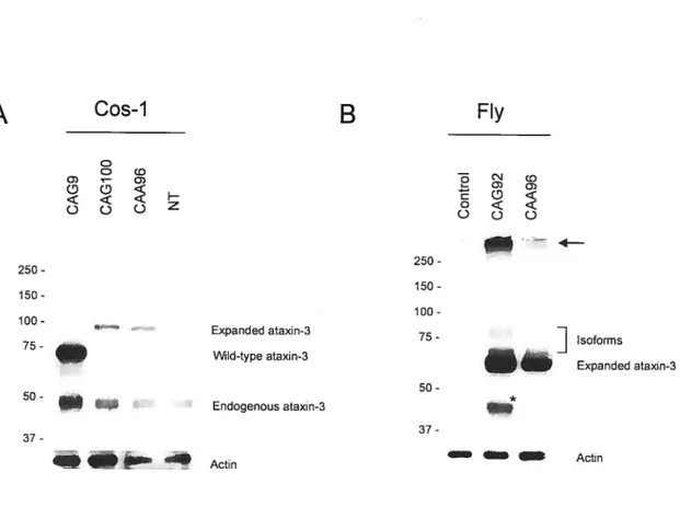
Figure 2.4 Expression of ataxin-3 protein 41
Figure 3.1 Frameshifled polyalanine products are detected in CAG100 56 Figure 3.2 Frequency ofribosomal siippage to the —1 frame 56 Figure 3.3 Secondary structure predictions for polyglutamine encoding repeats 58 Figure 4.1 Detection ofphosphorylated RNA polymerase II by H5-antibody 73 Figure 4.2 Phosphorylated RNA polymerase II protein levels 75
ListofTables
Table 1.1 Trinucleotide repeat expansion disorders 3
Abbreviations
ACA adenine, cytosine, adenine
Ala alanine
Asn asparagine
ATPase adenosine triphosphatase CAA cytosine, adenine, adenine CAG cytosine, adenine, guanine CBP CREB-binding protein CDX cyclin-dependent kinase
CREB cAMP response element binding protein CTD C-temiinal domain
CTD4H8 Purified mouse anti-RNA polymerase II monoclonal antibody DAPI 4’,6 diamidino-2-phenylindole
DM Dystrophia myotonica 1 DNA deoxyribonucleic acid
dNTP deoxyribonucleoside tri-phosphate
DRB 5 ,6-dichloro- 1 3-d-ribofuranosyl-benzimidazo1e DRPLA Dentatorubropallidoluysian atrophy
ECL enhanced chemiluminescence EGFP enhanced green fluorescent protein EYA eyes absent protein
Fcpl TFIW-associated CTD phosphatase FRAXE Fragile X syndrome E
FRAXF Fragile X syndrome A
FRDA Friedreich ataxia
GCA guanine, cytosine, adenine
GCG guanine, cytosine, guanine
Gin glutamine
HA hemaggiutinin
HD Huntington’s disease
HDAC histone deacetylase
HDL2 Huntington disease-like 2
His histidine
HRP horseradish peroxidase
Hsp Heat shock protein
“A hyperphosphoryiation state
ll hypophosphoryiation state
JD Josephin domain
MAML1 mastermind-like 1 MATi ‘menage à trois’ 1
MJD Machado-Joseph disease mRNA messenger ribonucleic acid MTOC microtubule organizing center Myc myelocytomatosis virus N2a mouse neuroblastoma celis
NGS normal goat serum
NI nuclear inclusion
NT non-transfected
OPMD ocu1ophargea1 muscular dystrophy ORF open reading frame
P/CAF p300/CBP-associated factor PABPN2 poly(A)-binding nuclear protein 2 PBS phosphate-buffered saline
PCR polymerase chain reaction PML Promyelocytic leukemia protein Pp 1 type 1 protein phosphatase PQBP-1 polyglutamine binding protein-1
PS2 polyclonal RNA polymerase II CTD (phospho S2) antibody pTEFb positive-acting transcription elongation factor-b
RIPA radioimmunoprecipitation RNAPII RNA polymerase II
RPIILS RNA polymerase II large subunit SMIA suberoylanilide hydroxamic acid SBMA Spinobulbar muscular atrophy SCA Spinocerebellar ataxia
Scpl small CTD phosphatase
Ser serine
SMN survival of motor neurons protein Spi Specificity protein 1 transcription factor TAF1113O TBP-associated factors
TBP TATA-binding protein
TFIIF Transcription initiation factor 11F
Thr threonine
tRNA transfer ribonucleic acid
UIM ubiquitin-interacting motifs YAC yeast artificial chromosome
Acknowledgments
Firstly, I would like to thank my supervisor, Dr. Guy A. Rouleau, for accepting me as a Master’s student and for giving me the opportunity to pursue research in an enriching environrnent under his guidance. I extend a very special thanks to Claudia Gaspar and Patrick Dion for more than generously providing their time, expertise and advice throughout my research. I am forever grateful for their help. I also thank Daniel
Rochefort for his tecimical assistance in a critical part ofmy project. In addition, I would like to thank Janet Laganière, Aida Abu-Baker, Liliane Karemera, and Adèle Salin Cantegral, members ofthe lab and friends, for their ldnd help and for making my time here a most enjoyable and memorable experience. Finally, I wish to thank my family for their support and encouragement tbroughout my graduate studies.
Chapter J: Introduction
1.1 Trinucleotide repeat expansion diseases
Trinucleotide repeat expansion diseases are due to unstable trinucleotide sequences which are repeated several times within a stretch ofDNA (Cummings and Zoghbi, 2000). The number ofrepeats is polymorphic in a normal population and no phenotype is observed until their expansion reaches a pathogenic length unique to each gene (Jasinska et al., 2003). These diseases are categorized into two subclasses depending on the location of the repeat with respect to the gene. The first subclass, accounting for seven diseases, has its repeats in non-coding sequences ofDNA (Jasinska et al., 2003; Di Prospero and Fishbeck, 2005). The second subclass, consisting ofup to nine diseases, is caused by an expansion ofa CAG trinucleotide repeat within the coding region ofthe causative gene (Pearson et al., 2005). This group includes spinobulbar muscular atrophy (SBMA), Huntington’s disease (HD), the spinocerebellar ataxias (SCAs) (1, 2, 3, 6, 7, 17), and dentatorubropallidoluysian atrophy (DRPLA) (Pearson et al., 2005). Because the CAG trinucleotide encodes the amino acid glutamine, these nine diseases are commonly referred to as polyglutamine diseases (Table 1.1).
Trinucleotide repeats are unstable in both somatic and germ cells (Pearson et al., 2005). The exact cause ofthe triplet expansion is unknown, but most likely invoïves one or more ofthe following processes: formation ofunusual DNA structures and DNA slippage during lagging-strand synthesis; aberrant repair of DNA mutagenic intermediates such as
double-strand or single-strand breaks, or endogenous DNA damage; or, recombination within the repeats by intrachromosomal strand annealing (Pearson et al., 2005).
Three possible mechanisms ofpathogenicity caused by triplet repeat expansions have been identified. The first, the gain-of-function mechanism for dominantly inherited diseases, such as in the expanded CAG repeat disorders, is based on abnormal protein products with expanded polyglutamine tracts which lead to misfolding and novel altered functions (La Spada and Taylor, 2003; Perutz et al., 1999). The second, the loss-of function mechanism for recessively inherited diseases, such as in fragile X syndrome and friedrich ataxia, is based on transcriptional interference of the affected gene leading to reduced levels oftheir protein products (Pieretti et al., 199f; Bardoni and Mandel, 2002). The third is also a gain-of-function mechanism for dominantly inherited diseases, but is based on abnormal interactions at the RNA level ofthe affected gene leading to disrupted processing and nuclear accumulation of the transcript, such as in the case ofmyotonic dystrophy (Wang et al., 1995; Taneja et al., 1995).
Table 1.1 Trinucleotide repeat expansion disorders. Adapted from Cummings and Zhoghbi, 2000; Di Prospero and f ischbeck, 2005; Evereil and Wood, 2004; Ranum and Day, 2002.
flisease Gene Locus Protein Repeat size
Normal Disease Non-coding repeats
Friedreich ataxia (FRDA) fXN 9q13-q21.l frataxin 7-34 >100 Fragile X syndrome A fMRI Xq27.3 Fragile X mental 6-53 >230
(FRAXF) retardation 1 protein
Fragile X syndrome E FMR2 Zq28 Fragile X mental 6-35 >200
(FRAXE) retardation 2 protein
Dysftophia myotonica 1 DMPK 19q13 Dystrophia myotomca 5-37 >50
(DM) protein kinase
Spinocerebellar ataxia 8 Antisense to 13q21 Undetennined 15-91 100-155
(SCA8) KLHLJ
Spinocerebellar ataxia 12 F?P2R23 5q3 1q33 Regulatory subunit of 7-28 66-78
(SCA12) the protein phosphatase
PP2A
Huntington disease-like 2 JPH3 1 6q24.3 Junctophilin 3 6-27 51-57 (HDL2)
Potygtutamiue disorders
Spinal and bulbar muscular AR Xql 3q2 1 Androgen receptor 9-36 3 8-62 atrophy (SBMA)
Huntington’s disease (HD) 1T15 4pl6.3 Hunfingtin 6-35 36-121
Dentatombral- DRFLA l2pl3.3l Atrophin 1 6-35 49-88
pallidoluysian atrophy (DRPLA)
Spinocerebellar ataxia 1 SCA I 6p23 Ataxin 1 6-44 3 9-82
(SCA 1)
Spinocerebellar ataxia 2 SCA2 12q24.1 Ataxin 2 15-31 36-63
(SCA2)
Spinocerebellar ataxia 3 SCA3/MJD 14q32.l Ataxin 3 12-40 55-$4 (Machado-Joseph
disease) (SCA3/MJD)
Spinocerebellar ataxia 6 CA CNA lA l9pl3 ŒIA-voltage-dependent 4-1 $ 21-33
(SCA6) calcium channel subunit
Spinocerebellar ataxia 7 SCA7 3pl2p13 Ataxia 7 4-35 37-306
(SCA7)
Spinocerebellar ataxia 17 TBP 6p27 TATA box binding 25-42 45-63
1.2 Pathogenesis of polyglutamine dïsorders
Nine neurodegenerative disorders (SBMA, HD, DRPLA and SCAs 1, 2, 3, 6, 7, 17), are caused by CAG repeat expansions within the coding sequence ofthe respective genes. Aside from their expanded polyglutamine tract, the mutant proteins do flot share any homology, but the diseases have similar features and probabiy share common mechanisms ofpathogenesis (Cummings and Zhoghbi, 2000).
The first simiiarity is the mode of inheritance. With the exception of SBMA, which is X linked, ail ofthese diseases are inherited in an autosomal dominant fashion.
Secondly, the progression and manifestations ofthe diseases are characterized by similar events. Neurodegeneration develops progressively, appears in midiife, and causes increasing neuronal dysfunction which eventually leads to neuronal loss 10-20 years following the onset of symptoms (Zoghbi and Off, 2000). Also, the age of onset is an exponential function that is inversely dependent on the number of the repeats; that is, longer expansions lead to an earlier age of onset with more severe manifestations (Zoghbi and Orr, 2000). Polyglutamine disorders also display one ofthe fundarnental features of neurodegenerative diseases: neuronal ce!! death is apparent only towards adulthood, despite expression ofthe mutant protein throughout life. This suggests that accumulation of the toxic product or cumulative cellular damage reaches a threshold beyond which the neuron cannot compensate (Paulson, 1999).
Thirdly, another phenomenon observed in affected families is genetic anticipation: over successive generations, the diseases present an earlier age of onset and progress more rapidly due to successive expansions ofthe CAG tract during transmission (Zoghbi and Off, 2000).
Fourthly, in ail of these diseases, only neuronal celis are specifically targeted by the mutant proteins. Moreover, only a selected subset ofneurons (specific for each disorder) is affected by apoptosis, despite widespread expression of mutant protein in the nervous system and throughout the body (Paulson et al., 1997a; Reddy and Housman, 1997; Zoghbi and Orr, 2000).
Fifthly, neuronal accumulation of mutant protein in nuclear or cytoplasmic inclusions is a common feature to ail polyglutamine diseases (Ross and Poirier, 2004; Michalik and van Broeckhoven, 2003). Mutant polyglutamine-containing proteins aggregate in vulnerable neurons and these aggregates are also positive for ubiquitin and proteasome components (Cummings and Zhoghbi, 2000). Though the presence ofthese nuclear inclusions may appear to be the cause of neuronal dysfunction, this link remains uncertain as several reports have indicated that they are neither necessary nor sufficient to initiate
polyglutamine-mediated disease (Cummings and Zhoghbi, 2000). Nevertheless, the commonalities of inclusion formation in many unrelated neurodegenerative diseases might reflect a shared coping response to diffuse toxic proteins and indicate a common pathological mechanism (Ross, 1997).
And finally, transcriptional dysregulation appears to represent a unifying pathogenic mechanism in polyglutamine disorders, as profound transcriptional changes are oflen noted in cadi case (Evert et ciL, 2003).
In spite ofthe many similarities among polyglutamine disorders, as reflected by the abundant publications looking into common mechanisms oftoxicity, it is important to note that each disease is nonetheless distinct and characterized by unique properties. for example, each is caused by a different gene, located on different chromosomes.
Furtherrnore, each possesses a characteristic critical threshold for glutamine repeat number, below which the disease does not occur (Gusella and Macdonald, 2000). Also, the brain regions affected are flot identical (Margolis, 2003). Therefore, the protein context and neuronal cell-type affected in the various disorders present differences unique to each disease and must be investigated individually.
1.3 Characterïstïcs ofspinocerebellar ataxia type-3 (SCA3)
1.3.1 Clinical and pathological features
SCA3 is a neurodegenerative disorder also called Machado-Joseph disease (MJD). The clinical features of SCA3 patients include cerebellar ataxia, oculomotor abnormalities, spasticity, peripheral neuropathy, and cognitive disturbances (Dûrretal., 1996). Core features include progressive ataxia, dysarthria, postural instability, nystagmus, eyelid retraction, and facial fasciculations. Dystonia is often prominent in younger patients (Sudarsky and Coutinho, 1995). five subtypes ofthe disease have been identified; the
most common, type 2, is associated with early adulthood to middle-age onset (20-45 years) and its clinical manifestations are cerebellar and pyramidal signs (Margolis, 2003).
The pathology of SCA3 is associated with neurodegeneration occurring mainly in the basal ganglia (substantia nigra), brain stem, spinal cord, pontine nuclei, and cerebellum with preservation of cerebellar cortex and inferior olive (DUrretal., 1996; Paulson et al.,
1997a; Sudarsky and Coutinho, 1995). The neuropathy involves both sensory and motor neurons in myelinated and unmyelinated fibers (Margolis, 2003).
Currently, there is no treatment or cure for SCA3, but identification of the mutant gene has been useful for diagnosis and genetic counselling (Colomer Gould, 2005).
1.3.2 Molecular genetics of SCA3
This disease is an autosomal dominantly-inherited ataxia originally described in people of AzoreanlPortuguese descent. The disorder has subsequently been identified in many other countries including Japan, Brazil, Australia, and China (Sudarsky and Coutinho,
1995).
The cause of SCA3, the most common dominantly inherited ataxia, was identified to be an unstable expansion of a CAG repeat in the human MJDJ gene (Kawaguchi et al., 1994), which had been previously mapped to chromosome 14q24.4-q32.1 (Takiyama et al., 1993). The MJD] gene encodes the cytoplasmic protein ataxin-3 of 42 kDa with
ubiquitous expression throughout the body. Despite this widespread expression, pathological sites are selective to certain brain regions only (Paulson et al., 1997a).
The number of normal CAG repeats ranges from 10-5 1, whereas the expanded CAG length is from 55-86; however, intermediate repeat lengths of 53 and 54 have been observed in SCA3 patients who display an abnormal phenotype (van Alfen et al., 2001; Zhou et aÏ., 1997). These expanded repeats are highly unstable and may expand from one generation to the next, which explains the anticipation observed in patients (Galvao et al., 2004). Large changes in CAG repeat number are also more frequently associated with paternal transmissions, suggesting the expansion occurs during the pre-meiotic proliferative stages of spermatogonial divisions (Galvao et al., 2004; Pearson et al., 2005).
An interesting observation is that despite being an autosomal dominant disorder, clinical studies have shown that SCA3 patients homozygous for the mutation display more severe symptoms at an earlier age than heterozygotes (Lerer et al., 1996; Sobue et al., 1996; Zlotogora, 1997). This suggests that in heterozygotes, the second non-mutant allele may act protectively to reduce the age at onset or that dosage ofrepeat-bearing proteins may be important for pathogenesis.
1.3.3 CeIlular features of the disease
1.3.3.1 Misfolded polyglutamine proteins
The polyglutamine proteins produced by the MJDJ gene with expanded-CAG repeats are prone to misfolding (Jana and Nukina, 2004). The first une ofcellular defence against these proteins involves molecular chaperones, which participate in refolding abnormal peptide conformations (McClellan and Frydman, 2001). Secondly, if this system fails, the defective proteins are tagged with ubiquitin and targeted for degradation by the proteasorne complex (Goldberg, 2003; Ciechanover and Brundin, 2003). Thirdly, if the misfolded proteins cannot be removed, they form aggregates which the ceil attempts to remove by autophagy (Ravikumar et al., 2004).
Lastïy, it was observed that when mutant ataxin-3 binds to the nuclear matrix, this causes a conformational change which exposes the polyglutamine tract (Perez et al., 1999). Upon exposure of the expanded tract, more aggregation and binding of other
polyglutamine-containing proteins occur. If removal of these aggregates is unsuccessful, they ultimately grow in size and form into larger nuclear inclusions (NIs), a hallmark of polyglutamine diseases (Jana and Nukina, 2004).
1.3.3.2 Inclusions in SCA3 affected brain regions
In SCA3 patients, the presence of visible nuclear inclusions (NIs) ofvarious sizes was detected in brain tissue (Paulson et al., 1 997a). Inclusions containing mutant ataxin-3 protein were observed in regions such as the cerebral cortex, striatum, thalamus, and
lateral geniculate body (Yamada et al., 2004). Interestingly, NIs were even detected in regions ofthe nervous system that showed no obvious neuronal loss (Yamada et al., 2001 a).
1.3.3.3 Formation of inclusions
Nuclear inclusions are most likely a manifestation ofmisfolding and aggregation ofthe mutant protein, and as a consequence, lead to aberrant protein-protein interactions within affected ceils (Chai et al., 2001). Studies suggest that NIs in polyglutamine diseases arise from nuclear bodies (NBs), which are intranuclear structures that function in
transcriptional regulation, growth suppression and apoptotic ccli death, and are fourni distributed throughout the nucieus in a speckled pattern (Seeier and Dejean, 1999; Zhong et al., 2000). The appearance of large macroscopic inclusions characteristic of SCA3 is preceded by the presence of insoluble, micro-aggregates of mutant ataxin-3 (Chai et al., 2001). These authors found that in SCA3, expanded polyglutamine co-iocalized with promyelocytic leukemia protein (PML), an essential component ofNBs (Takahashi et al., 2003). This suggests that NBs are gradually enriched in misfolded mutant proteins and eventuaily iose their characteristic structure due to the excessive accumulation of mutant proteins.
1.3.3.4 Mechanism of aggregation
Pemtz et al. (1994) proposed that the glutamine repeats act as polar zippers: they are capable of linking 3-strands together into sheets by networks of hydrogen bonds between
their main-chain amides and polar side chains; this cross-Ïinking would allow for the joining of complementary proteins, resulting in the formation of insoluble aggregates.
The expansion of glutamines might cause the mutated protein to acquire non-specffic or increased affinity for each other or for other glutamine-containing proteins, thereby causing pathological defects (Perutz et al., 1994; Stott et al., 1995). However, this cross linking in the presence ofthe mutant protein context was not as apparent (Perutz et al., 1994).
Altematively, Green H. (1993) proposed that aggregation was based on the activity ofa transglutaminase enzyme which links the amino groups of lysine residues in other proteins through isopeptide bonds to form covalently bonded aggregates. In fact, huntingtin, a polyglutamine disease protein, was shown to be a substrate for brain transglutaminase and its aggregation was dependent on Ca2 levels, which are linked to impulse-conduction in neurons (Kahiem et al., 1996; Kahlem et al., 1998).
Neither of these processes has been demonstratedin vivo. Whichever the mechanism may be, it appears that the proteins are destabilized and drawn into the nucleus, where ubiquitination and aggregation occur.
1.3.3.5 Components of inclusions
Inclusions are composed ofnot only the mutant protein, but also other cellular factors which might provide insights into the aberrant protein-protein interactions that occur prior to cell death. 0f interest is the presence of the wild-type ataxin-3 protein (with a
non-pathogenic polyglutamine length) within inclusions. Studies have found that wild type ataxin-3 can accumulate in eosinophilic nuclear inclusions, or Marinesco bodies, in the absence of expanded polyglutamines under stress conditions such as aging; in this case, wild-type ataxin-3 may play a role in the ubiquitination of other proteins
(Fuj igasaka et al., 2000). On the other hand, it can also be recmited to mutant protein containing inclusions in the presence ofpathologically expanded polyglutamine (Paulson et aï., 1997b; Uchihara et al., 2001).
As the inclusions contain misfolded proteins, it is common to also detect the presence of molecular chaperones, ubiquitin (a cellular modification used to tag proteins for
degradation), and proteasomal components, part ofthe cell’s protein degradation machinery (Paulson et al., 1997b; Chai et al., 1999b).
1.3.3.6 Fate of inclusions
Several reports have shown that inclusions are highly ubiquitylated and that the
proteasome complex redistributes into polyglutamine aggregates formed by the disease protein ataxin-3; this suggests proteasome function is closely linked to aggregate formation (Chai et al., 1999b; Schmidt et al., 2002). In addition, it was also observed that proteasomal inhibitors promoted aggregation of mutant ataxin-3, suggesting an involvernent of the protein surveillance machinery in suppression of aggregation (Chai et al. 1999b).
Evidence from SCA3 patient brains, obtained by immunohistochemical analysis of neuronal NIs in pontine neurons, revealed the presence of chaperones in the Hsp4O
family along with subunits of the proteasomal proteolytic machinery. As this rnachinery was detected in only a fraction ofthe pontine neurons, this suggests proteasomal
perturbations in specific regions in these brains (Schmidt et al., 2002). Overali, these resuits suggest that inclusions are ultimateÏy destined to undergo proteasome-mediated degradation.
Though the ubiquitin-proteasomal pathway is frequently referred to as the only pathway responsible for the clearance ofprotein aggregates, studies have shown that the
autophagy-lysosomal pathway is also involved in the removal of the aggregate-prone polyglutamine and polyalanine proteins (Ravikumar et al., 2002).
1.3.3.7 The role of inclusions
The role of inclusions in polyglutamine diseases has been extensively examined in many cases. Studies have demonstrated that inclusions can be harmful, coincidental, or even protective (Sisodia, 1998). The formation ofNls may be the underlying cause of neuronal dysfunction in polyglutamine diseases; however, these disruptions do flot invariably bring about celi death (Davies et al., 1997). In many of the cases studied, the correlation between the presence of nuclear inclusions and ceil death was poor, suggesting that inclusions are not necessarily the primary toxic insuit (Everett and Wood, 2004).
Moreover, it has been reported that in Huntington’s disease, inclusion formation actually promotes ccli suwival in neurons (Arrasate etal., 2004). These resuits indicate that inclusion formation might protect neurons by decreasing the levels oftoxic diffuse and soluble forms of mutant protein. Evidence in SCA7 demonstrated that extensive
inclusion formation leads to reduced symptoms, but is nonetheless insufficient to hait the disease, as the system is eventually overwhelmed and results in neuronal dysfunction (Bowman etal., 2005). When aggregates were present in one brain region (the pons), the size and shape ofthe neuronal nuclei were more preserved than in neurons without inclusions. This suggests that inclusions are not necessarily toxic to neurons ofdiseased brains (Uchihara et al., 2002). There is also a debate over whether large aggregates or small invisible aggregates directly cause ccli death (Zhoghbi and Orr, 2000).
Notwithstanding, the presence ofNls is critical, as it likely represents a biological marker for the underlying pathogenic process (Chai et al., 2001). Aggregation could help
explain why polyglutamine toxicity only affects neurons. Since these celis are post mitotic, the aggregate load is flot reduced when the nuclear membrane disintegrates, as it happens in dividing cells, where the aggregates are distributed to the cytoplasm of daughter cells (Michalik and Van Broeckhoven, 2003). It was shown in a SCA3 ccli culture model that ccli cycle arrest enhances toxicity (Yoshizawa et al., 2000); also, in a fty SCA3 model, ccli death was observed only in post-mitotic cells ofthe eyes, flot the diving ceils of imaginai discs (Warrick et al., 1998). As neurons are known to be arrested in G0 phase, these resuits possibly explain why they are more vulnerable than
other types of ceils in SCA3. The selective death of specific fleurons, however, remains unexplained.
1.3.4 Structure of ataxin-3 protein
The ataxin-3 protein, coded by the MJD] gene, is an intracellular protein of 42 kDa with mostly cytoplasmic expression, but is also found in neuronal nuclei of SCA3 patients (Paulson et al., 1 997a) and is therefore predicted to contain a nuclear localization signal (Tait et al., 1998). The protein contains an amino (N)-terminal Josephin domain (JD), two ubiquitin (Ub)-interacting motifs (UIMs), a polyglutamine stretch and a short variable C-terminal tau (Goto et al., 1997) (Figure 1.1). A rare spiice variant containing a third putative UIM in the carboxyl (C)-terminal was also identified by the same group. The ID appears to contain a catalytic cysteine protease with deubiquitinase activity (Scheel et al., 2003). The UIM is a conserved 15- amino acid motif for binding
polyubiquitin chains (Chai et al., 2004) and plays an important role in both the catalytic activity of ataxin-3 and the deubiquitinating reaction as a chain-editing enzyme that shortens Ub chains (Mao et al., 2005). Therefore, the UIM can bind to ubiquitinated proteins that accumulate when the proteasorne is inhibited and the ID can function to de ubiquitinate these proteins or even itseÏf in trans (Berke et al., 2005).
ubiquitin-protease poly-ubiquitin hinding domain dornain
I H
IHI INI
‘‘“‘“‘I0()I
j
w ii, i (360a.a.)
Josephin domain .
. 1IMM Alt
coiteci-ceil c]ornain
(373 a.a.)
Figure 1.1 Structure of the ataxin-3 protein containing an N-terminal Josephin domain with a predicted
ubiquitin-specific protease with catalytic triad (Cys’4,Histl9, and Asn’36), a coiled-coildornain, a
C-terminal ubiquitin-interacting motifs (UIMs), and a polyglutamine tau Q(n). Berke SJS et aÏ.,(2005) J. Biol. Chem. 280:32026-32034.
1.3.5 Functions of ataxin-3 protein
1.3.5.1 Protein maintenance activity
The physiological function of ataxin-3 is stiliunknown. There is strong evidence pointing towards its involvement in the ubiquitin-proteasome system (see figure 1.1). Ubiquitin-dependent pathways are responsible for degradation ofmost cytosolic proteins, including misfolded and damaged proteins, and aretherefore critical for proper protein quahty control (Ciechanover, 1994).
Studies demonstrate that the ataxin-3 protein can bind polyubiquitin chains of four or more ubiquitin tags through itsUIM and that binding is equally effective in both wild type and pathological ataxin-3 (Burnett etal.,2003). Moreover, UIMs are primarily responsible for inhibiting degradation through binding ofubiquitylated proteins and thus preventing access to the proteasome (Bumett and Pittman, 2005). The decreased
degradation by the proteasome is, therefore, indirectly mediated by alteration ofthe substrate via ataxin-3 and flot through a direct effect on the proteasomeitself(Bumett and
Pittman, 2005). The ataxin-3 protein also lias ubiquitin protease activity that removes polyubiquitin chains, with cysteine-14 being the catalytic site on the protein, and also binds an active site inhibitor of ubiquitin protease (Bumett et al., 2003).
More recently, Warrick et al., (2005), demonstrated that normal ataxin-3 was recruited to NIs and helped to reduce accumulation ofpathogenic protein in a Drosophila SCA3 model. Furthermore, this protective activity was mediated by the ubiquitin-proteasomal pathway. In addition, this intrinsic activity was even able to suppress toxicity of other polyglutamine disease proteins, such as huntingtin in Huntington’s disease and ataxin-1 in SCA1. These resuits may explain the fact that although SCA3 is an autosomal
dominant disorder, homozygotes display a more severe phenotype due to the cumulative effects ofthe loss ofthe protective function ofthe wild-type protein in conjunction with the gain of a novel function of the mutant protein when both alleles are mutated.
As for the role of mutant ataxin-3 in SCA3 pathology, evidence by Berke et al. (2005) shows that the proteasome efficiently degrades both normal and expanded ataxin-3 proteins and that proteasome inhibition by soluble expanded ataxin-3 does not appear to play a major role in SCA3 pathogenesis. In general, the presence of a polyglutamine expansion does flot dramatically impair proteasome activity under basal conditions, but does significantly impair its ability to respond to stress and increases stress-induced protein aggregation following stress (Ding et al., 2002), suggesting the pathology is complex and involves other cellular disruptions.
Although more dues are being revealed about the proteasomal inhibitory functions of this
protein, it is flot yet clear how essential wild-type ataxin-3 function is to the pathway, and
whether mutant ataxin-3 directly or indirectly alters its function, if at ail. Initial reports
suggest that the expansion modulates wild-type ataxin-3 protease activity andlor the range of its substrates, but whether or not this function is linked to the disease pathology
has yet to be determined (Bumett et al., 2003). It is further hypothesized that
pathological ataxin-3 and misfolded denatured proteins, coupled with decreased
proteasome activity due to aggregation, would increase the likelihood ofthis continued process and, over time, would resuit in inadequate degradation ofmany cellular
regulators resulting in cellular dysfunction (Bumett et al., 2003).
Another protein maintenance function of the endogenous ataxin-3 has to do with its association with histone deacetylase 6 (HDAC6), a protein that binds to polyubiquitinated
substrates (Kawaguchi et al., 2003), and the ATPase motor protein dynein that transports various cellular cargos along cytoskeletal microtobules towards the center ofthe ce!!
(Va!lee et al., 2004). The association ofthese three proteins- ataxin-3, HDAC6, and dynein - is responsible for trafficking aggregated proteins within the ce!! to form aggresomes (Bumett and Pittman, 2005).
Therefore, it appears that wild-type ataxin-3 is first recruited to bind and trim ubiquiting chains on misfo!ded ubiquitylated proteins, shie!ding them from the proteasome; then its
deubiquitin activity and UIMs are required for aggresome formation and for ataxin-3 to
microtubule organizing center (MTOC) for formation of aggresomes (Burnett and Pittman, 2005).
1.3.5.1 Transcriptional regulator activity
The second proposed function of ataxin-3 is its involvement in transcriptional regulation. Research by Li etal., (2002) has shown that endogenous ataxin-3 interacts with key regulators of transcription and acts to repress transcription via multiple mechanisms. The polyglutamine-containing C-terminus ofthe protein is able to repress transcription when targeted to chrornatin. Furthermore, the glutamine rich C-terminus binds to the
transcriptionai coactivators CREB-binding protein (CBP), p300, and PCAF, ail ofwhich have intrinsic histone acetyltransferase activity (Cho etal., 199$); ataxin-3 was shown to inhibit their abiiity to activate transcription. As an indication towards a pathological effect, the expanded polygiutamine repeats of ataxin-3 bind to transcription coactivators with a higher affinity than the normal iength polyglutamine repeats protein.
This group also identified a second mechanism of transcriptional repression by full length ataxin-3: the N-terminus of the protein was shown to bind and mask H3 and H4 histones and prevent acetylation by p300, thereby decreasing transcriptional accessibility. Despite what is known about its function, the specific targets of ataxin-3 in pathogenesis of SCA3 have yet to be determined. It is believed, however, that transcriptional
1.4 Objectives and thesis outiine
The purpose ofthis study is to gain further insight into specific aspects ofthe pathology
ofSCA3 at the cellular level. Following this introductory chapter (Chapter 1), this
rnanuscript is composed ofthree experirnental chapters, each with specific objectives designed to focus on one aspect ofthe disease. The first objective is to develop and
characterize SCA3 model systems to be used in the following chapters (Chapter 2). The
second objective is to build on the theory ofribosomal frameshifiing in SCA3
pathogenesis, an aspect that has been previously reported by our lab (Chapter 3). The
third objective is to investigate one mechanism of transcriptional dysregulation in the disease progression (Chapter 4). The last chapter is a general conclusion which
Chapter 2: Model systems of SCA3
2.1 Introduction: established models of SCA3
2.1.1 Mouse models
Mouse models are the preferred choice for the study ofhuman diseases because they are
a good represention ofthe mammalian systems; in addition, they are small and short lived, breed quickly to generate relatively large numbers, and can be used for invasive
procedures (Colomer Gould, 2005). Transgenic mice expressing mutant full-length and truncated (C-terminal fragments containing the glutamine tract) versions ofthe ataxin-3 protein have been generated for research purposes (flcedaetal., 1996; CernaI et al., 2002;
Goti et al., 2004). Particularly, the mice expressing the truncated version displayed
massive degeneration of the cerebellum and developed ataxia by four weeks of age. In
contrast, expression of fuil-length mutant ataxin-3 in mice failed to show any pathology (Ikeda et aÏ., 1996). From this mode!, researchers proposed that, since the truncated version was toxic, proteolytic cleavage of the mutant protein might occur to liberate the
polyglutamine tract, which leads to death ofneurons. further evidence by Wellington et
al. (1998) demonstrated that ataxin-3 can be cleaved by a caspase. The generation ofa
third mouse model led to different resuits through widespread expression ofhuman yeast artificial chromosome (YAC) constructs encompassing the MJDJ locus; cerebellar ataxia
and neurodegeneration were observed in the absence of a cleaved ataxin-3 fragment
(Cernai et al., 2002). A fourth mouse mode! developed by Goti et al. (2004) was established via the expression of full-iength hurnan mutant MJD]a cDNAs. These
animais displayed abnormai behaviour with prominent neuronal nuclear inclusions in selective brain regions and in the spinal cord. Unique to this iast model, however, was the finding that flot only the mutant ataxin-3, but aiso its putative-cleavage fragment, were enriched in nuclear fractions ofbrain homogenates. Aitogether, these mouse models have been important for the eiucidation ofthe mechanisms ofdisease pathogenesis and progression in human.
2.1.2 Invertebrate models: Drosophila
The fruit fly, Drosophila ,netanogaster, has been used as an experimentai organism for many human neurodegenerative diseases. The first polyglutamine disease modeled in Drosophila was SCA3. Warrick et al. (1998) expressed a truncated ataxin-3 protein (containing 12 amino acids ofataxin-3 upstream ofthe giutamine repeat, the expanded glutamine repeat, and the 43 amino acids comprising the carboxy-terminus of the protein) in fly eyes and observed both retinal degeneration and nuciear inclusions in the
photoreceptor cells of developing eye discs. Furthermore, they showed that the
deieterious effect ofthe expanded mutant protein could be prevented by co-expression of P35, an antiapoptotic agent, which partiaiiy restored pigmentation ofthe eye. Another group, Ghosh and Feany (2004), also modeled SCA3 in flics, but examined neurotoxicity in brain regions in an attempt to determine whether common pathways of toxicity govem neurodegeneration in the eye and brain. Using the same truncated ataxin-3 protein as the previous group, expression in this case showed ceii-type vuinerability and demonstrated retina-specific effects. Expression of expanded ataxin-3 in the brain caused toxicity to Kenyon ceils, the projection neurons ofthe mushroom bodies, structures impiicated in
leaming and memory. Using this model, the authors were able to show that
overexpression of the molecular chaperone Hsp7O can suppress polyglutamine-induced neurodegeneration. In addition, antiapoptotic therapies were shown to be beneficial to
the toxicity associated with SCA3 and possibly other neurodegenerative disorders. More
recently, Warrick et al., (2005) expressed a full-length ataxin-3 constnict in flues and found that ataxin-3 accumulated in ubiquitinated inclusions and that the full-length
protein is more selectively toxic to the nervous system. Each ofthese Drosophila models
lias provided unique insights to various aspects of the human disease.
2.1.3
In vitro or cellular modelsCeil models are often used to study specific aspects ofneurodegenerative diseases
because they are versatile and easy to manipulate; however, these models are unable to
reproduce the breadth of pathological events that happen in the human brain and thus remain of limited interest. Several groups were able to model SCA3 in ceil culture to illustrate the cellular expression and interactions of the mutant protein. Full-length
ataxin-3 expression appears in the cytoplasm and nucleus, where the protein is associated
with the nuclear matrix (ilceda et al., 1996; Paulson et al., 1997b; Tait et al., 199$). The mutant full-length ataxin-3 also forms aggregates in these ceil models, but with less efficiency and less toxicity than the truncated version. Also, transfected celis show
indentations ofthe nuclear envelope and undergo spontaneous non-apoptotic ceil death (Ikeda et al., 1996; Paulson et al., 19975; Evert et al., 1999). The importance ofnuclear
localization of the protein for aggregate formation was shown using fulI-length ataxin-3 which was recruited from the cytoplasm into nuclear inclusions by a non-reÏated
glutamine-rich repeat (Perez et al., 1998). Furthermore, the involvement ofthe
proteasomal degradation system was also identified in ce!! models and its co-localization to inclusions was demonstrated by others (Chai et al., 1999b). It was further shown that Hsp4O chaperones suppress ataxin-3 aggregation in neural and non-neural cells and that this reduction in aggregation was associated with a decrease in neurotoxicity (Chai et al.,
1999a). This evidence suggests a toxic role for aggregates in this cellu!ar model. And finally, a novel aspect of SCA3 was revea!ed in a Cos-1 ce!! culture model by Gaspar et al. (2000) and Toulouse et al. (2005) in our lab; it was demonstrated that ribosomal frameshifiing occurs in the long CAG repeats ofthe truncated MJDJ constructs which leads to the production ofpolyalanine-containing proteins. It is believed that these proteins, which were shown to be more toxic, play a role in pathogenesis ofthe disease. Toulouse et aï. demonstrated this toxicity by using two constructs with different repeats, CAG and CAA, both ofwhich code for polyglutamine; however, only the CAG construct can frameshift into the -1 frame to produce a polyalanine-encoding protein. Therefore, the practicality and simplicity ofthese cellular models are essential as a first step for studying disease processes in a control!ed and reproducible environment.
2.2 Objectives
The objective ofthis chapter is to establish a fuIl-length cellular SCA3 mode! by
generating three full-length MJD 1 constructs of different repeat lengths and types, tagged in the three reading frames, and to characterize their expression in Cos-1 cells. Aside from using the full-length of the gene, the tags used in this model will be more easily detectable than the ones used in our previous model established by Toulouse et al.,
(2005). Secondly, using a Drosophila model also expressing full-Ïength ataxin-3 previously established in our laboratory, we aim to characterize the phenotype ofthese flues so they can be used for further experiments and to test potential treatments.
2.3 Materïals and Methods
2.2.1 CeIlular model: generation of plasmid constructs
The original full-length MJDJ cDNA was obtained from a previous construct (Ichikawa et al., 2001). Modifications were made to generate the constructs used by Toulouse et al., (2005). Subsequent alterations were made by others in our lab. Here, we will only describe the modifications made to generate the constmcts used in this study.
The CAG9 expression construct was generated from a previously made construct in pEGFP vector. The sequence was amplified by PCR using primers 2491
pMJD 16(5_HindIll_1)F (5 ‘-AGAGACGAGAAGCCTACTTTAAGCTTCAGCA GCAACAGCAGCAGCAACAGCA-3’) and 2492 pMJD16(3_EcoRl_v3)R (5’-GGATGTGAATTCCTGTCCTGATAGGTCCCGCTGCTGCT-3’).
Full-length human MJD-] cDNAs with either 98 CAG or 94 CAA repeats from a
previously generated construct in pBSII (Bluescript vector) were amplified by PCR using primers 2637 MJD(BZO7BamHI)F (5 ‘-CGGTACCCGGGATCC-AGACAAATAA-3’) and 2638 MJD(BZO7 TAG-TAC)R (5 ‘-CGCGTCGACTCAGTGATGATGGTGGTG ATGATGATCCATTGAGGT-3’).
The PCR reaction conditions were the following: 1 Ong DNA template, 5 jtL 1 OX PCR buffer, dNTPs (25 mM each), Primers (100 ng!iL each), 0.5 L Native Ffu (Pyrococcus furiosus) DNA polymerase enzyme (Stratagene), and sterile water to a final volume of 50
tL. The touchdown PCR settings were 94°C for 45 sec, followed by 25 cycles of 94°C
for 45 sec, 63°C (decreasing at 0.5°C per cycle) for 45 sec, 72°C for 90 mm, followed by 10 cycles of 94°C for 45 sec, 58°C for 45 sec, 72°C for 90 mm, and finished with 72°C for 5 min. Products were visualized on 1% agarose gel. Bands were excised and purified
using QIAEX II Gel Extraction Kit (Qiagen). Purified band products were resuspended
in 20 jiL ofH2O and 2 L ofthis product was mn on a 1% agarose gel to confirm desired
products were obtained.
Addition of 3’ A-overhangs post-amplification for subcIoning was carried out by mixing
the remaining 1$ jiL with 3 tL of 1OXPCR buffer, 3.5 .tL dNTP, and Taq (Thermus
aquaticus) poiymerase (Stratagene) and incubating at 72°C for 15 min. The TOPO TA
Cloning Kit (Invitrogen) was used for the rest ofthe steps. The TOPO Cloning Reaction
was done using 4 .tL ofthe DNA with 3’ A-overhangs, 1 tL of sait solution, and 1 tL of the pCR2. 1-TOPO vector. Reactants were mixed gently and incubated at room
temperature for 30 min. Transformation using the One Shot DH5a-T1 Competent Ceils
was done by adding 2 jiL ofthe TOPO Cloning reaction to 50 !IL ofthe competent cells
and mixed gently. Cells were then placed on ice for 30 mm, heat-shocked at 42°C for 30 sec, placed on ice for 5 mm, and 300 iiL ofS.O.C. medium was added. Ceils were plated at 2 volumes (50 p.L and 350 tL) onto Luria-Bertani (LB) Medium plates (with 50
Analysis of positive clones was performed by selecting white colonies and culturing them in 3 mL of LB (with 50.tg/mL ampicillin) ovemight at 37°C. Isolation of plasmid DNA was done using the Qiafilter Plasmid Mini Kit (Qiagen). Transformants were analysed by restriction enzyme digestion with enzymes XhoI (Invitrogen) and SacI (New England BioLabs) for construct CAG9 (reaction conditions: 14 iL FI2O, 2 iL Buffer NEB2 (New
England BioLabs), 0.5 rL XhoI, 0.5 tL ScaI, and 3 tL DNA), and BamHI (Invitrogen) and SalI (New England BioLabs) for constructs CAG100 and CAA96 (reaction
conditions: 13.8 1iL E120, 2 1iL Buffer NEB3 (New England BioLabs), 10 mg!mL bovine serum albumin (BSA), 0.5 tL BamHI, 0.5 iL SalI, and 3 tL DNA). Reagents were incubated for 3 hrs at 37°C and analyzed on 1% agarose gel. Positive transformants were also analyzed by sequencing (2 j.tgofplasmid DNA in 50 jiL H20 with M13f and M13R primers).
Positively selected DNA plasmids in TOPO vector (for CAG1 00 and CAA96 constructs) were then subj ected to restriction enzyme digestion to cut out the insert of interest (using 2 ig ofDNA with enzymes BamHI (Invitrogen) and Sal I (New England BioLabs)), and so was the final pEGFP-Nlvector (Clontech Laboratories) (using 200 ng/iL ofvector with enzymes BglII (New England BioLabs) and SalI (New England BioLabs)).
Confirmation of digestion was visualized on 1% agarose gel. Ligation was carried out by combining the 3 iL digested vector, 6 tL insert, 4 tL 5X buffer, 6 jiL H20, and 1 jiL T4 Ligase enzyme (New England BioLabs), and incubating the reaction at room temperature for 4 hrs. Transformation was done by adding 3 iL of ligation product to 50 jiL of
competent E. cou celis, placed on ice for 30 mm, heat-shocked for 30 sec at 42°C, placed on ice for 2 mm, followed by addition of 450 tL of LB and incubation at 37°C for 1 hr. Ceils were then plated at 2 volumes (50 jiL and 450 tL) onto kanamycin plates (0.025 mg/mI) and grown ovemight at 37°C. Positive colonies were selected as previously described above and grown in 3 mL LB with kanamycin. DNA plasmids were extracted same as previously, and confirmation of insert incorporation was done by restriction enzyme digestion with NheI (New England BioLabs) and ApaI (New England BioLabs) enzymes. Positive colonies were then grown in 400 j.tL LB with kanamycin overnight and isolation ofplasmid DNA was done using Qiafilter Plasmid Maxi Kit (Qiagen). All plasmids were confirmed by sequencing.
Additional steps were required to generate the CAG9 construct. The pÏasmid DNA from the CAG9 construct in TOPO vector was subjected to DNA precipitation and PCR with Pfii polymerase with primers 2491 and 2492 to arnplify the insert (same conditions as previously described). This PCR product and the CAG100 construct in pEGfP vector were subj ected to restriction enzyme digestion with EcoRT (Invitrogen) and Hindiil (New England BioLabs) to generate the insert (with sticky ends) and vector, respectively. The insert and vectors were ligated, transformed, selected, and confirmed (same as
previously).
Upon sequence verification ofthe three constructs (CAG9, CAG100, and CAA96), it was noticed that the EGFP tag was in the -1 frame and not in the main reading frame.
0.5 jiL BamHI 2 jiL Buffer React 3 (Invitrogen)), and 15.5 iL H20), and incubating at 37°C ovemight. Blunt ends were fihled in to add 4 nucleotides (to place the tag in the main frarne) using 3 tL DNA, 3 jiL 1OXBuffer React 3 (Invitrogen), 0.5 jiL Klenow (Invitrogen), 5 jiL dNTP (1.25mM), and H20 to a final reaction volume of 30 jiL. The reaction was incubated at 25°C for 30min, stopped by adding 5 tL EDTA (0.5M) and incubated at 75°C for 20min. Constructs were then subject to ligation, transformation, selection, and sequence confirmation (same as previously).
Ail final constructs generated contained appropriate repeat lengths, EGFP and Myc epitope in the mainframe, hemaggiutinin (HA) tag in the -1 frame and His epitope in the +1 frame (f igure 2.1A).
2.2.2 CeIl culture and transient transfections
Ail ceil unes were cultured at 37°C in a humid atmosphere enriched with 5% C02. Cos-1 and HeLa cells were grown on tissue culture dishes and maintained in Dulbecco’s modified Eagle’s medium (Gibco-BRL) suppiemented with 10% fetal bovine semm (Gibco-BRL) and 1% PenicillinlStreptomycin (Gibco-BRL).
for transient transfections, Cos-1 ceils were plated at a density of 150,000 cells per well on coverslips in six-well plates 24 h before transfection. for protein extraction, celis were plated directly in six-well plates. Subsequentiy, the medium was removed and $00
iL of Opti-MEM was added to each well and retumed to the 37°C incubator. In parallel, for each transfection lug ofDNA was diluted in 100 jiL of Opti-MEM and 6 pL of
PLUS Reagent from the Lipofectamine PLUS Reagent kit (Invitrogen Life
Technologies). The mixture was allowed to stand for 15 min at room temperature. In a second tube, 5 tL of Lipofectamine Reagent was diluted into 100 iL of Opti-MEM. The two solutions were combined, mixed gently and incubated at room temperature for 15 min. For each transfection, the Lipofectamine PLUS Reagent-DNA complex mix solution was overlayed onto the celis and incubated for 3 hrs at 37C. Afler this period, 2 mL of DMEM supplemented with 10% fetal bovine serum and 1%
PeniciÏlinlStreptomycin was added. Celis were retumed to the 3TC incubator for an additional 4$ hrs.
2.2.3 Fluorescent visualization
Forty-eight hours after transfection, Cos-1 celis were washed in phosphate-buffered saline (P3 S), fixed for 15 min in 4% paraformaldehyde and washed three times in PBS and coverslips were mounted on siides using DAPI (4’,6 diamidino-2-phenylindole) Mounting Medium for Fluorescence (Vector Laboratories), and the localization of full Iength ataxin-3-EGFP fusion proteins was observed directly with a fluorescence microscope (Leica DM6000).
2.2.4 Quantification of inclusions
To quantitate the presence of inclusions, we counted ail cells that contained inclusions in random microscope fields at 200x magnification. Data are expressed as the percentage of transfected celis containing inclusions.
2.2.5 Statîstical analysîs
Analysis ofthe statistical significance ofthe frequency of inclusions in celis transfected
with the different constructs was performed using the One-way Anova of variance test.
2.2.6 CeIlular protein extraction and Western blot analysis
Forty-eight hours after transfection, Cos-1 celis were washed in ice-cold PBS and
solubilized for 20 min on ice in non-denaturing RIPA (radioimmunoprecipitation) buffer
(50mM Tris-HC1 pH 8, 150 mM NaC1, 1% NP-40 (20 mM Tris-HC1 pH 8, 137mM
NaCY, 10% glycerol, 1% nonidet P-40, 2 mM EDTA)), 0.5% sodium deoxycholate, 0.1%
SDS (sodium dodecyl suiphate) supplemented with protease inhibitors (Roche) and
phosphatase inhibitors (10 mM sodium fluoride and 1 mM sodium orthovanadate), and passed through a 23 gauge needle. Samples were centrifuged at 10 000g for 5 min at
4°C, the supematant was recuperated and protein concentration measured as follows:
Protein concentrations were determined by the Bradford method using Bio-Rad Protein Assay Dye Reagent Concentrate (Bio-Rad), O.D. values were plotted against a BSA
(New England BioLabs) standard curve and protein concentrations were calculated.
Thirty micrograms ofeach protein extract were aliquoted, boiled for 10 min in 1XSDS sample buffer (2.4 mL of 1M Tris-Cl pH 6.8, 3 mL of 20% SDS, 3 ml of 100% Glycerol,
1.6 mL ofi3-mercaptoethanol, 0.0006g Bromophenol blue), electrophoresed on an 8%
polyacrylamide gel (SDS-PAGE), and transblotted to a nitrocellulose membrane
in PBS-T (0.1% Tween-20 in PBS), incubated ovemight at 4°C with primary antibodies
(Mouse monoclonal anti-ataxin-3 antibody (1:500) (Chemicon International), and Mouse anti-actin monoclonal antibody (1:10 000) (Chemicon International)) in 5% milk, washed three times for 10 min in PBS-T, and incubated for 1 h at 4°C with secondary antibody
(Anti-mouse IgG HRP-conjugated antibody (1:10 000)(Cell Signaling Technology)),
followed by three 10 min washes in PBS-T. Immunodetection was performed using the enhanced chemiluminescence (ECL) system (Perkin Elmer Precisely). Membranes were exposed to Kodak BioMax MR Films (GE Healthcare).
2.2.7
Drosophila
stocks and transgenes
These transgenic flues have been previously characterized within the context of a parallel ataxin-3 study in our lab (Gasparetal., manuscript in preparation). Here, were will only
describe the procedures used to obtain and characterize the Drosophila lines necessary
for the present study. Flies bearing CAG92 ami CAA96 transgenic constructs in a
homozygous state were grown at 25°C (or 29°C as stated in the results section) on normal
fly food. Aduit male flues ofthese backgrounds were crossed to virgin gmr-GAL4 flues to
obtain unes driving the expression of the transgenic protein in the developing eye. For a control cross, male flues of a w1118 background (the original fly stock used for
microinjection of the MJDJ constructs) were crossed with virgin grnr-GAL4 flues to yield
a cross of+/gmr-GAL4 genotype. Full-length MIDi constructs used to make transgenic flues are shown in figure 2.1B.
2.2.8 Protein extraction from fly heads
For each fly une (control, CAG92, and CAA96), 30 heads were collected per sample, added to RIPA lysis buffer supplemented with protease and phosphatase inhibitors (same
as in celis), homogenized, sonicated twice for 10 sec each, and spun at 10 000g for 5 min at 4°C. The supematant was transferred to a new tube and protein concentration
rneasured as before. Western blot analysis was performed similarly as in celis.
2.2.9
Drosophila
model: phenotype characterization
For phenotypic characterization, aduit transgenic flues were observed upon eclosion
(ernergence ofthe aduit insect from the pupal case) and compared to control genotype fly unes. Aduit flues at day 10 from each constrnct and control were mounted onto a siide
with nail polish, observed on a Leica MZ 12 dissecting microscope and photos were taken on a Leica DFC 320 camera and digital images assembled using Adobe Photoshop
8.0.
2.4 Results
2.4.1 Distribution and subcellular Iocalization of ataxin-3 in Cos-J
cells
Using the constructs generated, we were able to obtain expression ofthe ataxin-3 protein
in Cos-1 celis. The subcellular localization ofthe enhanced green fluorescent protein
(EGFP)-tagged, full-length ataxin-3 was analyzed by fluorescence microscopy. The wild-type form of ataxin-3 (CAG9) showed expression in both the cytoplasm and the
nucleus (figure 2.2). Similarly, both expanded polyglutamine ataxin-3 constructs (CAG100 and CAA96) showed diffuse nuclear and cytoplasmic expression. In addition, celis expressing these two expanded constructs also contained inclusions ofvarious sizes both in the nucleus and perinuclear region, a cellular feature common to other
polyglutamine diseases. Furthermore, some nuclei ofthe expanded ataxin-3 (CAG100) expressing celis were ofirregular form, most likely representing dying cells. In other cellular models, expression of full-length ataxin-3 with normal and expanded repeats remained diffusely distributed in transfected cells, with most expression found in the cytoplasm and only a small amount in the nucleus (Paulson et al., 1997b; Perez et al.,
1998; Paulson et al., 1997a). These differences in subcellular localization probably illustrate that the process is complex and varies depending upon the ceil type and other cell type-specific factors (Perez et al., 1998). This being said, our full-length cellular model system, which shows aggregate formation, is in keeping with the mouse and Drosophila models expressing the full-length ataxin-3 protein; and it also reproduces the
observations made in patients.
2.4.2 Characterization of nuclear inclusions
Inclusion formation in this model was limited to the expanded MIDi constructs only, and was not detected with the construct of normal repeat length (Figure 2.2). Among cells expressing the expanded form, flot ail contained inclusions. Inclusions were observed in 24.0% and 14.6% oftransfected cells for the CAG100 and CAA96 constructs,
respectiveiy. The CAG100 expressing cells are thus significantly more prone to form large visible aggregates than their CAA96 expressing counterpart f(1,20) = 4.43 (P<
0.05). Both these frequencies are considerably higher than those from other studies, where full-Ïength expanded ataxin-3 was expressed in different celi types and obtained either no aggregate formation in 2931 ceils (Perez etal., 1998) or aggregates in less than 1% oftransfected HeLa ceils (Chai et al., 1999b). Furthermore, Toulouse et al., (2005) also experimented with several ccli types in their cellular model; both aggregation and frameshifling varied according to celi type. These discrepancies most likeiy reflect celi type specific differences, since it is known that aggregate formation and subsequent recruitment ofnuclear proteins to form inclusions can vary widely and thus may
contribute to selective toxicity in neurons observed in polyglutamine diseases (Chai et al., 2001). These observations underscore the importance ofmodeling the disease in diverse cdl types and mode! systems, in order to obtain maximum information on the
36
-
I
Figure2.1 Schematic representation of fufl-length MJDI constructs. (A) ful]-iength ataxin-3 protein with various polyglutamine repeat lengths in an EGfP-vector and tagged with three epitopes (HA, Myc, and His), one in each ofthe reading frames used in the cellular model. (B) FulI-length MJDI constructs used to generate transgenic flues.
Figure 2.2 Diffuse nuclear and cytoplasmic expression offull-length MiDI constructs. In Cos-l celis expressing the normal ataxin-3 protein, CAG9, no aggregates are formed. Incelis expressing the mutant ataxin-3 protein, CAG 100 and CAA96, polyglutamine inclusions form within the nucleus and in the
A
B
Frame +1 o CAG9 CAG1 00 CAA96 CAG92 CAA96 Frame O (Mvc GAG 9 GAG... GCA-Ala CAG-Gin GAG 100 GAG... GGA-Ala GAG-Gin GAA 96 ...GAA... AGA-Thr CAA-GinAGG -Ser AGG 4Ser AAC -Asn Frame +1 o —1 CAG9 CAG100 C/J96 perinuclear region.
2.4.3 Phenotypic characterization of transgenic flies
Expression ofthe polyglutamine expanded transgene CAG92 in flues disrupted eye morphology (rough eye phenotype) and pigmentation when directed to the developing
eye by using a specific promoter (Figure 2.3). We obsewed moderately abnormal eyes
with the CAG92 fly at 25°C, while at 29°C (a temperature which leads to higher
expression of the transgene) we observed severe discoloration of eye pigmentation and rough eye phenotype in flics expressing CAG92. It is important to emphasize that this
phenotypic presentation is progressive and ultimately resuits in almost complete ablation of the eye. Epon embedded sections prepared from the heads of these flues show severe degeneration and Ïoss ofommatidium cells ofthe eye (Gasparetal., manuscript in preparation). The CAA96 transgenic fly did flot show any phenotypic abnormality in either the eye morphology or pigmentation and was indistinguishable from the non
transgenic, control fly. The reason for this difference may be due to the inabiÏity ofthe
CAA96 constnict to -1 frameshifi during translation ofthe protein and generate a more toxic polyalanine species, as it happens with the CAG92 construct. This mechanism has been previously explored by our group and will be further discussed in Chapter 3 of this
manuscript (Gasparet al., 2000; Toulouse et aÏ., 2005; Gaspar et al., manuscript in
preparation). Altemately, an mRNA toxicity mechanism exclusive to CAG and not to
CAA may partly explain these observations, and is currently under investigation in our
Figure 2.3 Expanded-CAG polyglutamine protein causes aduit eye degeneration. Dissecting microscope pictures ofthe external eyes ofaduit flues expressing CAG92 or CAA96 in the eye at day W, compared to control flues. The eyes ofCAG92 expressing flues show moderate pigment degeneration compared to control and CAA96 expressing flues cultured at 25°C, while at 29°C (leading to higher expression ofthe transgene), they show severe discoloration ofeye pigmentation and rough eye morphology. The eyes of CAA96 expressing flues displayed no degeneration and appear normal, as in control flues.


![Figure 2.1 Schematic representation of fufl-length MJDI constructs. (A) ful]-iength ataxin-3 protein with various polyglutamine repeat lengths in an EGfP-vector and tagged with three epitopes (HA, Myc, and His), one in each ofthe reading frames used in the](https://thumb-eu.123doks.com/thumbv2/123doknet/2052529.5482/55.918.239.688.118.406/figure-schematic-representation-constructs-polyglutamine-lengths-epitopes-reading.webp)