Université de Montréal
Comparaison de deux méthodes de positionnement acétabulaire
lors de I’arthroplastie totale de la hanche
par
Pascal-André Vendittoli
Sciences BiomédicalesFaculté de Médecine
Mémoire présenté à la faculté des études supérieures en vue de l’obtention du grade de Maître
en Sciences Biomédicales (2-484-Ï-0)
Juin 2005
o
fII
de Montréal
Direction des bibliothèques
AVIS
L’auteur a autorisé l’Université de Montréal à reproduire et diffuser, en totalité ou en partie, pat quelque moyen que ce soit et sur quelque support que ce soit, et exclusivement à des fins non lucratives d’enseignement et de recherche, des copies de ce mémoire ou de cette thèse.
L’auteur et les coauteurs le cas échéant conservent la propriété du droit d’auteur et des droits moraux qui protègent ce document. Ni la thèse ou le mémoire, ni des extraits substantiels de ce document, ne doivent être imprimés ou autrement reproduits sans l’autorisation de l’auteur.
Afin de se conformer à la Loi canadienne sur la protection des
renseignements personnels, quelques formulaires secondaires, coordonnées ou signatures intégrées au texte ont pu être enlevés de ce document. Sien que cela ait pu affecter la pagination, il n’y a aucun contenu manquant. NOTICE
The author of this thesis or dissertation has granted a nonexciusive license allowing Université de Montréal to reproduce and publish the document, in part or in whole, and in any format, solely for noncommercial educational and research purposes.
The author and co-authors ifapplicable retain copyright ownership and moral rights in this document. Neither the whoie thesis or dissertation, nor substantial extracts from it, may be printed or otherwise reproduced without the author’s permission.
In compliance with the Canadian Privacy Act some supporting forms, contact
information or signatures may have been removed from the document. While this may affect the document page count, it does flot represent any loss of content from the document.
Université de Montréal Faculté des études supérieures
Ce mémoire intitulé:
Comparaison de deux méthodes de positionnement acétabulaire
lors de I’arthroplastie totale de la hanche
présenté par:
Pascal-André Vendittoli
a étéévalué par un jury composé des personnes suivantes:
Natalia Nuflo, président-rapporteur Nicolas Duval, directeur de recherche Jacques DeGuise, codirecteur Nicolas Newrnan, membredujuiy
Résumé
Le positionnement acétabulaire vertical est un aspect important du succès de l’arthroplastie totale de la hanche (ATH). Actuellement, la perception visuo-spatiale (PVS) du chirurgien est la technique conventionnelle utilisée pour positionner la composante acétabulaire lors de l’ATH. Une revue radiologique a démontré que les chirurgiens orthopédiques généraux obtenaient lors du remplacement total de la hanche primaire 17% de malpositions acétabulaires dans le plan vertical. Afin d’évaluer les bénéfices d’une nouvelle technique de positionnement acétabulaire avec un inclinomètre, une étude comparative sur cadavre a été réalisée. Lors de cette étude, l’utilisation d’un inclinomètre a permis à des médecins résidents en orthopédie d’éviter des erreurs de positionnement vertical acétabulaire dans 100% des cas. Toutefois, une évaluation clinique prospective et randomisée de l’inclinomètre n’a pu monter de différence entre la performance de chirurgiens d’expérience en arthroplastie utilisant l’inclinomètre ou non (PV$ seule) (p=0.536). L’inclinomètre serait donc un outil utile au positionnement vertical acétabulaire pour le chirurgien ne se spécialisant pas en arthroplastie totale de la hanche.
Mots clés
- acétabulum - hanche - positionnement - inclinomètre - arthroplastieAbstract
Vertical acetabular implant positioning is an important technical aspect in total bip arthroplasty (THA). Actually, surgeon’s visuo-spatial perception (VSP) is the conventional technique use to implant acetabular component in THA. A radiographic review showed that general orthopaedic surgeons are unable to position in correct vertical position 17% when implanting acetabular components. To evaluate the potential benefit of a new acetabular component positioning technique with an inclinometer, a comparative study on cadaver was undertaken. In this study, the inclinometer method allowed orthopaedic trainees to position the acetabular components in acceptable position in 100% of the cases. However, in a randomized clinical study involving experienced hip arthropÏasty surgeons, the inclinometer did not significantly reduce the variability of the cup abduction angle compared to the VSP method (p=O,536). When acetabular cup insertion is performed by orthopaedic surgeons not expert in hip arthroplastry, use of an inclinometer could increase the probability of positioning the cup within a vertical safe fange.
Key words
- acetabulum- hip - position
- inclinometer
Table des Matières
Page
Résumé et mots clés III
Abstract and key words W
Table des matières V
Liste des figures VII
Liste des tableaux IX
Liste des abréviations X
1- Introduction générale 1
la Causes d’échec de l’arthroplastie de la hanche 2 I b Positionnement des composantes prosthétiques un facteur clef 3 ic Optimiser le positionnement de la composante acétabulaire 5
idObjectifs 10
2- Matériel et Méthode globale 11
2a Description de l’étude 11
2b l étape : étude sur cadavre 11
2c 2êmeétape: étude clinique randomisée 13
3- Introduction aux deux articles scientifiques 21
4- Article I : Vertical acetabular positioning with an inclinometer in total hip arthroplasty (étude cadavérique)
5- Article 2: Randomised study comparing two methods ofacetabular
cup positioning in total hip arthroplasty (essai clinique randomisé 42
6- Discussion globale 61
7- Conclusion 65
Bibliographie générale 66
Annexe 1 formulaire de consentement, essai clinique randomisé Annexe 2 Fiche de collecte de données, essai clinique randomisé
Annexe 3 Accord des co-auteurs pour l’article : Vertical acetabular positioning with an inclinometer in total hip artbroplasty
Annexe 4 Permission écrite de l’éditeur pour l’article t Vertical acetabular
positioning with an inclinometer in total hip arthroplasty
Annexe 5 Accord des co-auteurs pour l’article t Randomised study comparing two
methods of acetabular cup positioning in total hip arthroplasty
Annexe 6 Accusé de réception au Journal ofArthroplasty pour l’article: Randomised study comparing two methods of acetabular cup positioning in total hip arthroplasty
Liste des figures
Page Figure 1: Prothèse totale de Chamley (métal-polyéthylène) 1
Figure 2: Positionnement acétabulaire peropératoire. 3
Figure 3 : Mesure de l’angle acétabulaire vertical selon
la technique de Lewinnek 4
Figure 4 Pourcentage de cupule acétabulaire en fonction 6 de l’angle vertical (nombre de cupules= 100)
Figure 5 Inclinomètre, vue de face 8
Figure 6 Inclinomètre,vue de profil 8
Figure 7 Inclinomètre attaché sur
Figures article 1: Vertical acetabular positioning with an inclinometer in total hip arthroplasty.
Figure 1: Radiographic review of n100 total hip 33 arthroplasties in 1997-9$
Figure 2 Inclinometer front view 34
Figure 3 Inclinometer lateral view 35
Figure 4 Percentage distribution of vertical acetabular 36 angles using VSP and Inclinometer techniques
(n number of implantations)
Figures article 2: Randomised study comparingtwo methods of acetabular cup positioning in total hip arthroplasty
Figure 1 Abduction angle acetabular cup in a retrospective 52 radiographie review of 100 total hip arthroplasties
Figure 2 Front view ofthe inclinometer 53
figure 3 Lateral view ofthe inclinometer 53
Figure 4 Abduction angle ofthe acetabular cup in a clinical trial 54 with and without the inclinometer
figure 5 Acetabular cup abduction angle in a cadaver study 55 with and without the inclinometer
Liste des tableaux
Article 2: Randomised study comparingtwo methods of acetabular cup positioning in total hip artliroplasty
Tableau I: Characteristics of THA by VSP and inclinorneter methods 51 (N = number of case)
Tableau II Number of implanted cup by VSP and inclinorneter rnethods 51
Tableau III Post-operative acetabular vertical angles according to the VSP 52 and inclinometer rnethod
Liste des abréviations
Abréviations françaises
- CH.U.M. : Centre hospitalier de l’Université de Montréal - H.M.R.: Hôpital Maisoimeuve-Rosemont
- H.N.D: Hôpital Notre-Dame
- H.S.L.: Hôpital St-Luc
-ECR: essai clinique randomisé - PV$: perception visuo-spatiale
- PTH: prothèse totale de hanche
- RX: radiographie simple
Abréviations anglaises:
-VSP : visuo-spatial perception
1- Introduction générale
La dégénérescence articulaire de la hanche est une maladie fréquente, limitant grandement la qualité de vie des individus atteints. Le remplacement articulaire total de la hanche est une chirurgie pratiquée pour traiter les patients atteints de dégénérescence articulaire sévère primaire ou secondaire. Cette intervention chirurgicale consiste au remplacement de la tête fémorale et de la cavité acétabulaire du patient par des implants prothétiques. En 1961, un chirurgien Anglais, Sir John Charnley, a révolutionné la chirurgie de remplacement articulaire de la hanche avec l’introduction du couple métal sur polyéthylène comme surface de charge (figure 1). Cette innovation a permis au remplacement articulaire de la hanche de devenir une chirurgie reproductible, donnant d’excellents résultats à long terme (77,5% de survie à 25 ans)5.
Au Canada, en 200 1-2002, 19 977 prothèses totales de hanche ont été implantées’. Les principaux diagnostics associés à ces chirurgies sont: l’ostéoarthrose (81%), l’ostéonécrose (7%), les maladies inflammatoires (5%), les maladies développementales (4%), la fracture de la hanche (6%), la dégénérescence post traumatique (1%) et autres causes (1%)’. L’arthroplastie de la hanche a démontré d’excellents résultats à long terme et le plus souvent permet à des patients avec un handicap fonctionnel majeur de retrouver une qualité de vie satisfaisante22’25’46. Quoiqu’elle soit le plus souvent pratiquée chez une population âgée de plus de 60 ans, cette chirurgie peut être pratiquée chez les gens de tout âge8’9”4.
la Causes d’échec de l’arthroplastie totale de la hanche
Malgré le succès de cette procédure, un taux d’échec significatif existe4’36’37. Au Canada en 2002-2003, 13.1% des remplacements total de hanche rapportés dans le Registre Canadien des Remplacements Articulaires étaient des procédures de révision’. Les principales causes de révision étaient : le descellement aseptique (55%), l’ostéolyse (33%), l’usure du polyethylène (30%), l’instabilité (17%), l’infection (10%), une fracture osseuse (8%), le bris d’un implant (4%) et autres causes (6%)l.
L’ostéolyse périprothétique et le descellement aseptique sont associés à l’usure de la surface de polyéthylène3’5. Les débris se dégageant de l’usure du polyéthylène engendrent une réaction inflammatoire locale responsable de 1’ ostéolyse periprothétique, et du descellement biologique des implants3’41. Les facteurs liés à l’usure des surfaces sont : la qualité du polyéthylène, le type d’alliage métallique utilisé pour la tête fémorale, la taille de la tête fémorale, la position des implants’5’23’4° et le degré d’activité du sujet porteur de la prothèse.
Plusieurs solutions ont été proposées pour réduire l’usure et la formation de débris. Les facteurs favorables, liés à la qualité du polyéthylène, ont été identifiés: procédé de fabrication par moulage sous presse (< compression molding
»),
stérilisation dans un milieu sans oxygène (pour éviter l’oxydation), augmentation du nombre dc liensintermoléculaires par irradiation et chaleur(« cross linked polyethylene
»),
etc. Les têtes fémorales de titane sensibles aux égratignures ont été retirées du marché et remplacées par des alliages de Chrome et Cobalt ou de l’acier inoxidable plus durs. La taille des têtes fémorales supérieures à 2$mm fut délaissée puisqu’elles étaient associées à une usure volumétrique augmentée du polyéthylène. Le degré d’activité des patients étant difficilement contrôlable, la recherche de surfaces de charge aux propriétés tribologiques supérieures fut entreprise. Certains ont suggéré l’utilisation de surface de charge alumine-alumine7 et d’autres métal-métal30’45. Des résultats encourageants à court terme ont été publiés chez des groupes de patients jeunes et actifs6”3.lb Positionnement des composantes prosthétigues : un facteur clef
Des erreurs ou difficultés techniques peropératoires sont parfois la cause d’une évolution non favorable’9’24’38. Certaines étapes cruciales comme l’insertion et le positionnement de la prothèse acétabulaire (Figure 2) sont déterminées en grande partie par l’expérience et la capacité d’orientation visuospatiale du chirurgien31. Le chirurgien doit orienter et implanter la composante acétabulaire en une position optimale dans l’espace tridimensionnel. Parmi les 3 dimensions, celle qui a le plus retenu l’attention de la littérature orthopédique est l’orientation verticale (abduction).
Figure 2 Positionnement acétabulaire peropératoire (image tirée de Campbell’s Operative Orthopedics, Tenth edition, p. 355)
-Plusieurs auteurs ont tenté de déterminer l’orientation verticale acétabulaire optimale: Lewinnek 30°- 50026 D’Lima450550h1,
Kummer 350_45024,
Salvatti 450
+1-Imura< 50’, Harkess 16et Seki 30°-50°42(Figure 3).
La mauvaise orientation verticale de la composante acétabulaire est reconnue comme un facteur affectant l’usure des composantes peu importe le type de surface utilisée (métal-polyéthylène, alumine-alumine et métal-métal)’°”2’1520232728 La verticalisation de la composante acétabulaire augmente la pression par unité de surface au niveau de l’implant acétabulaire28’38. Plusieurs études ont démontré une corrélation clinique entre l’angle acétabulaire vertical, l’usure des surfaces, l’ostéolyse prématurée et le descellement aseptique précoce’°”5’23’29.
Figure 3 : Positionnement acétabulaire peropératoire: inclinaison verticale (abduction) de la cupule
En plus d’augmenter l’usure des surfaces, le mauvais positionnement acétabulaire peut favoriser la luxation de la prothèse fémorale26-33, causer de l’accrochage entre les composantes2° et des douleurs secondaires’1.24 Certains mauvais positionnements commandent une chirurgie correctrice. Étant donné que toute nouvelle intervention chirurgicale comporte des risques et implique des coûts importants, il pourrait être avantageux de minimiser les complications postopératoires immédiates et possiblement d’augmenter la durée de vie moyenne d’une prothèse en assurant une installation optimale de la prothèse sans augmenter les risques peropératoires pour le patient.
ic Optimiser le positionnement de la composante acétabulaire
Conventionellement, le chirurgien utilise sa perception visuospatiale (PVS) pour déterminer le meilleur positionnement acétabulaire. C’est-à-dire qu’il tient compte de repères visuels anatomiques du patient: cotyle, épine iliaque antéro supérieure, ischion, etc. et de repères visuels structuraux de son environnement: plancher, table d’opération, murs, etc. pour déterminer la meilleure orientation spatiale de la composante acétabulaire.
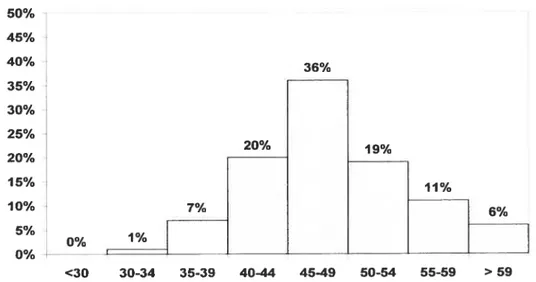
Afin d’évaluer la précision du positionnement acétabulaire vertical effectué selon la méthode de PVS, nous avons sélectionné au hasard 100 radiographies postopératoires de prothèses totales de hanche (PTH) effectuées en 1997-9$ par huit chirurgiens orthopédistes non spécialisés en remplacement total de la hanche (Hôpital de l’Enfant-Jésus, Québec). Sur ces radiographies, nous avons mesuré l’angle acétabulaire vertical à l’aide d’une ligne horizontale rejoignant les tubérosités ischiatiques selon la technique décritepar Lewinnek (voir f igure 3, page4)26
Les résultats ont démontré que l’angle acétabulaire vertical est très variable (Figure 4)44• Seulement 56% (n=56) des cupules présentaient un positionnement idéal*. 44% (n=44) des composantes ont été positionnées à moins de 40° ou plus de 49° et 17% (n17)
6
présentaient une erreur de positionnement (plus de 550
ou moins de 300). L’angle acétabulaire vertical moyen a été de 48.190 ± 6.74°. Ces résultats ont confirmé la faible précision du positionnement acétabulaire vertical à l’aide de la PVS. Nous croyons que la précision de cette technique peut être améliorée.
* Selon le travail de plusieurs auteurs qui ont tenté de déterminer l’orientation
verticale acétabulaire optimale: Lewinnek 3O0 50026 D’Lima 450550lt
Kummer 35045024, Salvati 45° +1- i0°, Imura < Harkess 45016 et Seki 30050042, nous avons déterminé que l’angle acétabulaire vertical idéal serait de 40° à 49° et qu’une erreur de positionnement serait un angle inférieur à 30° ou supérieur à 550 50% 45% 40% 36% 35% 30% 25% 20% 20% 19% 15% 11% 10% 6% 5% 0% 0% <30 30-34 35-39 40-44 4549 50-54 55-59 > 59
Figure 4: Pourcentage de cupules acétabulaires pour différentes tranches d’angle acétabulaire vertical (nombre de cupules l00)
La science médicale au fil de son évolution a favorisé le développement d’une multitude de moyens techniques pouvant augmenter la précision, la reproductibilité et l’efficacité du geste chirurgical. Par l’utilisation d’outils de précision, calibrés et validés, l’acte chirurgical peut aujourd’hui être plus reproductible et permettre aux
patients de bénéficier de résultats plus constants. Des efforts récents d’optimisation du positionnement acétabulaire à l’aide de systèmes de navigation assistés par ordinateur ont été publiés42. Ces techniques sont très prometteuses mais leur accessibilité est réduite en raison des coûts qui leurs sont associés et des restrictions budgétaires auxquelles font face les systèmes de santé.
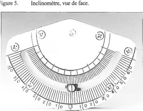
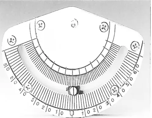
En 1997, nous avons conçu un inclinomètre en collaboration avec la compagnie Medical Design Inc (Figures 5 et 6). Cet inclinomètre est fait d’acier inoxydable et peut être stérilisé. Il est muni d’une pince permettant de le fixer sur la plupart des tiges d’insertion acétabulaire sur le marché. Un pendule influencé par la gravité détermine l’angle mesuré. Il est gradué aux 2 degrés de 00 à 70°. Lors de l’intervention
chirurgicale, le chirurgien peut associer l’inclinomètre à sa tige d’insertion acétabulaire (figures 7) et mesurer l’angle d’inclinaison avant de fixer la composante définitivement au bassin.
Présentement, aucune autre étude évaluant l’efficacité d’un guide d’insertion acétabulaire calibré avec un inclinomètre n’a été publiée. Nous croyons donc que cette étude aura le potentiel de déterminer si l’utilisation d’un inclinomètre permettrait d’augmenter la précision du positionnement acétabulaire et ainsi éviterait certains mauvais résultats du remplacement total de la hanche.
Figure 5. Inclinomètre, vue de face.
r
rA
‘7
iI
Figure 7 Inclinomètre attaché sur une tige d’insertion acétabulaire, vue de face et postérieure rti o y—
z
kld Objectifs
L’objectif principal de ce mémoire est de déterminer si l’utilisation de cet inclinomètre permet d’améliorer la précision du positionnement vertical de la composante acétabulaire lors du remplacement total de la hanche.
Pour atteindre cet objectif deux expériences sont nécessaires : une étude cadavérique et une étude clinique.
L’étude cadavérique a pour but de comparer la fréquence des positionnements verticaux acétabulaires dans une zone optimale avec l’inclinomètre et la perception visuospatiale (PVS), de déterminer si l’utilisation de l’inclinomètre permet d’éviter des erreurs de positionnement et d’évaluer la variabilité du positionnement vertical pour les insertions effectuées avec l’inclinomètre et avec la perception visuospatiale (PVS).
L’essai clinique randomisé (ECR) a pour objectif de comparer in vivo la précision du positionnement vertical de composantes acétabulaires de prothèses totales de la hanche implantées par des chirurgiens spécialisés en chirurgie de la hanche avec un inclinomètre à celle de composantes implantée selon la PVS. Cette étude veut aussi secondairement, déterminer la fréquence des positionnements verticaux obtenus entre 40° et 49° avec et sans l’utilisation de l’inclinomètre, déterminer la fréquence des erreurs de positionnement des deux méthodes: angle vertical de moins de 30 degrés ou plus de 55 degrés, déterminer la variabilité du positionnement entre les deux méthodes par rapport à l’objectif initial de positionnement désiré et déterminer si l’inclinomètre est un outil utile dans la pratique clinique d’orthopédistes spécialisés en remplacement prosthétique de la hanche.
2- Matériel et méthode globale
2a Description de l’étude (deux étapes):
La première partie, étude sur cadavre, a pour objectif d’évaluer la précision de l’inclinomètre en comparaison avec le positionnement par PVS dans un environnement contrôlé.
La deuxième portion met à l’épreuve l’inclinomètre dans la pratique quotidienne du chirurgien orthopédiste spécialisé en chirurgie de la hanche. Cet essai clinique permettra de vérifier si l’utilisation de l’inclinomètre est d’une précision égale ou supérieure à la PVS du chirurgien spécialiste du remplacement total de la hanche et s’il permet de minimiser les positionnements inadéquats.
2b 1èce étape : expérimentation sur cadavre
À
tour de rôle, nous avons demandé à des résidents en orthopédie de la région de Québec de positionner une composante acétabulaire à 400 d’angle vertical sur un cadavre selon leur PVS puis à l’aide de l’inclinomètre. Des clichés radiologiques ont été pris après chaque essai. Sur les radiographies (RX), l’angle vertical de la composante sera mesuré.Les insertions sont effectuées sur un cadavre frais
- positionné sur une table d’opération avec le bassin à 90° par rapport à la
table (non vérifié avec RX).
- dont l’approche chirurgicale, l’ostéotomie du fémur ainsi que l’alésage du
cotyle seront déjà effectués.
Matériel:
- Un cadavre male de poids normal
- Appareil de radiographie portatif
- Guide d’insertion acétabulaire traditionnel
-Inclinomètre maison
- Composante acétabulaire d’essai
(Interop: Suizer, Winterthur, Suisse)
- Participation de 14 résidents du programme d’orthopédie de
l’Université Laval et de 1 chirurgien orthopédiste.
Coordination:
1- Cadavre frais à l’hôpital
2- RX du bassin antero-posterieur (AP)
3- Positionnement sur table avec bassin à 90° par rapport à la table 4- Approche postéro latérale bilatérale
5- Ostéotomie du col fémoral et exposition de l’acétabulum. 6- Alésage du cotyle
7- Insertion à 40° d’angle vertical de l’acétabulum droit par PVS par chaque intervenant.
8- RX bassin AP (identifié A)
9- Insertion à 40° d’angle vertical de l’acétabulum droit avec l’inclinomètre par chaque résident.
10- RX bassin AP (identifié B)
11- Répéter la même séquence avec l’acétabulum gauche (no 6— 11)
Données:
- Angle vertical de la composante acétabulaire inséréeparPVS du coté
-Angle vertical de la composante acétabulaire insérée avec l’inclinomètre du coté gauche et droit mesuré sur RX.
Analyse
Comparaison entre les deux groupes: - angles moyens
- angles maximums et minimums
- variabilité des positionnements verticaux.
- nombre de positionnements non idéaux (<40°, >49°). - nombre de positionnement inadéquats (<30°, >55°)
NB s pour la description détaillée et les résultats de l’étape 1, voir section no 9- Article no 1: Vertical acetabular positioning with an inclinometer in total hip arthroplasty.
2c 2ême étape : Essai Clinique Randomisé
Population de l’étude
Cette étude a débutée dans le Service de chirurgie orthopédique adulte de 2 hôpitaux du Centre Hospitalier de l’Université de Montréal (C.H.U.M.) : Hôpital St-Luc (H.$.L.) et Hôpital Notre-Dame (H.N.D.). Jusqu’en décembre 2001, tous les patients traités par le Dr Nicolas Duval (H.N.D.) et par le Dr Luc Pilon (H.S.L) qui étaient en attente d’un remplacement total de hanche se sont fait offrir de participer à l’étude.
À
partir de 2002, l’étude fut poursuivie au sein du Service d’orthopédie de l’Hôpital Maisonneuve-Rosemont (H.M.R.). Les patients des Dr Pascal-André Vendittoli, Dr Martin Lavigne, Dr Robert Duchesne et Dr Alain Roy (H.M.R.) en attente d’une PTH, se sont fait offrir de participer à cette étude.Sélection des patients
Le diagnostic de la pathologie articulaire de la hanche est effectué selon les méthodes habituelles: histoire médicale, examen physique, examens de laboratoire, et imagerie. La décision d’offrir une chirurgie de PTH primaire ou de révision au patient est prise selon les critères habituels des chirurgiens impliqués dans l’étude. Tous les patients de plus de 1$ ans en attente pour une telle chirurgie se sont fait offrir de participer à l’étude. Aucun critère d’exclusion n’a été identifié pour cette étude.
Devis d’étude
Cette étude est un essai clinique randomisé, contrôlé avec deux groupes de traitement parallèles selon un devis standard.
Interventions
La chirurgie de remplacement total de la hanche est effectuée sous anesthésie générale ou régionale en décubitus latéral. Une approche postérieure standard ou une approche antérolatérale est utilisée à la convenance du chirurgien.
Dans le groupe contrôle, la composante acétabulaire est insérée avec la technique habituelle, i.e. la PVS de l’angle vertical par le chirurgien à l’aide d’un guide d’insertion standard. Selon différents critères chirurgicaux dont la stabilité articulaire, le recouvrement supérieur de la cupule, la géométrie du cotyle, etc., le chirurgien choisit un angle plus aigu ou plus obtus (idéalement entre 40 et 49 degrés). Une fois la composante fixée au bassin, le chirurgien estime l’angle vertical de la composante et l’inscrit surla fiche de recueil de données (annexe 2).
Dans le groupe expérimental, la composante acétabulaire est insérée à l’aide d’un inclinomètre à pendule ajouté sur le guide d’insertion acétabulaire. Le chirurgien décide alors de la position optimale de la composante (idéalement entre 40 et 49 degrés) en
mesurant l’angle vertical avec l’inclinomètre. Une fois la composante fixée au bassin, le chirurgien mesure l’angle vertical de la composante avec l’inclinomètre et l’inscrit sur la fiche de recueil de données (annexe 2).
Déroulement de l’étude
Une fois admis à l’hôpital, en prévision de sa chirurgie, les objectifs de l’étude, les risques et bénéfices sont expliqués au patient afin d’obtenir son consentement éclairé. Les sujets qui acceptent de participer à l’étude sont randomisés en deux groupes. La randomisation est sous la responsabilité de l’épidémiologiste responsable de l’étude (P.L.) et utilise le générateur de nombre aléatoire du logiciel S-Plus2.
Tel que fait dans la pratique courante, tous les patients ont une radiographie simple de la hanche avant l’opération. Une mesure de l’inclinaison verticale de leur acétabulum est effectuée. En post opératoire, tous les patients ont un RX du bassin. Sur ces RX, une mesure de l’angle vertical de la composante acétabulaire est effectuée. La mesure de l’angle acétabulaire s’établit en traçant une ligne horizontale reliant les échancrures acétabulaires et une seconde ligne suivant l’inclinaison de 1 ‘acétabulum
18.43
Toutes les mesures sur les RX sont prises par un des auteurs (P.A.V.) et un assistant de recherche à l’aide du même rapporteur d’angle gradué au degré. Lors de la lecture des RX, le groupe de randomisation (expérimental ou contrôle) n’est pas dévoilé auxévaluateurs et les lectures sont recueillies de façon indépendante. Les deux mesures sont saisies sur une fiche de collecte de données. La moyenne des deux mesures a été utilisée pour les analyses.
Taille de l’échantillon et puissance statistique
L’hypothèse principale de l’étude randomisée est que l’ajout d’un inclinomètre à pendule sur la tige d’insertion de la composante acétabulaire permet une meilleure
précision de l’angle de la composante verticale en comparaison à l’utilisation de la PVS La précision de l’angle d’insertion sera déterminée par rapport à l’angle visé par le chirurgien.
Atemoin=
Écart
entre l’estimation de l’angle vertical du cotyle par le chirurgien et la mesure sur le RX post opératoire.AExprimentaI
Écart
entre lamesure
de l’angle vertical du cotyle avec l’inclinomètre et lamesure surle RX post opératoire.
A= Écart minimal à détecter entre la mesure de l’angle de positionnement per opératoire et l’angle obtenu pour les deux méthodes. Déterminé à 4 degrés, pour que l’écart soit significatif cliniquement.
Groupe témoin:
Ai: Estimation de l’angle acétabulaire vertical per opératoire par le chirurgien A2 : Angle acétabulaire vertical mesuré sur RX post opératoire
Groupe expérimental:
A3 : Mesure de l’angle vertical à l’aide de l’inclinomètre A4 : Angle acétabulaire vertical mesuré sur RX post opératoire
= déviation standard des mesures d’angles: 6,74 (selon l’étude préliminaire: Revue de 100 radiographies à l’Hôpital de l’Enfant-Jésus)
Pour une erreur de type I u= 0,05 et une erreur de type II Ç3 = 0,2, n 45
Donc un nombre de 90 insertions devra être inclus dans l’étude. Pour palier à une perte de données en cours d’étude (RX manquants, etc.), nous allons inclure 100 sujets dans l’étude.
Évaluation de la puissance pour les issues secondaires
1- Hypothèse: la fréquence des positionnements verticaux adéquats (à 400 ou plus et moins de 50°) sera supérieure avec l’utilisation de l’inclinomètre à celle obtenue avec la PVS
Erreur de type I tx= 0,05 n=53
Pi (Groupe témoin): Proportion de positionnements adéquats prévue; estimée à 65 ¾ (selon étude cadavérique, estimation conservatrice puisque dans l’étude préliminaire (revue de 100 RX) la proportion est de 56%) P2 (Groupe expérimental): Proportion de positionnements adéquats
prévue; estimée à 95% (selon étude cadavérique: 100%)
Nous obtenons une puissance de 98,6 %
2- Hypothèse: la fréquence des erreurs majeures de positionnement (cotyle à moins de 30 degrés ou plus de 55 degrés) sera moindre avec l’utilisation de l’inclinomètre qu’avec la PVS.
Erreur de type I Π0,05 n=53
Pi: Proportion de positionnements inadéquats prévue (groupe contrôle); estimée à $ ¾ (selon étude cadavérique, estimation conservatrice puisque dans 1’ étude préliminaire (revue de 100 RX) la proportion est de 17%)
P2 : Proportion de positionnements inadéquats prévue (groupe expérimental); estimée à 0% (selon étude cadavérique)
Puissance obtenue : 57,4%
Si Pi = 15%, nous obtenons une puissance de 86,4%
Données Recueillies: - Chirurgien
- Diagnostique
- Type de chirurgie primaire! révision - Type d’approche
- Angulation verticale de la composante acétabulaire insérée avec le guide traditionnel.
- Angulation verticale de la composante acétabulaire insérée avec 1’ inclinomètre.
Analyses statistiques
L’homogénéité des deux groupes sera vérifiée en ce qui a trait à différentes variables pouvant avoir une influence sur les résultats i.e. la précision du positionnement: l’angle acétabulaire anatomique du patient (préopératoire) et le type de chirurgie (primaire vs révision).
Les proportions seront comparées avec le test du Chi-Carré de Pearson. Le test de Student sera utilisé pour évaluer les moyennes des deux groupes. La comparaison des variances sera effectuée avec le test de Levenes. Les intervalles de confiance présentés seront de 95% et l’erreur alpha acceptée comme seuil de signification sera de p <0.05 pour toutes les analyses statistiques.
Pour l’hypothèse principale (HI =l’ajout d’un inclinomètre à pendule sur la tige d’insertion de la composante acétabulaire permet une meilleure précision de l’angle de la composante verticale que l’utilisation d’un guide d’insertion standard seulement), si la distribution des résultats le permet, les différences moyennes d’angle entre le guide d’insertion standard et l’inclinomètre seront évaluées à l’aide du Students t-test. Pour les deux premières hypothèses secondaires (H1 La fréquence des positionnements adéquats sera supérieure avec l’utilisation de l’inclinomêtre comparée à la PVS; HI =
La fréquence des erreurs majeures de positionnements sera moindre avec l’utilisation de l’inclinomètre comparée à la PVS), l’étude des proportions de positionnements adéquats et des erreurs majeures sera effectuée avec le test du Chi Carré de Pearson.
Pour le troisième objectif secondaire, l’hypothèse est que la variabilité du positionnement par rapport à l’objectif visé sera moindre avec l’inclinomètre qu’avec la PVS. Le test de comparaison de variances de Levenes sera utilisé. Les analyses sont effectuées à l’aide du logiciel SPSS 10.03.
Considérations éthiques
Le projet de recherche a été soumis et approuvé par le comité scientifique et le comité d’éthique du C.H.U.M. et de 1’H.M.R.
Un document d’information a été remis et expliqué aux patients. Un consentement écrit éclairé a été rempli par le patient, le médecin traitant et un témoin afin que le patient soit conscient des risques et des bénéfices que comporte son inclusion dans l’étude.
Toutes les données recueillies sont traitées de façon strictement confidentielle. Aucune donnée nominative n’est utilisée dans l’analyse des données ou dans les publications ou présentations de cette étude.
NB : pour la description détaillée et les résultats de l’étape 2, voir section no 10-Article 2: Randomised study comparing two methods of acetabular cup positioning in total hip arthroplasty.
3- Introduction aux deux articles scientifiques
Le présent mémoire est présenté par articles scientifiques. Le coeur de cet ouvrage comporte deux études scientifiques: une étude cadavérique et une étude clinique qui font chacune l’objet d’un article. Le premier article tient lieu de Chapitre 4. Il décrit l’étude cadavérique et en livre les résultats. Il a permis de conclure que l’inclinomètre permet d’augmenter la fréquence de positionnement verticaux acétabulaire dans la zone optimale et d’éviter les erreurs de positionnement dans un environnement contrôlé. Suite à cette publication, il était primordial de démontrer l’application de l’inclinomêtre dans la pratique clinique de chirurgiens d’expérience.
L’étude de validation clinique, rendue nécéssaire par les conclusions de l’étude cadavérique, fait l’objet du deuxième article et compose le Chapitre 5. Ces deux articles permettent de répondre aux objectifs de recherche de ce mémoire. Une discussion et conclusion globales font suite à ces deux articles.
4- Article no 1:
Vertical
acetabular
positioning
witli
an
inclinometer in total hp arthroplasty
Auteurs:
Pascal-André Vendittoli, MD, fRCS(C)’ Chercheur principal
Nicolas Duval MD, FRC$(C)2
David lames Stitson, MBBS, FRCS(Orth), RAf3 Benoît Mâsse, PhD4
Departement of Orthopeadic Surgery, Centre Hospitalier Maisonneuve Rosemont (C.H.A.), Montréal, Québec, Canada
2
Department of Orthopaedic Surgery, Centre hospitalier de l’Université de Montréal-Hôpital Notre-Dame, Montréal, Québec, Canada
Departement of Orthopeadic Surgery, James Cook University Hospital, Middlesbrough, England
Epidemiology Research Group, Université Laval, Ste-Foy, Québec, Canada.
Mots clés: acetabulum, hip prosthesis, inclinometer, abduction angle
Article publié:
Vendittoli, P. A.; Duval, N.; Stitson, D. J.; and Masse, B.: Vertical acetabular positioning with an inclinometer in total hip arthroplasty. J Arthroplasty, 17(7): 936-41., 2002.
Reprint from Journal ofArthroplaty; vol 17, no 7: Vendittoli, P. A.; Duval, N.; Stitson, D. J.; and Masse, B., Vertical acetabular positioning with an inclinometer in total hip arthroplasty, Journal of Arthroplasty pp. 936-4 1, Copyright (2002), with permission from Elsevier (voir annexe 4).
Abstract
Vertical acetabular implant positioning is an important technical aspect in total hip arthroplasty. b evaluate the potential benefit of an inclinometer. 50 cup insertions were perforrned on a cadaver pelvis. Acetabular cup vertical angles averaged 44.4°±11.4° by visuo-spatial perception, and 42.2°±3.8° with the inclinometer. Ail cups were within the safe angle range of 40°-49° with the inclinometer compared to 64% of cups by visuo-spatial perception. Use ofthe inclinometer reduced variability by a factor of 2.0 to 4.5. The addition of an inciinometer for acetabular cup insertion increases the probability of positioning the cup within a vertical safe range during total hip arthroplasty on a cadaver pelvis, suggesting that it could be a useful adjunct in clinical practice.
Introduction
Total hip arthroplasty (THA) is one ofthe modem surgical procedures that oflen allows invalid patients to acquire a near normal quality of life’3. However, intra-operative difficulties or technical errors can significantly alter the outcome. It has been reported that acetabular component malpositioning is associated with an increased risk of dislocation46, limited range of motion and impingement7’°. An openly positioned cup (high vertical acetabular component angle) correlates with increased polyethylene wear rate, osteolysis and early aseptic loosening in metal-polyethylene surfaces”’5. Such malpositioning is also associated to premature acetabular aseptic loosening in ceramic on ceramic arthroplasty’6”7”8. This suggests that the observed phenomenon is one of component positioning rather than of the bearing surface chosen. The proposed explanation for early loosening, in this context, is abnormal joint loading”,t3. 19-21 These observations have prompted other authors to propose optimal ranges of vertical acetabular cup angles lying between 30° and 50°’22-27
It is common practice for orthopaedic surgeons to rely on visuo-spatial perception (VSP) when positioning the acetabular component during THA. The position of the cup is estimated by aligning the insertion rod to the body axis of the patient, to anatomical landmarks or to other reference points in the operating room(operating table, wall, floor, etc.)6. Some surgeons consider that it is easier to estimate a 90° angle than a 45° angle and add a fixed rod attached at 45° to the insertion rod28. Anatomical landmarks can also be used to improve VSP estimation6. We have observed, in a preliminary study, that vertical positioning of the acetabular component by VSP is variable and lacks precision (see Materials and Methods and Figure 1).
In an attempt to increase the probability of positioning the cup within a safe vertical range, we have designed an inclinometer that can be attached to any acetabular component insertion rod (Figures 2 and 3). This gives the surgeon the ability to accurately measure the vertical acetabular position in relation to the ground. The objective of this study was to determine if use of the inclinometer will result in a higher
number of cup implantations within a safe vertical fange Ofl a cadaver pelvis compared
to implantation using VSP alone.
Materials and Methods
Preliminary study: Radiographic review of 100 acetabular components positioned by VSP
A preiiminary study was performed to estimate the percentage of acetabular cups impianted within a safe vertical range during TJ-TA. A random sampie of 100 THA was obtained by computer selection with SP$S for Windows statistical package (version 8.0; $PS$, Chicago, Iliinois, 1997) from a database of 150 primary THA perforrned by eight surgeons in one teaching hospital between July 1997 and July 199$. Ail acetabular cups had been positioned by VSP alone, without a fixed angle rod attached to the insertion rod, and without any other aiignment adjuncts; as is common practice for the surgeons in this hospital. Vertical acetabular cup angle was measured on the postoperative antero posterior (AP) radiograph of the pelvis with a pencil and a goniometer from a reference une drawn tbrough the inferior border of the ischial tuberosities as described by Lewinnek et al.5.
Inclinometer
The inclinometer that has been developed is made of sterilizeable stainless steel and is designed for attachment to most acetabular insertion rods by hand-tightening a single screw. The instrument is scaled at 2° intervals from 0° to 70°. Display of the vertical angle value resuits from dispiacement of the pendulum within the inclinometer over the inclinometer scale under the influence of gravity (Figure 2 and 3).
Cadaver study
This study was conducted on a medium-size, fresh male cadaver in the right lateral decubitus position with the pelvis supported at approximately 90° to the plane of a “Maquet” operating table. The pelvis was stabilised with two table mounted supports applying pressure at the pubis and in the sacral area. Afier draping, a lefi lateral
Hardinge approach was performed foliowed by femoral neck osteotomy and acetabular reaming up to 54 mm. Exposure of the acetabulum was achieved with one Chamley spike in the postero-superior wall and one retractor hooked in the antero-inferior wail.
We used a trial of 56 mm acetabular cup mounted on a standard insertion rod without a fixed angle rod attached. For the VSP evaluation, each participant was asked to position the cup at 400 of vertical inclination, according to their usual technique, and to secure the cup in this position with a single hammer biow. Afier each insertion, an AP radiograph of the pelvis was taken with the triai cup and insertion rod in place. During each X-ray and between each triai, the cadaver remained in the same position on the operating table. X-ray cassettes on a support were positioned according to marks on the floor at the back of the cadaver pelvis. The X-ray beam was not moved throughout ail of the exposures. No manipulation of the cadaver, and in particular, its lower iimbs was undertaken between each triai. Afier the V$P triai, each participant repeated the same procedures with the aid of our inclinometer. We asked the participants to position the acetabuiar trial at 40° with the inclinometer attached to the insertion rod. Afier each trial insertion, an AP radiograph of the pelvis was taken according to the same protocol described for VSP. Compietion ofthe experiment by ail participants was foliowed by its repetition on the right hip. Thirteen orthopaedic residents and one orthopaedic surgeon participated in the study.
Thirteen orthopaedic residents and one orthopaedic surgeon took part in the study compieting a total of 50 triai insertions. Eieven residents compieted biiaterai V$P and inciinometer trials (44 insertions), whereas 2 residents and the orthopaedic surgeon compieted unilateral trials (6 insertions).
The vertical inclination of the cup was measured on plain radiographs with a pencii and a goniometer graduated every degree. The angle was measured between the horizontal inter-teardrop une and a une perpendicuiar to the insertion rod. This technique was preferred to the Lewinnek method since the presence ofthe insertion rod screwed to the cup during X-ray exposure gives an exact perpendicular reference to the vertical
orientation of the acetabular trial. Ail measurements were done by one of the authors blinded to the intervention group of each X-ray29’30•
Statistical analyses
Ail statisticai anaiyses were performed using S-PLUS 3•43t Proportions were compared by Fisher’s exact test. Mean differences between the VSP method and the inciinometer method were evaiuated by Student’s t-test for unequai variances. Comparisons of the equaiity of variances were conducted by Fisher’s test. Confidence intervais for the ratio of two standard deviations were computed by the F statistic. As number of triais was less than 30 and the data distributions had unequal variance, non-parametric tests, nameiy, the Mann-Whitney test for two-sampie tests, and the Wilcoxon signed rank test for one-sample tests were adopted for mean comparison. The confidence intervals presented are of 95%, and the significance ievei accepted was p<O.05 for ail statisticai analyses.
Results
Pre]iminary study: Radiographic review of 100 acetabular components posïtioned by VSP
Vertical positioning of the acetabuiar component with VSP aione was very variable (Figure 1). Forty-four per cent of the components were positioned at either more than 49° or iess than 40° of vertical inclination. Eighteen per cent were positioned at more than 55° or less than 35°. The average vertical inclination of the components was 48.2°± 6.7°.
Cadaver study
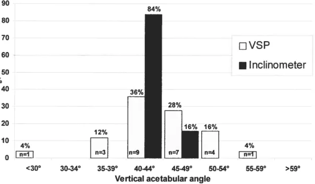
The anatomical vertical inclination of the cadaveric acetabulum used was 42°, as determined by radiographic measurements. The acetabular component vertical angle distribution is shown in Figures 4. The resuits of the 25 trials performed with VSP
alone showed that 64% (n=16) ofcomponents were positioned at angles of between 400 and 490
compared to 100% of 25 triaIs with the inclinometer (j=0.002). Eight per cent (n=two) of the positioning trials using VSP alone were at angles less than 30° or more than 55°, compared to none with the inclinometer (p=O.49O). The difference between mean angles of vertical positioning using both methods was of borderline significance (p= 0.083). Both means were significantly superior to 40° (VSP 44.4°, p<O.001; inclinometer 42.2°, p<0.001).
The 95% confidence interval of the ratio of the standard deviations of the two positioning methods showed that use of VSP alone was between 2.0 to 4.5 times more variable than positioning with the inclinometer (p<0.001). For a given patient with a target angle set at 40°, it is estimated that positioning precision using the inclinometer is 42.2°± 3.8°. compared to 44.4°± 11.4° with VSP alone.
Discussion
Acetabular component positioning is a very important step in the technique of total hip arthroplasty. There is probably a different ideal position for each patient according to individual anatomical peculiarities. This “perfect” position is somewhat impossible to achieve and most surgeon agree that position within a safe range of vertical angle is acceptable. Many authors recommend vertical angles of between 40-50° for metal polyethylene THA58’22, 25-27and 35-45° for ceramic-ceramic THA2324 to obtain the best results. From the literature, we do flot know precisely what vertical angles would compromise the results of THA. However, it is reasonable to assume that the further away from the safe range the cup is positioned, the greater the chance of a poor outcome.
b increase the accuracy of vertical acetabular positioning the use of a reference rod attached at 45° to the insertion rod is sometimes recommended to position the acetabular component in THA. This system assumes that it is easier for the surgeon to evaluate a 90° angle than a 45° angle. Only one study looked at the improvement offered by such a
reference rod28. four surgeons reported 30% of implanted acetabular cups at <40° or >49° and 8% at <30° or >60°. Our preliminary study invoiving the radiographic review
of 100 acetabular components positioned by VSP (f igure 1) and data from our cadaveric study produced resuits similar to those of other authors who also reported a wide distribution of vertical acetabular ang1es3235. These authors, however, did flot specify their positioning technique. The variability observed in these reports indicates that the accuracy of cup positioning could be improved upon.
We believe that improvement could be approached in three ways. One is to confirm positioning with a peroperative X-ray. No publications have been found that evaluate this technique, even though it is common practise in some orthopaedic centres. This technique has potential disadvantages that include: increased risk of contamination, increased operating time, and exposure of the patient and surgical team to radiation. The second potential improvement is computer-assisted technique. Seki et al. have reported encouraging resuits, but the technique is time-consuming and requires complex organisation making it too costly for routine use at present27. The third method is to improve visual positioning of the insertion rod with an inclinometer as demonstrated by our cadaver study.
In this study, ail the cups positioned with the inciinometer were within the safe vertical angle range of 40-49° compared to oniy 64% of the cups positioned by VSP. furthermore, 8% of the cups in the VSP group positioned at less than 30° or more than 55° of vertical inclination which couid be considered to lie outside of the safe range. With the inclinometer, we have been able to eliminate such malpositioning.
We have improved the variability of the acetabular cup positioning using the inclinometer by 2—4.5 times when compared to the variability of the cup positioned by VSP (p<0.001). The positioning precision for the inclinometer is 42.2°± 3.8°, compared to 44.4°± 11.4° by V$P. We conclude, therefore, that utilisation of an inclinometer can improve significantly the capacity of the surgeon to position the
acetabular component in a safe range of vertical angle compared the cup positioned by VSPduring total hip arthroplasty.
The mean vertical angle in this study is similar for both the VSP and Inclinometer groups however, this is of little interest because a data set with a wide variance (very high angles and low angles) and a data set with a narrow variance can resuit in similar mean values. The main objective in a clinical setting is to minimize the variability (standard deviation) between each implantation. This fact has been shown very well in the cadaver study; both means are similar but the distribution ofthe angle is much wider in the VSP group (figure 4). We noted that the average vertical acetabular angles with VSP and with the inclinometer were both significantly greater than 40° (VSP 44.4°, inclinometer 42.2°). The fact that these two values were significantly over 40° statistically, indicates that this positive variation could have been produced by a confounding factor that increased the vertical acetabular angle. This factor could be a more caudal pelvic position on the operating table or a non-horizontal operating table. With an inclinometer, the surgeon should pay special attention to vertical positioning of the patient pelvis on the operating table. since this tool uses a pendulum oriented by gravity.
The cadaver used in our study had radiographically-measured acetabular angles of 42°. Such non-dysplasic hips may have favoured good performance of the VSP method. In hip dysplasia and difficuit revision situations, it is likely that the VSP method would have given greater variability of resuits. Also, the application of a single task (positioning the acetabular component) compared to performance of multiple tasks (THA), and the desire to do well for the purpose of research may have favoured improved results during implantation by VSP.
The use of an inclinometer during THA will neyer replace careful pre operative surgical planning and systematic evaluation of cup position according to anatomical landmarks during surgery. This instrument represents a helpful adjunct to the surgeon, increasing the probability of positioning the cup within a safe range and thus, avoiding
malpositioning. This maybe especially useful when anatomical landmarks are deficient as is the case in hip dyspiasia or revision situations.
The addition of an inclinometer has been shown to increase the probability of positioning the acetabular cup within a safe vertical range during THA on a cadaver. A clinical trial is under way to assess its clinical performance before considering its potential commercialisation. We commend the inclinometer as a valuable adjunct to hip replacement surgery prior to wider availability of computer-assisted tecimiques.
Acknowledgments
We thank ail the residents who participated in this project, Dr. Gaston Paradis, Chief of the Department of Orthopaedics, Hôpital de l’Enfant Jésus (Québec), Claire Desgagné, orthopaedic nurse, Jean Montemillio and flavio Marinelli (Smith and Nephew), and The Hip Hip Hourra Foundation for their financial support. We acknowledge the editorial assistance of Ovid Da Silva, Éditeur-Rédacteur, Research Support Office, Research Centre, CHUM.
33
Figures pour l’article no 1: Vertical acetabular positioning wïth an inclinometer in total hip arthroplasty
Figure 1: radiographic review of n1 00 total hip arthroplasties in 1997-9$.
100 90 80 70 60 % 50 40 36 30 20 19 20 11 10 7 6 1
o
<350 35-39° 40-44° 45490 50-54° 55590 >590Figure 2. Inclinometer front view.
Figure 3 Inclinometer lateral view.
—
36
Figure 4: Percentage distribution of vertical acetabular angles using VSP and Inclinometer techniques (n= number of implantations).
90 80 50 -0! /0 40 30 Ao/ * 10 In=1I <30° AOl ‘t /0 n1 55590 70 60
EVSP
• Inclinometer
20 -10 28% 16% 16% 12%I
j
n=3 n=7 30-34° 3549° 40-44° 4549° 50-54°Vertical acetabular angle
References
1 Kavanagh Bf. Wallrichs S, Dewitz M, Berry D, Currier B. Ilstrup D, et al.: Chamley low friction artbroplasty of the bip: twenty-year resuits with cernent. J Arthroplasty 9:229-34. 1994
2 Wickland I, Romanus B: A comparison of quality of life before and afler arthroplasty in patient who had arthrosis ofthe bip joint. J Bone Joint Surg 73A:765-9, 1991
3 Laupacis A, Boume R, Rorarbeck C, f eeny D, Wong C, TugweÏl P, et al.: The effect of elective total hip replacement on health related quality of life. J Bone Joint Surg 75A:1619-26, 1993
4 O’Brien S, Engela DW, Beverland LS, Kemohan WG: A study of the factors in bip replacernent dislocation. Nurs Standard 11:39-42, 1996
5 Lewinnek GE, Lewis JL, Tarr R, Compere CL, Zimmerman JR: Dislocation afier total bip replacement arthroplasties. J Bone Joint Surg 60A:217-20, 1978
6 McCollum DE, Gray WJ: Dislocation afler total hip arthroplasty. Causes and prevention. Clin Orthop 261:159-70, 1990
7 D’Lima DD, Urquhart AG, Buehier KO, Walker RH, Coiweil CW: The effect of the orientation of the acetabular and femoral components on the range of motion of the hip at different head-neck ratios. J Bone Joint Surg $2A:315-21, 2000
8 Kummer fJ, Shah S, Iyer S, DiCesare PE: The effect of acetabular cup orientations on limiting hip rotation. J Arthroplasty 14(4):509-13, 1999
9 Bader R. Willmann G: Ceramic cups for hip endoprotheses. 6: Cup design, inclination and antetorsion angle modify range of motion and impingement. Biomed Tech (Beri) 44(7-8):209- 12, 1999
10 Bader R, Willmann G: Ceramic acetabular cups for hip endoprostheses. 7: How do position of the centre of rotation and the CCD angle of the shafi modify range of motion and impingement. Biomed Tech (Berl) 44(12):345-51, 1999
11 Kurtz 5M, Edidin AA, Bartel DL: The role of backside polishing, cup angle and polyethylene thickness on the contact stresses in metal backed acetabular component. J Biomechanics 30(6):639-42, 1997
l2Chen FS, Di Cesare PE, Kale AA, Lee JF, Frankel VH, Stuchin SA, Zuckerman JD: Resuits of cemented metal-backed acetabular components: a 10-year-average follow up study. J Arthroplasty 13(8):867-73, 199$.
13 Schrnalzried TP: The relationship between the design, position and articular wear of acetabular component inserted without cement and the development of pelvic osteolysis. J Bone Joint Surg 76A(5):677-8$, 1994
14 McBroom R., Muller M: Aseptic loosening: fifteen years experience with the Muller total hip arthroplasty. Procedings of the 39th Canadian Orthopeadic Association Meeting. Québec, 1983. J Bone Joint Surg 66B:300, 1984
15 Kennedy JG, Roger WB, Soffe KE, Sullivan RJ, Griffen DG, Sheehan Li: Effect of acetabular component orientation on recurrent dislocation, pelvic osteolysis, polyethylene wear, and component migration. J Arthroplasty 13(5):530-4, 199$
16 Kummer FJ, Stuchin SA, Frankle VH: Analysis of removed Autophore ceramic-on— ceramic components. J Arthroplasty 5(29):509-13, 1990
17 Dorlot JM. Christel P, Meunier A: Wear analysis of retrieved alumina heads and sockets of hip prostheses. J Biomed Mater Res: Appi Biomater 23(A3):299-3 10, 1989
1$ Hamadouche, M, Boutin, P, Daussange, J, Bolander, ME, Sedel, L: Alumina-on-alumina total hip arthroplasty: a minimum 1 $.5-year follow-up study. J Bone Joint Surg 84A(1): 69-77, 2002.
19 Dowiing JM, Atkinson JR, Dowson D, Charnley J: The characteristics of acetabular cups wom in the human body. J Bone Joint Surg 60-B(3):375-$2. 197$
20 Boutin P, Christel P, Dorlot JM, Meunier A, de Roquancourt A, Blanquaert D, Herman S, Sedel L, Witvoet J: The use of dense alumina-alumina ceramic combinations in total hip replacement. J Biomed Mater Res 22:1203-32, 1988
21 Maistrelli G, Gerundini M, Bombelli R: The inclination ofthe weight bearing surface in the hip joint. The clinical significance ofabnormal force. Orthop Rev 15(5):271-9, 1986
22 Salvati EA, 1m VC, Aglietti P, Wilson PD Jr: Radiology of total hp replacements. Clin Orthop 121:74-82, 1976
23 Sedel L, Kerbouil L, Christel P, Meunier A, Witvoet J: Alumina on alumina hip replacement: resuÏts and survivorship in young patients. J Bone Joint Surg 723:658-63, 1990
24 Sedel L: Editorial : ceramic hips. J Bone Joint Surg 79B:331, 1992
25 Imura S: Biomechanics of total hip arthroplasty. East Jpn J, Clin orthop 7:609-15, 1995
26 Harkess WJ: Arthroplasty of the hip. p. 535. In Crenshaw AH (ed): Campbell’s Operative Orthopaedics. Mosby; St. Louis, 199$
27 Seki M, Yuasa N, Ohkuni K: Analysis of optimal range of socket orientations in total hip arthroplasty with use of computer aided design simulation. J Orthopaed Res 16(4):513-7, 1998
28 Hassan DM, Johnston GH, Dust WN, Watson G, Dolovich AT: Accuracy of intraoperative assessment of acetabular prosthesis placement. J Arthroplasty
13(1):80-4, 1998
29 Herlin K. Petterson H. Sevlik G: Comparison of two and three dirnensional methods for assessment of orientation of total hip prosthesis. Acta Radiologica 29:357-61.
1988
30 SetIer R, Lyles D, Dorr L: The effect of pelvic rotation on alpha and theta angles in total hip arthroplasty. Contemp Orthop 17:67-9, 198$
31 Statistical Sciences, S-PLUS Guide to Statistical and Mathematical Analysis, Version 3.4, Seattle, StatSci, a division of Mathsoft, Inc., 1996
32 Pollard JA, Daum WJ, Uchida T: Can simple radiographs be predictive of total hip dislocation? J Arthroplasty 10(6): 800-4, 1995
33 Petterson H, Gentz Cf, Lindberg HO, Carlsson AS. Radiologic evaluation of the position of the acetabular component of the total hip prosthesis. Acta Radiologica Diagnosis 23(3A): 259-63, 1982
34 Del Schutte H, Lipman AJ, Bannar SM, Livermore JT, Ilstrup D, Morrey 3F: Effect of acetabular abduction on cup wear rates in total hip arthroplasty. J Arthroplasty
35 Nizard RS, Sedel L, Christel P, Meunier A, Soudry M, Witvoet J: Ten-year survivorship of cemented ceramic-ceramic total hip prosthesis. Clin Orthop 282:53-63, 1992
5- Article 2:
Randomised study comparing two methods of
acetabular
cup
positioning
in
total
hip
arthroplasty
Auteurs: Pascal-André Vendittoli, MD, FRCS(C) Chercheur principal Nicolas Duval, MD, FRCS(C)2 Pauline Lavoie, MD, MSc3 Martin Lavigne. MD, fRCS(C)1 Alain Roy MD, FRCS(C)’‘Service d’Orthopédie, Département de chinirgie, Hôpital Maisonneuve-Rosemont, Montréal, Québec, Canada
2
Département de chirurgie, Centre hospitalier de l’Université de Montréal(CHUM) Hôpital Notre-Dame, Montréal, Québec, Canada
Pavillon des Charmilles, Lavai, Québec, Canada
Mots clés: acetabulum, bip prosthesis, inclinometer, abduction angle
Abstract
Vertical acetabular implant positioning is an important tecimical aspect in total hip arthroplasty tillA). To evaluate the potential benefit of a new technique for acetabular cup positioning, 101 cup were randomized to be inserted with or without an inclinometer. Acetabular cup vertical angles averaged 43.6°± 6.8° with the inclinometer, and 42.7° ± 6.7° by visuo-spatial perception (VSP) (p=0.506). 57.4% of the cups were positioned within the desirable angle range of 400490 with the
inclinometer compared to 50.0% by VSP (p0.454). The proportion ofcup positioned outside a safe angle range of 30°-55° was low in both groups: 6.4% (n3/47) for the inclinometer group versus 3.8% (n=2/53) for the VSP group (p0.536). In THA performed by dedicated hip surgeons, the inclinometer did flot significantly reduce the variability ofthe cup abduction angle compared to the VSP method.
Introduction
Total hip arthroplasty (THA) is a very successftil surgical procedure’3. It is common practice for orthopaedic surgeons to rely on visuo-spatial perception (VSP) when positioning the acetabular component during THA. Cup abduction is estirnated by the insertion rod position according to patient’s body anatomical landmarks or other reference points in the operating room (operating table, wall, floor, etc.)4. Vertical positioning of the acetabular component by VSP is variable and lacks precision5’ 6.
7(Figure 1).
A significant number of THA revisions are performed every year, with many of them resulting from component malposition8. Acetabular component malposition is associated with an increased risk of dislocation4’ 9. 10
limited range of motion, and impingement11-14 A high vertical acetabular cup angle correlates positively with premature polyethylene wear, osteolysis and early aseptic loosening in metal poÏyethylene15’19 and ceramic-ceramic i221 The suggested explanation for this correlation is that a high vertical position augments the articular load per unit of surface area,15’ 17, 22-24 eventually leading to acetabular cup wear and supero-lateral migration of the femoral head. Many authors recommend positioning the acetabular cup within an optimal vertical angle range of 400 to49Q4 10-12, 25-30
To increase the probability of positioning the cup within a safe vertical angle range, we designed an inclinometer that can be attached to any acetabular component insertion rod (Figures 2 and 3). It allows the surgeon to accurately measure the vertical acetabular
position in relation to the ground. The objective ofthis study was to determine if use of the inclinometer could resuit in a higher number of cup implantations within a safe vertical range compared to implantation by VSP alone.
Materials and Methods
Inclinometer
This inclinometer is made of sterilizeable stainless steel designed for attachment to most acetabular insertion rods by hand-tightening a single screw. It is scaled at every 2° from
o
to 70°. Reading ofthe vertical angle value is the result ofdisplacement on the scale of a pendulum submitted to gravity within the inclinometer (Figures 2 and 3).Study
Seven surgeons performing THA regularly participated in the study. Patients were randomized to have their acetabular cup positioned with the inclinometer or by VSP (the usual method of the surgeons involved). Surgeries were performed on patients in lateral decubitus position with the pelvis fixed at approximately 90° to the plane of the operating table with 2 pelvic cushions attached to the table (one on the pubis and the other in the sacral area). A direct lateral (2 surgeons) or a posterior (5 surgeons) approach was performed. Afler routine acetabular preparation chosen acetabular cup was screwed to a standard insertion rod without a fixed angle rod attached. For VSP insertion, each acetabular cup was positioned by the surgeon’s usual manner. After fixation of the cup, the surgeon estimated the acetabular cup vertical angle. For the
inclinometer group. each surgeon performed the same procedure with the aid of the inclinometer. The surgeons were asked to position the acetabular cup at an angle of 40°-490
measurcd with the inclinometer attached to the insertion rod. Once the cup was fixed, the vertical angle red on the inclinometer was recorded.
An antero posterior radiograph of the pelvis taken postoperatively was used to measure the vertical angle of the cup according to the horizontal inter-teardrop une31’ 32 Ah measurements were made independently by 2 blinded observers. The average measurement of both observers was used for the analysis.
Statistical analyses
Ail statisticat analyses were performed using SAS sofware33. Proportions were compared by Pearson’s Chi-square test. Mean differences between the VSP and the inclinometer methods were evaluated by Students t-test for equal variances. Comparison of variances was conducted by Levenes’s test. The confidence intervals presented were 95%, and the significance level accepted was p <0.05 for ail statistical analyses.
Resu]ts
Type of surgery. operated side and pre-operative vertical acetabular angle (Sharp angle)34 were similar in both groups (Table 1). Table 2 presents the number of cup implantations per surgeon. With the VSP method, 50.9% (27/53) of components were
positioned at angles between 400 and 490
compared to 5 7.4% (27/47) with the inclinometer
(p=0A54).
3.8% (2/5 3) of the acetabular cups positioned by VSP were at angles less than 30° or more than 55° versus 6.4% (3/47) in the inclinometer group (pO.536, figure 4).There was no significant difference between mean vertical angles for the VSP and inclinometer groups (Table 3). No significant difference was found between the precision of both methods (vertical angle estimated per-operatively with the inclinometer or by surgeon V$P minus the vertical angle rneasured on post-operative radiographs): 5•40
(standard deviation 4.7) for the VSP group, and 4.8° (standard deviation 4.8) for the inclinometer group (pO.553).
The 95% confidence interval of the ratio of the standard deviations for both methods showed that the VSP method was flot more variable than positioning the cup with the inclinorneter (p0.880).
Discussion
As reported by Moran et al. orthopaedic surgeons are flot always accurate in estimating angles35. Preliminary investigation involving the radiographic review of 100 acetabular components positioned by VSP demonstrated a very wide vertical angle distribution7. Similar results have been published by other authors3639.
Many techniques have been developed to improve acetabular cup positioning during THA. Per-operative radiographic control is common practice in some orthopaedic
centres, especially with new, minimally-invasive techniques. Disadvantages of this technique include increased risk of contamination, longer operating time, and exposure of the surgical team and the patient to X-ray. Moreover, the precision of this technique has neyer been published. Another technique is a computer-assisted approach. Seki et al. reported encouraging results, but it is time-consuming and requires complex set-up that most orthopaedic centres cannot afford30.
A simpler and affordable way is to improve visual assessment of the insertion rod position. Some THA instruments offer a reference rod attached at 450
to the insertion rod to help the surgeon position the acetabular component5. This addition assumes that it is easier for the surgeon to evaluate a 900 angle than a 45° angle. One study looked at the improvement offered by such a reference rod. Four surgeons reported 30% of implanted acetabular cups at <40° or >49° and 8% at <30° or >60°.
In a cadaver study, the use of an inclinometer on the insertion rod was found to reduce the variability of vertical acetabular positioning by a factor of 2.0-4.5 in comparison to V$P7. No component was positioned less than 30° or more than 55° (Figure 5)7•
In the present clinical study, performed by dedicated hip surgeon, 6.4% (3/47) of acetabular components were positioned with the inclinometer at less than 30° or more than 550
of vertical angle, whereas 3.8% (2/53) of the components positionned by VSP were within the same angle range. In comparison, a group of general orthopaedic