Purification and Characterisation of a 31-kDa Chitinase
from the
Myzus Persicae Aphid: A Target for Hemiptera
Biocontrol
Frédéric Francis&Julien Saguez&Anas Cherqui&
Sophie Vandermoten&Charles Vincent&
Marie-France Versali&Jacques Dommès&
Edwin De Pauw&Philippe Giordanengo&
Eric Haubruge
Received: 16 May 2011 / Accepted: 20 December 2011 / Published online: 6 January 2012
# Springer Science+Business Media, LLC 2012
Abstract Hydrolytic enzymes involved in chitin degradation are important to allow moulting during insect development. Chitinases are interesting targets to disturb growth and develop alternative strategies to control insect pests. In this work, a chitinase from the aphid Myzus persicae was purified with a 36-fold purification rate in a three step procedure by ammonium sulphate fractionation, anion-exchange chromatography on a DEAE column and on an affinity Concanavalin A column. The purified chitinase purity assessed by 1D and 2D SDS–PAGE revealed a single band and three spots at 31 kDa, respectively. Chitinases were found to have high homologies with Concanavalins A and B, two chitinase-related proteins, a fungal endo-chitinase and an aphid acetylhydrolase by peptide identification by Maldi-Tof-Tof. The
F. Francis (*)
:
S. Vandermoten:
E. HaubrugeEntomologie Fonctionnelle et Evolutive, Gembloux Agro-Bio Tech, Université de Liège, Passage des Déportés 2, Gembloux, Liège, Belgium
e-mail: Frederic.Francis@ulg.ac.be J. Saguez
:
A. Cherqui:
P. GiordanengoUPRES EA 3900, Biologie des Plantes et Contrôle des Insectes Ravageurs, Université de Picardie Jules Verne, 33 Rue Saint Leu, 80039 Amiens Cedex, France J. Saguez
:
C. VincentCentre de Recherche et de Développement en Horticulture, Agriculture et Agro-alimentaire Canada, 430, Boulevard Gouin, Saint-Jean-sur-Richelieu, Québec J3B 3E6, Canada
M.-F. Versali
:
J. DommèsBiologie Moléculaire et Biotechnologie Végétales, Université de Liège, Boulevard du Rectorat 27, 4000 Liège, Belgium
E. De Pauw
efficiency of two potent chitinase inhibitors, namely allosamidin and psammaplin A, was tested and showed significant rate of enzymatic inhibition.
Keywords Aphid . Chitinase . Purification . Inhibitors . Biocontrol
Introduction
With a turnover of 10 gigatons per year, chitin is one of the most abundant natural polymers worldwide [20]. Absent from plants and vertebrates, this compound is present in numerous organisms, especially in the cell walls of fungi and in insect integuments, trachea and peritrophic membrane. Owing to the rigidity of their chitin-ous exosqueleton, the growth of arthropods implies a cyclic degradation and recycling of chitin. Synthesis and degradation of chitinous structures constitute a promising avenue of research to manage crop pathogens and pests.
Chitin turnover involves two types of enzymes: chitin synthase for its synthesis and chitinases for its degradation. In insects, chitinases (EC 3.2.1.14) are mainly synthesised just before ecdysis and are responsible of the degradation of old cuticlar structures. These hydrolytic enzymes are found in chitin-containing organisms as well as in organisms that do not contain chitin, such as plants, bacteria and vertebrates. In these organisms, chitinases may have a defensive role against pathogens or pests [15].
Chitinases from diverse origins were tested to be used in crop protection programmes by overexpression in transgenic plants to induce plant pathogen resistance [28] and to cope with insect attacks [19]. Although chitinases have a defensive role against plant pathogens, over-expression of chitinases from various origin, excluding those of insects, is not amenable to manage insects. Only chitinases from insects were shown to be toxic to insect pests, as Lepidopteran chitinases expressed in tobacco plants that induced deleterious effects on several Lepidopteran species [10,11,13].
Aphid attacks induce plant defences with the synthesis of PR proteins, among which are chitinases. However, aphid growth is undisturbed on plants that express high levels of chitinases. Furthermore, an insect and a bacterial chitinase induced probiotic effects on the green peach potato aphid Myzus persicae Sulzer (Homoptera, Aphididae) when respectively expressed in transgenic potato plants or delivered in vitro via an artificial diet [23]. Due to the poor aphicidal effects of chitinase, the relevance of chitinase-based strategy was ques-tioned to manage insects [24,25]. In contrast, two insect chitinase inhibitors, allosamidin [26] and psammaplin A [29], and an oligosaccharide that mimics allosamidin [5] have been shown to induce high mortality rates and reduce fecundity of M. persicae when fed with artificial diet [5,24]. Among all insect chitinase proteins isolated from Lepidoptera [1,16– 18], Coleoptera [12,33] and Diptera [7,33,34], only 14 protein sequences are available [31]. Moreover, most investigated chitinases were from Lepidopteran species such as Manduca sexta and Bombyx mori [16], two important economic Lepidopteran pests. Although aphids are very important economic pests by feeding on crop plants and by transmitting phytoviruses [3,4], no chitinase was characterised from Sternorrhyncha.
For the first time, we describe the purification of an aphid chitinase from M. persicae, one of the most polyphagous aphid. This purification constitutes a step that will allow: (1) the acquisition of original knowledge on aphid chitinases, (2) the determination of aphid chitinase nature and (3) the confirmation of the inhibitory activity of allosamidin and psammaplin A on aphid chitinase.
Materials and Methods
Plants and Insects
Potato plants (Solanum tuberosum cultivar Désirée) were grown in a controlled environ-mental room at 20 ± 1 °C and under a 16-h light/8-h dark photoperiod. These parthenogenesis-inducing conditions were used to maintain the colony of M. persicae on plants in the laboratory for several years.
Chitinase Purification
Whole aphids (3.6 g of fresh weight from nymphs and adults) were homogenised in a blender in 0.02 M Tris–HCl buffer at pH 7.4 on ice (here after crude extract or CE). The homogenate was filtered (45μm Millipore filter) and centrifuged for 15 min at 15,000 rpm at 4 °C. The supernatant was fractionated with ammonium sulphate. A first protein precip-itation at 35% salt concentration was removed and a second protein precipitating at 65% saturation was isolated, centrifuged for 15 min at 15,000 rpm at 4 °C and the supernatant removed. The precipitated protein pellet was suspended in 20 mM Tris–HCl buffer pH 8.8 (TB) and dialysed overnight against 1,500 volumes of TB, to remove the ammonium sulphate (step 1). The dialysate was then applied to a C10/20 column (GE Healthcare, UK) of DEAE-650 Fractogel column (VWR) previously equilibrated with TB. Chitinase was separated using a 0–0.4-M NaCl gradient in TB (step 2). Chitinase active fractions were pooled and applied to a Concanavalin A column (Sigma Sigma-Aldrich Co., USA) previ-ously equilibrated with a 0.02-mM Tris pH 7.4 including 0.5 M NaCl and 0.001 M of CaCl2,
MgCl2, MnCl2and DTT. Elution of chitinase active fractions was performed using the same
buffer including 0.65 M methylmannoside (step 3). An automatic collector (GE Healthcare) was used to obtain 1 mL fractions.
Enzymatic Chitinolytic Assays and Protein Quantification
Protein content was determined by the method of Bradford [4] using bovine serum albumin as a standard. Chitinase activity measurement was adapted from Wu et al. [32]. Briefly, chitinase activity was measured by monitoring the hydrolysis of CM-Chitin-RBV (2 mg/mL; LOEWE Biochemica GmbH, Germany) as substrate. Each assay was performed using 50μL of sample using a 1-mL reaction volume in a microtube containing 200μL citrate phosphate buffer (0.2 M; pH 4.5) and 200 μL CM-Chitin-RBV. After incubation (2 h at 37 °C), hydrolysis was stopped by adding 200μL of HCl (2 N) to precipitate non-degrading CM-Chitin-RBV and the sample was stored on ice for 15 min. After centrifugation (5 min at 12,000 rpm at 4 °C), the absorbance of the supernatant was monitored at 550 nm (Shimadzu UV-160A spectrophotometer, Japan) against a blank (prepared with all the chemicals without chitinase). One enzyme unit corresponds to 1 μmol of substrate hydrolysed per minute. Chitinolytic activity was calculated as the absorbance variation at 550 nm per minute (Δ550/min/mL). Specific activity was calculated as Δ550/min/mg.
Inhibitory Activity Assay
Allosamidin and psammaplin A were respectively provided by Dr. Sakuda (University of Tokyo, Japan) and Dr. Tabudravu (University of Aberdeen, Scotland, UK). Inhibition of purified chitinase M. persicae by allosamidin and psammaplin A was tested by incubating
5μg of purified chitinase mixed with different concentrations of each inhibitor (1, 10 and 50μg/mL) corresponding to the concentrations tested previously in aphids [24]. Residual chitinase activity was measured 1, 2 and 3 h after the addition of inhibitor as described in the previous section, using the colorimetric assay based on CM-Chitin-RBV hydrolysis.
1D and 2D Electrophoresis
For analytical SDS–PAGE, samples were diluted 1:2 (v/v) with a solubiliser (1% SDS; 0.02% bromophenol; 1% β-mercaptoethanol in Laemmli sample buffer) and boiled for 3 min before electrophoresis. For 1D electrophoresis, proteins and molecular weight stand-ards were loaded in a stacking gel (3.5% acrylamide, 0.5 M Tris–HCl buffer pH 6.8). Proteins were dissociated in a separation gel (12.5% acrylamide; 0.01% SDS; 1.5 M Tris– HCl buffer pH 8.8). Electrophoresis was carried out in Laemmli running buffer (0.2 M glycine; 0.1% SDS; 0.025 M Tris, pH 8.3), at 100 V and 50 mA for 2 h in a S-lab gel system (BioRad, USA). The LMW-SDS Marker Kit (GE Healthcare) was used for molecular mass standards. The gels were directly silver-stained.
The purified protein was submitted to a 2D electrophoresis to determine its isoelectric point (pI). For the first dimension, 5μg of protein was dissolved in rehydratation buffer (8 M urea; 4% CHAPS; 0.02 M DTT; 0.1% Bio-lytes ampholytes; 0.001% bromophenol blue) and loaded on IPG strips (7 cm, pH 4–7 ReadyStrip TM, Biorad) for IEF (Protean IEF Cell, BioRad). After an active rehydratation (50 V, overnight), strips were run at 20 °C with a stepwise increase of voltage (conditioning step 250 V for 15 min; rapid voltage ramping step 4,000 V for 2 h at 50μA/strip; final focusing step 10,000 V/h for 2.75 h at 50 μA/strip; hold step 500 V). Then, strips were soaked and incubated 15 min in an equilibrating buffer I (6 M urea; 2% SDS; 0.375 M Tris–HCl buffer pH 8.8; 20% glycerol and 0.130 M DTT) and 15 min in an equilibrating buffer II (6 M urea; 2% SDS; 0.375 M Tris–HCl buffer pH 8.8; 20% glycerol and 0.135 M iodoacetamide). For the second dimension, strips were loaded on separation gels and carried out as described before. After the 2D electrophoresis, proteins were stained with silver nitrate.
Protein Identification
Proteins from 1D and 2D gels were manually excised and gel plugs were washed three times with ammonium hydrogenocarbonate 0.05 M. Two additional washes were performed with 100% acetonitrile to dehydrate the gel before being rehydrated with freshly activated trypsin (10 ng/μL, ammonium hydrogenocarbonate 0.05 M) at 8 °C for 30 min. Trypsin digestion was performed for 3 h at 30 °C. Peptide extraction was performed with 10μL of 1% formic acid for 30 min at 20 °C. Protein digests were adsorbed for 3 min on prespotted anchorchips Bruker®. Spots were washed‘on-target’ (0.01 M dihydrogeno-ammonium phosphate; 0.1% TFA-MilliQ water) to remove salts. Peptide mass analysis was performed on an Ultraflex II system (Bruker). Mass data acquisition was performed in the mass range of 800 to 3,800m/z using the Standard Enhanced mode. Successful spectra per sample were treated using Flex Analysis 2.4 software (Bruker) and subsequently submitted in the batch mode of the Biotools 3.0 software suite (Bruker) with an in-house hosted Mascot search engine (MatrixScience.com) to two databases. The NCBInr database and a homemade aphid database constituted of available EST and contig sequences database were used. A mass tolerance of 80 ppm with close calibration and one missing cleavage site were allowed. Partial oxidation of methionine residues and complete carbamylation of cysteine residues were considered.
Results
Chitinase Purification
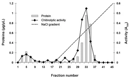
Step 1 allowed the elimination of a large amount of proteins and the recovery of more than 110% of the total chitinase activity from crude extract (Table 1). Protein fractions with chitinolytic activity were eluted with 0.3 M NaCl during the second step on an anion-exchange chromatography on DEAE-650 Fractogel column (Fig.1). At step 2, we obtained a 9.6-fold purification (based on a 1.00 value for the crude extract), allowing the recovery of more than 20% of chitinolytic activity. After step 3, chitinase-containing fractions were 36-fold purified compared with the crude extract.
1D and 2D Electrophoresis and Protein Identification
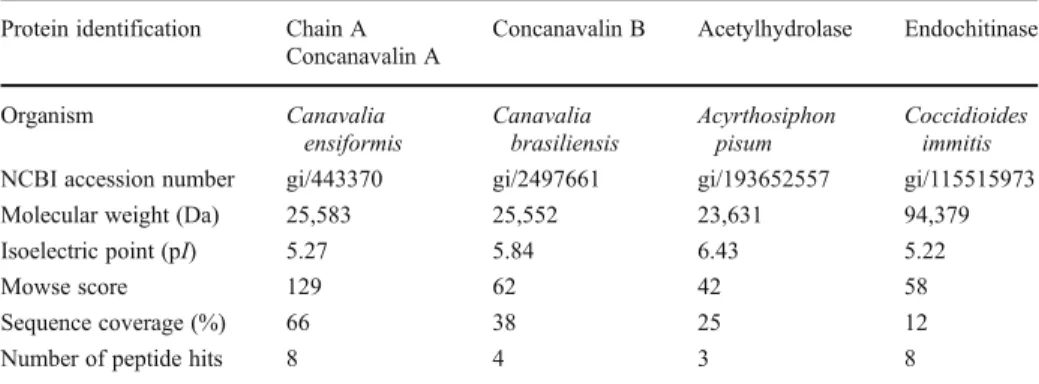
Each fraction resulting from each purification step of chitinase purification was analysed on 1D electrophoresis gels (Fig.2a). The purified chitinase fraction issued from step 3 revealed the presence of a single polypeptide band of approximately 31 kDa (Fig. 2a, lane 3). 2D electrophoresis revealed the presence of three major spots at 31 kDa with pI from 5.0 to 6.0 (Fig.2b). The peptides from the trypsin-digested protein excised from the gel matched with only four known proteins (Table 2). Concanavalin A and B from Canavalia spp., whose molecular weights are approximately 25.5 kDa, shared the highest sequence coverage with the peptides issued from the purified chitinase (respectively 66% and 38%). The purified chitinase also matched with 25% of a 23.6-kDa acetylhydrolase sequence from the aphid Acyrthosiphon pisum. The last protein is an endochitinase from the fungus Coccidioides immitis which shared 12% of protein sequence with the chitinase of M. persicae. All these proteins have isoelectric points comprised between 5.22 and 6.43, a range that corresponded to the three spots obtained with the chitinase of M. persicae (Fig.2b).
Chitinase Inhibition Assays
Inhibition of the purified M. persicae chitinase by allosamidin (Fig.3a) and psammaplin A (Fig. 3b) depended on the concentration of inhibitor. At 1 μg/mL, allosamidin induced higher inhibition than psammaplin after 1 h (respectively 20% and 0%) and 3 h (respectively 52% and 71%). In contrast, at 10μg/mL, psammaplin A inhibited more aphid chitinase than allosamidin after 1 h (respectively 47% and 36%) and 3 h (respectively 73% and 50%). At 50μg/mL, allosamidin and psammaplin A inhibited M. persicae chitinase in the same level and respectively induced 78% and 80% of inhibition, after 3 h.
Table 1 Purification steps of the chitinase from M. persicae Steps Purification steps (see‘Materials
and Methods’) Protein (μg/μL) Activitya Specific activityb % Recovery Fold purification Crude extract 2.73 0.027 0.010 100.0 1.00 1 Ammonium sulphate after dialysis 3.59 0.109 0.030 110.8 3.07 2 DEAE-650 Fractogel 0.90 0.091 0.095 20.4 9.63 3 Concanavalin A 0.08 0.029 0.361 15.3 36.53
a
Chitinolytic activity was calculated as the absorbance variation at 550 nm (Δ550/min/mL)
b
Discussion
Several chitinases were purified from bacteria and fungi for their potential use as biopesti-cide [13]. Although many chitinase genes have been described for trangenesis procedures, only few chitinases were purified from insects, mainly from Lepidopteran species [1,17]. To purify M. persicae chitinase, we tested several procedures after DEAE chromatography (step 2, data not shown). Chitin and chitosan, the usual substrates tested as affinity matrix, did not allow the purification of M. persicae chitinase which only bound on Concanavalin A affinity column. With a 36-fold purification factor and 15% of recovery yield, the efficiency of M. persicae chitinase purification was in the range of the few previous insect chitinase purifi-cations ranging from a 24 to 135 overall factor and 7% to 11% recovery rates, respectively [1,12,16]. On both 1D and 2D electrophoresis gels, the purified chitinase from M. persicae
Fig. 1 Elution profile of M. persicae chitinase on a DEAE-650 Fractogel column. The protein preparation (6 mL) applied to this column was the 60% fraction obtained from ammonium sulphate precipitation after dialysis. Proteins were eluted with a 0–0.4-M NaCl gradient, containing 0.02 M Tris–HCl buffer pH 8.8. Fractions of 1 mL were collected
Fig. 2 M. persicae chitinase purification by: a 1D electrophoresis gel—M molecular mass standards, CE crude extract, lane 1 sample after step 1, lane 2 fraction after step 2, lane 3 purified chitinase, after step 3; b 2D electrophoresis gel of the 31-kDa purified chitinase from M. persicae. Arrows indicate the spots from different isoelectric points
was estimated to be 31 kDa that represents an unusual low molecular weight compared with other insect chitinases. Molecular weights of insect chitinases range from 39 to 90 kDa. For example, in the lepidopteran B. mori, purified chitinases have molecular weights from 54 to 88 kDa [1]. A major 44-kDa digestive chitinase from Tenebrio molitor (Coleoptera) was also purified on SDS–PAGE [12]. The molecular weights estimated by SDS–PAGE for Tribolium castaneum (Coleoptera) and Drosophila melanogaster (Diptera) recombinant chitinases vary
Table 2 Identified proteins related to the purified chitinase from Myzus persicae Protein identification Chain A
Concanavalin A
Concanavalin B Acetylhydrolase Endochitinase
Organism Canavalia ensiformis Canavalia brasiliensis Acyrthosiphon pisum Coccidioides immitis NCBI accession number gi/443370 gi/2497661 gi/193652557 gi/115515973 Molecular weight (Da) 25,583 25,552 23,631 94,379 Isoelectric point (pI) 5.27 5.84 6.43 5.22
Mowse score 129 62 42 58
Sequence coverage (%) 66 38 25 12
Number of peptide hits 8 4 3 8
Fig. 3 Residual chitinolytic activ-ity of M. persicae-purified chiti-nase after 1, 2 and 3 h of incubation with 1, 10 and 50μg/mL of allosamidin (a) or psammaplin A (b)
from 39 to 83 kDa, whereas their theoretical weights ranged from 39 to 58 kDa [34]. On M. persicae chitinase-related 2D gel, a smaller spot was detected at around 10 kDa. This has been also observed with some chitinases from Lepidoptera [19] and Coleoptera [12] that are extensively glycosylated and produce several smaller forms than conventional ones by proteolysis.
The molecular weight of M. persicae chitinase determined by the mass spectrometry analysis is closer to plant proteins than other insect chitinases. In fact, M. persicae-purified chitinase did not cluster with other insect chitinases but matched with Concanavalin A and B from Canavalia spp., two chitinase-like proteins. This discrepancy where insect chitinase did not fit the systematic was also observed when a phylogenetic tree was built for T. molitor chitinase gene sequences [12]. Although Concanavalin B does not exhibit chitinase activity, this protein is known to share about 40% structural similarity with hevamine, a class III plant chitinase that belongs to the glycosyl hydrolase family 18 and has a molecular weight of 29 kDa [6,14,22,30]. We determined that the chitinase of M. persicae had 38% of sequence recovery with Concanavalin B. Hevamine, M. persicae chitinase and Concanavalin B could be distantly related proteins that have evolved and adapted to their respective functions. The absence of enzymatic activity in Concanavalin B can be explained by a difference of 3D structure related to the replacement of a glutamate residue, which is the most critical residue in the active chitinase motif. This resulted in total loss of activity. A wide variety of enzymatically active and non-active chitinases from GH18 family members, including growth factors and lectins in the holometabolous insects, appear to have evolved during development [2].
The presence of several chitinase-related proteins in aphids is not surprising. The screening of the entire genome of the pea aphid, A. pisum, detected nine genes encoding putative chitinase-like proteins, including six enzymatically active chitinases, one imaginal disc growth factor, and one endo-beta-N-acetylglucosaminidase. Quantitative reverse transcription-PCR demonstrated the expression of four chitinase and two distinct chitinase-like genes of A. pisum [21]. Here, M. persicae chitinase matched with an acetylhy-drolase found in A. pisum and platelet-activating factor acetylhyacetylhy-drolase (PAF hyacetylhy-drolase) from Drosophila, Aedes and Culex species. This kind of acetylhydrolase constitutes a subfamily of phospholipase specific for acyl chains [9]. The related phospholipids are key components of biological membranes and are the source of signaling molecules involved in many regulatory pathways associated with different physiological functions [8]. Particularly, the PAF factor binds to G protein-coupled receptors and activates a large range of cells [27]. Chitinases also induce the release of signaling molecules such as chito-oligosaccharides involved in physiological functions in plant meristems and embryos [13].
Mass spectroscopy analysis also revealed that the chitinase of M. persicae matched with a fungal endochitinase from C. immitis. Consequently, M. persicae chitinase appeared to be different from other insect chitinases. Hypothesis on the change of functional constraints were developed to explain the divergences in insect chitinases [9]. The match between M. persicae chitinase, an endochitinase and a Concanavalin B suggests that it belongs to chitinases from family 18 such as previous insect chitinases studied so far that all have been shown to possess signature motifs characteristic of family 18 glycosyl hydrolases [2]. The chitinolytic activity of M. persicae chitinase was evaluated by using allosamidin and psammaplin A, two important inhibitors of chitinases from family 18 including insect chitinases [25]. In a previous work, we reported the aphicidal effects (i.e. reduction of fecundity, increase of larval mortality, reduction of body size) of these two inhibitors [24]. Here, we confirm the direct and strong inhibition of M. persicae chitinase by allosamidin and psammaplin A, including at low concentrations (1 μg/mL). Allosamidin inhibitory activity was also tested on the plant-related chitinase, hevamine and the fungal C. immitis
chitinase. In both cases, this inhibitor interacts with the chitinases and inhibits them. Because hevamine and C. immitis chitinase are members of the family 18 of chitinases, these results also support that M. persicae chitinase belongs to this family.
This study reported the first isolation and purification of an aphid chitinase. The purification is the preliminary step for characterisation process which will improve the understanding of insect chitinases. Further investigations would allow the identification of protein and gene sequences, to confirm the‘membership’ of M. persicae chitinase to family 18 and to understand the differences between Lepidopteran, Coleopteran and Sternorrhyncha chitinases.
1D and 2D electrophoresis of M. persicae chitinase showed an unusual molecular weight compared with the other insect chitinases. This suggests that insect chitinases have evolved differently, opening the possibility to specifically and selectively target insect chitinases with appropriated inhibitors.
Acknowledgments We are grateful to Dr. S. Sakuda (Department of Applied Biological Chemistry, The University of Tokyo, Japan) and Dr. J.N. Tabudravu (Marine Natural Products Laboratory, Department of Chemistry, University of Aberdeen, Scotland, UK) for providing respectively allosamidin and psammaplin A. We thank the‘Service Régional de la Protection des Végétaux du Nord Pas-de-Calais’ for providing aphids and the‘Comité Nord Plants de Pommes de Terre’ for providing potato tubers. This work was supported by the Ministère Français de la Recherche, the Conseil Régional de Picardie, the Fonds Social Européen and also the Fond National pour la Recherche Scientifique from Belgium (FNRS) for its funding (FRFC project number 2.4561.06).
References
1. A-Banat, B. M. A., Kameyama, Y., Yoshioka, T., & Koga, D. (1999). Purification and characterization of a 54 kDa chitinase from Bombyx mori. Insect Biochemistry and Molecular Biology, 29, 537–547. 2. Arakane, Y., & Muthukrishnan, S. (2010). Insect chitinase and chitinase-like proteins. Cellular and
Molecular Life Sciences, 67, 201–216.
3. Blackman, R. L., & Eastop, V. F. (2000). Aphids on the world’s crops: An identification and information guide (2nd ed.). New York: Wiley.
4. Bradford, M. M. (1976). A rapid sensitive method for the quantitation of microgram quantities of protein utilizing the principle of protein-dye binding. Analytical Biochemistry, 72, 248–254.
5. Bultel, L., Saguez, J., Giordanengo, P. and Kovensky, J. (2007) Composé du type disaccharide, composition contenant un tel composé et procédé de fabrication d’un tel composé. French patent 07/08313 ref. F20027/SP. 6. Cederkvist, F. H., Saua, S. F., Karlsen, V., Sakuda, S., Eijsink, V. G., & Sørlie, M. (2007).
Thermody-namic analysis of allosamidin binding to a family 18 chitinase. Biochemistry, 30, 12347–12354. 7. Chen, A. C., Mayer, R. T., & DeLoach, J. R. (1982). Purification and characterization of chitinase from
the stable fly, Stomoxys calcitrans. Archives of Biochemistry and Biophysics, 216, 314–321.
8. Derewenda, Z. S., & Derewenda, U. (1998). The structure and function of platelet-activating factor acetylhydrolases. Cellular and Molecular Life Sciences, 54, 446–455.
9. Derewenda, Z. S., & Ho, Y. S. (1999). PAF-acetylhydrolases. Biochimica et Biophysica Acta, 1441, 229–236. 10. Ding, X., Gopalakrishnan, B., Johnson, L. B., White, F. F., Wang, X., Morgan, T. D., Kramer, K. J., & Muthukrishnan, S. (1998). Insect resistance of transgenic tobacco expressing an insect chitinase gene. Transgenic Research, 7, 77–84.
11. Fitches, E., Wilkinson, H., Bell, H., Bown, D. P., Gatehouse, J. A., & Edwards, J. P. (2004). Cloning, expression and functional characterisation of chitinase from larvae of tomato moth (Lacanobia oleracea): A demonstration of the insecticidal activity of insect chitinase. Insect Biochemistry and Molecular Biology, 34, 1037–1050.
12. Genta, F. A., Blanes, L., Cristofoletti, T., do Lago, C. R., Terra, W. R., & Ferreira, C. (2006). Purification, characterization and molecular cloning of the major chitinase from Tenebrio molitor larval midgut. Insect Biochemistry and Molecular Biology, 36, 789–800.
13. Giordanengo, P., Saguez, J., & Vincent, C. (2008). Chitinase et phytoprotection. In C. Regnault-Roger, B. J. R. Philogène, & C. Vincent (Eds.), Biopesticides d’origine végétale (2èmeth ed., pp. 341–354). Paris: Lavoisier Tech & Doc.
14. Hennig, M., Jansonius, J. N., Terwisscha van Scheltinga, A. C., Dijkstra, B. W., & Schlesier, B. (1995). Crystal structure of concanavalin B at 1.65 Å resolution. An “inactivated” chitinase from seeds of Canavalia ensiformis. Journal of Molecular Biology, 254, 237–246.
15. Jach, G., Görnhardt, B., Mundy, J., Logemann, J., Pinsdorf, E., Leah, R., Schell, J., & Maas, C. (1995). Enhanced quantitative resistance against fungal disease by combinatorial expression of different barley antifungal proteins in transgenic tobacco. The Plant Journal, 8, 97–109.
16. Kabir, K. E., Hirowatari, D., Watanabe, K., & Koga, D. (2006). Purification and characterization of a novel isozyme of chitinase from Bombyx mori. Bioscience, Biotechnology, and Biochemistry, 70, 252– 262.
17. Koga, D., Jilka, J., & Kramer, K. J. (1983). Insect endochitinase glycoproteins from moulting fluid, integument and pupal hemolymph of Manduca sexta L. Insect Biochem., 13, 295–305.
18. Koga, D., Sasaki, Y., Uchiumi, Y., Hirai, N., Arakane, Y., & Nagamatsu, Y. (1997). Purification and characterization of Bombyx mori chitinases. Insect Biochemistry and Molecular Biology, 27, 757–767. 19. Kramer, K. J., & Muthukrishnan, S. (1997). Insect chitinases: Molecular biology and potential use as
biopesticides. Insect Biochemistry and Molecular Biology, 27, 887–890.
20. Muzarelli, R. A. (1997). Human enzymatic activities related to the therapeutic administration of chitin derivatives. Cellular and Molecular Life Sciences, 53, 131–140.
21. Nakabachi, A., Shigenobu, S., & Miyagishima, S. (2010). Chitinase-like proteins encoded in the genome of the pea aphid (pp. 175–185). Insect Molecular Biology: Acyrthosiphon pisum.
22. Royer, V., Fraichard, S., & Bouhin, H. (2002). A novel putative insect chitinase with multiple catalytic domains: Hormonal regulation during metamorphosis. Biochemistry Journal, 366, 921–928.
23. Saguez, J., Dubois, F., Vincent, C., Laberche, J. C., Sangwan-Norreel, B. S., & Giordanengo, P. (2005). Differential aphicidal effects of chitinase inhibitors on the polyphagous homopteran Myzus persicae (Sulzer). Pest Management Science, 62, 1150–1154.
24. Saguez, J., Hainez, R., Cherqui, A., Van Wuytswinkel, O., Jeanpierre, H., Lebon, G., Noiraud, N., Beaujean, A., Jouanin, L., Laberche, J. C., Vincent, C., & Giordanengo, P. (2006). Unexpected effects of chitinases on the peach-potato aphid (Myzus persicae Sulzer) when delivered via transgenic potato plants (Solanum tuberosum Linné) and in vitro. Transgenic Research, 14, 57–67.
25. Saguez, J., Vincent, C., & Giordanengo, P. (2008). Chitinase inhibitors and chitin mimetics for crop protection. Pest Technology, 2, 81–86.
26. Sakuda, S., Isogai, A., Matsumoto, S., & Suzuki, A. (1987). Search for microbial insect growth regulators. II. Allosamidin, a novel insect chitinase inhibitor. Journal of Antibiotics, 40, 296–300.
27. Shukla, D. (1992). Platelet-activating factor receptor and signal transduction mechanisms. FASEBJ, 6, 2296–2301.
28. Stahl, E. A., & Bishop, J. G. (2000). Plant–pathogen arms races at the molecular level. Current Opinion in Plant Biology, 3, 299–304.
29. Tabudravu, J. N., Eijsink, V. G., Gooday, G. W., Jaspars, M., Komander, D., Legg, M., Synstad, B., & van Aalten, D. M. F. (2002). Psammaplin A, a chitinase inhibitor isolated from the Fijian marine sponge Aplysinella rhax. Bioorganic & Medicinal Chemistry, 10, 1123–1128.
30. Terwisscha van Scheltinga, A. C., Hennig, M., & Dijkstra, B. W. (1996). The 1.8 Å resolution structure of hevamine a plant chitinase/lysozyme and analysis of the conserved sequence and structure motifs of glycosyl hydrolase family 18. Journal of Molecular Biology, 262, 243–257.
31. Van Emden, H. F., Eastop, V. F., Hughes, R. D., & Way, M. J. (1969). The ecology of Myzus persicae. Ann. Rev. Entomol., 14, 197–270.
32. Wu, Y., Egerton, G., McCarthy, J. S., Nutman, T. B., & Bianco, A. E. (2003). Human immune responses to infective stage larval-specific chitinase of filarial parasite, Onchocerca volvulus, Ov-CHI-1. Filaria J., 14, 6–19.
33. Zhu, Q., Arakane, Y., Beeman, R. W., Kramer, K. J., & Muthukrishnan, S. (2008a). Characterization of recombinant chitinase-like proteins of Drosophila melanogaster and Tribolium castaneum. Insect Bio-chemistry and Molecular Biology, 3, 467–477.
34. Zhu, Q., Arakane, Y., Banerjee, D., Beeman, R. W., Kramer, K. J., & Muthukrishnan, S. (2008b). Domain organization and phylogenetic analysis of the chitinase-like family of proteins in three species of insects, Insect Biochem. Molecular Biology, 38, 452–466.