Direction des bibliothèques
AVIS
Ce document a été numérisé par la Division de la gestion des documents et des archives de l’Université de Montréal.
L’auteur a autorisé l’Université de Montréal à reproduire et diffuser, en totalité ou en partie, par quelque moyen que ce soit et sur quelque support que ce soit, et exclusivement à des fins non lucratives d’enseignement et de recherche, des copies de ce mémoire ou de cette thèse.
L’auteur et les coauteurs le cas échéant conservent la propriété du droit d’auteur et des droits moraux qui protègent ce document. Ni la thèse ou le mémoire, ni des extraits substantiels de ce document, ne doivent être imprimés ou autrement reproduits sans l’autorisation de l’auteur.
Afin de se conformer à la Loi canadienne sur la protection des renseignements personnels, quelques formulaires secondaires, coordonnées ou signatures intégrées au texte ont pu être enlevés de ce document. Bien que cela ait pu affecter la pagination, il n’y a aucun contenu manquant.
NOTICE
This document was digitized by the Records Management & Archives Division of Université de Montréal.
The author of this thesis or dissertation has granted a nonexclusive license allowing Université de Montréal to reproduce and publish the document, in part or in whole, and in any format, solely for noncommercial educational and research purposes.
The author and co-authors if applicable retain copyright ownership and moral rights in this document. Neither the whole thesis or dissertation, nor substantial extracts from it, may be printed or otherwise reproduced without the author’s permission.
In compliance with the Canadian Privacy Act some supporting forms, contact information or signatures may have been removed from the document. While this may affect the document page count, it does not represent any loss of content from the document.
o
7 ADUT 2008Université de Montréal
Structural bioinformatics analysis of the family of human ubiquitin-specifie proteases
par Xiao Zhu
Département de biochimie F acuIté de médecine
Mémoire présenté à la Faculté des études supérieures en vue de l'obtention du grade de
Maître ès sciences en bio-informatique
Décembre 2007
© Xiao Zhu, 2007
-Université de Montréal Faculté des études supérieures
Ce mémoire intitulé:
Structural bioinformatics analysis of the family of human ubiquitin-specifie proteases
présenté par :
Xiao Zhu
a été évalué par un jury composé des personnes suivantes :
Jurgen Sygusch président-rapporteur Traian Sulea directeur de recherche Robert Ménard codirecteur Simon Wing membre du jury
1
IV
Résumé
Les protéases spécifiques à l'ubiquitine (<< ubiquitin-specific proteases » ou USPs)
représentent un sous-ensemble important d'enzymes de dé-ubiquitination humaines (DUBs) qui catalysent la libération d'ubiquitine des protéines ubiquitinées. L'annotation fonctionnelle de ces enzymes est limitée aux méthodes de comparaison par séquences par l'absence de données structurelles. Ainsi, la fonction des régions longues en amont et en aval du domaine catalytique ainsi que de celles encastrées à
l'intérieur demeure inconnue. Dans l'article High Incidence of Ubiquitin-like
Domains in Human Ubiquitin-Specific Proteases ci-présenté, nous démontrons la présence de nouvelles régions structurées et fonctionnelles chez les USPs humaines. Afin d'élargir notre compréhension sur la fonction de ces enzymes, nous avons appliqué la méthode de prédiction de repliement par consensus aux régions non-annotées des USPs. Notre prédiction à haute fiabilité suggère que le domaine
apparenté à l'ubiquitine (<< ubiquitin-like» ou UBL) constitue le domaine le plus
fréquent dans la famille des USPs humaines. La présence des UBLs en copies multiples ainsi qu'insérés au sein du domaine catalytique de certaines USPs démontrent d'un haut degré de complexité structurale. Ces données complémentent nos connaissances actuelles sur l'organisation structurelle et fonctionnelle de l'ensemble des DUBs. Nos résultats suggèrent que la présence des UBLs est quasiment aussi fréquente que le domaine catalytique. Nous proposons diverses fonctions possibles pour des nouveaux domaines UBLs découverts chez les USPs humaines, telles que l'association avec le protéasome, le recrutement spécifique de substrats, ainsi que la distribution intracellulaire.
Mots clés: dé-ubiquitination, prédiction de repliement, protéasome, ubiquitine, UBL, USP.
v
Abstract
The family of ubiquitin-specific proteases (USPs) is the major member of the human deubiquitinating enzymes (DUBs) superfamily that specifically cleaves ubiquitin from ubiquitin-conjugated substrates. CUITent functional annotations of USPs is limited to sequence comparison methods and to the minimal availability of crystallographic data. Large regions both within and flanking the catalytic core which may explain substrate specificity and subcellular localization remain poorly defined.
In the article presente d, High Incidence of Ubiquitin-like Domains in Human
Ubiquitin-Specific Proteases, we provide evidence of the presence of novel structural
features and domains within the human USPs. Our methods utilize consensus prote in fold recognition techniques on stretches of un-annotated regions in the USPs in order to expand our CUITent understanding of the functional role of these enzymes. Among other interesting findings, we have discovered a high occurrence of reliably predicted ubiquitin-like (UBL) folds situated at both N- and C-terminal regions as weIl as embedded within the catalytic core of human USPs. The marked presence of multiple UBL domains as well as those integrated within the catalytic core present a new dimension in the structural complexity of these enzymes. Our results also suggest that the occurrence of UBL do mains in human USPs is a close second to the characteristic catalytic core. Several propositions for the functional implications including proteasome binding and substrate recognition of these UBLs are discussed.
Keywords: deubiquitination, consensus fold recognition, proteasome, ubiquitin, UBL,
vi
Table of Contents
Acknowledgements vii 1 2 3 4 5 Introduction Biological Background2.1 Ubiquiton - The Ubiquitin Superfold ••••••••••••••••
2.1.1 Type 1: Ubiquitin and Ubiquitin-Like Modifiers ••
2.1.2 Type 2: Integral Ubiquitin Motifs ••••••••••
2.2 Ubiquitin Modification Pathways •••••••••••••••••
2.2.1 Ubiquitination . . . . 1 4 4 5 6 6 6 2.2.2 Deubiquitination • • • • • • • • • • • • • • • • • • • • • 8
2.2.3 Deubiquitination and Diseases • • • • • • • • • • 10
Structural bioinformatics
3.1 CUITent Structural and Functional Repositories
3.2 3.3
Structural Classification of Proteins Prediction of Function
3.3.1 From Sequence to Function
3.3.2 From Sequence to Structure to Function ••••••
3.4 3D-Jury: A Consensus Fold Recognition Server •••••••••
Published article 4.1 Personal Contributions Conclusion References 12 12 12 13 13 14 16 18 30 31 34
vu
Acknowledgements
l' d like to thank my advisors Dr. Traian Sulea and Dr. Robert Ménard who, with
their ri ch and diverse expertise, have given me the opportunity to conduct this exciting research project. Throughout my journey at Biotechnology Research Institute (BRI), they gave academic guidance and mentorship that helped me to succeed. l was able to make critical decisions for my project and acquire independency in research. This valuable experience and many lessons learned throughout my stay have thoroughly prepared me for my scientific career.
ln addition, 1 would like to thank Dr. Enrico Purisima for allowing me to take part in the Computational Chemistry group at BRI and for securing funds to carry out this project. 1 am gracious for the invaluable advices and support 1 have received from colleagues such as Viktoria Lytvyn, Dr. Ratsavarinh Vongsamphanh, and Dr. Holger Lindner.
Chapter 1
Introduction
Post-translational modifications modulate enzyme activity and generate new dimensions to complex biological processes. In eukaryotes, the fate of a protein is often determined by attachment of ubiquitin moieties through covalent yet hydrolyzable enzyme mediated linkages. Polyubiquitination, the attachment of a polymerie ubiquitin chain, is classically known for its direct involvement in modulation of ubiquitin-mediated proteasomal degradation and, like the attachment of a single ubiquitin molecule, is involved in a multitude of biological functions from endocytosis to cell proliferation.
Ubiquitin is a small 76-residue prote in involved in post-translational modifications that help to modulate diverse biological pathways. The ubiquitin pathway includes two biochemical phenomena: ubiquitination and deubiquitination. Ubiquitination is a reversible post-translational modification that involves the covalent linkage of ubiquitin molecules to a target protein. Deubiquitination, performed by deubiquitinating enzymes (DUBs), can cleave the isopeptide bond between ubiquitin and the site of attachment. In addition to reversing ubiquitination, DUBs are also involved in the activation of ubiquitin and ubiquitin-like modifiers through C-terminal processing of their precursors. The ubiquitin pathway is involved in a wide array of biological functions including DNA repair, signal transduction, membrane protein trafficking, endocytosis, transcription, nuclear transport, and proteolysis. The family of human enzymes ubiquitin-specific proteases (USPs) constitutes the majority of DUBs. Although biological studies over the past decade have significantly contributed to the current structural and functional inventory of DUBs, we still know little about the physiological roles and mechanisms of intermolecular interactions of most USPs.
In human, there are currently 54 USPs known with high variance in length and domain architecture. Our current understanding of the structure and function ofUSPs is the result of a combination of bioinformatics predictions and experimental efforts. In addition to the evident presence of a catalytic core homologous to ubiquitin
C-CHAPTER 1 INTRODUCTION 2
terminal hydrolases (UCH), many USPs are found, mostly by sequence-based comparison techniques and probabilistic models such as BLAST, SMART, Pfam, and PROSITE, to harbor rather unrelated domain architectures outside the UCH domain (Nijman et al. 2005). For example, zinc-fingers are observed at the N-terminal region of sorne USPs (USP3, 5, 13, 16, 20, 22,33,44, and 49) while others have ubiquitin-associated do mains (UBA) placed in tandem at the heart of the catalytic core. Despite current efforts in the annotation and characterization of these enzymes, large portions of the N- and C-terminal extensions of human USPs remain structurally and functionally un-annotated. In addition, the length of their catalytic core domains varies from approximately 300 to 800 residues due to large uncharacterized insertions which may play important physiological roles. This lacuna in functional assignment is a direct indication that ev en the most sensitive evolution-based sequence comparisons may not be sufficient to infer functional homologies.
The objective of this study is to develop and apply a methodology to structurally annotate previously un-annotated regions of the 54 putative human USPs. Possible functions of the resulting predictions will be inferred and discussed from predicted folds. Given the currently known and anticipated biological roles for USPs, our comprehensive structural annotation of these members of the DUB family provides important stepping stones towards elucidating their precise involvements in many hum an diseases.
In order to better understand the objectives and results, biological and computational background related to the subject will be presented over the next two chapters. First, the fundamentals of the prominent ubiquitin pathway will be discussed in Chapter 2. The next section, Chapter 3, confers the fundamentals of the bioinformatics aspect of our study to the reader. From sequence alignment to structure prediction, key information conceming classical and structural-bioinformatics give the reader the fundamentals necessary to comprehend our choice of method and the power of our predictions. In addition, the fundamentals of the key tool employed in our study will be presented. The pub li shed article will then be presented in Chapter 4.
Chapter 2
Biological Background
In this chapter, an overview of structural and functional properties of type 1 and type 2 ubiquitin-like (UBL) modifiers will first be presented. Next, the two fundamental processes that constitute the ubiquitin modification of proteins will be explored: ubiquitination, the attachment of ubiquitin molecules cnte a protein target, and deubiquitination, their removal. Structural and functional aspects of deubiquitinating enzymes (DUBs), the focus of present study, will be presented in greater detail.
2.1 Ubiquiton: the ubiquitin superfold
Ubiquitin and UBL structures belong to the ~-grasp c1ass of structural folds. It boasts a beta-sheet wrapped around a central a-helix mu ch like the grasp of a hand (Figure 1). The central helix flanked by two upstream and three downstream consecutive beta-sheets is characteristic to secondary structure arrangements of ubiquitin and consensus of its umbrella family: ubiquitons. A three-residue 3 w-helical turn often follows immediately after the central a-helix in many UBLs.
Figure 1: Cartoon representation ofubiquitin (PDB: lAAR). AlI 7 lysine residues are shown (red).
CHAPTER 2 BIOLOGICAL BACKGROUND 4
ln biological systems, the functional role of a prote in is not only controlled by its level of expression but also by post-translational processes during which chemical modifications of the polypeptide modulate its activity, sub-cellular localization, and elimination. In eukaryotes, there exists a post-translational process widely known as ubiquitination, which is analogous to, but biologically distinct from, phosphorylation, and which consists in the addition of ubiquitin moieties to protein targets. Molecules that have a covalent mode-of-action resembling the C-terminal attachment of ubiquitin are commonly termed UBL molecules or type 1 ubiquitons. In contrast, there also exist UBL structures that are embedded within a larger protein, which
constitute the type II class of uhiquitons.
2.1.1 Type 1: ubiquitin and ubiquitin-like modifiers
Ubiquitin (Ub) is a small and highly conserved molecule abundant in eukaryotes. Its 3D structure is representative of the UBL fold, which is shared by a large number of protein sequences showing remarkable structural similarities while displaying divergent sequence homology (Kiel, Serrano 2006). This 76-residue polypeptide acts as a signaling marker that effectively guides the target protein through various biological pathways. The Ub-Iabeling of a target prote in is usually a
result of the formation of a covalent linkage between the C-terminus of Ub and the
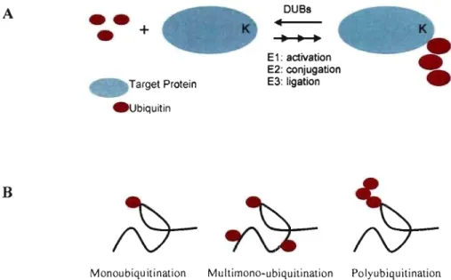
E-amino group of a substrate lysine residue (Figure 2A). This reversible process is commonly known as ubiquitination and is govemed by the sequential action of three enzymes: El, E2 and E3, as will be described in the following section. There also exist several small proteins of close structural resemblance to ubiquitin, which have rather divergent sequence similarities and functions, but share with Ub the same 3D-structural. fold and the covalent mode-of-action via a reactive C-terminus glycine (Welchman et al. 2005). The most studied type 1 UBL molecules are SUMO isoforms, NEDD8, ISG15 and FATIO with functional roles ranging from transcriptional regulation (SUMO) to E3 regulation (NEDD8) to immune response (lSG 15) to apoptosis (Welchman et al. 2005).
CHAPTER 2 BIOLOGICAL BACKGROUND A B
••
•
+
Target Protein . Ubiquitin DUBs E 1: activation E2: conjugation E3: ligationMonoubiquitination Multimono-ubiquitination Polyubiquitination
5
Figure 2: Target labeling by ubiquitin(s). (A) The pro cess of ubiquitination is guided by the sequential actions of E 1, E2, and E3 enzymes. (B) There exist three modes of Ub attaclunent: mono-, multi-, and polyubiquitination.
2.1.2 Type II: integral ubiquitin motifs
Ubiquitin-like structures also exist as integral elements of larger proteins. These genetically built-in UBL domains can be located anywhere in the sequence, although N-terminal UBL structures are prevalent in CUITent knowledge bases. Type II UBL structures generally play integral roles in protein-protein interaction, subcellular localization, as weIl as intrinsic modulation of enzyme activity. Integral UBLs have also been shown to associate with the 19S subunit of the 26S proteasome primarily by interacting directly with ubiquitin-interacting motifs (UlM) of the proteasome. For example, UBL domain-containing protein Rad23 , which also contains a regulatory
ubiquitin-associated domain (UBA), was shown to deliver polyubiquitinated
substrates to the proteasome for degradation. (Walters et al. 2004)
2.2 Ubiquitin modification pathways
The previous section described the two types of ubiquitons. We will now introduce the biochemical mechanism which underlies different mode of attaclunent ofubiquitin and ubiquitin-like molecules.
CHAPTER 2 BIOLOGICAL BACKGROUND 6
2.2.1 Ubiquitination
Ubiquitination is defined by the covalent linkage of Ub onto a target prote in via formation of a bond typically between the C terminus (Gly76) of Ub and the E-amino
group of a substrate lysine residue. It is a three-part process mediated by sequential
action of El activating, E2 conjugating, and E3 ubiquitin-protein ligating enzymes and initiates all known types of ubiquitination . The carboxyl group of Gly76 forms a thiol ester with El, activating the C terminus of Ub for a nucleophilic attack. This activated Ub is transiently carried by an E2 to an E3, which in turn specifically recognizes and facilitates transfer of the Ub to a substrate (Pickart, Eddins 2004).
In eukaryotes, there exist three modes for ubiquitin modification, each distinguished by its characteristic pattern of attachment which triggers distinct biological pathways. First, monoublquitination is defined by the linkage of a single Ub molecule to a single site on a target protein. Second, multi-monoubiquitination involves monoubiquitination at multiple sites. Last, polyubiquitination consists of the addition of a polyubiquitin chain constituting two or more covalently linked Ub molecules (Figure 2B).
AlI 7 lysine residues of the 76 amino-acid Ub offer potential linkage sites for chain extension (Figure 1). In nature, however, Lys48 and Lys63 linkages are more commonly observed. Formation of Lys48-linked polyubiquitin chains is precursor to signaling a target prote in for proteasomal degradation and plays important roles in DNA repair and signal transduction. While Lys48-linked polyubiquitin chains can effectively target a protein to the proteasome for degradation, Lys63-linkage topology typically does not. The latter configuration is structurally different in comparison with Lys48-linkage and is generally recognized in DNA repair, signal transduction, and non-Iysosomal degradation. (Kerscher et al. 2006) In addition, Lys63-linked chains activates NF-lcB signaling pathway involved in inflammation, apoptosis, and tumorigenesis (Nijman et al. 2005).
The general response of a cell following exposure to chemical and mechanical stress is an increase in intrinsic proteolysis. Stimulation of ubiquitin-dependant proteolytic system is one of the important elements in the proteolytic cascade and is shown to respond to a host of cellular stresses caused by viral infection, heat shock,
CHAPTER 2 BIOLOGICAL BACKGROUND 7
and protein damage (Wilkinson 1995). This destructive pathway is govemed by proteasomal degradation of polyubiquitinated target proteins. Once covalently tagged with a Lys48-linked polyubiquitin chain, the marked proteinis able to localize to the ubiquitin-interacting motif (UIM)-containing 198 subunit of the 268 proteasome and undergo proteolytic disassembly. The UlM is a short segment of about 20 residues that recognizes and binds to Ub and is often present in three consecutive copies within proteins, especially those involved in ubiquitin pathways.
Monoubiquitination and multi-monoubiquitination are commonly associated with non-degrading processes such as endocytosis, regulation of transcription, and sorting of target proteins to the vacuole (Loayza, Michaelis 1998;80etens et aL 2001). Monoubiquitination was previously demonstrated in yeast and vertebrate cells to be sufficient to induce endocytosis of membrane-bound proteins and receptors. (Hicke, Dunn 2003;Peschard, Park 2003;Holler, Dikic 2004). Monoubiquitination is aiso involved in subcellular localization and in recruiting Ub binding proteins to specifie interacting partners (Nijman et al. 2005).
2.2.2 Deubiquitination
The reversaI of ubiquitination during which Ub moieties are excised is termed deubiquitination, a process mediated by members of the deubiquitinating enzyme (DUB) superfamily. Ubiquitin-specific proteases (U8P) are cysteine proteases and form a family of ubiquitin-processing enzymes that belong to the DUB superfamiIy. To date, more than 54 human U8Ps have been reported and summarized in recent research publications and reviews (Nijman et aL 2005;Quesada et aL 2004). These enzymes possess a catalytic core homologous to that of the papain superfamily of cysteine proteases. In human, the USP catalytic core domain varies from 300 to 850 residues flanked by highly variable N- and C-terminal domain structures. Within the human USP family, homology exists primarily in the Cys-box and His-box regions harboring the catalytic residues. The highly divergent N- and C-terminal regions containing a variety of structural domains and functional motifs (e.g., zinc finger domains, EF-hand domains, UIMs) indicate an elevated structural and functional complexity within the USP family of DUBs. The large number of both validated and
CHAPTER 2 BIOLOGICAL BACKGROUND 8
predicted USPs suggests that these enzymes may exhibit selectivity for specific target proteins as weIl as the type of Ub conjugation. The current functional understanding ofUSPs relies largely on sequence-based comparisons.
Despite the importance ofUSPs, which play key regulatory roles in a multitude of processes, our knowledge of their substrate specificity and precise mode of regulation is surprisingly scant compared to the sequence and structural landscape of this class of enzymes (Nijman et al. 2005). Human USPs harbor divergent domain structures in regions extending the catalytic core. Such diversity allows for narrow specificities for the target protein and substrate ubiquitin or ubiquitin chains as weIl as for modulation of activity of the se enzymes (Nijman et al. 2005). The dual property of target and substrate recognition of a particular prote in and ubiquitin branching, respectively, may further increase the degree of specificity of USPs. The ubiquitin pathway is weIl known for its key role in Ub-directed proteolysis of target proteins. Crystallographic studies have confirmed that USP14, which resides in and is regulated by the 26S proteasome, contains a previously identified N-terminus UBL domain that specifically associates with the 19S regulatory particle (Borodovsky et al. 2001 ;Hu et al. 2005;Nijman et al. 2005). Because oftheir structural dissimilarities, recognition of Lys48 and Lys63 linked poly-Ub chains often do not overlap. Amongst other branching-specific USPs, USP8 and USP14 exclusively cleave Lys48 but not Lys63 linked poly-Ub chains. Such specificity may be acquired from regions flanking the catalytic core of USPs. In fact, the ubiquitin-interacting zinc finger (Znf-UBP) is one of the factors in USP 15 essential for disassembly of Ub polymers (Hetfeld et al. 2005). Interestingly, USPs are also engaged in systems that involve modifications via UBL moieties. USP21 is an example of dual recognition of both Ub and NEDD8 while USP 18 is shown to cleave and maintain cellular levels of ISG 15 (Gong et al. 2000;Malakhov et al. 2002).
In addition to Ub substrate specificity, regions outside the conserved catalytic core ofUSPs may play roles in target recognition as weiL In fact, many E3 ligases are targets for USPs. NRDP1, which ubiquitinates and promotes degradation of ErbB3, an epithelial growth factor receptor (EGFR), is deubiquitinated and stabilized by USP8 (Qiu et al. 2004;Wu et aL 2004). USP8 contains a rhodanese-like domain
CHAPTER 2 BIOLOGICAL BACKGROUND 9
which, in conjunction to its catalytic core, contributes to the recognition of polyubiquitinated NRDPl (Avvakumov et al. 2006). USP7, also known as HAUSP, can stabilize the E3 Mdm2, a p53 suppressor and lead to attenuation of p53 activity (Li et al. 2004). Disruption ofUSP7 activity was shown to effectively suppress tumor growth (Cummins et al. 2004). Moreover, USP7 can also deubiquitinate and stabilize
p53, thereby possessing dual roles in p53 regulation (Li et al. 2004). Interestingly,
USP7 harbors a TRAF-like MATH domain in the N-terminal region preceding the catalytic core that recognizes and binds to both p53 and Mdm2 (Hu et al. 2006). The primary objectives of the present study are to explore the sequence-to-structure-to-function paradigm by applying structural bioinformatics tools on un-annotated regions of the 54 known human USP sequences to uncover novel information elusive to simple sequence comparison methods.
2.2.3 Deubiquitination and diseases
The implication of DUBs in human diseases has drawn significant attention and research interest to this class of enzymes in recent years. To demonstrate the extent of implications of USPs and other DUBs in human diseases, sorne examples are presented.
The first direct evidence of the role of ubiquitination in tumor suppressor p53
downregulation originated from human papillomavirus (HPV) studies. It was shown
that degradation of turnor suppressor p53 was induced by an ubiquitin ligase complex in HPV. In fact, p53 is polyubiquitinated in cells infected by HPV leading to its proteasomal degradation (Scheffner et al. 1990;Scheffner et al. 1992). As previously noted, regulation of p53 is also modulated by Mdm2, a RING finger E3 that inhibits p53 activation function and downregulates its expression via polyubiquitination. Conversely, Mdm2 is itself regulated by p53 and these enzymes together form an auto-regulatory negative feedback loop in cell proliferation (Pickart, Eddins 2004). The USP7 enzyme, also known as herpesvirus-associated USP, or HAUSP, was originally observed to associate with herpes simplex virus-type 1 immediate-early Vmwll0, a RING finger protein required for efficient initiation of viral lytic cycle (Everett et al. 1997;Sacks, Schaffer 1987). Interestingly, USP7 specifically stabilizes
CHAPTER 2 BIOLOGICAL BACKGROUND 10
p53 via deubiquitination therefore inducing p53-mediated cell growth arrest and apoptosis (Cummins et al. 2004;Kim et al. 2003). In recent studies, the full length isoform of USP2 (USP2a) has been identified as an oncogenic enzyme that stabilizes fatty acid synthase (FAS) and Mdm2 via deubiquitination (Graner et aL 2004;Priolo et aL 2006;Stevenson et aL 2007). USP2a overexpression induces tumorigenesis via a mechanism opposing that ofUSP7. In contrast, the truncated form ofUSP2 promotes apoptosis when overexpressed in several cancer cell Hnes (Gewies, Grimm 2003). USP2 isoforms share a common catalytic core and differ only in the length of the N-terminal regions.
A significant increase in USP4 mRNA levelhas been reported in small cell tumors and in adenocarcinomas of the lung. USP4 has been shown to direcdy associate with retinoblastoma protein (PRb), a tumor suppressor protein known to be dysfunctional in a number of types of cancer, but does not exhibit deubiquitination activity (Blanchette et al. 2001). Nevertheless, A2Areceptor, involved in endoplasmic reticulum (ER) quality control, is a deubiquitination substrate for USP4 (Milojevic et aL 2006). In addition, reciprocity in activity between USP4 and its E3 ligase R052 was also identified (Wada, Kamitani 2006). Further investigation is required to understand the precise implications of USP4 in oncogenesis. USP 15 also possesses a pRb-interacting motif and may be involved, in cell growth regulation via deubiquitination and thus stabilization of pRb. (Kim et aL 2003)
Emerging evidence suggests that USP8 is involved in cellproliferation by inducing degradation of EGFR and growth factor receptor tyrosine kinases (RTK) (Daviet, Colland 2007). Initially identified as a cell growth regulator that modulates the ubiquitination state of several key proteins in proliferation, USP8 is able to cleave both linear and isopeptide-linked ubiquitin chains (Naviglio et aL 1998) to rescue and recycle Ub at late endosome. Several interaction partners and substrates were identified. In mouse, Ras-guanine nucleotide exchange factor CDC25 is
deubiquitinated and stabilized by USP8. An Hrs-binding protein, Hbp, which is
involved in receptor endo- and exocytocis binds to USP8 via a Src homology domain 3 (SH3). USP8 was also shown to mediate T -cell anergy by preventing self-ubiquitination and degradation of the transmembrane RING finger E3 ligase GRAIL,
CHAPTER 2 BIOLOGICAL BACKGROUND Il
which is cIosely linked to endocytic pathways. In a similar fashion, the catalytic core and rhodanese domains ofUSP8 were shown to bind to and stabilize NRDPl, an E3 . ligase that mediates EGFR stabilization. (reviewed in Daviet, Colland 2007)
The first study demonstrating the relationship between . DUBs and
neurodegenerative diseases was the identification of the association of autosomal dominant point mutation in UCH-Ll with Parkinson's disease. Another ex ample of implications of DUBs in neurological dysfunction is that in mice, a single homozygous mutation in the· ataxia gene encoding USP 14 leads to severe tremors followed by hind limb paralysis and death (D'Amato, Hicks 1965). In fact, reinstatement of USPI4, for which abolishment of activity resulted in 35% decrease in Ub mono mers in most of the tissue, restored Ub levels and reinstated motor functions (Anderson et al. 2005;Crimmins et al. 2006). In addition, mouse homolog of USP25 shows connection with the expression of proliferative neuroepithelial cells and post-mitotic neurons. In brain cells of Down syndrome patients, the expression of USP25 was decreased 2-fold therefore providing cIues that USP25 may be involved in Down syndrome pathogenesis. (Cummins et al. 2004;Kim et al. 2003)
USPs are also involved in spermatogenesis. Genetic screening of 576 infertile and 96 fertile men revealed the link between USP9 and male infertility. A 4-bp deletion in the y chromosome-linked USP9 (USP9Y) was determined to be responsible for the absence of sperm in the semen of azoospermie men (Sun et al. 1999).
These deubiquitination-related diseases further emphasize the CUITent need. for functional and structural annotations of USPs. The present study provides a first comprehensive examination of human USPs by looking at information beyond what the sequences alone can provide. Our computational approaches and methodology are further described in Chapter 3 and in the published paper.
Chapter 3
Structural Bioinformatics
This section will provide fundamental understanding of current tools in domain annotations and functional predictions. Sequence-based comparison methods will be briefly described. Fold recognition by threading methods will also be presented. Functional inference from a sequence-based and fold recognition methods will be discussed. The primary tool structure prediction methods used in this study, 3D-Jury, will be introduced in more detail.
3.1
Current structural and functional repositories
There exist several databases for the functions and structures of gene products. The NCBI (at the National Center for Biotechnology Information) is without question the pinnacle of all databases. Amongst other features, the GenBank database homed at NCBI contains sequence information, functional annotations, and have many useful cross-references. Sequence query in these databases is generally the first approach to annotating a new gene product with unknown functions. (Jenuth 2000) The Swiss-Prot prote in knowledgebase is another weIl known database dedicated to proteins and contains cross-reference to a wide array of functional and structural databases including NCBI and RCSB's Protein Data Bank (PDB) (Bairoch et al. 2004). The latter database is the primary repository for atomic-resolution experimental structures of proteins and protein complexes. InterPro is the main portal to structural-functional
repositories, including Pfam, SMART, ProDom, PRINTS, UniProt and ProSite. It
also contains references to structural classification databases SCOP and CATH.
3.2
Structural classifications
The RCSB Protein Data Bank (PDB) is a weIl maintained and up-to-date collection of published structures of biomolecules determined either by NMR or X-ray crystallography. There has been an exponential growth in the number of protein structures in the PDB over the past decade. Each protein structure is classified into
CHAPTER 3 STRUCTURAL BIOINFORMATICS 13
superfamilies of characteristic protein folds and further sorted into unique classes of closely related folds by two major classification methods: SCOP and CA TH. Structural Classification of Proteins (SCOP) distributes protein domains from structural classes down to folds regardless of sequence homology (Murzin et al. 1995). SCOP is considered as a standard in classification of prote in folds and relies largely on expert Interpretations of prote in structures. CATH is an acronym for the four main levels of hierarchical organization of structural folds: Class Architecture Topology Homology. Structural classification by CATH is a semi-automated consensus driven process guided by both sequence and structural homology information. Expert Interpretations occur at the final A-stage classification via visual inspections and cross-references (Orengo et al. 1997). However, as noted in the Introduction section, the growth in the number of new folds defined by these structure classifiers has reached a staIl. Therefore, it is believed that the structural classes at the present time form the basis of a great majority ofprotein structures in the PDB.
Ubiquitin and UBL structures belong to the ~-grasp class of structural folds
(SCOP entry d.15), which consists of a beta-sheet wrapped around a central a-helix much like the grasp of a hand (Figure 1). A central alpha-he li x tlanked by two upstream and three downstream consecutive beta-strands is characteristic to the secondary structure arrangement of ubiquitin and ubiquitons. A three-residue 310-helical turn often follows immediately after the central a-helix in many UBLs.
3.3
Prediction of function
One of the main purposes of bioinformatics is to develop and implement computational methods to annotate the function of a given, typically newly discovered, gene product. Depending on the sequence identitylhomology to existing functionally annotated prote in sequences, functional Inference can be attempted by classical sequence-based approaches or/and via a structural bioinformatics route centered on fold detection methods.
CHAPTER 3 STRUCTURAL BIOINFORMATICS 14
The classical approach to functional inference essentially relies on the pairwise alignment of the new (query) sequence with those in functionally annotated prote in sequence databases (described in Section 3.l), aiming to identify hits with global or even local similarities that can provide hints about its functions. Another type of sequence-based comparison utilizes probabilistic models such as the hidden Markov chain (HMM) to compare a query sequence with a signature of probabilities for a particular sequence to occur rather than pairwise comparison of two sequences. Profile comparison is another widely used statistical approach that aligns a query sequence with a pre-determined sequence pattern characteristic to a group of sequences notably from the same family (Krogh et al. 1994). Profile building and comparison methods are more sensitive than direct pairwise alignment in detecting homology. These traditional evolutionary-based approaches to predict the function of a prote in generally yield reliable results when statistically significant homology exists between the query and one or more database sequences.
The downfall of sequence-based functional assignment is that the relationship
between sequence and function is neither unique nor straightforward. It is widely
known that proteins exercising similar functions may exhibit divergent sequences, and vice versa. This can be explained through observations of structural similarities between two proteins even in the absence of sequence similarities, which brings about another phenomenon that similar structures may display similar functions. Therefore, structural homologies that are undetectable with sequence-based comparisons may be overlooked in classical functional annotation protocols still widely used today. Increasing cases of proteins with similar structure and functions, but undetectable sequence similarity have driven development of more sensitive methods for structure-function prediction (Brenner et al. 1998;Martin et al. 1998).
3.3.2 From sequence to structure to function
The fold recognition concept, initially implemented to identify analogous proteins with undetectable sequence homologies, has gained popularity in the 1990s. Fold detection appeared as a necessary tool due to the observation of structurally related proteins with dissimilar primary sequences. This may have arisen from convergent
CHAPTER 3 STRUCTURAL BIOINFORMATICS 15
evolution oftwo initially unrelated genes due to pressure exercised by external factors from habitats of two different species, or from rising demand of a cell to perform a given task. Fold detection in cases of low sequence homology thus opened new avenues for function inferences, however with the caveat that similarly folded protein structures with completely different biochemical functions do exist.
The traditional approach to fold prediction is based on sequence-sequence or sequence-profile comparisons. Homology modeling for example utilizes the evolutionary information obtained from sequence alignments and an associated scoring matrix to predict the spatial arrangement of each residue. An immediate extension to the sequence-sequence comparison approach has emerged with the implementation of threading methods. This technique utilizes a template structure to compute position-specific alignment scores based on iterative calculation of substitution scores by replacing the side-chain of a residue with all other 19 naturally occUITing side-chains. Another approach to fold prediction is based solely on a first-principle physical, but time-consuming, treatment. These ab initia methods take advantage of physical properties of atoms in order to calculate the free energy over an ensemble of prote in conformations and to simulate prote in folding. CUITent limitations in computation power, however, do not allow sufficient sampling flexibility to effectively predict protein folds via ab initia methods. A final dass of prote in structure prediction methods, termed meta-predictors, applies statistical methods to improve the accuracy of a collection of prote in structure predictions over that of individual methods.
Meta-prediction is a simple yet powerful approach (Ginai ski et al. 2005) that utilizes a diverse collection of prediction algorithms as the basis for arriving at a consensus 3D-structural prediction for a query prote in sequence. Meta Servers belong to an online framework of meta-predictors that gather or implement, and then analyze models predicted by individual servers or methods. The principle behind meta-predictors relies on observations that the most abundant low-energy conformation (from simulated structures generated by ab initia prediction protocols) is doser to the native structure than the conformation with the lowest energy (Ginai ski et al. 2003). This statement translates into the philosophy behind meta-predictors by inferring that
CHAPTER 3 STRUCTURAL BIOINFORMATICS 16
most abundant high-scoring models are closer to the native structure than the model with highest score (GinaI ski . et al. 2003). The consensus approach has experienced great success in this domain and is included in a series of biennial benchmark studies, CASP, a large-scale experiment launched in 1994 to assess protein structure prediction methods and presently at version 7. AlI results obtained by the published
)
CASP6 (version 6 of CASP) experiment, which was completed for the period ending in December 2004, indicate that meta-predictors are more accurate than any independent fold recognition methods (Wang et al. 2005).
There exists several fully automated Meta Servers for consensus prediction. HistoricaIly, the first server, Pcons, implemented a neural network to uniformly scale the confidence score of models from various methods based on the expected accuracy of individual models (GinaI ski et al. 2005;Lundstrom et al. 2001). This approach
outperformed any individual method included by Pcons by generating ~8-1 0% more
correct predictions and with a significantly higher specificity. A second consensus prediction is 3D-Jury which, unlike Pcons, solely relies on the statistical significance of predicted models (GinaI ski et al. 2003).
3.4 3D-Jury - a consensus fold recognition server
3D-Jury (GinaI ski et al. 2003) is a simple yet powerful meta-predictor and is currently part of continuously-run structure prediction benchmark LiveBench and in
CAF ASP evaluation of fully-automated fold prediction servers (Boume
2003;Rychlewski, Fischer 2005). By including results from other meta-predictors in its consensus calculations, 3D-Jury has also eamed the name "meta-meta-predictor". During the course of this project, the 3D-Jury server included the Pcons meta-server for fold prediction (Wallner et al. 2003). In a similar fashion as in clustering of similar structures from ab initia simulations, 3D-Jury identifies, through a simple normalized summation over similarity scores between each model and aIl other input
models, the best structure at the center of aIl predicted models. It thus can be
considered as a non-energetic prediction method, since model ranking relies on the repeated occurrence of low-energy models rather than on the scores of the se models from independent prediction methods. A similarity score between a pair of
3D-CHAPTER 3 STRUCTURAL BIOINFORMATICS 17
models is defined as the number of corresponding Co.-atom pairs within 3.5
A
of eachother after optimum 3D-superimposition of the model. A similarity score of 40 represents a threshold for reliable structural predictions, based on the observation that it corresponds to a -90% confidence that the underlying 3D-structures belong to the same class of protein fold (Ginalski et al. 2003). Two modes are implemented in 3D-Jury: 1) The all-mode1 mode considers ail predicted models, white 2) the best-model mode discards aIl but the model from each server with the best similarity score with aIl other models. The set of models generated from the selected mode are then used for calculating the 3D-Jury score. At the time of this study, results from 15 servers from sequence-based comparisons to threading methods to meta-servers were gathered in the 3D-Jury system (http://bioinfo.pl/meta/). Data collected from servers harboring threading algorithms (mGenThreader, INUB, Sam-T02, FUGUEv2, and 3D-PSSM), highly accurate sequence-only analyses (FF AS(03), Meta-BASIC(3), BasicDist, ORFeus2, Psi-BLAST, and Superfamily), and secondary structure predictors (PROFsec, PSI-PRED) were used as jury.
In LiveBench6 experiments, 3D-Jury demonstrated high sensitivity on difficult targets, which are outliers from structural alignment of PDB entries within the same fold class (Rychlewski et al. 2003). In the same study, 3D-Jury was shown to produce the highest number of correct predictions in both difficult and easy targets using variations in the subset of model predictions.
18
Chapter 4
Published Article
l'f:lWIlEY
l,nterScienceo
PROTEINS
PREDICTION REPORT
High incidence of ubiquitin-like domains
in human ubiquitin-specific proteases
Xiao ZhU,I,2 Robert Ménard,l.2 and Traian Suleah
1 8iolechnology Ro('arch InstÎlutC'. National Rocarcb Council of Canada, Montreal. Qlk"bc-c H4P 2Rl. Canada 2Department of Biochernitnry. Univt'rKÏlc- de Montréal. Montreal. Quebo: H3C 3'7, Canada
ABSTRACT
Ulriqllitin-sp«ific prat_ (USPr) emerge lU
Irq reguImors of numerous uIhJar , ,
-and accmmt for tW buDc of Iamum
üubiquiti-fUlting enzymG (DUlb). TIoeir modullrr _
l>Irc, moltly _ _ al br Mtf'"'JU homo~
is believed tu deknrrine ~ reugraitiDn
and subulluIar loadimtion. Currmtly, /1 1arge proportion of known launan USE' ~
~ not llruwflltal ei/her stnIctIlrIIlly or ~
tionally, induding ~gions bo/h witlrin and
flanking Iheirclllll1yt1c cores. To extend the cvr-~t Ilndemanding of 1aunan USPs, we applied
œnsemw fold ~œpitiOll tu tW ~
œntmt of /he human USP firmlly. The moIt
intemting discovery _ the marlced p - "
of rdiably prftlicted ubiquili"..1ike (UBL)
domairu in tlris family of enzymes. The UBL
domain thus apf1e1ll'S tu be tW moIt frequatly occurring domain in the 1umum USP flltflilJl afier /he chllrllcteristic C'IItalylic dornain. The
p - " of multiple UBL domabu per USP protein, lU weil lU of UBL domairu embeddaI
in /he USP cIIIII1yt1c œ~, a.dd tu tW stnlctllrlll œmplaity cunmtIy
rtIC08"=
for ma"" DUBs. Pouib1e frmaional roIa of the lIeW/ylI1ICOVeml UBL domains of Iamum usp"
including p r o _ binding, and sulntrtm
and protein target specificilies, lin discwsed.
Proteiru 2007; 69;1-7. ID 2007 Wil~-Li5s. Ine
Key words: consensus fold reœpitimr;
tÜu-b/quitl .... tionj proùwome; ubilfllltln; UBlo USP.
INTRODUCTION
Post-translational ubiquitination of proteins in eukaryotes govems cellular activities ranging from selective protein degradation by proteasomes to membrane protein trafficking. signal transduction. transcription. nuclear transpon. autophagy. and immune responses,l-4 Protein ubiquitination is catalyzed by the sequential action of El. E2. and E3 enzymes that activate and transfer ubiquitin or ubiquitin-like moditiers to the ~-amino group of an internallysine residue of target proteins. 5•6 Ubiquitination is a reversible process. The isopeptide bond between ubiquitin and a substrate protein, or between ubiquitin molecules in a polyubiquitin chain, can be deaved by deubiquitinating enzymes (DUBs). which are also responsible for the activa-tion of ubiquitin and ubiquitin-like modifiers by C-terminal processing of their precursors.7 A large number of DUEs have been disrovered and repre-sent an emerging dass of ubiquitin pathway regulators. predominantly from eukaryotes.8 but also of bacterial and viral origins.9- 11
New insights into molecular structures. biochemical activities. substrate speciticities and functions have been gained for the current inventory of DUBs over the past decade.7.8 Most known cellular DUEs are cysteine pro-teases. induding those rrom the ubiquitin-specitic proteao;e (USP) structural class. which represents the bulk (over 50) of DUBs enroded in the human genome.8•12 Linle is known about the physiological function of most human USPs. and specitic substrates remain dusive. The current view is that the modular. multidomain architecture of USPs contributes to their speciticity with respect to the type of ubiquitin polymer and moditier. but perhaps more imponantly. to the targe! protein pan of the substrate.8 Human USPs have highly variable amino acid sequences upstream and! or downstream of the catalytic core. A number of domains have been
The" Supplcmentary Matnial refC'TTC'd 10 in this arridC' CilIl lx-found al http-J/www.inteT'lfCienlX'.wilC'"(. com/jpag .. /0887-3585/.uppmati
-CorrC'Spon<kncr 10: Dr. Traian SWC'3. Biotechoology Re.urch InstltulC'. National R('SClrch C.ouncil of Canada. 6100 Royalmount Avmue. Montrca.l. Quebcc H4P 2Rl. Canada.
E-mail:
Rc.:civ<d 9 D.<embtr 2006; R",,"<d 19 Febru:"y 2007: Ae<tpl<d 19 March 2007 PublLshed onlinC' 27 June' 2007 in Wiley InlC'rSciC'nCt' (www.intC.rsciC.nCt..wiley.com). 001: 10.1002/prol.21546
() 2007 GoYC'rnmC'nl of C:m;l(ia. ExcllBi\'(, woridwidC' publication righb: in the' article' have' b«n transfern-d to Wi1er-U!iIl. In~ PRUfEINS 1
CHAPTER 4 PUBLISHED ARTICLE 19
X. Zhu et el.
annotated in these regions based on sequence homolob'Y,8,12 . some alrt'ady' confinnl'Cl experilIIentaJly, for cxample, the ll{AF-like do main of human USP7, the DUSP domain of human USP1S, and the CS dnmain of human USPI9. However, a large proportion of the N- and C-terminal extensions of human USPs relllain structurally ,1I1d func-tionally unannotated. Also, the size of their catalytic core domains varies from '"'-'300 to 1100 residues due to large sequences llncharacterized structurally, which may play fllnctional roles.
Given the currently known and expected important (Cliniar l'oies of USPs, a detailed structural annotation of indiyidual family members of this dass of DUBs is an important step toward e1ucidating their molecular func-tions in human health and disease. On this account, we have subjeeted the currently unannotated content of human us]> f.1fnily to advanccd structural bioinfonnatics techniques. The most impressive tinding of this predic-tion exercise is the abund anee of ubiquitin-like (U 13l.) domains in this family of enzymes, both within and out-side USP catalytic core domains. The newly uncovered UBL domains arc likdy to play important functional roles toward the substrate and target protein specifieities of human USPs.
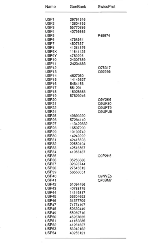
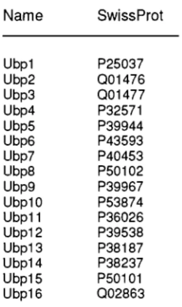
MATERIALS AND METHDDS Sequences of the currentJy known human USPs corre-sponding to the C19 f.unily of the MEROPS peptidasc database (http://merops.sanger.ac.ukJ) were collected from the Genl3ank (http://www.ncbi.nlm.nih.gov/Gen-bankl) and SwissProt (http://www.cxpasy.org/sprotll data bases. Only one sequence was sdected from those of multiple. isoforms reported for some USPs (gene.rally nearly identical mutation isoforms, othenvise the longest sequence was selected), thus leading 10 a nonredundant set of S4 distinct human USP sequences (see Supplemen-tary Materia!). The boundaries of their catalytie core, as weil as ail their currently annotated domain.~ outside this core domain were obtained from the pfam (http:// www.sanger.ac.uklSoftware/Pfam/) and InterPrn (http:// www.ebi.ac.uklinterproJ) dat,lbascs and confirmed, when-eyer ayailable, with actual structures retrie\'ed from the l'mtein Data Bank (PDB, http://www.rcsb.org/). The remaining unannotated sequence content was therefore defined by the sequences flanki ng or between the cur-rently annlltated domains, as weil as inscI1ed in the cata-Iytic core. ln the latter case, locating such insertions required (i) a multiple sequence alignment of ail S4 cata-Iytic core sequences, which was perfor1l1ed with the MAFIT5 algorithl11,13 and (ii) comparisons with the minimal catal}"tic core domain dclineated by its available crystal structures from several human USPs (with l'DB IDs): 2 (2HDS), 7 (INBS, lNBF, 2F1Z), 8 (2GFO). and 14 (2AYN, 2AYO).
2 PROTEINS
Structural domain detection of the currently unanno-tated content of the human USP family was carried out at the Structure Prediction Meta Server (http://meta. bioinfo.pll), which assembles state-of-the-art fold recog-nition methods, and provides a consensus scoring of the three-dimensional structure predictions generated for a given query sequence hy independent algorithms, using the 3D- Jury meta-predictor. 14 Short sequence stretches «40 residues) were not considered. Overly long contih'1l-ous sequences (>800 residues) were split into shortcr fragments prior to fold recognition calculations. l1lis splitting was donc in two W,lyS: (i) generating thrce equal-length sequences corresponding to tJle N- and C-termin;!l hah'es plus the central region of the same length, and (ii) following the consensus predictions of domain boundaries generated by the Meta-DP meta-server (http://meta-dp.cse.buff.llo.eduJ).15 Considering the possibility of e1l1bedded domain folds, newly identi-lied domains were excised out of the original query sequence (typically longer), and the resulting flanking regions were 1l1erged and subjected to a new round of fold detection. Finally, the excised sequences of ail newly mapped domains were resubmitted to the. Structure Pre-diction Meta Sen'er to obtain the final template ranking, rc1iability indicators, query-to-te1l1plate sequence align-ments and secondary structure predictions.
The reliability of fold assignment was based primarily on the 3D-Jury confidence score, which was calculated using the standard settings under which the score was found to correlatc to the number of correctly predicted residues. 16 Accordingly, a confidence threshold of 50 for the 3D-jury score translates into a prediction reliability of over 90%. Fur shorter sequences « 100 residues), the 3D-jury contidence cut-off was lowered to 40. A qualita-tive evaluatÎon of the qucry-to-tcmplate sequence and secondary structure alignments was also carrÎed out to support the assessment uf each top-ranked structural assignment.
Secondary structure predictions were based on four methods: PROFsec,17 PSI-PRED.18 and SAM-TOZ with DSSP and STRIDE alphabets. l9 A consensus was th en derived for each sequence by (i) majority voting over ail four methods for a-helices and J3-strands, and (ii) SAM-T02 predictions of G-helices, a secondary structure not available from the other prediciion methods. The multi-ple sequence alignment of the identitied UBL domains was assembled starting from individual query-to-telllpJate sequencc alignlllents top-r,1I1ked by 3D-jury consensus fold recognition. This preliminary alignment was furtller refincd by: (i) considering the structure-based sequence alignment between tbe top-ranked UBL templates, which was gencrated with the Expresso (3D-Coffee) program,20 and (ii) minor local improvcments in the sequence und secondary structure alignments among predicted UBL dOlllains. Sequence homology-based dustering of prc-dicted UBL domains of USPs was derived with the Clustal
CHAPTER 4 PUELISHED ARTICLE
20
Deteot:ion of UBL Domaine ln Human USPe
W program21 uSÎng the PAM350 scoring matrix, given the seqllence divergence of thi.' UBL fi)ld.
RESULTS
One approach loward extending the Cl1rrent sequence-homology-based dc)main ,lI1notation of human USPs is to detect structural relationships that have only remote or no underlying sequence horn<llogy. 'fhis is the
objec-tive of fnld recognition ll1ethods, Thus, wc suiJjeeted thc unannotated sequence content of the human USP family to the consensus protein structure prediction method 3D- lury.l4, 16 This widdy lIsed melil-prcdktor performs consensus scoring over the 3D model, generated hy state-of-the-art fold recognition algorithms, <lnd ranked as a top-pcrformer at the lMest CAS!', CAFASP, and Live-Bench prediction contests.22,23 We also have reecntly employed 3D-Jllry to predict the USP-like structure and infer the deubiquîtinating activily for the SARS coronavi-rus papain-like protease,24,25 predictions whkh wert: experilllentally confirmed bu th functiol1ally and ,tmctur·
<lUy.26-28
One of the most interesting result, ,lemming oui of Ihi, analysis was the prediction of llhiquitin-like (UBL) dornains in an unexpectcdly high Ilumber of hum,ll1 USP, (Fig, 1 Al, These UBL Jorn,lins were predkted with high reliability as judged by the statistically significant 3D- fury scores obtained for the correspollding USP sequences af,rainst nUrllcrolls UBL templates (see Slip ple-mentaiT Materia\). Consistent with the fold recognition data, the newly identilÎed UBL dOllwins fo!low the con-sensus sccondary structllre and the common lÎngerprint sequence characteristic to the ubiqllitin sllperfold (Fig. 1 B).29
As shown schematically in Figure 1 A, the prevÎollsly Ilnannotated UBL domains detected for various human USPs by our structural bioinforl1latics analysi, are pres-ent both imide and outside their catlllyrk core domains. Ubiquitin-like domains nested imide catalytic core dom.lins are found in the human USPs 4, 6, Il, 15, 19, 31, 32, and 43, ln ail these enzyme" the UBL domain insertion occurs at highly homologous positions, spedfi-cully, in the middle of the cin:ularly pl'rmutcd Zn-tlnger-like dOl1ll1in, itself nested within the catalytic core between the IWo sub-domains of the papain-like fold. 30 The nested UBL dumain would !Je inscrted in thes.; USPs between the f3-strancl and the u -hdix that arc graftcd onto the four-stranded f3-ribbon of the drcularly per-l1luted Zn-finger and are lltilized for its attachment to the C-terminal sub-dOllli\Ïn of the papain-like fold (Fig. 1 Cl, as obscrvcd in the crysml structures of several USp,) [-33 ln ('ach case, the inserted UBL domain is clin:,tly fullowed bya region of about lïO·240 residut'l:l
(depending on the enzyme) beforc the remaincler of the circularly pcrmuted Zn·finger-like f(Jld (Fig, tA). No fold
DOl lO, 1()()2/l'ro!
similarity l'ouM !Je detected for any of these regions, which, for most parts, lack prcdicted secondary structure clements. An interesting "\Illriation is observed for USP19, where an annotated MYND Zn-linger domain of about 45 residues i5 intercalatccl immediately after the nested UBL domain and before the large, mostly llilstructured, region.
The remuinder of the newly identified UBL domains are located outside the boundarics of the catalytic core dOl11uin (Fig. lA), Ubiquitin-Iike donmins N-terminal to the catalytic core are detected in human US!'s 4, 9X, 9Y, 11, 15, 24, 32, 34, and 47. Thus, USPs 4, Il, 15, and 32 teature two UBL domains, one inside and the other one immediately upstream to the catalytk çore domain. Interestingly, the predicted N-temlinal URL domain in ail these four USP, is preceded by a DUSP domain.34 'Ille single N-terminal UBL domains of US!>, 9X, 9Y, 24, and 34 are predicted to be l1anked on both sides by ,111-œ·helical domains (not shown), Multiple UBL domains were detected in the C-tcrminal extensions relative to the cataly1ic core of human USPs 7 (four domains), 40 (two domains), and 47 (three domains; a fOllrth UBL domain is prcdicted upstre,lm to the catalytic core).
DISCUSSIDN
Based 011 primary sequence homo)ogy, UBL domains have been previously detected on'Iy in the N-termimll part nf human USP14,35 and in the C-terminal end of human USP4S.12 ln the former case, the solution NMR structure of the UBL do main of 11l0use USPI4 (l'DB ID: lWGG; 97°/t. sequence identity to the human domain) c(mfirms this structural assignment. The exquisite prom· i,cu ity of the ubiquitin superfold to variations in primary sequence,29 may have preduded the detection of most UBL domains b}' simple applications ~1f standard homot-ogy tooL~ sucll as PSI-BLAST,36 possib)y leading to their llnder-representation in the currently <lV'dilablc public annotations of USPs. Supporting thi, idea, the only other previously reported UBL domain of il lm man USP, that
nI' USP9Y, resultcd from a fold recognition-based annota-tion study targeted to the male·spedfic- region of the human y chromosome.37
The present structural bioinformatics i.lnalysis of the (urrentl)' unannotated content of the endre h\umm USP f<lmily significantly augments tlle existing annotation with the addition of 26 UBL domains From 15 distinct human USPs. Thus, the UBL domain ean be regarded as the most frequentl)' occurring domain in the hllman USP famil)', atter the characteristic pl:ote\lse core domain.S,12 We "mnot exclude the possibility that a few other UBL domains, perhaps more remotely related to the currently known mcmbers of the ubiquÎtin superfold,29 l1<1ve escaped our fold detection employing the existing bcst-performing algorithms and the currcnt PDB content. For
CHAPTER 4 PUBL/SHED ARTICLE A,----= _ _ _