The following scientific article was officially published in the journal Spine Deformity, (the official journal of the Scoliosis Research Society), published by Elsevier. This article’s citation is as follows:
Ramsay, J., Joncas, J., Gilbert, G., Trop, I., Cheriet, F., Labelle, H. and Parent, S. “Is Breast Asymmetry Present in Girls with Adolescent Idiopathic Scoliosis?” Spine Deformity, Vol. 2, No. 5, (2014) pp.374-379.
doi: http://dx.doi.org/10.1016/j.jspd.2014.05.002
The manuscript, as submitted by the authors, is reproduced here as it appears in the first author’s Master’s thesis, entitled L’asymétrie mammaire chez les adolescentes présentant une scoliose idiopathique. The thesis citation is as follows:
Ramsay, Joyce. "L’asymétrie mammaire chez les adolescentes présentant une scoliose idiopathique." M.Sc., Université de Montréal, 2013.
Joyce Ramsay, 2013
© 2013 Joyce Ramsay. This work is licensed under the Creative Commons Attribution-NonCommercial-NoDerivatives 4.0 International License. To view a copy of this license, visit:
CHAPITRE 5
Article 1 : Is Breast Asymmetry Present in Girls with
Adolescent Idiopathic Scoliosis ?
Cet article a été l’objet d’une présentation orale à la 19è conférence internationale IMAST le 11 juillet 2013 à Vancouver, CB, Canada.
Le manuscrit de l’article a également été soumis à la revue scientifique de la SRS, Spine Deformity en décembre 2013. La version soumise, conforme aux normes de présentation de cette revue, est présentée aux pages suivantes.
ABSTRACT
Study design: Cross-Sectional Descriptive StudyObjectives: To characterize breast asymmetry (BA), as defined by breast volume difference, in girls with significant Adolescent Idiopathic Scoliosis (AIS), using MRI.
Summary and background: BA is a frequent concern among girls with AIS. It is commonly believed that this results from chest wall deformity. While many women exhibit physiological BA, the prevalence is not known in adolescents and it remains unclear if it is more frequent in AIS. Breasts vary in shape and size and many attempts at measuring them have been explored. MRI shows the highest precision at defining breast tissue.
Methods: 30 patients were enrolled on the basis of their thoracic curvature, skeletal and breast maturity, without regard to their perception on their BA. MRI acquisitions were performed in prone with a 1.5 Tesla system using a 16-channel breast coil. Segmentation was achieved using the ITK-SNAP 2.4.0 software and subsequently manually refined.
Results: The mean left breast volume (528.32 cc ± 205.96) was greater compared to the mean right breast volume (495.18 cc ± 170.16) with a significant difference between them. The mean BA was found to be 8.32% ± 6.43 (p< .0001). A weak positive correlation was observed between BA and thoracic Cobb angle (0.177 p=0.349) as well as thoracic gibbosity angle (0.289, p=0.122). The left breast was consistently larger in 65.5% of the patients. 20 patients (66.7%) displayed BA ≥ 5%.
Conclusions: We have described BA in patients with significant AIS using MRI. This method is feasible, objective and very precise. The majority of patients had a larger left breast, which could compound the apparent BA secondary to trunk rotation. In many cases, BA is present independently of thoracic deformity. This knowledge will assist in counselling AIS patients in regards to their concerns with BA.
Is Breast Asymmetry Present in Girls with
Adolescent Idiopathic Scoliosis ?
Joyce Ramsay, MD, PTa,e , JulieJoncas, RN, BSca, Guillaume Gilbert, PhDb,c,
Isabelle Trop, MD, MPHc, Farida Cheriet, PhDa,d, Hubert Labelle, MDa, Stefan Parent, MD, PhDa,e*
aSainte-Justine University Hospital Research Center, 3175, Chemin de la Côte-Sainte-Catherine, Montreal, Quebec, H3T 1C5, Canada
bMR Clinical Science, Philips Healthcare, 281 Hillmount Road, Markham, Ontario, L6C 2S3, Canada
cHôtel-Dieu University of Montreal Hospital Center, 3840 Saint Urbain Street, Montreal, Quebec, H2W 1T8, Canada
dPolytechnique Montreal, 2500, Chemin de Polytechnique, Montreal, Quebec, H3T 1J4, Canada
*Corresponding Author and Reprints: Stefan Parent, MD, PhD
Division of Orthopedics
Sainte-Justine University Hospital, 3175 Chemin de la Côte-Sainte-Catherine Montreal, (Quebec) H3T 1C5 Canada Phone: 514-345-4876 Fax: 514-345-4755 E-mail:
IRB approval/Research Ethics Committee:
Sainte-Justine University Hospital Ethics Committee approval # 3532 University of Montreal Hospital Center Ethics Committee approval # 12.143
Funding sources:
Academic Research Chair in Pediatric Spinal Deformities of CHU-Sainte-Justine
Acknowledgment:
This study is a result of collaboration between three university institutions in Montreal, Canada. It was supported in part by the Academic Research Chair on Pediatric Spinal Deformities of CHU-Sainte-Justine and MENTOR, a strategic training program of the Canadian Institute of Health Research.
Introduction
Breast asymmetry is a common complaint among girls with adolescent idiopathic scoliosis (AIS). AIS is a complex tridimensional deformity of the spine that arises in 1-3% of otherwise healthy children between 10 years of age and skeletal maturity [1]. Patients with scoliosis are generally screened, evaluated and if necessary, surgically corrected using a posterior approach. However, it is the anterior aspect of the deformity that often concerns girls with AIS, in particular, the asymmetry of their breasts. It is commonly believed that the breast asymmetry is secondary to the chest wall deformity (e.g. left breast larger in a right thoracic scoliosis) [2]. Breast asymmetry is defined as a difference in shape, position and/or volume of the breast or the nipple-areola complex, and may be primary or secondary to a thoracic deformity. Some patients with scoliosis experience breast asymmetry, but its relationship with the thoracic deformity remains unknown [3,4]. Most women have some degree of minor physiological asymmetry (as high as 88% in some studies) [5,6] but this has not been previously described in the adolescent population.
Multiple methods for measuring the breast have been attempted, but none yields consistent results. The breast is an organ of varied size, shape, contour, width, height, projection, composition, volume, nipple level and position on the chest wall. When performing aesthetic or reconstructive breast surgery, the plastic surgeon relies mostly on his/her aesthetic opinions and artistic skills as well as his/her experience [7]. Various methods to measure breast volume have been reported: Archimedian methods based on the water displacement principle [8-10], 3D thermoplastic casting [11-13], direct (from body surface) [14-18] or indirect (from 2D imaging such as photographs, mammograms or ultrasounds) [19-21] anthropometric measures,
modern radiologic procedures (CT and MRI) and 3D body surface reconstruction obtained from biplanar images such as stereo photography, laser or phase shifted interferometry 3D surface scanning [22-41]. To date, MRI measurements show the highest level of precision and are considered the gold standard for breast volume measurement [32,42-44].
It is unclear if breast asymmetry is more frequent in patients with AIS. Very few studies have evaluated the relationship between scoliosis and breast asymmetry [3,45]. The goal of this study is to describe breast asymmetry, as defined by breast volume difference, using MRI in a series of patients with significant AIS.
Materials and Methods
From July 2012 to January 2013, 30 consecutive female patients with AIS were recruited from the scoliosis outpatient clinic at Sainte-Justine’s University Hospital in Montreal, Canada. These patients were enrolled on the basis of their skeletal (Risser 4-5, > 1 year post menarche) and breast maturity, and their thoracic curvature, without regard to their subjective opinion on their breast asymmetry. Tanner breast stage was assessed by observation [46]. We included patients with significant thoracic curvatures (Cobb angle ≥ 30°). Patients with congenital, juvenile idiopathic or neurologic scoliosis were excluded. All patients and parents of minors agreed to participate and gave informed consent. The study was approved by the Ethics Committee of both Sainte-Justine’s University Hospital and the University of Montreal Hospital Center.
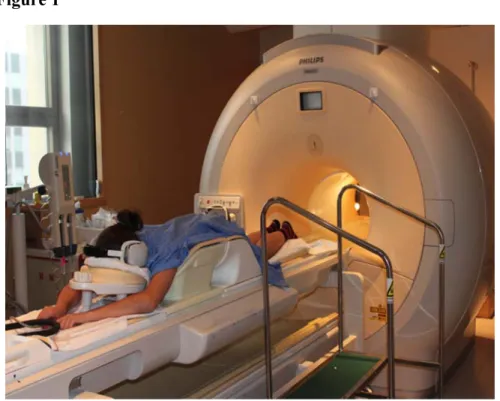
All MRI acquisitions were performed at one of our university institutions with a 1.5 Tesla system (Achieva XR, Philips Healthcare, Best, The Netherlands) using a dedicated 16-channel breast coil. The patient was positioned prone with the arms resting overhead and the breast hanging freely in the breast coil to optimize its natural contour. No intravascular contrast
agent was used. A 3D gradient-echo sequence without fat suppression was used with the following parameters: repetition time (TR): 7.6 ms; echo time (TE): 4.6 ms; flip angle: 12 degrees; field-of-view (FoV): 340 mm x 340 mm x 220 mm (adjusted on a subject by subject basis to cover the complete torso at the breast level); acquisition resolution: 1 mm x 1 mm x 2 mm; and reconstruction resolution: 1 mm x 1 mm x 1 mm. Images were exported from the scanner using the DICOM format and converted to the Analyze format using dcm2niigui software (MRIcron, McAusland Center for Brain Imaging, University of South Carolina, http://www.mccauslandcenter.sc.edu/mricro/mricron/index.html). The images were automatically reformatted to a resolution of 1 mm x 1mm x 3 mm prior to segmentation to reduce the amount of data to process. Segmentation was performed using the software ITK-SNAP 2.4.0 (Penn Image Computing and Science Laboratory (PICSL), Philadelphia, PA, www.itksnap.org) [47]. A preliminary segmentation of each breast was performed using a semi-automatic 3D active contour method. These segmentations were then manually refined by the same evaluator (JR) before being used for the final volume estimates. The nipple and skin were included in the breast volume calculation. The superior and inferior borders were demarcated using the sagittal view, whereas the medial, lateral and posterior borders were precisely defined using the axial view (Figure 2). The pectoralis major muscle was used to outline the posterior plane of the breast. The 3D mesh (Figure 3) also provided a clear representation of the segmented area and was used to smooth the contour of the breast.
Statistical Analysis
The results for continuous variables are expressed as means with standard deviations. To determine the breast volume asymmetry, we considered the absolute value of the difference between (LBV/((LBV+RBV)/2)) and (RBV/((LBV+RBV)/2)). RBV being the right breast
volume and LBV being the left breast volume, expressed in cubic centimeters (cm3 or cc). A
two-tailed paired T test was used to test the hypothesis that this difference in breast volume differed from zero. Also, a Pearson correlation was computed to determine the correlation between breast asymmetry and thoracic Cobb angle as well as thoracic gibbosity. All analyses were performed with SPSS software, release 21.0 (IBM Corporation, Armonk, New York, USA) and conducted at the 0.05 significance level.
Results
Thirty young (age 15.7 ± 1.4 years) skeletally mature (Risser 4-5) girls with AIS (29 right thoracic scoliosis and one left thoracic scoliosis) and a significant thoracic Cobb angle (46 degrees; range 26 to 81) and mature breasts (Tanner stage 5) underwent a breast MRI in order to objectively measure their breast asymmetry (Table 1). Among these, 18 were treated with a brace at some point in time and 15 had planned spine surgery.
The mean left breast volume (528.32 cc 205.96) was higher than the mean right breast volume (495.18 cc 170.16) and the difference was statistically significant (33.14 cc 60.38) (p=0.0054). The mean breast asymmetry, computed as (LBV - RBV)/((LBV + RBV)/2), was 8.32% 6.43 (p<0.0001) (Table 2). A positive correlation was found between breast asymmetry and thoracic Cobb angle (r=0.177, p=0.349) and between breast asymmetry and thoracic gibbosity (r=0.289, p=0.122) (Table 3).
The left breast was consistently larger in 19 out of the 29 patients (65.5%) presenting a right thoracic scoliosis. In contrast, the patient with a left thoracic scoliosis had a larger right breast. Twenty patients (66.7%) displayed a breast asymmetry ≥ 5% and only two patients (6.7%) with one < 0.5%.
Discussion
Although relatively costly [48] and less accessible, MRI allowed us to revisit a common belief in the scoliosis community: that unequal breast size is an illusion induced by the scoliosis and trunk rotation. This imaging technology in combination with the image segmentation enabled us to define with accuracy the posterior limit of the breast while taking into account the chest wall deformity. The mean breast asymmetry in this cohort (8.32 %, CI 5.92-10.72) was found to be statistically significant (p<0.0001) but the clinical relevance of this difference is unclear. If we arbitrarily set the threshold for non-physiologic breast asymmetry at 5%, we obtain a rate of 66.7 % of our cohort. It would therefore be interesting to evaluate the breast volumes in a cohort of young adolescents without any chest or trunk deformities to determine a reference threshold for breast asymmetry in the adolescent population.
Most of the patients in this series had a larger left breast. Shepherd presents similar results in a group of young girls (n=18, age 13-14 years) using dual-energy X-ray absorptiometer [49]. However, the majority of his patients had not reached breast maturity as described by Tanner stages and it is also the only study to our knowledge using this breast volume measuring method. In the adult population, some authors agree on this laterality fact [12,26], while others not [15,50,51]. When only considering studies using MRI as a measuring method [50,51], the tendency favors a larger right breast volume.
It is possible that a developmental abnormality in the arterial blood supply to the chest wall may impact the developing breast [52] but it would not explain completely the asymmetry observed. Therapeutic braces may also play a role. Repetitive local trauma to the breast tissue
may contribute to breast asymmetry. We believe that a well-made brace concentrates the pressure points in the sagittal plane and aims at relieving pressure on the breast. Furthermore, only a third of our cohort wore a rigid brace at a given moment. In our cohort with AIS and thoracic wall deformity, we would expect shape or positional breast asymmetry rather than volume asymmetry. In the majority of cases, breast asymmetry is present independently of the thoracic deformity.
We found a weak positive correlation between breast asymmetry and thoracic Cobb angle (r=0.177, p=0.349). These results do not corroborate with Tsai (r=0.90) using an anthropomorphic method in a sub-cohort of patients (n=60) undergoing augmentation mammoplasty [4]. The author does not report the mean Cobb angle in his study, which appears to be minor since he declares that the majority of the patients were not aware of the scoliosis diagnosis. We feel confident with the precision of our results using the MRI and believe that thoracic Cobb angle only partially explains the breast asymmetry observed. We also studied the correlation between breast asymmetry and the thoracic gibbosity angle as it was demonstrated by Seoud to be more closely correlated to vertebral rotation than the Cobb angle [53]. No statistical significant correlation was found between breast asymmetry and thoracic gibbosity angle (0.289, p=0.122), further reinforcing the fact that breast asymmetry is independent of spine and thoracic deformity.
Some limitations of our study must be considered. In some patients, the completion of breast maturity could have been overestimated. It is known that in some cases, one breast develops more rapidly than the other, leading to an asymmetry in the shape, volume or position of the breasts or the nipple-areola complex. This asymmetry usually attenuates with time and eventually disappears [54]. Furthermore, Tanner staging has its own limitations as far as
predicting the end of the breast development [46].
Manual segmentation can also be subject to individual variability. In some patients, the pectoralis fascia was not clearly defined in all imaging sections. In others, especially in small breasts, the medial and lateral borders could be challenging to identify. To limit the variability in breast volumes, all sixty segmentations were meticulously performed by a single evaluator (JR). Furthermore, the segmentation process was facilitated by the 3 axis views and the visualization of the 3D mesh. Also, very slight differences in volume estimates were found between gross (semi-automatic) and refined (manual) segmentations. The ease in identifying anatomical landmarks on the MRI makes the segmentation process very accurate. For these reasons, as well as considering the length of time involved in the segmentation process (approximately 15-20hrs/patient), we opted not to perform a reproducibility study.
The prone position used for the MRI acquisitions enhanced the breast contour, but it did not depict exactly how the upright patient perceives herself. A study comparing the breast volumes in both postures would help us appreciate the discrepancies, if any exist. We could also correlate the results obtained using the MRI with the patient’s perception of breast asymmetry by submitting the Truncal Anterior Asymmetry Scoliosis Questionnaire (TAASQ) to our participants [55].
This study described breast asymmetry in an AIS cohort using MRI. This method is feasible, objective and very precise. It will help us to advise girls with AIS regarding their concerns with breast asymmetry. The results will serve as reference values to study a measuring technique using 3D body surface imaging for a faster, less costly and more accessible tool to use in the clinical setting.
References
[1] Parent S, Newton PO, Wenger DR. Adolescent idiopathic scoliosis: etiology, anatomy, natural history, and bracing. Instr Course Lect 2005;54:529-36.
[2] Mao S-h, Qiu Y, Zhu Z-z, et al. Clinical evaluation of the anterior chest wall deformity in thoracic adolescent idiopathic scoliosis. Spine 2012;37:E540-8.
[3] Normelli H, Sevastik JA, Ljung G, et al. The symmetry of the breasts in normal and scoliotic girls. Spine 1986;11:749-52.
[4] Tsai F-C, Hsieh M-S, Liao C-K, et al. Correlation between scoliosis and breast asymmetries in women undergoing augmentation mammaplasty. Aesthetic Plastic Surgery 2010;34:374-80.
[5] Rohrich RJ, Hartley W, Brown S. Incidence of breast and chest wall asymmetry in breast augmentation: a retrospective analysis of 100 patients. Plastic & Reconstructive Surgery 2006;118:7S-13S; discussion 4S, 5S-7S.
[6] Medard de Chardon V, Balaguer T, Chignon-Sicard B, et al. [Constitutional
asymmetries in aesthetic breast augmentation: incidence, postoperative satisfaction and surgical options]. Annales de Chirurgie Plastique et Esthetique 2009;54:340-7.
[7] Maliniac JW. Evaluation of principal mamma-plastic procedures. Plast Reconstr Surg (1946) 1949;4:359-73.
[8] Bouman FG. Volumetric measurement of the human breast and breast tissue before and during mammaplasty. Br J Plast Surg 1970;23:263-4.
[9] Tegtmeier RE. A quick, accurate mammometer. Ann Plast Surg 1978;1:625-6.
[10] Schultz RC, Dolezal RF, Nolan J. Further applications of Archimedes' principle in the correction of asymmetrical breasts. Ann Plast Surg 1986;16:98-101.
[11] Ingleby H. Changes in breast volume in a group of normal young women. Bull. Int. Assoc. Med. Museums 1949;29:87-92.
[12] Campaigne BN, Katch VL, Freedson P, et al. Measurement of breast volume in females: description of a reliable method. Ann Hum Biol 1979;6:363-7.
[13] Edsander-Nord A, Wickman M, Jurell G. Measurement of breast volume with thermoplastic casts. Scandinavian Journal of Plastic & Reconstructive Surgery & Hand Surgery 1996;30:129-32.
[14] Smith DJ, Jr., Palin WE, Jr., Katch VL, et al. Breast volume and anthropomorphic measurements: normal values. Plastic & Reconstructive Surgery 1986;78:331-5. [15] Qiao Q, Ling Y, Zhou G, et al. Breast volume measurement in 125 young Chinese
women. Chin Med Sci J 1992;7:44-8.
[16] Westreich M. Anthropomorphic breast measurement: protocol and results in 50 women with aesthetically perfect breasts and clinical application. Plastic & Reconstructive Surgery 1997;100:468-79.
[17] Brown TP, Ringrose C, Hyland RE, et al. A method of assessing female breast morphometry and its clinical application. Br J Plast Surg 1999;52:355-9.
[18] Brown RW, Cheng YC, Kurtay M. A formula for surgical modifications of the breast. Plastic & Reconstructive Surgery 2000;106:1342-5.
[19] Malini S, Smith EO, Goldzieher JW. Measurement of breast volume by ultrasound during normal menstrual cycles and with oral contraceptive use. Obstetrics & Gynecology 1985;66:538-41.
[20] Loughry CW, Sheffer DB, Price TE, Jr., et al. Breast volume measurement of 248 women using biostereometric analysis. Plastic & Reconstructive Surgery 1987;80:553-8.
[21] Kalbhen CL, McGill JJ, Fendley PM, et al. Mammographic determination of breast volume: comparing different methods. AJR Am J Roentgenol 1999;173:1643-9. [22] Galdino GM, Nahabedian M, Chiaramonte M, et al. Clinical applications of
three-dimensional photography in breast surgery. Plastic & Reconstructive Surgery 2002;110:58-70.
[23] Nahabedian MY, Galdino G. Symmetrical breast reconstruction: is there a role for three-dimensional digital photography? Plastic & Reconstructive Surgery
2003;112:1582-90.
[24] Lee HY, Hong K, Kim EA. Measurement protocol of women's nude breasts using a 3D scanning technique. Appl Ergon 2004;35:353-9.
[25] Garson S, Delay E, Sinna R, et al. [3D evaluation and mammary augmentation surgery]. Annales de Chirurgie Plastique et Esthetique 2005;50:643-51.
[26] Losken A, Fishman I, Denson DD, et al. An Objective Evaluation of Breast Symmetry and Shape Differences Using 3-Dimensional Images. Annals of Plastic Surgery
2005;55:571-5.
[27] Losken A, Seify H, Denson DD, et al. Validating three-dimensional imaging of the breast. Ann Plast Surg 2005;54:471-6; discussion 7-8.
[28] Isogai N, Sai K, Kamiishi H, et al. Quantitative analysis of the reconstructed breast using a 3-dimensional laser light scanner. Ann Plast Surg 2006;56:237-42.
[29] Kovacs L, Yassouridis A, Zimmermann A, et al. Optimization of 3-dimensional imaging of the breast region with 3-dimensional laser scanners. Ann Plast Surg 2006;56:229-36.
[30] Kovacs L, Eder M, Hollweck R, et al. New aspects of breast volume measurement using 3-dimensional surface imaging. Annals of Plastic Surgery 2006;57:602-10. [31] Tepper OM, Small K, Rudolph L, et al. Virtual 3-dimensional modeling as a valuable
adjunct to aesthetic and reconstructive breast surgery. American Journal of Surgery 2006;192:548-51.
[32] Kovacs L, Eder M, Hollweck R, et al. Comparison between breast volume
measurement using 3D surface imaging and classical techniques. Breast 2007;16:137-45.
[33] Moyer HR, Carlson GW, Styblo TM, et al. Three-dimensional digital evaluation of breast symmetry after breast conservation therapy. J Am Coll Surg 2008;207:227-32. [34] Sinna R, Garson S, Taha F, et al. [Evaluation of 3D numerisation with structured light
projection in breast surgery]. Annales de Chirurgie Plastique et Esthetique 2009;54:317-30.
[35] Paul SM, Chamberlin AP, Hatt C, et al. Reliability, validity, and precision of an active stereophotogrammetry system for three-dimensional evaluation of the human torso. Medical Engineering & Physics 2009;31:1337-42.
[36] Liu C, Luan J, Mu L, et al. The role of three-dimensional scanning technique in evaluation of breast asymmetry in breast augmentation: a 100-case study. Plastic & Reconstructive Surgery 2010;126:2125-32.
[37] Gabriel A, Fritzsche S, Creasman C, et al. Incidence of breast and chest wall asymmetries: 4D photography. Aesthet Surg J 2011;31:506-10.
[38] Eder M, v Waldenfels F, Sichtermann M, et al. Three-dimensional evaluation of breast contour and volume changes following subpectoral augmentation mammaplasty over 6 months. Journal of Plastic, Reconstructive and Aesthetic Surgery 2011;64:1152-60. [39] Tebbetts JB. Correction of breast asymmetry does not exist, and the role of
three-dimensional imaging remains a question. Plastic & Reconstructive Surgery 2011;128:824-5.
[40] Eder M, Waldenfels FV, Swobodnik A, et al. Objective breast symmetry evaluation using 3-D surface imaging. Breast 2012;21:152-8.
[41] Becker H. The role of three-dimensional scanning technique in evaluation of breast asymmetry. Plastic & Reconstructive Surgery 2012;130:893e-4e; author reply 4e-6e. [42] Palin WE, Jr., von Fraunhofer JA, Smith DJ, Jr. Measurement of breast volume:
comparison of techniques. Plastic & Reconstructive Surgery 1986;77:253-5. [43] Bulstrode N, Bellamy E, Shrotria S. Breast volume assessment: comparing five
different techniques. Breast 2001;10:117-23.
[44] Herold C, Reichelt A, Stieglitz LH, et al. MRI-based breast volumetry-evaluation of three different software solutions. J Digit Imaging 2010;23:603-10.
[45] Denoel C, Aguirre MF, Bianco G, et al. Idiopathic scoliosis and breast asymmetry. Journal of Plastic, Reconstructive and Aesthetic Surgery 2009;62:1303-8.
[46] Marshall WA, Tanner JM. Variations in pattern of pubertal changes in girls. Arch Dis Child 1969;44:291-303.
[47] Yushkevich PA, Piven J, Hazlett HC, et al. User-guided 3D active contour segmentation of anatomical structures: significantly improved efficiency and reliability. Neuroimage 2006;31:1116-28.
[48] Caruso MK, Guillot TS, Nguyen T, et al. The cost effectiveness of three different measures of breast volume. Aesthetic Plastic Surgery 2006;30:16-20.
[49] Shepherd JA, Malkov S, Fan B, et al. Breast density assessment in adolescent girls using dual-energy X-ray absorptiometry: a feasibility study. Cancer Epidemiology, Biomarkers & Prevention 2008;17:1709-13.
[50] Hussain Z, Roberts N, Whitehouse GH, et al. Estimation of breast volume and its variation during the menstrual cycle using MRI and stereology. Br J Radiol 1999;72:236-45.
[51] Koch MC, Adamietz B, Jud SM, et al. Breast volumetry using a three-dimensional surface assessment technique. Aesthetic Plast Surg 2011;35:847-55.
[52] Iliopoulos P, Korovessis P, Koureas G, et al. Asymmetric evolution of anterior chest wall blood supply in female adolescents with progressive right-convex thoracic idiopathic scoliosis. European Spine Journal 2007;16:1343-7.
[53] Seoud L, Dansereau J, Labelle H, et al. Multilevel analysis of trunk surface
measurements for noninvasive assessment of scoliosis deformities. Spine (Phila Pa 1976) 2012;37:E1045-53.
[54] Eidlitz-Markus T, Mukamel M, Haimi-Cohen Y, et al. Breast asymmetry during adolescence: physiologic and non-physiologic causes. Isr Med Assoc J 2010;12:203-6. [55] Lonner B, Shah S, Toombs C, et al. What an AIS Patient Sees in the Mirror: Validation
of the Truncal Anterior Asymmetry Scoliosis Questionnaire (TAASQ). Paper presented at: Scoliosis Research Society, 48th annual international meeting & course2013; Lyon, France
FIGURES
Figures Legend
Figure 1 Illustration of the positioning of a subject in the breast coil prior to the insertion in the MRI magnet.
Figure 2 Axial cut at the nipple level depicting the segmented breasts in this AIS patient. Figure 3 Axial cut of the right breast showing its medial, lateral and posterior borders. Figure 4 3D meshes of the left and right breasts obtained with manual segmentation.
Figure 2
TABLES
Table 1 Descriptive Statistics
n = 30 Mean Standard
Deviation Min Max
Age (years) 15.7 1.4 13.5 18.9
Weight (kg) 55.1 10.9 34.1 84.7
Height (cm) 161.0 5.6 149.0 173.5
Menarche (years) 13.0 1.1 11.0 15.2
Thoracic Cobb angle (°) 46.1 14.6 26.0 81.0
Gibbosity angle (°) 7.0 5.4 -11.7 16.5
Table 3 Pearson correlation coefficients with breast asymmetry.
n = 30 r p
Thoracic Cobb angle (°) 0.177 0.349
Table 2 Breast volumes and breast asymmetry obtained from MRI segmentation.
n = 30 Mean Standard
deviation Min Max 95% CI p
Right Breast Volume (cc) 495.18 170.16 194.29 979.57 431.64-558.71
Left Breast Volume (cc) 528.32 205.96 188.91 1178.09 451.41-605.22
Difference of volume (cc) 33.14 60.38 -198.50 91.72 10.59-55.69 < .01