HAL Id: hal-02694515
https://hal.inrae.fr/hal-02694515
Submitted on 1 Jun 2020HAL is a multi-disciplinary open access archive for the deposit and dissemination of sci-entific research documents, whether they are pub-lished or not. The documents may come from teaching and research institutions in France or abroad, or from public or private research centers.
L’archive ouverte pluridisciplinaire HAL, est destinée au dépôt et à la diffusion de documents scientifiques de niveau recherche, publiés ou non, émanant des établissements d’enseignement et de recherche français ou étrangers, des laboratoires publics ou privés.
Systemic lipopolysaccharide influences rectal sensitivity
in rats: role of mast cells, cytokines, and vagus nerve
Anne-Marie Coelho, Jean Fioramonti, Lionel Bueno
To cite this version:
Anne-Marie Coelho, Jean Fioramonti, Lionel Bueno. Systemic lipopolysaccharide influences rectal sensitivity in rats: role of mast cells, cytokines, and vagus nerve. AJP - Gastrointestinal and Liver Physiology, American Physiological Society, 2000, 279 (4), pp.G781-G790. �hal-02694515�
279:781-790, 2000.
Am J Physiol Gastrointest Liver Physiol
You might find this additional information useful...
44 articles, 9 of which you can access free at:
This article cites
http://ajpgi.physiology.org/cgi/content/full/279/4/G781#BIBL 11 other HighWire hosted articles, the first 5 are:
This article has been cited by
[PDF] [Full Text] [Abstract] , August 18, 2004; 292 (7): 852-858. JAMA H. C. Lin Syndrome
Small Intestinal Bacterial Overgrowth: A Framework for Understanding Irritable Bowel [PDF] [Full Text] [Abstract] , August 1, 2005; 289 (2): G254-G260.
Am J Physiol Gastrointest Liver Physiol
B. Wang, J. Glatzle, M. H. Mueller, M. Kreis, P. Enck and D. Grundy vitro: role of prostaglandins
Lipopolysaccharide-induced changes in mesenteric afferent sensitivity of rat jejunum in [PDF] [Full Text] [Abstract] , December 1, 2005; 94 (6): 3815-3825. J Neurophysiol
D. A. Bereiter, K. Okamoto, A. Tashiro and H. Hirata Caudalis Neurons
Endotoxin-Induced Uveitis Causes Long-Term Changes in Trigeminal Subnucleus [PDF] [Full Text] [Abstract] , June 1, 2006; 1 (1): 37-48.
Soc Cogn Affect Neurosci
E. P. M. Vianna, J. Weinstock, D. Elliott, R. Summers and D. Tranel Increased feelings with increased body signals
[PDF] [Full Text] [Abstract] , April 1, 2007; 580 (1): 347-356. J. Physiol.
Fioramonti and L. Bueno
F. Barreau, C. Cartier, M. Leveque, L. Ferrier, R. Moriez, V. Laroute, A. Rosztoczy, J. deprivation: corticotrophin-releasing factor and nerve growth factor interplay
Pathways involved in gut mucosal barrier dysfunction induced in adult rats by maternal
on the following topics:
http://highwire.stanford.edu/lists/artbytopic.dtl can be found at
Medline items on this article's topics
Physiology .. Rats Physiology .. Nerves
Veterinary Science .. Vagus Nerve Oncology .. Interleukin-1
Physiology .. Mast Cells
Biochemistry .. Lipopolysaccharides
including high-resolution figures, can be found at:
Updated information and services
http://ajpgi.physiology.org/cgi/content/full/279/4/G781
can be found at: AJP - Gastrointestinal and Liver Physiology
about
Additional material and information
http://www.the-aps.org/publications/ajpgi
This information is current as of September 6, 2010 .
http://www.the-aps.org/. Society. ISSN: 0193-1857, ESSN: 1522-1547. Visit our website at
American Physiological Society, 9650 Rockville Pike, Bethesda MD 20814-3991. Copyright © 2000 by the American Physiological abnormal function of the gastrointestinal tract, hepatobiliary system, and pancreas. It is published 12 times a year (monthly) by the
publishes original articles pertaining to all aspects of research involving normal or AJP - Gastrointestinal and Liver Physiology
on September 6, 2010
ajpgi.physiology.org
Systemic lipopolysaccharide influences rectal sensitivity
in rats: role of mast cells, cytokines, and vagus nerve
ANNE-MARIE COELHO, JEAN FIORAMONTI, AND LIONEL BUE´ NO Neuro-Gastroenterology and Nutrition Unit, Institut National
de la Recherche Agronomique, 31931 Toulouse, France
Received 20 September 1999; accepted in final form 17 April 2000 Coelho, Anne-Marie, Jean Fioramonti, and Lionel
Bue´no. Systemic lipopolysaccharide influences rectal
sensi-tivity in rats: role of mast cells, cytokines, and vagus nerve.
Am J Physiol Gastrointest Liver Physiol 279: G781–G790,
2000.—Intraperitoneal lipopolysaccharide (LPS) produces somatic hyperalgesia, releases interleukin (IL)-1 and tumor necrosis factor-␣ (TNF-␣), and activates vagal afferents. The aim of this study was to evaluate the effect of peripheral LPS on rectal sensitivity and to specify the mechanisms involved. Abdominal muscle contractions were recorded in conscious rats equipped with intramuscular electrodes. Rectal disten-sion (RD) was performed at various times after LPS or experimental treatments. In controls, RD significantly in-creased the number of abdominal contractions from a thresh-old volume of distension of 0.8 ml. At the lowest volume (0.4 ml), this number was increased after administration of LPS (3, 9, and 12 h later), recombinant human IL-1 (from 3 to 9 h), recombinant bovine TNF-␣ (from 6 to 9 h), and BrX-537A (from 6 to 12 h), a mast cell degranulator. The effect of LPS was reduced by doxantrazole, Lys-D-Pro-Thr, and solu-ble recombinant TNF receptor. Vagotomy selectively ampli-fied the response to LPS. We conclude that, in vivo, intra-peritoneal LPS lowers visceral pain threshold (allodynia) through a mechanism involving mast cell degranulation and IL-1 and TNF-␣ release and that the vagus nerve may exert a tonic protective role against LPS-induced rectal allodynia. endotoxins; rectal allodynia; mast cells; interleukin-1; tu-mor necrosis factor-␣; subdiaphragmatic vagotomy
THE GASTROINTESTINAL TRACTrepresents one of the body’s
largest interfaces with the outside environment. It possesses a complex immune system providing its de-fense against environmental threats including infec-tion by viruses, bacteria, and parasites. Approximately one-third of patients with bacterial gastroenteritis de-velop chronic abdominal symptoms and signs of sen-sory changes in the gut (1). This entity, called “postin-fectious irritable bowel syndrome” (PI-IBS), accounts for ⬃30% of all irritable bowel syndrome (IBS) pa-tients. The major symptoms observed in patients with IBS include disordered colonic motility and acute or chronic abdominal pain. These patients exhibit an ex-aggerated sigmoid motor response to a variety of stim-uli (41) and also have a lowered visceral sensory threshold to pain caused by balloon distension (35).
Unfortunately, the pathophysiology of visceral hyper-sensitivity in patients with both IBS and bacterial infection is not precisely known.
Lipopolysaccharide (LPS), also known as endotoxin, is a cell wall Gram-negative bacteria component. It induces a wide array of effects after bacterial infection, including fever (18), sickness behavior (17), and hyper-algesia. Indeed, several studies have shown that an intraperitoneal injection of LPS enhances pain respon-siveness to various somatic stimuli (see Refs. 43 and 46). Moreover, LPS-induced alterations in nociception depend on proinflammatory cytokines released from monocytes and macrophages under LPS stimulation such as interleukin (IL)-1, IL-6, and tumor necrosis factor-␣ (TNF-␣). Indeed, an important role of inflam-matory cytokines at the peripheral level has been re-cently recognized in sensory hypersensitivity (44). For example, cutaneous hyperalgesia can be produced by intraperitoneal injection of IL-1 (22) and TNF-␣ (42). Moreover, after peripheral administration, LPS-, IL-1--, and TNF-␣-induced hyperalgesia requires vagal integrity because it is blocked by subdiaphragmatic vagotomy (43, 44). This result agrees with the obser-vation that brain release of proinflammatory media-tors, including cytokines, is mediated in part by vagal afferents (19), even though it does not appear to be the only route for LPS/cytokine-to-brain communica-tion (10).
Kanaan et al. (15) showed that intraplantar injection of endotoxin produces local inflammation and delayed somatic hyperalgesia, mediated locally by IL-1, nerve growth factor (NGF), and PGE2(39). This hyperalgesic
effect starts 1–2 h after intraplantar endotoxin injec-tion and peaks at 9 h in rats and 24 h in mice (16). Similarly, we recently reported (3) that experimental mast cell degranulation in vivo induces a delayed (6– 12 h) increase in sensitivity (allodynia) to rectal dis-tension in awake rats. Indeed, mast cells are involved in postinfectious (24) and stress-induced (13) hyperal-gesia, and their density is altered in functional bowel disorders where, for example, an accumulation of mast cells in the ileum has been demonstrated (45). The anatomic arrangement of mast cells places them in the
Address for reprint requests and other correspondence: L. Bue´no, INRA, NGN Unit, BP 3, F-31931 Toulouse, France (E-mail: lbueno@toulouse.inra.fr).
The costs of publication of this article were defrayed in part by the payment of page charges. The article must therefore be hereby marked ‘‘advertisement’’ in accordance with 18 U.S.C. Section 1734 solely to indicate this fact.
Am J Physiol Gastrointest Liver Physiol
279: G781–G790, 2000.
0193-1857/00 $5.00 Copyright©2000 the American Physiological Society
http://www.ajpgi.org G781
on September 6, 2010
ajpgi.physiology.org
first line of defense against injury or infection, partic-ularly for skin, airways, and gastrointestinal tract, sites that interface directly with the external environ-ment. These cells are well suited to initiate an acute inflammatory process and, through interaction with other tissue cells, to continue to maintain or modulate later response.
No studies have investigated the influence of sys-temic LPS on visceral sensitivity, as was previously established for somatic sensitivity. Consequently, the present study was designed to evaluate whether intra-peritoneal administration of endotoxin can initiate vis-ceral allodynia to rectal distension in rats and to de-termine the role of peripheral IL-1 and TNF-␣, the involvement of mast cells, and the neuronal (vagus and/or other) pathway in LPS-related allodynia.
MATERIALS AND METHODS General Surgical Procedure
Animal preparation. Male Wistar rats (Harlan, Gannat,
France), initially weighing between 200 and 250 g, were surgically prepared for electromyography according to a pre-viously described technique (38). Rats were anesthetized by intraperitoneal injection of acepromazine (Calmivet, Veto-quinol, Lure, France) and ketamine (Imalgene 1000, Rhoˆne-Me´rieux, Lyon, France) at doses of 0.6 and 120 mg/kg, re-spectively. Three groups of three electrodes of NiCr wire (60-cm length and 80-mm diameter) were implanted bilater-ally in the abdominal external oblique musculature just superior to the inguinal ligament. Electrodes were exterior-ized on the back of the neck and protected by a glass tube attached to the skin. Animals were individually housed in polypropylene cages and kept in a temperature-controlled room (21°C). They were allowed free access to water and food (UAR pellets, Epinay, France). All protocols were approved by the Local Animal Care and Use Committee of Institut National de la Recherche Agronomique.
Electromyographic recording. Electromyographic
record-ings began five days after surgery. The electrical activity of abdominal striated muscles was recorded with an electroen-cephalograph machine (Mini VIII, Alvar, Paris, France) us-ing a short time constant (0.03 s) to remove low-frequency signals (⬍3 Hz) and a paper speed of 3.6 cm/min.
Distension procedure. Rats were placed in plastic tunnels
(6-cm diameter and 25-cm length) in which they could not move, escape, or turn around, to prevent damage to the balloon. Rats were exposed to this procedure over 3 days before rectal distension (RD) to minimize stress reactions during experiments. The balloon used for distension was an arterial embolectomy catheter (Fogarty, Edwards Laborato-ries, Santa Ana, CA). RD was performed by insertion of the balloon (2-mm diameter and 2-cm length) in the rectum, at 1 cm of the anus, the catheter being fixed at the tail with adhesive tape. The balloon was inflated progressively, in 0.4-ml steps, from 0 to 1.6 ml, each step of inflation lasting 5 min. To detect possible leakage, the volume of water intro-duced into the balloon was checked by complete removal with a syringe at the end of the distension period.
Temperature Recording
To measure body temperature (Tb), a thermistor probe
(NTC type, code 10K3A1, Farnell, Villefranche sur Saoˆne, France) was placed in the peritoneal cavity, using a
previ-ously described technique (26). Tbwas recorded 5 days after
surgery by connecting the thermistor probe to an electronic thermometer developed in our laboratory. It was calibrated to give an initial output of 0 V at 35°C with a sensitivity of 200 mV/°C. The temperature was monitored on a potentio-metric recorder (L6514, Linseis, Selb, Germany) with a paper speed of 2 cm/h.
Subdiaphragmatic Vagotomy
Surgery. Seven days before implantation of abdominal
electrodes, abdominal vagotomies were performed as follows. Rats were anesthetized with ketamine and acepromazine (120 and 0.6 mg/kg ip, respectively). After midline laparot-omy, the stomach and lower esophagus were visualized. The stomach was gently retracted beneath the diaphragm to clearly expose the ventral and dorsal trunks of the vagus nerve and covered with saline-moistened sterile gauze. Each vagal trunk was dissected from the esophagus and sectioned. The stomach was then returned to its normal position, and the incision was closed. Sham vagotomies consisted of the same operative procedure except that the vagal trunks were neither tied nor sectioned. Animals were returned to their home cages after the operation and were provided with food and water ad libitum. Normal food intake resumed within 3–5 days after vagotomy.
Verification procedure. The effectiveness of vagus nerve
section was assessed 14 days after vagotomy by the sulfated cholecystokinin (CCK-8S) satiety test. Subdiaphragmatic va-gotomy suppresses the blockade of food consumption induced by CCK-8S. Consequently, CCK-8S or saline was injected at the dose of 4g/kg ip after 20 h of food deprivation, and food intake was measured 1 h after injection.
Histological Mast Cell Counting Method
Intestinal tissue samples were put in Carnoy’s solution immediately after the animals were killed, fixed for 24 h at room temperature, and then embedded in paraffin blocks using routine techniques. Sections were cut at a thickness of 5 mm and stained with hemalun-eosin for routine histologi-cal analysis or with Alcian blue-Safranin O for identification of intestinal mast cells. Three sections for each sample and each animal were analyzed by light microscopy, and the number of intact mast cells was counted for each section. For each animal, the number of intact mast cells per square millimeter of intestinal tissue was the mean of the values obtained for the three sections.
Chemicals
LPS (from Escherichia coli, serotype 0111:B4; L3024, lot no. 38H4065) was purchased from Sigma-Aldrich (St. Quen-tin Fallavier, France) and was dissolved in saline (NaCl 0.9%) at a concentration of 1 mg/ml. BrX-537A (bromolasalo-cid ethanolate) was kindly supplied by Roche Laboratories (London, UK) and was dissolved in DMSO at a concentration of 2 mg/ml. Doxantrazole was obtained from Wellcome Re-search Laboratories (lot no. 59C72, Beckenham, UK) and was dissolved in DMSO (5 mg/ml). Recombinant human IL-1 (rhIL-1) and recombinant human dimeric soluble TNF re-ceptor (rhuTNF:Fc molecule p75, linked to the Fc portion of the human IgG1; sTNFR) were kindly provided by Dr. Mic-kael B. Widmer (Immunex, Seattle, WA). They were dis-solved in saline and Tris-NaCl, pH 7.4, respectively. The construction and production of sTNFR has been previously described (27). Recombinant bovine TNF-␣ (rboTNF-␣) was kindly provided by Sandoz (Basel, Switzerland) and
on September 6, 2010
ajpgi.physiology.org
solved in saline at a final concentration of 150g/ml. The tripeptide H-Lys-D-Pro-Thr-OH was purchased from Bachem AG (H-7230, lot no. 122401, Budendorf, Switzerland) and dissolved in saline at a concentration of 10 mg/ml. In all experiments, intraperitoneal injections of drug or vehicle were given in a volume of 1 ml/kg.
Experimental Protocol
Effect of LPS and role of mast cells. These studies
deter-mined the effects of intraperitoneal injection of LPS on rectal sensitivity and the involvement of mast cells using both pharmacological and histological methods. In a first series of experiments, two groups of rats were used. In the first group (n ⫽ 6–8), rats were injected intraperitoneally with BrX-537A vehicle (DMSO) and, 48 h later, received BrX-BrX-537A (mast cell degranulator; 2 mg/kg ip). RDs were performed before (⫺1 h, control) and 3, 6, 9, 12, and 24 h after BrX-537A or vehicle administration. The dose of BrX-537A (2 mg/kg) has been found active in a model of rectal sensitivity (3). Eight days later, the same animals were injected intraperi-toneally with LPS vehicle (saline) and, 48 h later, received LPS (1 mg/kg ip). RDs were performed before (⫺1 h, control) and 3, 6, 9, 12, and 24 h after LPS or vehicle administration. The dose of LPS was chosen according to preliminary exper-iments showing less reproducible and significant data at lower doses (i.e., 0.1 and 0.5 mg/kg ip; Coelho et al., unpub-lished observations). In the second group (n⫽ 8), rats were injected, in a randomized order, with doxantrazole (5 mg/kg ip), a mast cell stabilizing agent, or its vehicle 20 min before LPS or its vehicle. RDs were performed before (⫺1 h, control) and 3, 6, 9, and 12 h after LPS administration. An 8-day interval was observed between two single LPS administra-tions. The dose of doxantrazole (5 mg/kg ip) was chosen according to its efficacy in preventing mast cell degranula-tion-induced rectal allodynia (3). The time chosen between two single administrations of LPS (8 days) was judged to be the minimum necessary time for a complete recovery from each LPS treatment to eliminate a tolerance parameter of our LPS treatments.
In a second series of experiments, five groups of eight male Wistar rats weighing 250–300 g were used for histological studies. Group 1 was used as control. Groups 2 and 3 received BrX-537A (2 mg/kg ip). Groups 4 and 5 received LPS (1 mg/kg ip). Rats in groups 2 and 4 were killed 1 h after administra-tion; those in groups 3 and 5 were killed 5 h later. Tissue samples of ileum and proximal colon (2–3 cm from the ceco-colonic junction) were collected and prepared for mast cell counting.
Effect of LPS on Tb and behavior. Tbwas recorded in a
group of five rats. On day 1, animals were injected intraperi-toneally with vehicle (saline, 1 ml/kg) after 1 h of tempera-ture control recording. Temperatempera-ture was monitored for a period of 24 h to establish a baseline. On day 2, the same animals were injected intraperitoneally with LPS (1 mg/kg) and Tbwas recorded for 24 h. All measurements were
per-formed at a subthermoneutral ambient temperature of 21.0 ⫾ 1.0°C. All animals received saline or LPS between 8:30 and 9:00 AM.
Role of IL-1. Two groups of rats were used to evaluate the
role of IL-1. Group 1 (n ⫽ 9) was given rhIL-1 at a dose (10 g/kg ip) known to induce sickness behavior (44). Vehicle was injected intraperitoneally for control purposes. RDs were performed 3, 6, 9, and 12 h after rhIL-1 or vehicle admin-istration. Group 2 (n⫽ 5) received, in a randomized order, intraperitoneal injection of tripeptide Lys-D-Pro-Thr (or
ve-hicle) 30 min before LPS injection, at the dose (10 mg/kg)
known to antagonize IL-1-induced hyperalgesia (7) and to significantly reduce the hyperalgesic effect of intraplantar LPS (39). The same group received an injection of tripeptide alone. RDs were performed before (⫺1 h, control) and 12 h after LPS (or vehicle) injection. Eight days separated LPS/ vehicle and LPS/tripeptide randomized treatments.
Role of TNF-␣. Two groups of rats were used. Group 1 (n ⫽
6) was injected intraperitoneally with rboTNF-␣, or its vehi-cle, at a dose (150g/kg) found to be active in a model of somatic hyperalgesia (42). RDs were performed 3, 6, 9, and 12 h later. Group 2 (n⫽ 6) was injected twice intraperitone-ally with sTNFR (total dose 2 mg/kg) or vehicle; the first injection (1 mg/kg) was performed immediately before LPS (1 mg/kg ip) or saline, and the second injection (1 mg/kg) was performed 90 min later. RDs were performed before (⫺1 h, control) and 12 h after LPS or saline injection. The delay of 90 min for the second injection was chosen according to the efficacy of sTNFR, a procedure previously validated in mice (27). Eight days separated LPS/vehicle and LPS/sTNFR ran-domized treatments.
Effect of subdiaphragmatic vagotomy on LPS effect. Four
groups of rats were used to determine the role of vagal nerves: sham vagotomy⫹ vehicle (n ⫽ 5), sham vagotomy ⫹ LPS (n ⫽ 8), vagotomy ⫹ vehicle (n ⫽ 7), and vagotomy ⫹ LPS (n⫽ 7). Rats were injected intraperitoneally with LPS (1 mg/kg) or its vehicle. RD was performed 12 h after vehicle or LPS injection.
Statistical Analysis
Statistical analysis of the number of abdominal contrac-tions for each 5-min period during RD was performed by one-way ANOVA followed by Student’s unpaired or paired
t-test where relevant. Values are expressed as means⫾ SE.
Tbvalues are presented as means⫾ SE and were compared
by one-way ANOVA followed by Student’s paired t-test. Mast cell numbers per square millimeter were analyzed using the Mann-Whitney U-test for unpaired data, and values are expressed as means⫾ SE. All differences were considered significant at P⬍ 0.05.
RESULTS
Effect of Intraperitoneal Injection of LPS on Rectal Sensitivity
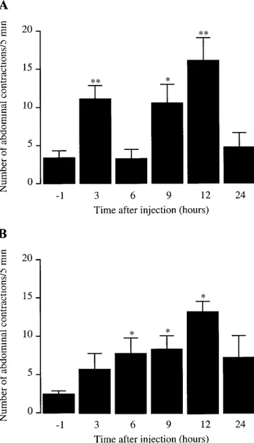
Gradual RD increased the frequency of abdominal contractions in a distension volume-dependent man-ner. A volume of 0.8 ml was determined as the thresh-old at which RD induced a significant increase of the number of abdominal contractions compared with the predistension level (29). Saline-treated rats and un-treated controls responded similarly to RD regardless of volume (0–1.6 ml) and time of distension (3, 6, 9, 12, and 24 h). LPS (1 mg/kg ip) increased the number of abdominal contractions for the 0.4-ml volume 3 (11.0⫾ 1.8 contractions/5 min), 9 (10.5 ⫾ 2.5 contractions/5 min), and 12 (16.1⫾ 3.0 contractions/5 min) h after its administration compared with 3.4⫾ 0.9 contractions/5 min for the control RD performed 1 h before LPS (Fig. 1). At other times (6 and 24 h) and other volumes (0.8–1.6 ml), abdominal responses were unaffected (P⬎ 0.05) by LPS treatment (data not shown). On the basis of these data, we used the time of 12 h (maximal effect) to perform RDs in subsequent pharmacological G783
LIPOPOLYSACCHARIDE AND VISCERAL HYPERSENSITIVITY
on September 6, 2010
ajpgi.physiology.org
investigations except for kinetic studies and determi-nation of mast cell involvement.
Effect of Intraperitoneal Injection of LPS on Tband Behavior
The basal core temperature of rats was 37.8⫾ 0.2°C. Intraperitoneal injection of vehicle (saline, 1 ml/kg) did not significantly (P ⬎ 0.05) modify the profile of Tb
during the daytime period of recording. All rats dis-played normal circadian changes in Tb, with lower
daytime and higher nighttime Tb values (data not shown). LPS (1 mg/kg ip) significantly increased (P ⬍
0.05) Tb between 1.5 and 9 h after injection. The Tb
increase was characterized by the occurrence of two peak elevations, a first maximal Tbrise that peaked 2 h
later (38.5⫾ 0.2 vs. 37.5 ⫾ 0.1°C of vehicle control) and a second maximal Tb rise that appeared 5.5 h later
(38.4⫾ 0.2 vs. 37.2 ⫾ 0.1°C) (Fig. 2). Between 10 and 24 h, there were no differences in Tb between vehicle
and LPS treatments. Concerning behavioral effects, rats injected with 1 mg/kg LPS showed a few signs of illness such as piloerection and lack of activity, but these did not last longer than 24 h. At the high dose used, no deaths were noted.
Involvement of Mast Cells in LPS-Induced Rectal Hypersensitivity
As previously described (3), the number of abdominal contractions observed at the lowest volume of disten-sion (0.4 ml) was significantly increased 6, 9, and 12 h after BrX-537A (2 mg/kg ip; 7.7⫾ 2.0, 8.2 ⫾ 1.8, and 13.1 ⫾ 1.4 contractions/5 min, respectively, vs. 2.4 ⫾ 0.5 for control RD) (Fig. 1). Doxantrazole (5 mg/kg ip) or its vehicle was given 20 min before LPS, and RD were performed 3, 6, 9, and 12 h after LPS. As in the previous series of experiments, animals receiving LPS after vehicle treatment showed an increase in the num-ber of abdominal contractions only for the threshold volume of 0.4 ml at 3, 9, and 12 h after LPS (Fig. 3). Prior administration of doxantrazole (5 mg/kg ip) sup-pressed the abdominal response observed 3–12 h after LPS; compared with vehicle, doxantrazole significantly (P⬍ 0.05) decreased the number of abdominal contrac-tions at the 0.4-ml volume of distension, 3 (3.7⫾ 1.7 vs. 11.0 ⫾ 1.8 contractions/5 min for LPS ⫹ vehicle con-trol) and 12 (5.7 ⫾ 2.2 vs 16.1 ⫾ 3.0) h after LPS administration (Fig. 3).
Fig. 2. Effect of LPS on body temperature. LPS (1 mg/kg ip) induced a biphasic fever with 2 maximal peritoneal temperature rises, 2 and 5–6 h after injection, respectively. Values are means⫾ SE; n ⫽ 5 rats. *Significant difference (P⬍ 0.05) from corresponding vehicle values.
Fig. 1. Kinetics of lipopolysaccharide (LPS; A) and BrX-537A (B) effects on abdominal responses to rectal distension at the 0.4-ml volume in awake rats. Both LPS (1 mg/kg ip) and BrX-537A (2 mg/kg ip) increased the number of abdominal contractions elicited by the lowest volume of distension at 3, 9, and 12 h and 6, 9, and 12 h after injections, respectively. Values are means ⫾ SE; n ⫽ 6–8 rats. Significant difference from control value measured 1 h before injec-tions: *P⬍ 0.05, **P ⬍ 0.01.
on September 6, 2010
ajpgi.physiology.org
Mast Cell Numbers After LPS Administration
To confirm the involvement of mast cells in LPS-induced visceral hypersensitivity and to compare the time course of mast cell degranulation with BrX-537A, histological examination of mucosal mast cells in the ileum and proximal colon was performed on rats treated with either BrX-537A or LPS. The number of intact mucosal mast cells in control animals was 215.1⫾ 33.1 and 133.0 ⫾ 17.9 cells/mm2in the ileum and proximal colon, respectively. When rats were in-jected with BrX-537A (2 mg/kg ip), this number was significantly reduced (107.2 ⫾ 17.6 and 81.2 ⫾ 38.9 cells/mm2; P ⬍ 0.05) 1 h after BrK-537A administra-tion. In contrast, rats given LPS presented a lower number of intact mucosal mast cells only 5 h (131.0⫾ 22.3 and 72.8⫾ 23.7 cells/mm2) after LPS administra-tion (Table 1).
Involvement of IL-1 in LPS-Induced Delayed Visceral Allodynia
After intraperitoneal injection of rhIL-1 (10 g/kg), a significant (P ⬍ 0.05) increase of the number of abdominal contractions generated by the 0.4-ml vol-ume of distension, compared with vehicle, was ob-served 3, 6, and 9 h after rhIL-1 administration (Table 2). No significant increase of abdominal contrac-tions was observed after intraperitoneal injection of saline for the same volume of distension, compared with baseline contractions, at any time of RD after saline. The tripeptide Lys-D-Pro-Thr (10 mg/kg ip) did
not affect per se the abdominal response induced by RD at any volume of distension applied. However, when injected 30 min before LPS (1 mg/kg ip), the tripeptide significantly decreased (P ⬍ 0.05) the number of ab-dominal contractions at the distension volume of 0.4 ml (Fig. 4, A and B) 12 h after LPS (7.2⫾ 2.0 vs. 15.8 ⫾ 3.7 for LPS⫹ vehicle control).
Involvement of TNF-␣ in LPS-Induced Delayed Visceral Allodynia
Similarly to rhIL-1, systemic administration of rboTNF-␣ (150 g/kg ip) regenerated rectal allodynia; intraperitoneal administration of rboTNF-␣ increased the number of abdominal contractions induced by RD at the 0.4-ml volume by 67% and 58% at 6 and 9 h after administration, respectively, compared with control animals treated with vehicle (Table 2). sTNFR (2 mg/kg ip) had no effect per se on basal rectal
sensitiv-Fig. 3. Effect of the mast cell stabilizer doxantrazole on LPS-induced increase of abdominal contractions in response to rectal distension (0.4 ml). Doxantrazole (5 mg/kg ip) reduced the increase in the number of abdominal contractions induced by LPS 3 and 12 h after injection. Values are means⫾ SE; n ⫽ 8 rats. Significant difference from the control value (⫺1 h): *P ⬍ 0.05, **P ⬍ 0.01. †Significant difference (P⬍ 0.05) from corresponding LPS ⫹ vehicle value.
Table 1. Number of intact mast cells identified in ileum and proximal colon 1 and 5 h after BrX-537A or LPS administration
Control
BrX-537A (2 mg/kg ip) LPS (1 mg/kg ip)
⫹1 h ⫹5 h ⫹1 h ⫹5 h
Ileum 215.1⫾33.1 107.2⫾7.2† 155.3⫾9.4 204.2⫾23.9 131.0⫾9.1*
Proximal colon 133.0⫾17.9 81.2⫾15.9* 130.8⫾22.4 130.0⫾20.5 72.8⫾9.7*
Values (no. of intact mast cells/mm2) are means⫾ SE; n ⫽ 6–8 rats. LPS, lipopolysaccharide. *P ⬍ 0.05, †P ⬍ 0.01 significantly different from corresponding control values.
Table 2. Kinetics of rhIL-1 and rboTNF-␣ effects on abdominal response to gradual
distension in awake rats
3 h 6 h 9 h 12 h Vehicle (0.3 ml/rat) 3.9⫾1.2 4.2⫾1.0 6.9⫾1.5 7.2⫾2.0 rhIL-1 (10 g/kg ip) 14.0⫾3.0* 11.9⫾3.1* 14.1⫾2.2* 9.6⫾2.2 Vehicle (0.3 ml/rat) 3.7⫾2.2 3.5⫾2.6 4.8⫾3.4 5.5⫾2.5 rboTNF-␣ (150g/kg ip) 12.3⫾5.7 19.0⫾2.0† 16.8⫾2.9* 7.0⫾4.5 Values are means⫾ SE of the number of abdominal contractions during 5-min periods at the 0.4-ml volume of distension; n ⫽ 6–8 rats. Recombinant human interleukin-1 (rhIL-1) and recombinant bovine tumor necrosis factor-␣ (rboTNF-␣) increased the number of abdominal contractions elicited at the 0.4-ml volume of distension from 3 to 9 h and from 6 to 9 h after injections, respectively. * P ⬍ 0.05, † P ⬍ 0.001 significantly different from corresponding vehicle values.
G785 LIPOPOLYSACCHARIDE AND VISCERAL HYPERSENSITIVITY
on September 6, 2010
ajpgi.physiology.org
ity, but, when injected before LPS, sTNFR significantly (P ⬍ 0.05) reduced the LPS-induced increase in the number of abdominal contractions observed at the 0.4-ml volume (Fig. 5, A and B) 12 h after LPS admin-istration (4.7⫾ 0.9 vs. 15.5 ⫾ 2.1 contractions/5 min for LPS⫹ vehicle control).
Involvement of Vagus Nerves in LPS-Induced Delayed Visceral Allodynia
CCK-8S significantly (P⬍ 0.05) inhibited food intake in sham-vagotomized but not in vagotomized animals, thereby confirming the role of the vagus nerve in the CCK satiety test. Food intake was significantly de-creased in CCK-injected sham-vagotomized rats com-pared with saline control sham-operated animals (1.8 ⫾ 0.5 vs. 4.5 ⫾ 0.4 g), whereas CCK-8S did not modify food intake in vagotomized animals compared
with corresponding saline-injected vagotomized ani-mals (3.7⫾ 0.7 vs. 4.4 ⫾ 0.4 g).
After vehicle treatment, subdiaphragmatic truncal vagotomy did not affect the number of abdominal con-tractions compared with sham-vagotomized animals at any volume of RD (Fig. 6). In sham-vagotomized ani-mals, LPS increased the number of abdominal contrac-tions 12 h after its administration at the RD volume of 0.4 ml (14.7 ⫾ 1.4 vs. 4.9 ⫾ 2.3 abdominal contrac-tions/5 min for vehicle control), similarly to what was observed in intact animals. Surprisingly, marked dif-ferences occurred between sham-vagotomized and va-gotomized groups after LPS injection (Fig. 6): the num-ber of abdominal contractions at the lowest volume of distension (0.4 ml) reached 23.9 ⫾ 4.4 contractions/5 min in vagotomized animals, a value significantly higher than that observed in sham-vagotomized
ani-Fig. 4. Effect of tripeptide Lys-D
-Pro-Thr on LPS-induced increase of number of abdominal contractions in response to rectal distension at different volumes of distension (A) and at the lowest vol-ume of distension (0.4 ml; B). Lys-D -Pro-Thr (10 mg/kg ip) significantly re-duced the number of abdominal contractions at the 0.4-ml volume of distension 12 h after LPS administra-tion. Values are means ⫾ SE; n ⫽ 5 rats. **Significant difference (P⬍ 0.01) from corresponding control values. †Significant difference (P⬍ 0.05) from corresponding LPS⫹ vehicle value.
Fig. 5. Effect of peripheral administra-tion of soluble tumor necrosis factor re-ceptor (sTNFR) on LPS-induced increase of number of abdominal contractions in response to rectal distension in awake rats at different volumes of distension (A) and at the lowest volume of disten-sion (0.4 ml; B). Note that sTNFR (2 mg/kg ip) significantly reduced the num-ber of abdominal contractions elicited by LPS injection only at the volume of 0.4 ml 12 h after LPS. Values are means⫾ SE; n⫽ 6 rats. *Significant difference (P ⬍ 0.05) from corresponding control values. ††Significant difference (P ⬍ 0.01) from LPS ⫹ vehicle-treated ani-mals.
on September 6, 2010
ajpgi.physiology.org
mals (14.7 ⫾ 1.4 contractions/5 min for sham ⫹ LPS value).
DISCUSSION
The present experiments provide new insights re-garding the effects of LPS on visceral sensitivity as previously investigated for somatic pain (43, 46). First, intraperitoneal injection of LPS triggers a delayed low-ering (9–12 h) of the threshold of rectal distension-induced nociception in rats. Second, among the various proinflammatory mediators released in response to LPS, IL-1 and TNF-␣ appear to have a critical role in the genesis of LPS-induced rectal allodynia. Third, the nociceptive response observed after LPS administra-tion is attenuated by doxantrazole, a mast cell stabi-lizer, and histological studies confirmed gut mucosal mast cell degranulation after LPS injection. Fourth, subdiaphragmatic vagotomy, surprisingly, increases the magnitude of rectal allodynia induced by LPS. Together, these data lead to the conclusion that intra-peritoneal LPS evokes a mast cell- and cytokine-depen-dent delayed rectal allodynia controlled by vagus nerves.
In the first part of the study, we demonstrated that intraperitoneal LPS decreases rectal pain threshold but does not modify the magnitude of response to noxious volumes of distension. These data suggest that LPS released during infection favors an abnormal vis-ceral sensitivity to mechanical stimuli, corresponding to a lowered threshold of barosensitivity, without af-fecting the visceral pain intensity evoked by noxious stimulation. The same observations were described previously after inflammation of colorectal mucosa by
trinitrobenzene sulfonic acid in ethanol (29). The mech-anisms evoking such abnormal visceral sensitivity are not yet fully understood. Such abnormal pain sensa-tion, known as allodynia, could be related to a sensiti-zation of low-threshold primary afferents, known to transmit nonpainful sensations in normal conditions and to be able to trigger postsynaptic nociceptive mes-sages in inflammatory conditions. Such alterations have already been observed after nerve injury and cutaneous inflammation in somatic sensitivity (2, 12, 21). In the present study, the absence of change in the abdominal response for higher volumes may be related to the lack of sensitization of high-threshold afferents during the 12 h after LPS administration. Such a hypothesis might explain why LPS turns rectal percep-tion into an abnormal one (allodynia) without modify-ing the pain response magnitude evoked by noxious stimuli.
We have also observed that intraperitoneal LPS en-hances rectal sensitivity in two distinct periods, i.e., 3 h (early phase) and from 9 to 12 h (late phase) after its administration. We can attribute the early phase to a direct effect of endotoxin or pronociceptive mediators, such as PGE2 released from macrophages, acting on primary afferent terminals and the late phase to the subsequent development of inflammation with an in-tense activation of primary afferent fibers and changes in spinal or central processing (25, 32). In agreement with such a hypothesis, most reports related to endo-toxin-induced somatic hyperalgesia have measured a decrease in the latency to cutaneous nociceptive stim-ulus that occurred in the first hour after intraperito-neal injection of LPS (44, 46). In addition, a model of localized inflammatory hyperalgesia was recently de-veloped in rats using intraplantar injection of endo-toxin in the hind paw (15). In this study, Kanaan et al. (15) also observed a peak of hyperalgesia 9 h after endotoxin injection with complete recovery 24 h later, and they explained this delayed response by a similar time-related occurrence of a localized inflammatory reaction.
Different patterns of body temperature profile (monophasic fever, biphasic fever, and hypothermia/ hyperthermia) have been described depending on the dose of LPS used (36). At a high dose (ⱖ1 mg/kg), LPS triggers first a 1- to 3-h decrease of body temperature (hypothermia) followed by a long (6–24 h) period of fever (37, 47). In the present experiments, intraperito-neal administration of a high dose of LPS, i.e., 1 mg/kg, evokes a biphasic fever with two peaks of hyperthermia 2 and 5–6 h later, but we have never observed an initial hypothermia in the first hour after injection. Such a discrepancy may be related to LPS preparation or serotype, rat strain, or route of administration, as previously reported (11, 15).
In the third part of the study, we showed that LPS-induced delayed allodynia is attenuated by an IL-1 receptor antagonist, the tripeptide Lys-D-Pro-Thr. This
result agrees with previous observations in which LPS-induced somatic hyperalgesia was also abolished by intraperitoneal administration of the IL-1 receptor
an-Fig. 6. Influence of vagotomy vs. sham operation on LPS-induced delayed (12 h) rectal hypersensitivity to distension. The response to LPS is amplified in vagotomized animals. Values are means⫾ SE; n, no. of rats. Significant difference from corresponding vehicle control value: *P⬍ 0.05, **P ⬍ 0.01. †Significant difference (P ⬍ 0.05) from corresponding sham⫹ LPS value.
G787 LIPOPOLYSACCHARIDE AND VISCERAL HYPERSENSITIVITY
on September 6, 2010
ajpgi.physiology.org
tagonist (22) and by this tripeptide (39). Moreover, Kanaan et al. (15) also suggested the involvement of IL-1 in the mediation of both endotoxin-induced ther-mal and mechanical hyperalgesia. Moreover, intraperi-toneal IL-1 reproduces the nociceptive response of LPS on the pain threshold to rectal distension with a time course of 3–9 h. Similarly, sTNFR, which acts as a functional TNF antagonist, reduces LPS-induced de-layed allodynia (12 h), suggesting that TNF-␣ partici-pates in the delayed decrease of rectal pain threshold after LPS. These results are also in agreement with data obtained for somatic pain. Indeed, the cutaneous hyperalgesic effect of LPS can be blocked by TNF-␣ binding protein, which acts as a functional TNF antag-onist (42, 44). Intraperitoneal TNF-␣ administration can also reproduce LPS nociceptive response with a delayed maximal response appearing between 6 and 9 h. A long-lasting somatic hyperalgesia after in-traplantar TNF-␣ administration has also been re-ported (4).
Changes in visceral sensitivity related to LPS could be secondary to the activation of chemosensitive noci-ceptors by inflammatory and/or proalgesic mediators (32). Several chemicals produced by local cells are capable of changing the sensitivity of nociceptors, in-cluding bradykinin, histamine, 5-hydroxytryptamine, neuropeptides such as substance P and calcitonin gene-related peptide, prostaglandins (5), and also cy-tokines (4, 7). LPS stimulates the expression of a large number of cytokines, particularly IL-1 and TNF-␣, that may act directly on receptors found on neurons or indirectly by stimulating the release of proalgesic sub-stances that can act on neurons in a cascadelike man-ner (40). For example, IL-1, as well as LPS, stimulates the arachidonic acid cascade, resulting in prostanoid production and release (28) that sensitize primary af-ferent nociceptors and augment the excitability of sen-sory afferents (5). However, other mediators such as bradykinin can initiate both the cascade of cytokine release that mediates hyperalgesic response to endo-toxins (9) and activation and sensitization of pain no-ciceptors (14). In contrast to these data, IL-1 can also induce hyperalgesia by acting directly on high-thresh-old mechanoreceptors, leading to a decrease in latency of neuronal discharges, to a lowering of the threshold, and to an increase of neuronal response to mechanical and thermal stimulation (10). In fact, these observa-tions suggest a dual action of IL-1 on different struc-tures such as immune cells and the endings of terminal neurons, depending on the pathophysiological context. Concerning the potential involvement of TNF-␣ in the LPS cascade, several lines of evidence suggest that TNF-␣ can exert its effects through a cascade of cyto-kine release rather than by a direct stimulation of sensory afferent neurons. Indeed, TNF-␣ is the first cytokine released after LPS administration, and it reaches a maximal plasma concentration after 1 h (48). In fact, TNF-␣ may produce hyperalgesia by inducing the secondary release of IL-1, because its effect can be blocked by an IL-1 receptor antagonist (42). In carra-geenan-evoked somatic hyperalgesia, the same
mech-anism has been reported: bradykinin induces the re-lease of TNF-␣, which in turn stimulates the release of other hyperalgesic cytokines (IL-1, IL-6, and IL-8) responsible for the generation of cyclooxygenase prod-ucts and sympathomimetic amines (8). In consequence, these two cytokines seem to play a role in LPS-induced visceral hypersensitivity at different levels and in a cascade.
Doxantrazole, a mast cell stabilizer, when injected before LPS, prevents both the early (3 h) and the late (12 h) phase of LPS-induced rectal allodynia. Conse-quently, mast cell activation appears to be involved in the cascade of reactions leading to the nociceptive response related to LPS administration. Concerning the early phase (3 h), a previous report showed that LPS can directly degranulate mast cells with produc-tion of cytokines without substantial release of pre-formed mediators by exocytosis (20). Moreover, other previous studies report the importance of various im-mune cells, particularly resident macrophages, from the liver in the production of somatic hyperalgesia appearing early within 1 h after intraperitoneal LPS administration (43, 44). Together, these observations permit us to suggest that the early phase could be linked to the activation of immune cells, and particu-larly mast cells, present in sites other than the gut wall because this early nociceptive response is abolished by previous treatment with a mast cell stabilizer, dox-antrazole, and because no immediate mucosal mast cell degranulation is observed histologically in the gut. Concerning the late phase (12 h), we suggest that this phase is related to the development of an inflammatory reaction triggered by LPS and involving a delayed gut mucosal mast cell degranulation. Indeed, our histolog-ical study shows a decrease in mucosal mast cell num-ber occurring 5 h after LPS administration, and this phase is abolished by doxantrazole. Moreover, this hypothesis is in agreement with a previous study show-ing that BrX-537A, a potent gut mucosal mast cell degranulator (30), promotes only a delayed rectal allo-dynia observed from 6 to 12 h after its administration (3). Furthermore, we have shown here that BrX-537A triggers an immediate (⬍1 h) mucosal mast cell de-granulation in the gastrointestinal tract. Conse-quently, we can hypothesize that the late phase of rectal allodynia, consecutive to intraperitoneal LPS administration, is linked to resident mast cell degran-ulation localized in the intestinal tract.
Vagus nerve serves as an informational highway for inflammatory signals from the periphery, and bilateral truncal vagotomy abolishes a wide range of behavioral and neural effects of peripheral administration of LPS. A part of the “illness” signals elicited by intraperito-neal cytokines or LPS is relayed directly to brain pri-marily by the vagus nerve, which activates a centrifu-gal pain facilitatory pathway. Indeed, proinflammatory cytokines (IL-1 and TNF-␣) produce somatic hyperal-gesia by activating vagal afferents (42, 44). In contrast, our data demonstrate that total subdiaphragmatic va-gotomy amplifies LPS-induced rectal allodynia and thus that the vagus nerve has a protective effect
on September 6, 2010
ajpgi.physiology.org
against LPS-induced visceral allodynia. Chemical, electrical, or physiological activation of cardiopulmo-nary, diaphragmatic, or subdiaphragmatic vagal affer-ents can result in either facilitation or inhibition of nociception in some species, depending on the intensity of stimulation (for review, see Ref. 31). For example, high-intensity stimulation of vagal afferents activates spinal descending inhibitory systems, whereas low in-tensity stimulates pain facilitatory circuits (34). There-fore, it can be postulated that, according to the inten-sity of the stimulus applied in our study, vagal afferent fibers can participate in a feedback loop directly con-trolling chemical nociceptive inputs from the periphery by activating descending antinociceptive pathways. However, there is substantial evidence that sensory neurons have a protective role against injury to the gut (6, 33). Indeed, worsening of inflammation has been observed in acute colitis models after perivagal capsa-icin pretreatment, suggesting a direct protective func-tion of vagal afferents on colonic mucosa against in-flammation (23). Thus we can hypothesize that similar processes may underlie the protective action of vagus nerve against local inflammatory reactions resulting from LPS administration. However, it can also be hy-pothesized that the ability of total subdiaphragmatic vagotomy to reduce LPS hyperalgesia, in some studies, or to amplify the same response, as seen in our data, may be due to differences in the dose of LPS applied. Indeed, the dose of 1 mg/kg is very high compared with doses used in somatic models (16, 43), inducing, in particular, fever and behavioral or locomotive distur-bances. In consequence, the vagus nerve appears to possess a powerful role in mediating peripheral im-mune signals to the brain, but its role must be different according to the nature and/or the amplitude of the aggression and the pathophysiological context devel-oped after LPS infection. It is clear, however, that multiple circuits mediating pain responses exist and are activated under different circumstances.
In summary, the present study indicates for the first time that peripherally injected LPS lowers the visceral pain threshold to rectal distension, this allodynia being observed between 9 and 12 h, and that this effect is mediated by two cytokines, IL-1 and TNF-␣, and involves mast cell degranulation. Because intraperito-neal administration of LPS is associated with inflam-matory reactions in the gut, this result adds some insights into possible mechanisms by which immune reactions of the brain-gut axis may participate in the genesis of visceral allodynia.
We thank C. Betoulieres, L. Ressayre, and I. Lorette for technical assistance and Institut National de la Recherche Agronomique and Solvay-Pharma Laboratories for financial support.
This work was presented in part at the 16th International Sym-posium on Gastrointestinal Motility, Lorne, Victoria, Australia, Feb-ruary 15–20, 1998 (3a).
REFERENCES
1. Bergin AJ, Donnelly TC, McKendrick NW, and Read NW. Changes in anorectal function in persistent bowel disturbance following salmonella gastroenteritis. Eur J Gastroenterol
Hepa-tol 5: 617–620, 1993.
2. Cervero F and Laird JMA. Machanisms of touch-evoked pain (allodynia): a new model. Pain 68: 13–23, 1996.
3. Coelho AM, Fioramonti J, and Bue´no L. Mast cell degranu-lation induces delayed rectal allodynia in rats: role of histamine and 5-HT. Dig Dis Sci 43: 727–737, 1998.
3a.Coelho AM, Fioramonti J, and Bue´no L. Lipopolysaccharide induces a delayed rectal allodynia via mast cell degranulation in rats (Abstract). Neurogastroenterol Motil 10: 65, 1998.
4. Cunha FQ, Poole S, Lorenzetti BB, and Ferreira SH. The pivotal role of tumor necrosis factor␣ in the development of inflammatory hyperalgesia. Br J Pharmacol 107: 660–664, 1992. 5. Dray A. Inflammatory mediators of pain. Br J Anaesth 75:
125–131, 1995.
6. Evangelista S and Meli A. Influence of capsaicin-sensitive fibres on experimentally-induced colitis in rats. J Pharm
Phar-macol 41: 574–575, 1989.
7. Ferreira SH, Lorenzetti BB, Bristow AF, and Poole S. Interleukin-1 as a potent hyperalgesic agent antagonized by tripeptide analogue. Nature 334: 698–700, 1988.
8. Ferreira SH, Lorenzetti BB, Cunha FQ, and Poole S. Bra-dykinin release of TNF-␣ plays a key role in the development of inflammatory hyperalgesia. Agents Actions 38: C7–C9, 1993. 9. Ferreira SH, Lorenzetti BB, and Poole S. Bradykinin
ini-tiates cytokine-mediated inflammatory hyperalgesia. Br J
Phar-macol 110: 1227–1231, 1993.
10. Fukuoka H, Kawatani M, Hisamitsu T, and Takeshige C. Cutaneous hyperalgesia induced by peripheral injection of inter-leukin-1 in the rat. Brain Res 657: 133–140, 1994.
11. Gracely RH, Lynch SA, and Bennett GJ. Painful neuropa-thy: altered central processing maintained dynamically by pe-ripheral input. Pain 51: 175–194, 1992.
12. Gue´ M, Del Rio-Lache`ze C, Eutame`ne H, Theodorou V, Fioramonti J, and Bue´no L. Stress-induced visceral hyper-sensitivity to rectal distension in rats: role of CRF and mast cells. Neurogastroenterol Motil 9: 271–279, 1997.
13. Handwerker HO and Reeh PW. Pain and inflammation. In:
Proceedings of the Sixth World Congress on Pain, edited by Bond
MR, Charlton JE, and Woolf CJ. Amsterdam: Elsevier Science, 1991, p. 59–70.
14. Horan MA, Little RA, Rothwell NJ, and Strijbos PJLM. Comparison of the effects of several endotoxin preparations on body temperature and metabolic rate in the rat. Can J Physiol
Pharmacol 67: 1011–1014, 1989.
15. Kanaan SA, Saade´ NE, Haddad JJ, Abdelnoor AM, Atweh SF, Jabbur SJ, and Safieh-Garabedian B. Endotoxin-in-duced local inflammation and hyperalgesia in rats and mice: a new model for inflammatory pain. Pain 66: 373–379, 1996. 16. Kent S, Bluthe´ RM, Kelley KW, and Dantzer R. Sickness
behavior as a new target for drug development. Trends
Pharma-col Sci 13: 24–28, 1992.
17. Kluger MJ. Fever: role of pyrogens and cryogens. Physiol Rev 71: 93–127, 1991.
18. Laye´ S, Parnet P, Goujon E, and Dantzer R. Peripheral administration of lipopolysaccharide induces the expression of cytokine transcripts in the brain and in the pituitary of mice.
Mol Brain Res 27: 157–162, 1994.
19. Leal-Berumen I, Conlon P, and Marshall JS. IL-6 produc-tion by rat peritoneal mast cells is not necessarily preceded by histamine release and can be induced by bacterial lipopolysac-charide. J Immunol 152: 5468–5476, 1994.
20. Ma QP and Woolf CJ. Progressive tactile hypersensitivity: an inflammation-induced incremental increase in the excitability of the spinal cord. Pain 67: 97–106, 1996.
21. Maier SF, Wiertelak EP, Martin D, and Watkins LR. Inter-leukin-1 mediates the behavioral hyperalgesia produced by lith-ium chloride and endotoxin. Brain Res 623: 321–324, 1993. 22. Mazelin L, The´odorou V, More´ J, Fioramonti J, and Bue´no
L. Protective role of vagal afferents in experimentally-induced colitis in rats. J Auton Nerv Syst 73: 38–45, 1998.
23. McLean PG, Picard C, Garcia-Villar R, More´ J, Fioramonti J, and Bue´no L. Role of kinin B1 and B2 receptors and mast cells in post intestinal infection-induced hypersensi-tivity to distension. Neurogastroenterol Motil 10: 499–508, 1998.
G789
LIPOPOLYSACCHARIDE AND VISCERAL HYPERSENSITIVITY
on September 6, 2010
ajpgi.physiology.org
24. McMahon SB, Lewin GR, and Wall PD. Central hyperexcit-ability triggered by noxious inputs. Curr Opin Neurobiol 3: 602–610, 1993.
25. Million M, Fioramonti J, Zajac JM, and Bue´no L. Effects of neuropeptide FF on intestinal motility and temperature changes by endotoxin and platelet-activating factor. Eur J Pharmacol 334: 67–73, 1997.
26. Mohler KM, Torrance DS, Smith CA, Goodwin RG, Strem-ler KE, Fung VP, Madani H, and Widmer MB. Soluble tumor necrosis factor (TNF) receptors are effective therapeutic agents in lethal endotoxemia and function simultaneously as both TNF carriers and TNF antagonists. J Immunol 151: 1548–1561, 1993. 27. Morrison DC and Rayn JL. Endotoxins and disease
mecha-nisms. Annu Rev Med 38: 417–432, 1987.
28. Morteau O, Hachet T, Caussette M, and Bue´no L. Experi-mental colitis alters visceromotor response to colorectal disten-sion in awake rats. Dig Dis Sci 39: 1239–1248, 1994.
29. Pearce FL, Befus AD, Gauldie J, and Bienenstock J. Mu-cosal mast cells. II. Effects of anti-allergic compounds on hista-mine secretion by isolated intestinal mast cells. J Immunol 128: 2481–2486, 1982.
30. Randish A and Gebhart GF. Vagal afferent modulation of nociception. Brain Res Rev 17: 77–99, 1992.
31. Reeh PW. Chemical excitation and sensitization of nociceptors. In: Cellular Mechanisms of Sensory Processing, edited by Urban L. Berlin: Springer, 1994, p. 119–270.
32. Reinshagen M, Patel A, Sottili M, Nast C, Davis W, Mueller K, and Eysselein VE. Protective function of extrinsic sensory neurons in acute rabbit experimental colitis. Gastroenterology 106: 1208–1214, 1994.
33. Ren K, Randich A, and Gebhart GF. Vagal afferent modula-tion of spinal nociceptive transmission in the rat. J Neurophysiol 62: 401–415, 1989.
34. Ritchie J. Pain from distension of the pelvic colon by inflating a balloon in the irritable bowel syndrome. Gut 14: 125–132, 1973. 35. Romanovsky AA, Shido O, Sakurada S, Sugimoto N, and Nagasaka T. Endotoxin shock: thermoregulatory mechanisms.
Am J Physiol Regulatory Integrative Comp Physiol 270: R693–
R703, 1996.
36. Romanovsky AA, Simons CT, Szekely M, and Kulchitsky VA. The vagus nerve in the thermoregulatory response to
sys-temic inflammation. Am J Physiol Regulatory Integrative Comp
Physiol 273: R407–R413, 1997.
37. Ruckebusch M and Fioramonti J. Electrical spiking activity and propulsion in small intestine in fed and fasted rats.
Gastro-enterology 68: 1500–1508, 1975.
38. Safieh-Garabedian B, Kanaan SA, Haddad JJ, Abou Jaoude P, Jabbur SJ, and Saade´ NE. Involvement of inter-leukin-1, nerve growth factor and prostaglandin E2in endo-toxin-induced localized inflammatory hyperalgesia. Br J
Phar-macol 121: 1619–1626, 1997.
39. Safieh-Garabedian B, Poole S, Allchorne A, Winter J, and Woolf CJ. Contribution of interleukin-1 to the inflammation-induced increase in nerve growth factor levels and inflammatory hyperalgesia. Br J Pharmacol 115: 1265–1275, 1995.
40. Sullivan MA, Cohen S, and Snape WRJ. Colonic myoelectri-cal activity in irritable-bowel syndrome. Effect of eating and anticholinergics. N Engl J Med 298: 878–883, 1978.
41. Watkins LR, Goehler LE, Relton J, Brewer MT, and Maier SF. Mechanisms of tumor necrosis factor-␣ (TNF-␣) hyperalge-sia. Brain Res 692: 244–250, 1995.
42. Watkins LR, Wiertelak EP, Goehler LE, Mooney-Heiberger K, Martinez J, Furness L, Smith KP, and Maier SF. Neurocircuitry of illness-induced hyperalgesia. Brain Res 639: 283–299, 1994.
43. Watkins LR, Wiertelak EP, Goehler LE, Smith KP, Martin D, and Maier SF. Characterization of cytokine-induced hyper-algesia. Brain Res 654: 15–26, 1994.
44. Weston AP, Biddle WL, Bhatia PS, and Miner PB. Terminal ileal mucosal mast cells in irritable bowel syndrome. Dig Dis Sci 38: 1590–1595, 1993.
45. Wiertelak EP, Smith KP, Furness L, Mooney-Heiberger K, Mayr T, Maier SF, and Watkins LR. Acute and conditioned hyperalgesic responses to illness. Pain 56: 227–234, 1994. 46. Yirmiya R, Rosen H, Donchin O, and Ovadia H. Behavioral
effects of lipopolysaccharide in rats: involvement of endogenous opioids. Brain Res 648: 80–86, 1994.
47. Zuckerman SH, Shellhaas J, and Butler LD. Differential regulation of lipopolysaccharide-induced interleukin 1 and tu-mor necrosis factor synthesis: effects of endogenous and exoge-nous glucocorticoids and the role of the pituitary-adrenal axis.
Eur J Immunol 19: 301–305, 1989. on September 6, 2010
ajpgi.physiology.org