FABRICATION AND SURFACE MODIFICATION OF POLY L-LACTIC ACID
NANOSTRUCTURES FOR GROWTH FACTOR IMMOBILIZATION AND
NEURAL STEM CELL DELIVERY
TANIT HADDAD
DÉPARTEMENT DE GÉNIE CHIMIQUE ÉCOLE POLYTECHNIQUE DE MONTRÉAL
MÉMOIRE PRÉSENTÉ EN VUE DE L’OBTENTION DU DIPLÔME DE MAÎTRISE ÈS SCIENCES APPLIQUÉES
(GÉNIE CHIMIQUE) AOÛT, 2014
UNIVERSITÉ DE MONTRÉAL
ÉCOLE POLYTECHNIQUE DE MONTRÉAL
Ce mémoire intitulé:
FABRICATION AND SURFACE MODIFICATION OF POLY L- LACTIC ACID NANOSTRUCTURES FOR GROWTH FACTOR IMMOBILIZATION AND NEURAL STEM
CELL DELIVERY
présenté par : HADDAD Tanit
en vue de l’obtention du diplôme de : Maîtrise ès sciences appliquées a été dûment accepté par le jury d’examen constitué de :
M. VIRGILIO Nick, Ph. D, président
M. AJJI Abdellah, Ph. D, membre et directeur de recherche
M. DE CRESCENZO Gregory, Ph. D, membre et codirecteur de recherche M. DUBOIS Charles, Ph. D, membre et codirecteur de recherche
ACKNOWLEDGEMENTS
I would like to thank my Masters' director M. Abdellah Ajji for this opportunity, help and support. I would like to thank Rouwayda El Ayoubi as well as my co-directors Gregory De Crescenzo and Charles Dubois for all their help, suggestions and guidance throughout this Masters degree.
I would like to thank all of the staff and members of the laboratories at École Polytechnique de Montréal. I would particularly like to thank Loïc Binan, Samantha Noel, Benoît Liberelle, Mehdi Pakravan, Fouzia Ajaja and Afra Hajizadeh.
I would also like to thank my spouse, family (parents, siblings, spouse’s family) and friends.
RÉSUMÉ
Les accidents vasculaires cérébraux, le cancer et la maladie d’alzheimer sont les
principales causes de décès au Canada. Les lésions nerveuses ou les nerfs endommagés dans le système nerveux central peuvent détruire la qualité de vie des survivants, présentent de grands couts pour la société et peuvent entraîner la mort. Il n’existe actuellement aucun traitement efficace encore pour aider à la régénération des tissus nerveux.
Les stratégies de régénération des tissus nerveux ont été étudiées, cependant, de nombreuses limites ont été rencontrées. Par exemple, l'injection directe de cellules souches neuronales dans le système nerveux central a donné lieu à la formation de tumeurs. L'injection directe de facteurs de croissance n’a révélé aucun bénéfice avant sept jours de traitement continu. En outre, l'encapsulation des facteurs de croissance a été précédemment étudiée et possède des inconvénients majeurs. Des limites telles que, la difficulté à maintenir leur livraison à long terme sur une gamme de concentration définie, leur courte demi-vie ainsi qu’une courte distance de migration ont été observées. La production in situ et la distribution de facteur de croissance par l’intermédiaire de cellules telles que les fibroblastes modifiés génétiquement pour exprimer le BDNF ainsi que les cellules fabriquées en coculture ont aussi démontré des inconvénients majeurs. En effet l’immunosuppression était constamment requise et le taux de survie des cellules très faible.
Les biomatériaux tels que les hydrogels ont été largement étudiés et présentent des difficultés de manipulations, une faible adhérence, des couts élevés, des difficultés à incorporer des médicaments sur leur structure ainsi que de présenter des difficultés lors de leurs stérilisations.
Cette étude vise donc à optimiser les stratégies de régénération des tissus nerveux existants. Les biomatériaux électrofilés ont montré des résultats prometteurs dans la littérature en raison de leur porosité, haut rapport surface-volume, interconnexion des pores ainsi que de leur topographie imitant la matrice extracellulaire (ECM) du cerveau. Ces biomatériaux électrofilés
ont donc été utilisés dans ce projet. Le procédé d'électrofilage a été utilisé dans cette étude afin d'obtenir des fibres dans la gamme nanométrique présentant une topologie à haute porosité, le diamètre des fibres idéales ainsi qu’une résistance mécanique adéquate. L’acide poly lactique-L (PLLA) est étudié dans cette recherche; il s'agit d'un polymère couramment utilisé dans l'ingénierie tissulaire neurale étant donné qu'il est autorisé par la FDA et a été utilisé sous la forme de nanofibres électrofilées.
Des études antérieures ont utilisé d'autres protéines pour l'immobilisation tels que la laminine et le collagène. Ces résultats, cependant, n'étaient pas prometteurs et nécessitaient l'addition de facteurs de croissance dans les médias. Par conséquent, dans ce travail, les nanofibres de PLLA électrofilées ont été optimisées par greffage de manière covalente du facteur de croissance épidermique (EGF) puisque EGF a montré des résultats prometteurs dans des études précédentes.
Les nanofibres de PLLA électrofilées ont d'abord été fonctionnalisées avec polyallylamine pour introduire des groupes amine. Ensuite, le greffage de l'EGF par l'intermédiaire d'un ester de glycol de bis-N-succinimidyle pentaéthylène (PEG) espaceur est effectué. Le substrat est resté physiquement intact et le diamètre moyen de fibres ainsi que la porosité est restés inchangés après la fonctionnalisation de groupes d’amine. Ceci est en contraste frappant avec les protocoles d'amination se fondant sur un traitement au plasma qui a rapporté une dégradation du PLLA ou aminolyse au moyen de la petite molécule ethlynediamine (EtDA).
Des cellules souches d’ingénérie ressemblant à des cellules souches neuronales (NSLC) ont ensuite été ensemencées sur les substrats modifiés et se sont avérées viables jusqu'à 14 jours. Leur prolifération ainsi que leur propagation ont été observées. L'adhérence cellulaire et la prolifération sont supérieures lorsque les substrats ont été greffés avec de l'EGF en comparaison à des substrats qui ont été seulement aminés. Comme témoin positif, la prolifération des NSLC a été caractérisée sur des nanofibres de laminine dans du milieu dépourvu de EGF. Aucune différence significative dans la prolifération des cellules entre les substrats de type EGF greffé et le témoin positif n’a été observée. Par conséquent, ce nouveau biomatériau fonctionnalisé et
greffé du EGF démontre une adhésion cellulaire efficace, une prolifération ainsi que la viabilité des cellules jusqu'à 14 jours et présente une avenue prometteuse dans le traitement de la régénération de cellules souches.
ABSTRACT
Damaged nerves and nerve injuries in the Central Nervous System (CNS) diminish the quality of life of survivors, are costly to society, and can cause death. Strokes, cancer, and Alzheimer’s Disease (AD) are the leading causes of death in Canada. There is currently still no efficient treatment to aid in the neural tissue regeneration of damaged nerve cells.
Previous nervous tissue regeneration strategies have been studied, however, many limitations were encountered. For example, direct injection of neural stem cells into the central nervous system (CNS) has resulted in tumor formation. Direct injection of growth factors showed no benefits until after seven days of continuous treatments. Furthermore, the encapsulation of growth factors was previously studied and demonstrated major drawbacks. Some limitations, such as the difficulty to maintain long-term delivery within a defined concentration range, as well as the short half-life of the growth factors and short migration distance were observed. Additionally, the in situ production and delivery of growth factors using cells, such as the transplantation of genetically modified fibroblasts to express BDNF, as well as the co-culturing of cells have also seen some drawbacks. Immune suppression was constantly needed and low cell survival rate was observed. Furthermore, biomaterials such as hydrogels have been widely studied and proved to be difficult to handle and sterilize, load drugs and nutrients, are non-adherent, and expensive.
This study thus aims at optimizing existing nervous tissue regeneration strategies. Electrospun biomaterials have shown promising results in literature due to its high porosity, high surface area-to-volume ratio, interconnected pores, and topography that mimic extra cellular matrix (ECM) in the brain. The electrospinning process was used in this study to obtain the desired qualities mentioned, as well as to obtain fibers in the nano-metric range. Poly L-lactic acid (PLLA) is a polymer commonly used in neural tissue engineering, since it is FDA-approved and was used as a form of electrospun nanofibers in this study.
Previous studies have used other proteins for immobilization such as laminin and collagen. These results, however, were not promising and most often required the addition of growth factors in the media as well. Therefore, in this work, the electrospun PLLA nanofibers were optimized by covalently grafting epidermal growth factor (EGF) since EGF has shown promising results in previous studies.
The electrospun PLLA nanofibers were first functionalized with polyallylamine to introduce amine groups, before EGF grafting via a bis-N-succinimidyl-pentaethylene glycol ester (PEG) linker. The substrate remained physically intact, the average fiber diameter (AFD) and porosity also remained unchanged following amine functionalization. This is in stark contrast with amination protocols relying on plasma treatment that has been reported to degrade PLLA or aminolysis using the small molecule ethylenediamine (EtDa).
Engineered neural stem-like cells (NSLC) were then seeded onto the modified substrates and were shown to be viable up to 14 days, while proliferating and spreading. Cell adhesion and proliferation was improved when substrates were grafted with EGF, when compared to substrates that were only aminated. As a positive control, NSLC proliferation was also characterized on laminin-coated mats in EGF-free medium where no significant differences in cell proliferation were observed between the EGF-grafted substrates and the positive control. Therefore, this new functionalized and EGF-grafted biomaterial has achieved efficient cell adhesion, proliferation as well as cell viability for up to 14 days and has promising use in stem cell regeneration therapy.
TABLE OF CONTENT
ACKNOWLEDGEMENT………...………...………iii RÉSUMÉ………...……….……….iv ABSTRACT………...……….…………...vii TABLE OF CONTENT………...………...ix LIST OF TABLES………...………..xii LIST OF FIGURES………...xiiiLIST OF ACRONYMS AND ABBREVIATIONS…...………xv
LIST OF APPENDICES………...……….……….xviii
INTRODUCTION………...………..………...1
CHAPTER 1 GENERAL PROCEDURE……….7
CHAPTER 2 LITERATURE REVIEW………...8
2.1 Direct injection of stem cells for in situ tissue regeneration ... 8
2.2 Direct injection of growth factors for in situ tissue regeneration ... 10
2.3 Encapsulated Growth factors ... 10
2.4 In situ production and delivery of growth factors using cells ... 11
2.5 Scaffolds for stem cell therapy ... 12
2.5.1 Hydrogel as a scaffold ... 14
2.5.2 Electrospun Fibers ... 15
2.6 Electrospinning ... 15
2.6.1 Polymer solution conditions ... 16
2.6.2 Electrospinning process conditions ... 19
2.7 Tissue engineering ... 20
2.8 Poly lactic acid (PLA) and poly L-lactic acid (PLLA) ... 21
2.8.1 General ... 21
2.9 Surface modification ... 23
2.9.1 Aminolysis ... 24
2.9.2 Surface coating/physical adsorption of proteins ... 24
2.9.3 Chemical grafting of proteins ... 25
2.9.4 Proteins for surface modification ... 25
2.10 Mechanical properties ... 28
2.11 Problem identification ... 28
2.12 General Objective ... 29
2.12.1 Specific Objectives ... 29
3.1 Presentation ... 30
3.2 Article 1: Fabrication and Surface modification of poly L-lactic acid (PLLA) scaffolds for growth factor immobilization and neuronal stem cell delivery. ... 30
3.2.1 Introduction ... 32
3.2.2 Materials and methods ... 34
3.2.3 Results ... 40 3.2.4 Discussion ... 44 3.2.5 Conclusion ... 47 3.2.6 Acknowledgement ... 48 3.2.7 References ... 49 3.2.8 Figures ... 51 3.2.9 Table ... 57
CHAPTER 4 GENERAL DISCUSSION………...58
PROPOSED IMPROVEMENTS - FUTURE WORK DIRECTIONS………..62 APPENDICES………64
LIST OF TABLES
Table 1: Properties of pristine and aminolysed PLLA nanofibers. ... 57 Table 2: Average fiber diameter (AFD) and porosity for aligned, random, and beaded
nanofibers ... 66 Table 3: Summary of some growth factors in neural tissue engineering ... 71
LIST OF FIGURES
Figure 1-1: Cross-section of unmyelinated axons being embraced by Schwann cells. Axons are marked by numbers and Schwann cells by letters. A capital letter denotes a Schwann cell in contact and a lower case letter a Schwann cell out of contact with an axon. [3] ... 2 Figure 1-2: Schematic representation comparing injury at the CNS vs. PNS. [9] ... 3 Figure 1-3: Hydrolysis of Poly L-Lactic acid to L-Lactic acid ... 4 Figure 1-4: Chemical structures of Polyethylene terephthalate (PET) and Poly L-Lactic acid (PLLA) ... 5 Figure 2-1: Neural stem cell niche with NSC (in blue) [39] ... 8 Figure 2-2: Schematic illustration of typical tissue engineering approaches. Cells are obtained from a small biopsy from a patient, expanded in vitro, and transplanted into the patient either by injection using a needle or other minimally invasive delivery approach, or by implantation at the site following an incision (cut) by the surgeon to allow placememnt. [75] ... 13 Figure 2-3: Hydrogel fabrication and application. [76] ... 14 Figure 2-4: Electrospinning set-up. [93] ... 16 Figure 2-5: Taylor cone shown at top of images. Lower voltages result in thinner fluid jets as well as less fluid being drawn from syringe where the interference colors provide information on jet diameter. [102] ... 18 Figure 2-6: Synthesis of PLA [118] ... 22 Figure 3-1: PLLA aminolysis reaction involving (A) polyallylamine (PAAm) and (B) ethylenediamine (EtDA). ... 51 Figure 3-2: SEM imaging of (A) pristine and (B-D) PAAm-treated PLLA nanofibers. PAAm grafting was carried out at pH 12.5 and 60 deg. C for (B) 1 h, (C) 3 h, and (D) 20 h. ... 52 Figure 3-3: Characterization of the EGF grafting on PLLA nanofibers by direct ELISA. Optical densities (O.D.) corresponding to PAAm-covered mats treated with (a) PEG linker, EGF and ethanolamine (for deactivation of unreacted PEG). Mats covered with PEG linkers that
were deactivated before EGF incubation (c) or without any EGF incubation (d) were used as negative controls. As reference, the O.D. value obtained on (d) unmodified PAAm-covered mats is presented. Statistical differences are noted ** (p < 0.01). Experiments were performed using four samples (n = 4). ... 53 Figure 3-4: NSLC proliferation on pristine, PAAm-grafted, EGF-grafted and laminin-coated PLLA mats. Cells were exposed to basal medium (denoted ‘-’) or basal medium supplemented with FGF only or a mix of FGF and EGF. Statistical differences noted * correspond to p < 0.05 (n = 3). ... 54 Figure 3-5: Fluorescence microscopy images of NSLC after 10 days of cell culture onto various surfaces. Scale bars correspond to 400 µm. ... 55 Figure 3-6: Fluorescence microscopy images of NSLC at days 5 (A-D) and day 14 (E-H) in culture on nanofibers with grafted EGF or pristine nanofibers. Scale bars correspond to 100 µm. ... 56
LIST OF ACRONYMS AND ABBREVIATIONS
antiNgR Nogo-66 receptor- antibody
AD Alzheimer’s disease
AFD Average fiber diameter APP Amyloid precursor protein BDNF Brain derived neurotrophic factor
Bis(NHS)PEG5 Bis-N-succinimidyl-(pentaethylene glycol) ester
BSA Bovine serum albumin CNS Central Nervous System CNTF Ciliary neurotrophic factor DCM Dichloromethane
DRG Dorsal root ganglion neurons ECM Extra cellular matrix
EDC 1-ethyl-3-(3-dimethylaminopropyl) carbodiimide EGF Epidermal growth factor
EGFR Epidermal growth factor receptor ELISA Enzyme linked immunosorbent assay EtDA Ethylenediamine
EtOH Ethanol
FGF Fibroblast growth factor
GDNF Glial cell-line derived neurotrophic factor HA Hyalorunic acid
HCl Hydrochloric acid
LC-SPDP Succinimidyl 6-[3-(2-pyridyldithio)-propionamido]hexanoate MAG Myelin-associated glycoprotein
MeOH Methanol
MSC Mesenchymal stem cell NDC Normal donkey serum NGF Nerve growth factor
NPMM Neural progenitor maintenance medium NSC Neural Stem Cell
NSLC Engineered Stem Like Cell NSPC Neural stem/progenitor cells NT-3 Neurotrophin 3
NT-4/5 Neurotrophin – 4/5
OGmp Oligodendrocyte myelin glycoprotei OPCs Oligodendrocyte precursor cells PAAm Polyallylamine
PBS Phosphate buffered saline PC-12 Pheochromocytoma cells PCL Poly (e-caprolactone) PD Parkinson’s disease
PDGF Platelet-derived growth factors PET Polyethylene terephthalate
PHEMA Poly(2-hydroxyethylmethacrylate) PLA Poly lactic acid
PLL Poly-L-lysine PLLA Poly L-lactic acid
PMMA Poly(methyl methacrylate) PNS Peripheral Nervous System PVam Polyvinylamine
SCI Spinal cord injury
SEM Scanning electron microscopy
STAT-3 Signal transducer and activator of transcription TUJ-1 β-III-tubulin staining
TFA Trifluoroacetic acid
TH Tyrosine hydroxylase
LIST OF APPENDICES
APPENDIX A Optimization of the topology of the PLLA scaffolds: Fiber orientation and bead presence………...……….………..65
APPENDIX B Summary of growth factors……….…………72
INTRODUCTION
Strokes, cancer, trauma, degenerative diseases in the brain (ex. Parkinson’s (PD) and Alzheimer’s diseases (AD)), and nerve injuries are some of the many sources of damage or diseases inflicting change in the nervous system that cause cell death. They can destroy the quality of life of survivors, are costly to society and are the leading causes of death and injuries in North America. [1] For example, 16 million first-ever strokes occur annually worldwide causing 5.7 million deaths with an estimated cost of $65.5 billion in 2008 in the United States only. [2] 1.4 million Americans endure brain injuries annually of which 50,000 patients die. [1]
It is widely known that when damaged, there is almost no regeneration or repair of cells that occur in the central nervous system (CNS) since the neuron’s cell body has been affected (unlike in the peripheral nervous system (PNS) where injured axons are able to self-repair and restore function). Damaged axons in the PNS regenerate and recover by proliferating Schwann cells and phagocytosing myelin by macrophages or monocytes. [1] Damaged neurons in the CNS (brain and spinal chord), however, have difficulty in restoring function due to the lack of Schwann cells. Schwann cells are glial cells present in the PNS; they contain neurotrophic and growth factors that form myelin sheaths on axons. They range in length from 220-400 micrometers with a thickness of 2-5 micrometers. [3] In a cross-section (Figure 1-1), several unmyelinated axons are embraced by Schwann cells, while surrounded by basal lamina. This complex structure is the result of overlap and regrouping of adjoining Schwann cells. [3]
Furthermore, the CNS lacks macrophages and monocytes, therefore are unable to clear the myelin debris when an axon injury occurs. Additionally, after CNS injury, glial scar tissue form due to the presence of astrocytes and act as a barrier to regenerating neurons. Inhibitory molecules such as myelin-associated glycoprotein (MAG) and oligodendrocyte myelin glycoprotein (OGmp) are also present in the CNS that inhibits axon regeneration. [4-8] A schematic diagram of CNS and PNS injury is shown in Figure 1-2. [9]
Figure 1-1: Cross-section of unmyelinated axons being embraced by Schwann cells. Axons are marked by numbers and Schwann cells by letters. A capital letter denotes a Schwann cell in contact and a lower case letter a Schwann cell out of contact with an axon. [3]
Figure 1-2: Schematic representation comparing injury at the CNS vs. PNS. [9]
The possibility of using biomaterials as a cell-scaffold construct in neural tissue engineering has been explored as an alternative to current therapeutic strategies. The ideal material should have excellent cytocompatible properties, otherwise they may induce severe inflammation or infection and may fail to improve neural cell growth. [1] Electrospun nanofibers have shown to be cytocompatible while stimulating cell proliferation. [10, 11] Haibin et al [11] have seeded MC3T3-E1 cells (cells commonly used to assess cytotoxicity of potential substrates for cell growth) on poly lactic acid (PLA) fibrous scaffolds, and after a 48 hour incubation, have seen that the cells were able to spread and extend on the scaffolds as well as seep through the meshes on the scaffolds.
The use of polymer fibers, such as polyesters, as scaffolds has been tested and has given promising results for tissue regeneration. [12-14] Li et al [14] demonstrated that BALB/c C7 mouse fibroblasts adhered and spread on the surface of the PLGA fiber network while cells grew
in the direction of fiber orientation. This favorable cell–scaffold interaction supports the biocompatibility of the structure. Ishuag et al [13] have investigated the use of 3-D poly (DL-lactic-co-glycolic acid) foam scaffolds for transplanting autogenous osteoblasts to regenerate bone tissue. The authors reported that the scaffolds supported the proliferation and differentiation of cells.
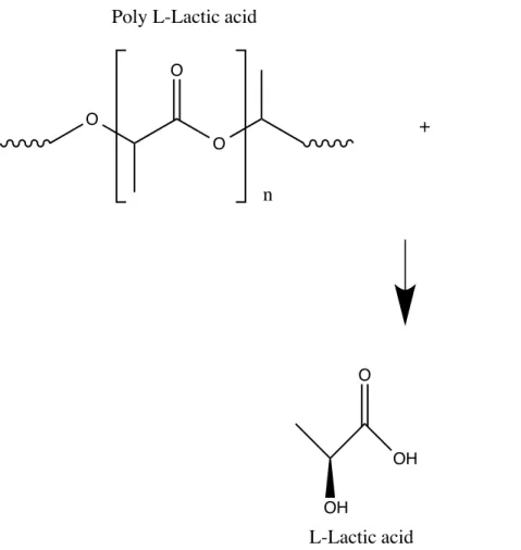
Among polyesters, poly L-lactic acid (PLLA) seems to be an ideal FDA-approved biomaterial due to its non-toxicity and resemblance to the extra-cellular matrix in the brain, thus its potential in neural cell regeneration. [15, 16] An image of glial cells and extra-cellular matrix is shown in Appendix C (Figure C5). PLLA is biodegradable; PLLA is naturally degraded to L-lactic acid via hydrolysis of the ester linkages (Figure 1-3). L-L-lactic acid is eventually metabolized in the body to carbon dioxide and water. [17]
Figure 1-3: Hydrolysis of Poly L-Lactic acid to L-Lactic acid O
O
O
n Poly L-Lactic acid
+ H2O
OH O
OH
Furthermore, growth factor signaling has been shown to have a significant role in tissue repair. In the last few decades, our understanding on the role of growth factors on the CNS has also greatly increased. [18-20] Furthermore, the inclusion of growth factors within scaffolds made of biodegradable and biocompatible polymers is desirable in order to retain them at the site of transplantation and control their spatiotemporal delivery to the damaged region of the CNS. [21-23] Epidermal growth factor (EGF), a soluble 6kDa polypeptide that is naturally present in the CNS, is known to promote the oligomerization and phosphorylation of cell surface EGF receptors (EGFRs) leading to cell proliferation and was used in this study. [24-28] Of interest, when tethered onto a scaffold, EGF has been shown to promote rapid expansion of neural stem cells (NSCs). [24, 29, 30] Amine functionalization on polyethylene terephthalate (PET) with ethylenediamine (EtDA) and polyvinylamine (PVam) has been previously reported [31] PLLA and PET are two different polymers with different chemical structures as shown in Figure 1-4. In this sudy, PLLA nanofibers were found to be very fragile and the necessary adjustments were performed and adapted from the above-mentioned protocol.
PET
PLLA
Figure 1-4: Chemical structures of Polyethylene terephthalate (PET) and Poly L-Lactic acid (PLLA) O O O O O n O O O n
The main objective of this work is to fabricate a PLLA scaffold by electrospinning, to establish the optimum topological features of nanofibrous mats for neural stem cells and to subsequently functionalize it for improved neural stem cell transplantation. More specifically, we will investigate EGF grafting onto the PLLA scaffold after surface modification via aminolysis. This strategy will combine the advantages of both the PLLA polymer as a cell-scaffold (mechanical strength, high porosity, biodegradability and biocompatibility, etc) as well as the benefits of tethered EGF. The impact of EGF-grafted PLLA nanofiber scaffolds upon neural stem-like cells (NSLC) adhesion, viability and proliferation will be tested. Neural stem-like cells (NSLC) were provided by NewWorld Laboratories Inc. They were created from somatic cells through reprogramming by NWL. Neural Stem-Like Cells (NSLCs) were engineered from human fibroblast cells by transient reprogramming using defined factors. These reprogrammed cells are epigenetically stable. They have global gene- and protein-expression profiles that are similar to Neural Stem Cells.
A literature review is presented in the first part of this thesis showing the current strategies documented for neural stem cell regeneration.
Based on the literature review, the second part of the thesis presents, in our opinion, the most promising strategy for fabricating and modifying a substrate to promote cell proliferation for neural tissue regeneration. An extensive cell culture study was performed on fibers grafted with EGF under different media conditions.
CHAPITRE 1
GENERAL PROCEDURE
A literature review summarizing all pertinent work performed in neuronal tissue engineering is presented. The literature review consists of the different methods currently reported for tissue regeneration. Such strategies include direct injection of stem cells and/or growth factors, encapsulated growth factors, hydrogels as scaffolds, etc as well as the surface modification of polymeric scaffolds using different bio-macromolecules. The advantages and disadvantages of the strategies presented in the literature review are analyzed in an attempt to develop an optimized strategy.
In a second section, an article entitled ‘Fabrication and Surface modification of poly L-lactic acid (PLLA) scaffolds for growth factor immobilization and neuronal stem cell delivery’ is presented. This article was submitted to the Journal of Biomedical Materials Research - Part B, a highly interdisciplinary peer-reviewed journal whose common focus is on biomaterials applied to the human body and covers all disciplines where medical devices are used.
The second part of this thesis presents the step- by- step process of the fabrication of PLLA electrospun scaffolds, followed with an optimized amine functionalization, and EGF grafting. An extensive cell culture study was performed with EGF-grafted fibers under different media conditions. Negative controls involving aminated nanofibers and pristine nanofibers were performed for comparison, as well as positive control (nanofibers that were only coated with laminin). These cell culture assays were used to test for cell adhesion, viability and proliferation using our new strategy.
Finally, future work directions and improvements are proposed such as a grafted-EGF density-dependent study, EGF tethering to the PLLA scaffold in an oriented fashion for improved bioactivity, and the study of progenitor cell differentiation to specific and appropriate lineages.
CHAPITRE 2
LITERATURE REVIEW
2.1 Direct injection of stem cells for in situ tissue regeneration
Contrary to what was once believed, studies in the last two decades have shown that the CNS does indeed contain neural stem cells and has potential for extensive cellular replacement. The discovery of neural stem cells has therefore opened up the development of therapeutic strategies for CNS using stem cells. [32] Even though neural stem cells are present in the CNS, the quantity of active neural stem cells seems to be very low. Neural stem cells range in size from 4-50 µm. [33-38] Schematic drawing [39]of NSC and its environment is shown below.
Figure 2-1: Neural stem cell niche with NSC (in blue) [39]
The injection of additional stem cells at the site of injury for cell regeneration has thus been studied. In an animal model where aspects of multiple sclerosis were reflected, adult neural stem cell cultured neurospheres were injected in the CNS. The injected cells migrated to the damaged areas of the CNS and the animals displayed significant functional recovery. [32, 40]
However, the animal experiment may not be translated to humans since multiple sclerosis in the human CNS is a chronic and inflammatory disorder that involves both glial and axonal changes and its treatment is different from the animal experiment. [32] Another obstacle for the implementation of treatments based on direct cell injection is the lack of appropriate factors in the host tissue to accommodate the injected stem cells. Altered pathological tissue due to Alzheimer’s disease is no longer able to present factors that promote proper differentiation hence making it difficult for the injected stem cells to treat the disease. [41, 42] Furthermore, Hansmann et al [43] have implanted immortalized oligodendrocyte precursor cells (OPCs) where it has been reported that cell injection may result in tumor formation. That is, it has been suggested that a failure of molecular control mechanisms in murine glial precursor cell line BO-1 cells, responsible for a shift from differentiation to proliferation, led to tumor formation. [43] The signal transducer and activator of transcription (STAT)-3 (member of the Janus Kinase (Jak)/STAT-pathway downstream of the epidermal growth factor receptor (EGFR)) had been used as a tumor suppressor in the PNS but its function in glial tumors is still unknown. [43, 44] The presence of CNS stem cells was first discovered when growth factor responsive cells from both embryonic and adult CNS stem cells were isolated. [19, 20] Furthermore, studies on stem cells have shown that growth factors play a significant role in controlling the fundamental process of development. [32] It has been previously reported that direct implantation of human mesenchymal stem cells (MSCs) stimulated the synthesis of neuronal survival factors; increased endogenous expression of nerve growth factor (NGF), vascular endothelial growth factor (VEGF), ciliary neurotrophic factor (CNTF), and basic fibroblast growth factor-2 (FGF-2) as well as cell proliferation. [45] These studies showed that the benefits coming from direct stem cell injections may be due to their ability to directly produce growth factors and thus provide the appropriate cues for proper development/repair. This suggests that the direct injection of growth factors may be sufficient instead of stem cell injection. [46] This could explain why, in contrast to the CNS that lack Schwann cells, injured peripheral neurons are able to regenerate their axons because of their Schwann cells that provide neurotrophic factors. [46]
2.2 Direct injection of growth factors for in situ tissue regeneration
Growth factors are able to maximize the intrinsic regenerative potency of endogenous progenitor cells as well as aid exogenous stem cell proliferation and differentiation. [21, 47] It is now known that peripheral nerve bridges contain large quantities of NGF, BDNF, neurotrophin-3 (NT-3), ciliary neurotrophic factor (CNTF) and glial cell-line derived neurotrophic factor (GDNF) secreted by Schwann cells, which is the cause of the stimulation of axon growth regeneration. [48] Tuszynski et al [49, 50] have genetically modified suspensions of autologous fibroblasts to secrete NGF and these modified cells were injected into the central gray matter of the non-lesioned thoracic spinal cord. The results demonstrated that axons grew in significantly larger numbers when presented with an environment containing growth factors. [49, 50] Kobayashi et al [51] have shown that brain-derived neurotrophic factor (BDNF) and neurotrophin-4/5 (NT-4/5) prevented atrophy of rat rubrospinal neurons and promoted axonal regeneration. Furthermore, Boyd and Gordon [52] have shown that long-term continuous treatment with exogenous GDNF significantly increased the number of motoneurons that regenerate their axons. However, no significant benefits were observed from the combined GDNF and BDNF treatment on the axonal regeneration of motoneurons until after seven days of continuous treatment. Vejsada et al [53] have also shown that continuous neurotrophic factor delivery was essential in order to promote the long-term survival of axotomized neonatal motoneurons. Furthermore, the short half-lives of growth factors, their relatively large size, slow tissue penetration and potential toxicity at systematic level exposures limits their use by direct injections. [54] A summary of the various growth factors mentioned is presented in Appendix B.2.3 Encapsulated Growth factors
One of the most common approaches used to get controlled drug release is to embed the drug into a hydrophilic or hydrophobic matrix; a matrix defined as a three-dimensional network containing the drug, polymer, solvents, etc. [55] The drug is then released and the chemical kinetics are affected by many factors such as polymer swelling, polymer erosion, drug dissolution (re-crystallization), drug diffusion characteristics, particle size distribution, drug-polymer interaction, drug distribution inside the matrix, drug/polymer ratio and geometry. [55, 56] Biocompatible and biodegradable biomaterials are excellent for the gradual release of a drug or
for tissue scaffolds. When designing the biomaterials to be used, many factors are taken into consideration such as the degradation rate and the appropriate materials, conditions, dimensions, and geometries are chosen for specific applications. [57] Wang et al reported the encapsulation of vascular endothelial growth factor (VEGF) and BDNF in PLGA microspheres using the
water-in-oil-in-water emulsion technique. The PLGA microspheres were then embedded within a
cross-linked hyaluronic acid (HA) hydrogel as a delivery system. [58] Although hydrogels are widely used for encapsulating growth factors, they release their growth factors in an initial outburst that is higher than physiological levels, therefore affecting the duration of the delivery. [46, 59] Lam et al [60] have encapsulated and stabilized NGF into Poly (lactic-co-glycolic) (PLGA) acid microspheres by spray-freeze-drying method. First, the human NGF formulations were prepared in two different buffer solutions and lyophilized: one buffer system consisting of histidine and the other consisting of sodium bicarbonate and zinc acetate. For the spray-freeze drying technique, the human NGF formulations were pumped into an ultrasonic spray nozzle with a peristaltic pump. The protein was then sprayed into a flask containing liquid nitrogen and merged into a liquid nitrogen bath. The frozen protein droplets were poured into a stainless steel tray and dried by lyophilization (primary temperature at -25°C for 30 h, followed with secondary drying at 20°C for 10 h). The NGF released from the PLGA microspheres degraded at physiological temperature. In order to prevent NGF degradation and aggregation during microencapsulation and release, PEG or surfactant (pluronic F68) was added to the formulation, however, the integrity of the NGF released was not improved.
2.4 In situ production and delivery of growth factors using cells
The grafting of primary fibroblasts that are genetically modified ex vivo to produce BDNF or NT-3 has been previously reported.[61, 62] However, immune suppression was needed in order to prevent the rejection of the grafts. In order to overcome this need for immune suppression, previous reports have developed a method based on alginate encapsulation for grafting non-autologous BDNF producing fibroblasts into the injured spinal chord.[63] Tobias et al [64] have also shown that alginate encapsulation can protect non-autologous BDNF producing fibroblasts (Fb/BDNF) from rejection after transplantation into a site of injury and promoted behavioral recovery. They have further shown that the grafting of encapsulated Fb/BDNF containing cells into a subtotal cervical hemi-section resulted in partial recovery of forelimb
usage as well as axonal growth.[64] These cell-containing scaffolds have shown their capability to survive transplantation into the injured spinal cord for at least one month in the absence of immune suppression.[63] Furthermore, alginate encapsulation was able to protect the Fb/BDNF for two months after grafting.[64, 65]
Layers of cells may be used where the bottom layers are first cultured and can provide the requirements needed to the cells of interest (cultured on top) resulting in the regeneration of neuronal structures ex vivo. [46] Zeng et al [66] have cocultured cells from the bone-marrow derived stromal cell line PA6 to induce differentiation of human embryonic stem cells (hESCs). The production of dopaminergic neurons at a high frequency from hESCs was observed as well as the survival of transplanted, tyrosine hydroxylase (TH)-positive cells after 3 weeks of differentiation. A bottom layer of PA6 cells was also used as a feeder layer to initiate the neural differentiation of BG01 cells. An outgrowth of elongated cells was observed after 6 days in culture. However, the survival of dopaminergic neurons after transplantation was limited suggesting this method is not ideal. It has also been shown that the co-culturing with PA6 cells is simple and fast and can be used with cells from subhuman primates.[67] Other studies reported the administration of NGF to the brain using ex vivo gene delivery. [68-72] Genetically modified autologous fibroblasts were used to produce and secrete NGF. This method of delivery sustained NGF production for at least 18 months, prevented cholinergic degeneration, stimulated cholinergic function, and improved memory.[68-72] Tuszynski et al have also applied this procedure to humans where genetically modified NGF-producing fibroblasts were injected into the brains of 8 subjects suffering from early-stage probable Alzheimer disease.[73] Evidence showed that growth factor delivery in this approach has the potential to modify neurological disease progression.
2.5 Scaffolds for stem cell therapy
In the case of neurodegenerative diseases, the injected stem cells cannot properly differentiate because the pathological tissue does not contain the necessary factors.[46] NSC transplantation may not be effective because the amyloid precursor protein (APP) metabolism is altered and might lead to excessive gliogenesis. Stem cell therapy is challenging due to the blood-brain barrier that makes it difficult for the neurotrophic factors to diffuse. Also, other causes that may explain why the NSCs are not able to regenerate properly in the damaged brain are hostility
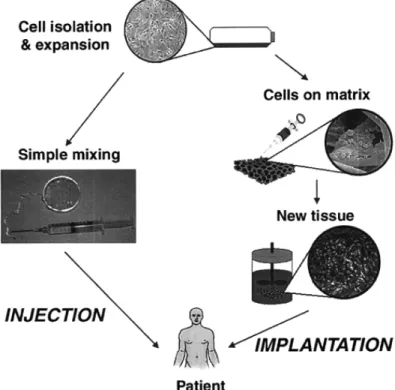
of the environment; inflammation, glial scar formation, release of inhibitory molecules, and the absence of growth-guiding astrocytes. [74] As a result, and because of the fragility of the cells, a biomaterial to be used as a scaffold to transplant the stem cells into the host target area appears to be highly desirable. Figure 2-2 shows a schematic of a classic tissue engineering process in which cells can be isolated from the patient (from a biopsy) and expanded.[75] Two distinct strategies are described where one corresponds to the direct injection of cells whereas the other is the use of a polymer scaffold. Hydrogels can also be directly injected, in combination with cells, into the host environment.[75]
Figure 2-2: Schematic illustration of typical tissue engineering approaches. Cells are obtained from a small biopsy from a patient, expanded in vitro, and transplanted into the patient either by injection using a needle or other minimally invasive delivery approach, or by implantation at the
2.5.1 Hydrogel as a scaffold
Hydrogels have shown promising characteristics as they are relatively easy to create, have a 3D structure, and the best material can be chosen based on the desired mechanical structures as shown in Figure 2-3. [76]
Figure 2-3: Hydrogel fabrication and application. [76]
Numerous studies have been performed on the use of hydrogels as a cell scaffold. Hyaluronic acid (HA) hydrogels modified with laminin, harboring similar mechanical and rheological properties as brain tissue were shown to inhibit glial formation and support angiogenesis. [77, 78] Wei et al have [79] demonstrated that when hyaluronic acid (HA)-based hydrogels modified with poly-L-lysine (PLL) and a nogo-66 receptor- antibody (antiNgR) fusion protein (the whole structure being referred to HA-PLL/antiNgR) was administered to rats,
inhibition of glial scar formation was observed. HA, however, is highly hydrophilic; in order to undergo cell proliferation, cells need a substrate that is neither too hydrophilic nor too hydrophobic. [80] Woerly et al [81] have evaluated the application of Poly[N-(2-hydroxypropyl) methacrylamide] (PHPMA) hydrogels for the promotion of axonal regeneration in the transected rat spinal cord. Following implantation, the hydrogel was able to bridge tissue defects, favored cell ingrowth, angiogenesis and promoted axonal growth. Their similarity in viscoelastic properties to neural tissue (as well as large surface area) contributes to their success as a strategy for neural tissue regeneration. Type I collagen has been used as an injectable hydrogel while inhibiting glial scar formation after spinal cord injury. [82] However, telopeptide regions are present during gel formation for crosslinks between molecules. These telopeptide regions are implicated in antigenicity and render the gels weaker when removed. [83]
Furthermore, some limitations of hydrogels as potential scaffold for neural tissue engineering include their high cost and low mechanical strength. [75, 84]
2.5.2 Electrospun Fibers
A variety of methods exist for the fabrication of PLLA nanofibers; they can be fabricated by liquid-liquid phase separation, [85] or thermally induced phase separation method [86], as well as melt spinning method (melt extrusion and hot draw). [87] Recently, electrospun scaffolds have been assayed as a substrate to support cell regeneration. [10, 16, 88, 89] Electrospun fibers have been fabricated for the generation of scaffolds destined to neural tissue engineering because of their interconnected pores, high porosity, high surface area-to-volume ratio, and topography that mimic the natural extracellular matrix (ECM). Electrospun fibers have been demonstrated to support the attachment and differentiation of various neuronal cells, such as dorsal root ganglion neurons (DRG), Schwann cells, hippocampal neurons, as well as PC12 cells. [90]
2.6 Electrospinning
Electrospinning is a fabrication technique that produces polymeric fibers using electrostatic force. It is possible to obtain fibers from a wide variety of materials such as poly (e-caprolactone) (PCL), poly lactic-co-glycolic acid (PLGA), poly lactic acid (PLA), etc. Fiber properties such as diameter, porosity, and alignment can also be tuned during the electrospinning process. This technique consists of placing a polymer solution in a syringe where a strong
difference of potential is applied between a metallic collector and the metallic needle. A droplet charged with static electricity in the shape of a Taylor cone is then created. This droplet is attracted by the electric field in the direction of the collector where charge repulsion exceeds the surface tension, [91] and evaporates during its flight, creating fibers with a diameter varying from tens of nanometers to a few micrometers. [92] Electrospun fibers are attractive for scaffold production due to the relative simplicity of its fabrication, (summarized in Figure 2-4), its tunability and its cost-effectiveness.
Figure 2-4: Electrospinning set-up. [93]
2.6.1 Polymer solution conditions
A suitable solvent that is able to dissolve the polymer while possessing suitable vapor pressure is usually chosen. A good solvent is one that evaporates quickly enough for the fiber to maintain its integrity when it reaches its target but not too quickly in order to allow the fiber to harden before reaching the collector. Furthermore, the quality of the solvent [94], as well as the temperature of the solution has an effect on fiber diameter. [95] There are also many other conditions that can influence the nanofiber topography and morphology (e.g., beaded, random, or aligned, etc) such as polymer concentration, molecular weight, solution viscosity, surface tension,
solution conductivity, dielectric effect of solvent, voltage, feed rate, collector speed, distance between tip and collector, ambient temperature and humidity.
2.6.1.1 Polymer concentration, molecular weight, and solution viscosity
Polymer concentration has an effect on fiber diameter as well as fiber morphology. Generally, higher polymer concentrations yield nanofibers of larger average diameters. [96] Another factor affecting the viscosity of the solution is the molecular weight (Mw) of the polymer. The molecular weight represents the length of the polymer chain and determines the amount of polymer entanglements in a solution. Low Mw polymer solutions are not able to form fibers when electrospun. [93] Furthermore, Mw directly affects the viscosity, which is understandable since the amount of polymer chain entanglements in the solvent are determined by polymer length. Usually, a low viscosity solution tends to result in bead formation in the fiber structure. [93, 97] Also, when the viscosity of the solution is low, there may be a secondary jet that erupts from the main jet and cause a variety of different fiber diameters. [93, 98] When too viscous, solutions can also dry at the tip of the needle, preventing electrospinning. [10, 99] Additionally, at very low humidity, the solvent evaporation rate is higher causing clogging to occur at the needle tip. [93]
2.6.1.2 Surface tension
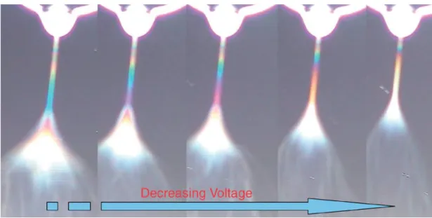
The surface tension is defined by the tractional force, γ, acting across any unit length of line on the interfacial membrane (i.e. liquid and vapour). [100] To begin the electrospinning process, the charged solution must first overcome its surface tension, allowing the stretching of the polymer solution to reach the collector without the formation of beads. [93] The Coulomb repulsion between the charged ions in a polymer solution favors the creation of a jet, whereas the surface tension of the solution favours a sphere-like shape. When the electrical potential of the surface is increased, the electrical forces dominate the surface tension of the solution and a charged jet of fluid (referred to as the Taylor cone) is released as shown in Figure 2-5 where the Taylor cone is shown at the top of the illustrations. [101]
Figure 2-5: Taylor cone shown at top of images. Lower voltages result in thinner fluid jets as well as less fluid being drawn from syringe where the interference colors provide information on jet diameter. [102]
2.6.1.3 Solution conductivity
The conductivity of a solution is a measure of its ability to conduct electricity. The presence of ions has an effect on the conductivity; many of the solvents used in electrospinning possess a certain level of conductivity. The stretching of the polymer solution from the syringe is caused by the repulsion of the charges at the polymer surface. An increase in the stretching of a solution results from the increase of charges, leading to bead-less fibers. An increase in polymer solution stretching also results in fibers with smaller diameters. [93, 99]
2.6.1.4 Dielectric effect of solvent
The dielectric constant (a quantity measuring the ability of a substance to store electrical energy in an electric field) of a solution plays a major role in electrospinning. A solvent with a high dielectric constant will result in a fiber with smaller diameters and less beads. [93]
2.6.2 Electrospinning process conditions
External factors such as voltage, feed rate, temperature of solution, ambient temperature, humidity, type of collector, diameter of needle, and distance between tip and collector also play a role in the outcome of fiber morphology.
2.6.2.1 Voltage
Taylor cones define the onset of extensional velocity gradients in the process of forming fibers. [103] The polymer jet exits the syringe needle and forms a Taylor cone when a high voltage is applied and the electrostatic force overcomes the surface tension of the solution; the coulombic repulsive force in the jet then stretches the solution. Generally, a high voltage leads to smaller fiber diameters; this is due to the fact that a higher voltage will lead to an increase in stretching of the solution. Lower voltages result in thinner fluid jets and less fluid is drawn from the syringe as shown in Figure 2-5 where the interference colors provide information on jet diameter. [101]
A higher voltage can cause instability of the jet and Taylor cone, causing them to recede into the syringe needle, resulting in beaded fibers. [93]
2.6.2.2 Feed rate
The feed rate cannot be too high in order to give enough time for the solvent to evaporate during flight. When the feed rate is increased, the fiber diameter or bead size is also increased. [93]
2.6.2.3 Effect of collector
The collector plate is usually made out of a conductive material such as aluminum foil and/or a metal plate, which is electrically grounded. Once the voltage is turned on, there is an electric field between the source and the collector, hence attracting the polymer solution towards the collector. [93]
2.6.2.4 Distance between tip and collector
The distance between tip and collector has an effect on resulting fibers. For example, beads can be observed when the distance is too low. [104] This is due to the fact that decreased
distance causes an increase in field strength. A distance that is too high may either decrease or increase the fiber diameter [93] therefore an optimal distance is necessary in order to achieve the desired fiber morphology.
2.6.2.5 Ambient humidity and temperature
Water condensation can form on fibers when the humidity is too high and this can affect the fiber morphology. [93, 104] The humidity can affect the diameter and porosity of the fibers. [105, 106] The humidity and temperature have a significant effect on average fiber diameter. At high ambient humidity (higher than the optimal range of 20-40 % relative humidity), the fiber diameter of a polymer can decrease and form beads as represented in Appendix C (Figure C3), which can pose a potential challenge. [107] Furthermore, the solution absorbs ambient water at high humidity leading to the fusion of fibers at the collector [107] as demonstrated in Appendix C (Figure C4).
2.7 Tissue engineering
Tissue engineering is the application of the principles of engineering as well as life sciences to make a biological substitute, which often contains cells and biodegradable scaffolds co-cultured in vitro, that can restore, maintain or improve tissue function. In tissue engineering procedures, artificial extracellular matrices (ECM), i.e. biomaterials, are often used as scaffolds for cells to grow in. Cells interact with the biomaterials where their initial interaction is determined by the chemical composition, surface energy, roughness, and surface topography that are in direct contact with cells.[108] Cell adhesion, migration, differentiation and proliferation depend on biological cues such as growth factors and cytokines; they also depend on chemical and physical characteristics of the biomaterial surface.[108] Polyesters such as poly lactic acid (PLA) are FDA-approved; they are biodegradable, biocompatible, and non-toxic. Polyester surfaces have poor hydrophilicity, however, and lack functional groups that may ease chemical tailoring of their surface. Hence, the introduction of such functional groups is usually needed. Many different surface modification methods have been reported such as plasma treatment[109, 110], surface hydrolysis[111], chemical grafting[112], physical adsorption[113] and self-assembly technology.[114]
2.7.1 Biological response to biomaterials
In order to develop an efficient biomaterial, the understanding of cell interactions with the extracellular matrix (ECM) is important. In a normal biological situation, cell-cell interactions are mediated and modulated by signaling complexes that occur through adhesion receptors on cell surfaces when cells interact with the ECM. Cells adhere to their ECM through their cytoskeleton actin filaments. Cell membrane receptors, called integrins, are what mediate this adhesion. [108] The ECM also acts as a reservoir for growth factors whose diffusion (and thus availability) is controlled by the ECM physical properties. The ECM protects the growth factors from degradation and regulates their synthesis. [115]
Once the biomaterials are implanted in the body, the first step that occurs is water adsorption, followed by protein adsorption prior to cell attachment. The surface properties of biomaterials are important in that they determine the binding strength and structural arrangement of water molecules, which may affect later protein-surface, and thus cell-surface interactions. The porosity of a biomaterial also has an effect on cell behavior. Gugala et al reported the superiority of porous membranes to non-porous ones from poly (L/D-lactide) 80/20% for the growth and osteoblastic differentiation of rat bone marrow stromal cells in vitro. [108]
2.8 Poly lactic acid (PLA) and poly L-lactic acid (PLLA)
2.8.1 General
Poly(α-hydroxy acids) such as poly(glycolic acid) (PGA), poly(lactic acid) (PLA) and their copolymers have been widely used as biomaterials. [116, 117] PLA is a thermoplastic, high strength, high-modulus aliphatic polyester made from renewable resources, more specifically α-hydroxy acids, which include polyglycolic acid or polymandelic acid. PLA is formed by direct condensation polymerization of lactic acid. Similarly, poly L-lactic acid is formed by polycondensation of the L enantiomer. The stereochemical structure of poly lactic acid can be modified by polymerizing a controlled mixture of the L- or D-isomers in order to yield either amorphous or crystalline, high molecular weight polymers. [118] Lactic acid has been first isolated in 1780 from sour milk and has many food-related applications. It is used as a buffering agent, acidic flavoring agent, acidulant and bacterial inhibitor in many processed foods and can
be manufactured by carbohydrate fermentation or chemical synthesis. [118] As mentioned earlier, lactic acid (2-hydroxy propionic acid) exists in two optically active configurations; the L(+) isomer is produced in humans and other mammals, whereas the D(-)- and L(+)-enantiomers are produced in bacterial systems. There are two different routes for PLA polymerization. The first route is through polycondensation from lactic acid to yield low molecular weight PLA. The second route is from lactide through ring-opening polymerization process to yield poly lactic acid as shown in Figure 2-6.
2.9 Surface modification
The chemical structure, hydrophobicity, ionic groups, morphology, and topography of the biomaterial have an effect on the interaction with surrounding biological environment. Furthermore, the surface properties such as wettability, surface charge and topography affect cell adhesion. [108] Additional factors that influence the cell interactions with the scaffold include the structure of the biomaterial, porosity, pore size, geometry, distribution, surface texture such as roughness, pattern, orientation, and surface chemical properties such as free energy and surface micro-morphology. The porosity of a scaffold plays an important role in directing tissue formation and function. [119, 120] A high porosity (higher than 80%) [85] is necessary in a scaffold for homogeneous cell distribution and interconnection as well as diffusion of nutrients and oxygen. [121] The stiffness of a material also decreases as the porosity is increased. [121]
Pore size also has an impact on the amount of contraction a scaffold will undergo after implantation. [121] For example, a pore diameter ranging from 20–125 µm was needed for contraction-inhibiting activity to be observed in collagen–glycosaminoglycan graft copolymers used for dermal repair. [122] Choice of pore size is also related to the cell type to be cultured. [121] Furthermore, the maximum fiber thickness recommended for tissue engineering scaffolds is 150-200 µm due to the inability of and insufficient oxygen and nutrient transport within the deeper compartments of the biomaterial. [121, 123]
Surface modifications of biomaterials in an alkaline solution could be used for the production of hydrophilic and rough surfaces. [108] Cell adhesion to ECM proteins is mediated by integrins where they organize into complexes that contain structural and signaling proteins. There has been a large development to the modification of polyester surfaces. Several methods of surface modification are possible such as chemical grafting, surface coating, entrapment, and electrostatic self-assembly.
2.9.1 Aminolysis
Aminolysis can be used prior to the grafting of bioactive molecules where primary amines are introduced by thermally induced aminolysis. The aminolysis reaction is based on the nucleophilic attack of an amine onto the ester bonds of a polymer chain via polymer-chain scission and has been widely used in literature for the addition of amine groups. Fadeev et al have added amine groups on PET surfaces using polyamines. [124] Furthermore, Yang et al [125] reported UV-light-induced surface aminolysis reactions. The tertiary amines are functionalized onto the PET surface, are easily protonated and used for electrostatic immobilization of proteins. Studies have also reported the addition of amine groups using a small diamine, ethylenediamine (EtDA)[31, 126] as well as an aminated polymer, polyvinylamine (PVam). [31, 127] The aminated PET films were followed with the functionalization of Succinimidyl 6-[3-(2-pyridyldithio)-propionamido]hexanoate, SPDP linker, an amino-reactive molecule. The LC-SPDP functionalization of substrates could be followed with the oriented immobilization of cysteine-tagged proteins. [128, 129]
2.9.2 Surface coating/physical adsorption of proteins
Most materials need to be coated in order to improve cell adhesion, even electrospun fibers. [37, 130] This is due to the poor hydrophilicity of polyester surfaces. [108] Biomaterials as scaffolds are limited by the ability of attached cells to dynamically repopulate. [131, 132] Surface coating with, for example, collagen and laminin provide an adhesive interface between biomaterial surfaces and cells. Garic et al modified PLA surfaces by creating tiny cavities by sodium hydroxide, as well as coating with type-I collagen for comparison. [133] Results showed that human keratinocytes preferred growing on partially hydrolyzed surfaces. This study has suggested that other surface modifications can further improve keratinocyte culture on PLA. However, it has been reported that surface coating of proteins via adsorption may affect the topography of the scaffold and thus cell fate. [16] For this reason, other methods of incorporating proteins and growth factors have been explored.
2.9.3 Chemical grafting of proteins
Soluble growth factors have been used to help regeneration after spinal chord injury using mini pump technology. [134] This method has shown the importance of a stable gradient for guidance; it has also shown a limitation where the soluble growth factors need to be constantly replenished. Protein grafting was therefore attempted in vivo. Since surface immobilized growth factors are biomimetic [135-139], it has been hypothesized that they can guide neurites similarly to soluble growth factors. [134] Kapur et al [134] have photochemically bound nerve growth factors (NGF) via photoreactive PAAm onto microporous poly(2-hydroxyethylmethacrylate) (PHEMA) gels. Results showed that 30 ± 7% of the pheochromocytoma (PC-12) cell population responded to bound NGF, similarly in comparison to cells cultured on collagen in the presence of 40 ng/ml soluble NGF (39 ± 12%). Zhu et al showed a method for the photochemical immobilization of gelatin and chitosan onto polyesters. [140] Results showed that fibroblast cells did not spread on the chitosan-PLGA or pure PLGA surface after 2h but spread on the gelatin-chitosan-PLGA surface.
2.9.4 Proteins for surface modification
2.9.4.1 CollagenCollagen adsorption
Some natural materials such as collagen, fibronectin, and some peptides have been reported as scaffold modifiers. [134] Tjia have shown the benefits of adsorbed collagen onto a scaffold. [141] They have reported that, in the absence of any treatment, keratinocyte migration on PLGA substrates was poor. However, substrate conditioning with type I collagen can enhance the rate of migration significantly. Furthermore, the combined presence of substrate-adsorbed collagen in conjunction with a cell-derived protein (fibronectin) can activate keratinocyte migration onto PLGA substrates. [141] Additionally, He et al [142] have coated collagen on electrospun nanofibers and reported an enhanced spreading, attachment, and viability for human coronary artery endothelial cells.
Collagen grafting
Li et al have used electrospun nanofibers on which collagen had been immobilized for neural stem cell culture. [143] Collagen was grafted onto a copolymer of methyl methacrylate (MMA) and acrylic acid (AA) nanofiber surface via EDC/NHS activation. It was reported that the cell viability and cell proliferation were improved on the collagen-modified surfaces when compared to that on the unmodified surfaces. Ito et al [144] have immobilized collagen onto poly(methyl methacrylate) (PMMA) films, which has resulted in the flattening of cells during early cell adhesion but did not enhance cell growth significantly. [145]
2.9.4.2 Laminin Laminin adsorption
Laminin plays an important role in cell migration, differentiation, and axonal growth. [146] It is one of the proteins present in the ECM and is continuously synthesized after nerve injury in the PNS. [147] Also, myelination in the PNS is not observed in the absence of laminin. [148, 149] Laminin adsorption onto scaffolds has been shown to promote neurite outgrowth through directional guidance. [150, 151]
Laminin grafting
Koh et al [152] have compared the addition of laminin to nanofibers using covalent binding, physical adsorption, and blended electrospinning. In general, they have shown that laminin modified nanofibers supported cell adhesion, cell viability and proliferation as well as neurite outgrowth when nerve growth factors were introduced. More laminin were found in the blended laminin-PLLA nanofibers than immobilized laminin and physically adsorbed laminin. This could be the reason why more neurite outgrowth of PC12 cells was observed on blended laminin-PLLA than on the other modified nanofibers. However, this technique was not efficient at achieving high cell viability. Laminin has a higher tendency to promote neurite outgrowth than to enhance the viability of nerve cells. [152, 153] Another drawback is that the approach undertaken in this study still required the use of nerve growth factors (NGF) in the medium. Some studies have shown that NGF plays a role in the maintenance of central neurons and in the control of laminin production. [153, 154]
2.9.4.3 EGF EGF adsorption
EGF is known to be a functional target for the clinical treatment of damaged tissues. [155] As previously discussed, EGF plays an important role in cell proliferation, differentiation and migration, thus aiding in wound healing and tissue regeneration. Non-specific adsorption of the epidermal growth factor (EGF) onto a substrate has been shown to dramatically decrease its bioactivity. [156] This is most likely due to the inappropriate orientation of EGF or possible adsorption-induced conformational changes of EGF. [156] Some reports have also shown that physically adsorbed EGF on insoluble glass substrates showed no activity. [157] This could mean that receptor motility is important in the signaling for both the mitogenic and morphologic response or that the conformation of physically adsorbed EGF is unsuitable for stimulating the EGFR. [157] These results showed that the mere presence of EGF on the surface is insufficient to trigger a biological response. [157]
2.9.4.4 EGF grafting
When soluble EGF is used, it is difficult to control its local concentrations due to cellular uptake and diffusion. [158] For the cells to properly respond to growth factors, the time-span of their presence in the desired target tissues is important. [159] When a controlled release system is used, maintaining a signal at the cell surface is difficult due to endocytosis of growth factor-receptor complexes. [158] Therefore, new strategies are needed to overcome these obstacles. This is where the immobilization of growth factors comes into play; covalent immobilization of EGF onto a scaffold is non-endocytosible and non-diffusible, allowing cell surface events involving signal transduction to occur. Furthermore, immobilization can be used in order to spatially control EGF densities or concentrations.[158] To further support the use of tethered EGF, it has been reported that adsorbed EGF shows very low, if not non-existent biological activity. [157, 160] Furthermore, surface coating of proteins via adsorption may affect the topography of the scaffold and thus cell fate. [16] Grafting of proteins enable long-term delivery, allows proteins to remain stable and functional for several months, and have shown positive results in activating intracellular signaling pathways thus influencing cell fate. [161] Therefore, the purpose of this study is to optimize current neural tissue regeneration strategies by immobilizing EGF onto a PLLA scaffold.

![Figure 1-2: Schematic representation comparing injury at the CNS vs. PNS. [9]](https://thumb-eu.123doks.com/thumbv2/123doknet/2332341.32008/21.918.180.758.133.602/figure-schematic-representation-comparing-injury-cns-vs-pns.webp)

![Figure 2-1: Neural stem cell niche with NSC (in blue) [39]](https://thumb-eu.123doks.com/thumbv2/123doknet/2332341.32008/26.918.279.648.418.809/figure-neural-stem-cell-niche-nsc-blue.webp)
![Figure 2-3: Hydrogel fabrication and application. [76]](https://thumb-eu.123doks.com/thumbv2/123doknet/2332341.32008/32.918.129.777.293.802/figure-hydrogel-fabrication-and-application.webp)
![Figure 2-4: Electrospinning set-up. [93]](https://thumb-eu.123doks.com/thumbv2/123doknet/2332341.32008/34.918.151.746.346.714/figure-electrospinning-set-up.webp)
![Figure 2-6: Synthesis of PLA [118]](https://thumb-eu.123doks.com/thumbv2/123doknet/2332341.32008/40.918.248.675.330.831/figure-synthesis-of-pla.webp)
