Université de Montréal
Regulation of excitotoxicity in thiamine deficiency: role of
glutamate transporters.
par
Shivraj Singh Jhala
Programme de Sciences biomédicales, PhD Faculté de Médecine
Thèse présentée à la Faculté des études supérieures et postdoctorales en vue de l’obtention du grade de Philosophie Doctoral (Ph.D.)
en sciences biomédicales
Août 2012
Université de Montréal
Faculté des études supérieures et postdoctorales
Cette thèse intitulée:
Regulation of excitotoxicity in thiamine deficiency: role of
glutamate transporters.
présentée par : Shivraj Singh Jhala
a été évaluée par un jury composé des personnes suivantes :
Dr. Pierre Blanchet, président-rapporteur Dr. Alan Stewart Hazell, directeur de recherche
Dr. Guylaine Ferland, membre du jury Dr. Raghu Vemuganti, examinateur externe Dr. Eugenio Rasio, représentant du doyen de la FESP
RÉSUMÉ
L’excitotoxicité est un mécanisme physiopathologique majeur impliqué dans la pathogenèse de la déficience en thiamine (DT). Dans les régions cérébrales vulnérables à la DT, on observe une mort cellulaire induite par excitotoxicité dont l’origine semble être la conséquence d’une perturbation du métabolisme énergétique mitochondrial, d’une dépolarisation membranaire soutenue et d’une diminution de l’absorption du glutamate par les astrocytes suite à la diminution de l’expression des transporteurs EAAT1 et EAAT2. Il est clairement établi que le glutamate joue un rôle central dans l’excitotoxicité lors de la DT. Ainsi, la mise en évidence des mécanismes impliqués dans la diminution de l’expression des transporteurs du glutamate est essentielle à la compréhension de la physiopathologie de la DT.
L’objectif de cette thèse consiste en l’étude de la régulation des transporteurs astrocytaires du glutamate et la mise au point de stratégies thérapeutiques ciblant la pathogenèse de l’excitotoxicité lors de l’encéphalopathie consécutive à la DT.
Les principaux résultats de cette thèse démontrent des perturbations des transporteurs du glutamate à la fois dans des modèles animaux de DT et dans des astrocytes en culture soumis à une DT. La DT se caractérise par la perte du variant d’épissage GLT-1b codant pour un transporteur du glutamate dans le thalamus et le colliculus inférieur, les régions cérébrales affectées lors d’une DT, en l’absence de modification des niveaux d’ARNm. Ces résultats suggèrent une régulation post-transcriptionnelle de l’expression des transporteurs du glutamate en condition de DT.
Les études basées sur l’utilisation d’inhibiteurs spécifiques des facteurs de transcription NFkB et de l’enzyme nucléaire poly(ADP)ribose polymérase-1 (PARP-1) démontrent que la régulation de l’expression du transporteur GLT-1 est sous le contrôle de voies de signalisation NFkB dépendantes de
PARP-1. Cette étude démontre une augmentation de l’activation de PARP-1 et de NFkB dans les régions vulnérables chez le rat soumis à une DT et en culture d’astrocytes DT. L’inhibition pharmacologique du facteur de transcription NFkB par le PDTC induit une augmentation des niveaux d’expression de GLT-1, tandis que l’inhibition de PARP-1 par le DPQ conduit à l’inhibition de l’hyperactivation de NFkB observée lors de DT. L’ensemble de ces résultats met en évidence un nouveau mécanisme de régulation des transporteurs du glutamate par l’activation de PARP-1.
L’accumulation de lactate est une caractéristique de la DT. Un traitement avec le milieu de culture d’astrocytes en condition de DT sur des cultures d’astrocytes naïfs induit une diminution de l’expression de GLT-1 ainsi qu’une inhibition de la capacité d’absorption du glutamate par les astrocytes naïfs. En revanche, l’administration de lactate exogène ne modifie pas le niveau d’expression protéique de GLT-1. Ainsi, des facteurs solubles autres que le lactate sont sécrétés par des astrocytes en condition de perturbation métabolique et peuvent potentiellement réguler l’activité des transporteurs du glutamate et contribuer à la pathogenèse du syncytium astroglial.
En outre, la ceftriaxone, un antibiotique de la famille des β-lactamines, augmente de façon différentielle l’expression du variant-d’épissage GLT-1 dans le colliculus inférieur chez le rat DT et en culture d’astrocytes DT. Ces résultats suggèrent que la ceftriaxone peut constituer une avenue thérapeutique dans la régulation de l’activité des transporteurs du glutamate lors de DT.
Pour conclure, la mort cellulaire d’origine excitotoxique lors de DT survient en conséquence d’une dysfonction mitochondriale associée à une perturbation du métabolisme énergétique cérébral. La modification de l’expression des transporteurs du gluatamate est sous le contrôle des voies de signalisation NFkB dépendantes du facteur PARP-1. De plus, l’inhibition métabolique et l’augmentation des sécrétions de lactate observées lors de DT peuvent également constituer un autre mécanisme physiopathologique expliquant la diminution d’expression des transporteurs de glutamate. Enfin, la ceftriaxone pourrait représenter une stratégie thérapeutique potentielle dans le traitement de la régulation
de l’expression des transporteurs du glutamate et de la perte neuronale associés à l’excitotoxicité observée lors de DT.
Mots-clés : Excitotoxicité, transporteurs du glutamate, syndrome de Wernicke-Korsakoff, déficience en thiamine, stress oxydatif, mort neuronale.
ABSTRACT
Excitotoxicity has been implicated as a major pathophysiological mechanism in the pathogenesis of thiamine deficiency (TD). Excitotoxic-mediated cell death is localized in areas of focal vulnerability in TD and may occur as a consequence of impairment in mitochondrial energy metabolism, sustained cell membrane depolarization and decreased uptake of glutamate by astrocytes due to the loss of excitatory amino acid transporters, (EAAT1 and EAAT2). Over the years, a number of studies have identified glutamate as being a major contributor to excitotoxicity in the pathophysiology of TD. Thus, downregulation of astrocytic glutamate transporters resulting in excitotoxicity is a key feature of TD and understanding the regulation of these transporters is essential to understanding the pathophysiology of the disorder.
The objective of the present thesis project was to examine the underlying basis of astrocytic glutamate transporter regulation during TD encephalopathy.
Major findings of the studies presented in this thesis project provide evidence for glutamate transporter abnormalities in TD animal models and astrocyte cultures exposed to TD. TD results in the loss of the glutamate transporter splice variant-1b (GLT-1b) in vulnerable areas of brain, i.e. thalamus and inferior colliculus, with no significant alteration in the mRNA levels of the transporters, suggesting that glutamate transporter regulation under conditions of TD is a posttranscriptional event.
Studies using a specific inhibitor of the transcription factor, Nuclear factor-kappa B (NF-κB) and a nuclear enzyme poly (ADP)ribose polymerase-1 (PARP-1) provided evidence for the regulation of GLT-1 by PARP-1 dependent NF-κB signalling pathways. The major findings of this study suggested an increase in the activation of PARP-1 and NF-κB molecule in the vulnerable areas of TD rat brain and TD astrocyte cultures. Pharmacological inhibition of NF-κB showed an increase in the levels of GLT-1, while inhibition of PARP-1 using a specific PARP-1 inhibitor, DPQ inhibited the increased activation of NF-κB that was observed during TD. Overall results of this finding provided evidence for a mechanism
involving PARP-1 activation in the regulation of glutamate transporters.
Given the increased lactate accumulation as a classical feature of TD, we studied the effect of soluble factors produced by astrocytes on glutamate transporter function. Treatment of naïve astrocyte cultures with TD conditioned media resulted in decreased levels of GLT-1 and inhibition of glutamate uptake capacity concomitant with a loss of mitochondrial membrane potential. Administration of exogenous lactic acid produced a similar reduction in glutamate uptake to that resulting from conditioned media. However, lactic acid treatment did not result in a change in GLT-1 protein levels. In addition, the pro-inflammatory cytokine TNF-α was shown to be increased in astrocytes treated with TD along with elevated levels of the phospho-IκB fragment, indicative of increased activation of NFκB. Inhibition of NFκB led to an amelioration of the decrease in GLT-1 that occurs in TD, along with recovery of glutamate uptake. Thus, soluble factors released from astrocytes under conditions of metabolic impairment such as lactate and TNF-α impairment appear to exert a regulatory influence on glutamate transporter function.
Ceftriaxone, a β-lactam antibiotic, has the ability to differentially stimulate GLT-1b (splice-variant) expression in the inferior colliculus in TD rats and under in vitro conditions with TD astrocyte cultures. Thus, ceftriaxone may be a potential therapeutic strategy in the regulation of glutamate transporter function during TD.
In summary, excitotoxic cell death in TD occurs as a consequence of mitochondrial dysfunction associated with cerebral energy impairment and abnormal glutamate transporter status. A major underlying mechanism for glutamate transporter abnormalities is mediated by PARP-1 dependent NF-κB signaling pathways. In addition, metabolic inhibition with substantial production of lactate and TNF-α may be perhaps another mechanism responsible for glutamate transporter downregulation in TD. Keywords: Excitotoxicity, glutamate transporters, Wernicke-Korsakoff syndrome, thiamine deficiency, oxidative stress, neuronal cell death.
TABLE OF CONTENTS
RÉSUMÉ……… III ABSTRACT………... VI TABLE OF CONTENTS……….. VIII LIST OF FIGURES ………... XII LIST OF TABLES………... XV LIST OF ABBREVIATIONS……….. XVI ACKNOWLEDGMENTS………... XXI CHAPTER I. INTRODUCTION
1.1 THIAMINE DEFICIENCY ENCEPHALOPATHY……… 2
1.1.2 Thiamine, the vitamin……….. 2
1.1.2.1 History……….... 2
1.1.2.2 Chemical properties………... 2
1.1.2.3 Physiology and metabolic function……….... 3
1.1.2.3.1 Thiamine transport and phosphorylation in brain……….. 3
1.1.2.3.2 Role of thiamine in brain metabolism……….... 5
1.1.3 Etiology and epidemiology of thiamine deficiency……….... 5
1.1.3.1 Lack of thiamine intake………. 5
1.1.3.2 Increased metabolic consumption……….. 6
1.1.3.3 Increased thiamine depletion………. 6
1.1.3.4 Decreased absorption………. 6
1.1.3.5 Consequences of chronic alcoholism………. 6
1.1.4 Major disorders of thiamine deficiency………. 8
1.1.4.1 Sub-clinical thiamine deficiency……….... 8
1.2 PATHOPHYSIOLOGY OF THIAMINE DEFICIENCY
ENCEPHALOPATHY……….... 10
1.2.1 Neuroanatomical damages………. 10
1.2.2 Effects of thiamine deficiency on enzyme activity……….... 13
1.2.3 Lactic acid accumulation………... 15
1.2.4 Disruption of membrane potential ……….... 15
1.2.5 Blood brain barrier disruption……….... 15
1.2.6 Oxidative stress……….. 17
1.2.7 Excitotoxicity………. 18
1.2.8 Inflammation……….. 19
1.2.9 Induction of immediate early genes………... 21
1.3 GLUTAMATE-MEDIATED EXCITOTOXICITY………. 21
1.3.1 Cerebral vulnerability to glutamate mediated excitotoxicity………. 21
1.3.2 Glutamate transport and release ……….... 23
1.3.3 Glutamate receptor and excitotoxicity………... 24
1.3.4 Glutamate transporters………... 26
1.4 REGULATION OF GLUTAMATE TRANSPORTERS ……….... 28
1.4.1 Transcriptional regulation……….. 29
1.4.1.1 Neuron derived factors………... 29
1.4.1.2 Growth factors……… 30
1.4.1.3 Nuclear factor kappa B (NF-κB) Signalling pathways……….. 31
1.4.1.4 Pituitary adenylate cyclase-activating polypeptide (PACAP)………... 32
1.4.1.6 Steroids………... 33
1.4.2 Translational regulation……….. 34
1.4.2.1 Glycosylation and maturation……….... 34
1.4.2.1.1 EAAT1/GLAST………. 35
1.4.2.1.2 EAAT2/GLT1……… 35
1.4.2.1.3 Glycosylation of human excitatory transporters……… 36
1.4.2.2 Protein targeting and stabilization……….. 36
1.4.2.2.1 Membrane domains……… 36
1.4.2.2.2 Excitatory transporters interacting proteins………... 37
1.4.2.2.2.1 EAAT2/GLT1 interacting protein……….. 38
1.4.2.2.2.2 EAAT1/GLAST interacting protein……….. 38
1.4.2.3 Glutamate transporter trafficking……….. 39
1.4.3 Glutamate transporters modification……… 41
1.4.3.1 Arachidonic acid……… 41
1.4.3.2 Sulfhydryl based redox mechanisms………. 41
1.4.4 Summary of glutamate transporter regulation………. 42
1.5 THE GLUTAMATE HYPOTHESIS OF TD……… 44
1.7 OBJECTIVE AND HYPOTHESIS 46 CHAPTER 2. Loss of the glutamate transporter splice-variant GLT-1b in inferior colliculus and its prevention by ceftriaxone in thiamine deficiency………..…. 47
CHAPTER 3
Astrocytic PARP-1 regulates glutamate transporters levels via NF-κB in thiamine
deficiency……… 75
CHAPTER 4. Astrocytic soluble factors disrupt mitochondrial membrane potential and downregulate GLT-1 in thiamine deficiency………. 108
CHAPTER 5. GENERAL DISCUSSION………. 144
CHAPTER 6. CONCLUSION………... 160
LIST OF FIGURES
CHAPTER 1
Figure1. Chemical structure of thiamine……… Figure 2 Thiamine metabolism in brain………. Figure 3. Schematic of pathophysiological mechanisms during TD………... Figure 4. Glutamate trafficking and its pathophysiologic consequences in neurons and astrocytes………. Figure 5. Suggested pathways effecting glutamate transporter expression……… Figure 6. Excitotoxic mechanism in thiamine deficiency………..
3 4 14 27 32 45 CHAPTER 2
Figure1. Histological analysis of the inferior colliculus………... Figure 2 Real-time PCR analysis of the splice-variant glutamate transporter GLT-1a…… Figure 3(i). Effect of ceftriaxone on GLT-1b splice variant in the vulnerable areas of TD rat brain ………. Figure 3(ii). Effect of ceftriaxone on GLT-1b splice variant in the non-vulnerable areas of TD rat brain ………. Figure 4. Effect of ceftriaxone on GLT 1b protein levels in astrocyte cultures treated with TD……….. Figure 5. Immunohistochemical staining of the glutamate transporter splice-variant GLT-1b in the inferior colliculus TD rat brain………
69 70 71 72 73 74
CHAPTER 3
Figure1. Increased activation of PARP-1 in TD astrocytes……….. Figure 2 Increased PARP-1 activation and its inhibition after NAD+ and DPQ
administration……… Figure 3. Flow cytometry analysis of phopho-IkB and glutamate transporters in TD induced astrocyte culture……….. Figure 4. Pharmacological inhibition of PARP-1 by DPQ significantly blocks the NFκB activation………. Figure 5. NF-κB inhibition blocks the downregulation of GLT-1b splice variant during TD………. Figure 6. Inhibition of PARP-1 and NFκB molecules restored the glutamate transporters in the astrocytes under conditions of TD………... Figure 7. PARP-1 and NFkB inhibition significantly restored the glutamate uptake capacity of the TD astroyctes ………...
101 102 103 104 105 106 107 CHAPTER 4
Figure 1. Representative cultures of astrocytes immunostained for GFAP………. Figure 2. Effects of TD conditioned media treatment on glutamate uptake capacity of the naïve astrocytes………. Figure 3. Effect of TD conditioned media on glutamate transporter levels in naïve astrocytes………. Figure 4. Effect of lactate on glutamate transporters integrity in astrocytes………....
136
137
138 139
Figure 5. Analysis of mitochondrial membrane potential in astrocytes with TD or after treatment of naïve astrocytes with TD conditioned media……… Figure 6. Influence of antioxidants on the effects of TD conditioned media on glutamate transport function in astrocytes……….. Figure 7. Involvement of TNF-α dependent NFκB activation in glutamate transport dysfunction……….... Figure 8. Involvement of TNF-α dependent NFkB activation in glutamate transporter dysfunction in naïve astrocytes treated with TD conditioned media……….
140
141
142
LIST OF TABLES
CHAPTER 1Table 1. Organ Systems of the body affected in thiamine deficiency………. Table 2. Nomenclature and expression pattern of glutamate transporters………..
9 26
LIST OF ABBREVIATIONS
AA AD ALS AMPA AQP-4 ATP Aβ APP BBB BCSFB bFGF 4-CIN CNQX CNTF CT DAG dBC Dlg-1 DPQ EAAT-1 EAAT-2 EBP-β : : : : : : : : : : : : : : : : : : : : : : Arachidonic acid Alzheimer’s diseaseAmyotrophic lateral sclerosis;
2-amino-3-(5-methyl-3-oxo-1,2- oxazol-4-yl)propanoic acid Aquaporin-4
Adenosine triphosphate Amyloid-β peptide
Amyloid precursor protein Blood-brain barrier
Blood-cerebrospinal fluid barrier Basic fibroblast growth factor alpha-Cyano-4-Hydroxycinnamate 6-cyano-7-nitroquinoxaline-2,3,-dione Ciliary neurotrophic factor
Computed tomography Diacylglycerol
dibutyryl-cAMP
Drosophila disc large tumor suppressor
3,4-Dihydro-5[4-(1-piperindinyl)butoxy]-1(2H)-isoquinoline Excitatory amino acid transporter-1
Excitatory amino acid transporter-2 Enhancer-binding protein- β
EBP-δ EGF eNOS Egr-1 GABA GFAP GLT-1 GLT-1a GLT-1b GLAST GPS-1 Gro1 GSH HD IEG IFNs IFN-γ IkBs IKK IL-1β IL-6 IL-18 iNOS : : : : : : : : : : : : : : : : : : : : : : :
Enhancer-binding protein beta- δ Epidermal growth factor
Endothelial nitric oxide synthase Early growth response protein-1 γ-aminobutyric acid
Glial fibrillary acidic protein Glutamate transporter-1
Glutamate transporter-1splice variant a Glutamate transporter-1splice variant b Glutamate/Aspartate transporter G protein pathway suppressor-1 Growth-regulated oncogene-1 Glutathione Huntington’s disease Immediate-early gene Interferon’s Interferon gamma NFkB inhibitor proteins IkB kinase Interleukin-1β Interleukin-6 Interleukin-18
JM4 K+ KGDHC Kif-4 MCP-1 mGluRs MIP-1α MIP-1β MK-801 MMP-9 MPT MRI NeuN NCM NFκB NGF NMDA NHERF-1 NHERF-2 PAR PARP-1 PACAP PD : : : : : : : : : : : : : : : : : : : : : : : Jena muenchen-4 Potassium
α-ketoglutarate dehydrogenase complex Kinesin superfamily proteins-4
Monocyte chemoattractant protein 1 Metabotropic glutamate receptors Macrophage inflammatory protein-1α Macrophage inflammatory protein-1 β
Methyl-dihydro-dibenzocyclohepten imine hydrogen maleate Matrix metalloproteinase-9
Mitochondrial permeability transition Magnetic resonance imaging
Neuronal nuclear antigen protein/neuronal marker Neuron conditioned media
Nuclear factor-kappa B Nerve growth factor N-methyl-D-aspartate
Na+/H+ exchanger regulatory factors-1 Na+/H+ exchanger regulatory factors-2 Poly(ADP)Ribose
Poly(ADP)Ribose polymerase-1
Pituitary adenylate cyclase-activating polypeptide Parkinson’s disease
PDGF PDTC PET PKC PKA PSD ROS RTK SPECT SNARE TCA TD TDP TGF- α TK TKs TNF-α TPP TSPO VGLUTs VAChT VMATs VGATs : : : : : : : : : : : : : : : : : : : : : : :
Platelet-derived growth factor Pyrrolidine dithiocarbamate Positron emission tomography Protein kinase A
Protein kinase C
Post synaptic density protein Reactive oxygen species Receptor tyrosine kinase
Single photon emission computed tomography
Soluble N-ethylmaleimide attachment protein receptors Tricarboxylic acid cycle
Thiamine deficiency Thiamine diphosphate
Transforming growth factor α Transketolase
Tyrosine kinase
Tumor necrosis factor- α Thiamine pyrophosphate Translocator protein
Vesicular glutamate transporters Vesicular acetylcholine transporters Vesicular monoamine transporters Vesicular GABA transporters
VIAAT WE WKS ZO-1 : : : :
Vesicular inhibitory amino acid transporters Wernicke’s encephalopathy
Wernicke-Korsakoff syndrome zonula occludens-1 protein
ACKNOWLEDGEMENTS
I would like to thank the many people who made this thesis possible. Foremost, I would like to express my sincere gratitude to my supervisor, Dr. Alan Stewart Hazell for providing me such a great opportunity to realize my vision in neuroscience research. I would like to thank Dr. Alan for his continuous support of my Ph.D. studies and research, for his patience, motivation, enthusiasm, and immense knowledge. His guidance helped me in all the time of research and writing of this thesis.
I would like to thank Dongmei Wang, who consistently guided me in the right direction for the past four years of my PhD study. Her help has been indispensable.
I would also like express my gratitude to Dr. Christopher Rose for the support and encouragement whenever I was in need. I am thankful to Mireille and Mélanie for their help and support during my tenure at Saint-Luc Hospital. I would like to thank all my colleagues and friends, including Anne and Christina, for their help and motivation, stimulating discussions, for the sleepless nights we were working together, and for all the fun we have had in the last four years.
I am thankful to the University of Montreal and Hospital Saint-Luc, CRCHUM, for providing me with such a great research environment during my graduate studies.
Thanks to my wife who lifts my spirits and generously supports me in all my endeavours, and inspired and helped me to remain focused and finish my studies.
From the heart, I would like to thank my mother and my elder brother for their unconditional beliefs in my initiatives and ambitions. You have always been my constant source of inspiration.
1.1 THIAMINE DEFICIENCY ENCEPHALOPATHY 1.1.2 Thiamine, the vitamin
1.1.2.1 History
Thiamine or "thio-vitamine" ("sulfur-containing vitamin") is a water-soluble vitamin of the B complex. Also named aneurin for its detrimental neurological effects. Phosphate derivatives of thiamine are involved in many cellular processes. The best-characterized form is thiamine pyrophosphate (TPP), a coenzyme essential for the catabolism of sugars and amino acids. Deficiency of thiamine has been implicated in the variety of metabolic and anatomic disturbances. First described in the Dutch East Indies during the 1600's, beriberi became a serious health problem in the 1900's with the advent of steam powered rice mills which produced widely consumed 'polished' rice devoid of the vitamin rich husk. A series of studies by Peters in the 1930s indicated that birds fed polished rice resulted in TD and the accumulation of lactate in the brainstem, addition of a small quantity of crystalline thiamine led to normalization of lactate levels and improved the polyneuritis in birds. This interesting finding at the time led to formulation of the concept of a biochemical lesion in TD (Kinnersley and Peters, 1930; Peters, 1936). However, administration of thiamine did not completely eliminate the features of beriberi and it was quickly realized with the neurological manifestation persisting despite supplementation with thiamine (Singleton and Martin 2001).
1.1.2.2 Chemical properties
The thiamine molecule (Fig. 1) is a water soluble, white crystalline solid. In the crystallized state or in an acid solution the stability of thiamine is good. In a neutral or alkaline solution thiamine is unstable and sensitive to heat, oxygen and ultraviolet light.
Figure1. Chemical structure of thiamine 1.1.2.3 Physiology and metabolic function
Thiamine is actively absorbed from the small intestine and is transformed within the body by phosphorylation into active co-enzyme thiamine pyrophosphate. It is because of the inability of the body to produce thiamine and can only store up to 30mg of it in tissues and also the vitamin has a high turnover rate, that a continuous supply of the vitamin is needed. The limited stores may be depleted within 9-18 days on a thiamine-free diet, with the appearance of the clinical signs (Ariaey-Nejad et. al., 1970; McCormick et. al., 1988; Rosen and Barkin, 1998). In addition, the body is readily depleted of thiamine by fever and other metabolic stress. The heart, kidney, liver and brain have the highest concentrations, followed by the leukocytes and the red blood cells (F. Hoffman-LaRoche, 1994). Dephosphorylation of thiamine occurs in kidney and excess free vitamin is rapidly excreted in the urine. The urinary excretion depends on the urine volume and during diuresis large amounts of thiamine may be lost. Small quantities of thiamine are excreted in sweat (Marks, 1975).
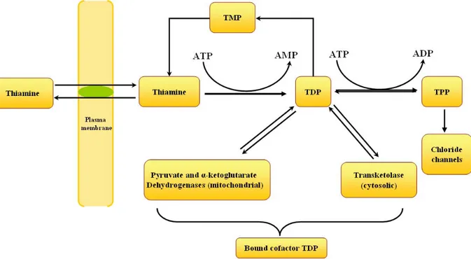
1.1.2.3.1 Thiamine transport and phosphorylation in brain.
Transport of thiamine across the blood-brain barrier is conducted through a carrier-mediated system (Spector 1976; Greenwood et al., 1982). This system appears to be independent of energy metabolism (Greenwood et al., 1986). Sharma and Quastel (1965) presented evidence for a saturable and energy-requiring thiamine uptake in cortex
slices, suggesting that nerve cells actively pump thiamine. Moreover, neuroblastomas as well as glial cells possess a high-affinity thiamine transport system [Km of 35 nM for thiamine] (Bettendorff and Wins, 1994). This high affinity carrier is responsible for the transport of thiamine across the cell membrane and thus contributes to the homeostasis of intracellular thiamine at high extracellular concentrations (Bettendorff, 1994). Thiamine taken up by neuroblastoma cells is rapidly phosphorylated to TDP by thiamine pyrophosphokinase (EC 2.7.6 .2) (Bettendorff and Wins, 1994). Phosphorylation of thiamine and subsequent binding of TDP to cytoplasmic transketolase (EC 2 .2.1 .1) and mitochondrial pyruvate (PDH, EC 1 .2.4.1) and a-ketoglutarate (KGDH, EC 1.2.4.2) dehydrogenases are responsible for secondary active accumulation of thiamine compounds in neuroblastoma cells (Fig. 2).
1.1.2.3.2 Role of thiamine in brain metabolism
The metabolically active form of thiamine is thiamine pyrophosphate (TPP). Essentially TPP is required for several biochemical reactions involved in the breakdown of glucose to liberate energy. It acts as a co-enzyme in oxidative decarboxylation and transketolase reactions. Mainly there are three major enzyme systems that require TPP as a cofactor: pyruvate dehydrogenase (EC 1.2.4.1) complex, an organized enzyme assembly that connects glycolysis with the tricarboxylic acid (TCA) cycle, α-ketoglutarate dehydrogenase (EC 1.2.4.2) complex (KGDHC), a multicomponent enzyme complex associated with the TCA, and transketolase (EC 2.2.1.1), a key participant in the pentose phosphate shunt which is involved in nucleic acid and lipid biosynthesis. Thiamine is thus an important factor in carbohydrate metabolism and hence in thiamine deficiency (TD), blood pyruvate and often blood lactate levels rise abruptly (Combs, 1992).
1.1.3 Etiology and epidemiology of thiamine deficiency
Vitamin B1 deficiency can occur because of many different causes. The most basic form of TD results because of impaired nutritional status associated with chronic diseases, such as alcoholism, gastrointestinal diseases, HIV-AIDS, and persistent vomiting. Following are the major factors responsible for TD:
1.1.3.1 Lack of thiamine intake
Lack of thiamine intake can occur with food containing a high level of thiaminases such as milled rice, shrimp, mussels, clams, fresh fish, and raw animal tissues, food high in anti-thiamine factors and processed food with a content high in sulfite, which destroys thiamine. In addition, diet-related factors such as alcoholic state,
starvation state or gastro-intestinal disorder can also reduce thiamine intake (Masumoto et. al., 2009; Matrana et. al., 2009; Ahmed et. al., 2011).
1.1.3.2 Increased metabolic consumption
Increased metabolic consumption of thiamine may occur because of pregnancy, hyperthyroidism, lactation, fever, severe infection and increased physical exercise. (Anderson et al, 1985; Braverman and Utiger, 1996).
1.1.3.3 Increased thiamine depletion
Increased thiamine depletion may occur in complications such as diarrhea, diuretic therapies, peritoneal dialysis, hemodialysis, hyperemesis gravidarum (Indraccolo et. al., 2005; Al-Attas et. Al., 2011).
1.1.3.4 Decreased absorption
The major factors responsible for the decreased thiamine absorption are chronic intestinal disease, alcoholism, malnutrition, gastric intestinal disorder, malabsorption syndrome; celiac and tropical sprue (Anderson et al, 1985).
1.1.3.5 Consequences of chronic alcoholism
The relation between alcohol intake and thiamine deficiency is well established and has been investigated in the past. Up to 80% of alcoholics in general population have a deficiency in thiamine (Morgan 1982), some of them will go on to develop WE and/or Korsakoff psychosis (WKS). However, the most debated issue regarding alcoholism in clinical setting is the occurrence of TD related brain damage with or without alcoholism (Charness 1993; Joyce1994; Butterworth 1995). In general, cerebral damage due to thiamine deficiency progresses with alcohol toxicity and vice versa. Therefore, alcoholism does not directly cause TD (Phillips et al. 1981), though it may induce such
deficiency because of its normal association with malnourishment and its related mechanism. Particularly, the low thiamine absorption rate at the mucosal level, the impaired hepatic function, and the raised alcohol-related thiamine metabolism may lead to the development of chronic thiamine deficiency (Harper 2009). Consequently, the effect of alcohol was investigated in metabolism (Ammon et al. 1965; Lindros 1982), oxidative stress, excitotoxicity (Lovinger 1993). Furthermore, ethanol and its more toxic oxidative metabolite acetaldehyde is involved directly or indirectly in brain damage, and it may be that high levels of acetaldehyde play a role in producing TD. Non-oxidative metabolites of alcohol metabolism particularly fatty acid ethyl esters are found increased in ethanol induced brain damage. Interactions between these conditions influence brain damage in alcoholism. Moreover, chronic alcohol administration accelerates the lesions of experimental TD, by up regulating NMDA receptor expression and excitotoxicity (Harper and Matsumoto 2005). Furthermore, the most frequently accessible target for the alcohol to trigger a detrimental effect in the brain, are the blood-brain barrier (BBB) and blood-CSF barrier (BCSFB) (Nixon et al. 2008). It is well established that the alcohol intoxication and thiamine deficient glucose metabolism increases the permeability of the blood brain barrier (BBB) leading to instability in the osmotic gradient (Calingasan et al. 1995; Nixon et al. 2008). This results in the swelling of intra- and extracellular spaces. Also, observed in the periventricular regions, the BBB is physiologically less tight and there is a high rate of thiamine-related glucose and oxidative metabolism. Among the neurological manifestations, ataxia was positively associated with alcoholism without any obvious cerebellar lesions, while infratentorial signal-intensity alterations were only observed in non alcoholics. Alternatively, choroid plexus (CP) is hypothesized as the
other target of alcohol intoxication and it is postulated that CP is the primary source of brain pathology in TD and alcohol toxicity (Nixon et al. 2009). Impairment of the BCSFB and BBB are not considered as a primary factor in the pathogenesis of WE or of ethanol intoxication, only for the reason that there has been insufficient assessment of the BCSFB in these conditions. Thus, it can be concluded that alcohol alone is not responsible for TD and its related brain damages, however, alcohol abuse does potentiate the neurological impairment in TD encephalopathy.
1.1.4 Major disorders of thiamine deficiency
TD progresses with a variety of clinical signs brought about by the presence of complicating factors, such as infections, or by the presence of symptoms from multiple deficiencies such as other B vitamins, vitamin C and minerals as well as the effects of stresses of many kinds, such as physical labour and pregnancy. However, vitamin B1 deficiency manifests itself principally with changes involving the nervous system, the cardiovascular system, and also the gastrointestinal tract (Williams, 1961; Sebrell, 1962; Sauberlich, 1967).
1.1.4.1 Sub-clinical thiamine deficiency
The factors responsible for the occurrence of sub-clinical or mild thiamine deficiency are intake of high carbohydrate food and low thiamine intake, raised physiological or metabolic demand, primarily due to pregnancy and lactation, heavy physical exertion, inter-current illness (cancer, liver diseases, infections, hyperthyroidism), surgery, and wherever absorption is reduced by regular high blood alcohol levels, gastrointestinal disease; dysentery, diarrhoea, nausea/vomiting (Anderson et al, 1985).
Table 1 attempts to summarize some of the typical lesions seen in specific organ systems of the body as a result of thiamine deficiency.
Table 1. Organ Systems of the body affected in thiamine deficiency
Nervous system WE and WKS; Polyneuritis (multifactorial); autonomic, sensory and motor nerves are affected; paraesthesia and hyperesthesia, loss of ankle and knee jerks with muscle wasting and paralysis -typically wrist- and foot-drop (symmetrical).
Eye Nutritional amblyopia
Heart and blood vessels Enlarged heart. Congestive heart failure which is one of the contributory causes of peripheral oedema and results in increase in circulating blood volume.
Gastrointestinal tract Constipation (rarely diarrhea) with abdominal distension and colicky pains, anorexia, nausea, vomiting.
The symptoms of mild thiamine deficiency are elusive and can be attributed to other problems therefore diagnosis is often difficult. However, anorexia, which is one of the early symptoms of subclinical thiamine deficiency, is regarded to be a protective phenomenon since a high-carbohydrate diet is more dangerous in the presence of thiamine deficiency (Lonsdale et al, 1980).
1.1.4.2 Wernicke’s Korsakov Syndrome (WKS)
WKS is characterized by symmetric hyperaemic brain lesions with glial proliferation, capillary dilatation, and perivascular haemorrhage. The syndrome is manifested by a confusional state, disorientation, ophthalmoplegia, nystagmus, diplopia, and gait ataxia (Wernicke’s encephalopathy, WE), with severe loss of memory for recent events and confabulation (Korsakov’s psychosis) occurring following recovery. It
appears that the disorder can have an autosomal recessive inheritance but is expressed as a clinical disease only in the event of TD.
Up to 80-90 % of patients with WE go on to develop the more debilitating chronic Korsakoff’s psychosis. The first reports of the important role of TD in the etiology of WKS were described by Alexander and colleagues (1938) and Bowman et al (1939). DeWardner and Lennox (1947) later observed 52 malnourished prisoners of war and further established the important link between TD and WKS in humans. WKS is most commonly observed in alcoholics, the majority of them with liver disease, and is often precipitated abruptly by administration of glucose to patients severely deficient in thiamine. If untreated, death is common; even with treatment, 17% die within 3 weeks (Feldmann, 1988).
1.2 PATHOPHYSIOLOGY OF THIAMINE DEFICIENCY ENCEPHALOPATHY 1.2.1 Neuroanatomical damage in WE and TD
In TD, damage to the brain is focal in nature. Typically, vulnerable areas include the mammillary bodies, thalamus, inferior colliculus, brainstem, and cerebellum (Troncoso et al., 1981; Langlais et al., 1996). Although the thiamine content of the brain is almost uniform (13 µg/g dry weight) (Dreyfus et al., 1959; Cooper and Pincus, 1979), vulnerable areas at risk of damage show marked alterations in levels depending on the metabolic rate. Each organ or part of the nervous system appears to have its particular thiamine levels for depletion (Sharma and Quastel, 1965). Cerebral levels of thiamine are highest in the cerebellar vermis followed by caudate nucleus, brain stem, periaqueductal region, mamillary region and thalamus (Dreyfus et al., 1959). However, the precise
relationship between thiamine levels and subsequent damage during its depletion remains a mystery.
WE patients and TD animals characteristically display gross neuropathological changes that include brain atrophy, hemorrhages (Victor et al., 1989), edematous necrosis (Watanabe et al., 1981), white matter damage (Yamashita and Yamamoto, 1995; Langlais and Zhang, 1997), gliosis and significant neuronal loss (Witt, 1985; Todd et al., 1999; Mulholland, 2006). Typically, mammillary bodies as well as the medial, midline and intralaminar nuclei of the thalamus, inferior colliculus, periaqueductal area and floor of the fourth ventricle show severe damage. In addition, brainstem nuclei, cerebellum, cranial nerve nucleus, pretectal regions, and locus coeruleus are affected in WE (Torvik, 1987; Victor et al., 1989), with cerebellar damage being most prominent in the anterior superior vermis and more severe in alcoholics with WE (Phillips et al., 1990). Microscopically, loss of Purkinje cells and shrinkage of the molecular and granule cell layers have been identfied (Phillips et al., 1987), with marked reduction in Purkinje dendritic arborization commonly observed both in alcoholics and TD-dependent brain damage (Terasawa et al., 1999). Such occurrence of cerebellar degeneration in alcoholics is relatively common (Kril, 1996), and due to the similarity of this damage in both alcoholics and TD, it has been hypothesized that alcoholic cerebellar degeneration is nutritional in origin (Terasawa et al., 1999). Experimentally, the thalamus and, in particular, the medial dorsal nuclei, represents the area of brain most widely studied in TD (Langlais et al., 1992; Hazell et al., 1998a), with other affected areas including the anterior nuclear group, geniculate body, ventral posterior medial nucleus, dorsal nucleus, and lateral posterior nucleus. Moreover, the hypothalamus also displays abnormalities in
areas such as the suprachiasmatic nucleus, supraoptic nucleus, and medial preoptic nucleus. Evidence also indicates that regions of the brain such as the cerebral cortex, previously considered being unaffected, also sustaining damage in TD and WE.
Histological evidence of damage to the peripheral nervous system following metabolic and toxic insults has led many investigators to focus on the role of thiamine and the effects of alcohol on the progression of these diseases. Chronic TD and alcohol toxicity are often causes of peripheral neuropathy (Swank et al., 1940; Victor et al., 1989), with the most common being distal axonopathies, also termed “dying back” disease. Although the precise mechanism for this type of pathology had not yet been established, it was envisioned that damage to the axonal transport system and metabolic disturbances were the causative factor for the progression of this neuropathy (Schoental and Cavanagh, 1977). While 80% of the patients with WKS observed by Victor had evidence of a peripheral neuropathy, many of these cases exhibited alcohol-dependent TD, suggesting an involvement of the toxic effects of alcohol in the progression of peripheral neuropathy. To date, animal models of alcoholic neuropathy and its relationship to TD have yet to be established.
Many cases with TD have been shown to exhibit more subtle neurological signs and symptoms, including the abnormalities in brain regions such as, cerebellum, inflammation or degeneration of peripheral nerves (neuropathy) as well as changes in behavior and problems with learning, memory, and decision making (Torvik 1987). Autopsy studies have found that a region of the cerebellum known as the anterior superior cerebellar vermis most frequently exhibits TD–induced damage (Baker et al. 1999; Lavoie and Butterworth 1995; Victor et al. 1989). TD contributes to a reduction in
the number and size of a certain cerebellar cell type called Purkinje cells in parts of the cerebellar vermis (Philips et al. 1987). Cerebellum is involved primarily in muscle coordination and it is also recognized for its role in various aspects of cognitive and sensory functioning (Parks et al. 2003). Accordingly, cerebellar degeneration is associated with difficulties in movement coordination and involuntary eye movements, such as nystagmus. Cerebellar degeneration is found both in alcoholics with WKS and alcoholics alone, but because WKS patients typically have a higher degree of cerebellar atrophy, it appears likely that TD also is the predominant cause of cerebellar degeneration.
In addition to the cerebellum, numerous other brain regions and structures are damaged in people with WKS. Although animal studies have suggested that thiamine deficiency may contribute to damage to these structures, the exact role of TD and the level of sensitivity of these structures to thiamine deficiency have not yet been determined. Further studies are certainly needed in this area.
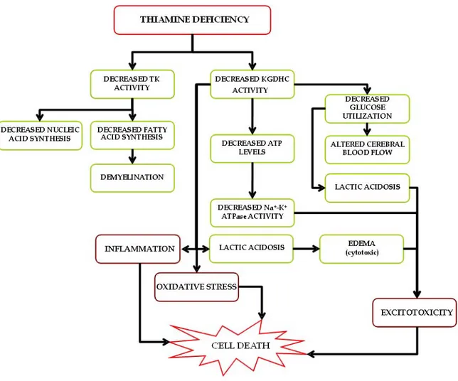
1.2.2 Effects of thiamine deficiency on enzyme activity
The pathophysiological mechanisms involved in TD are complex. Compromised brain energy metabolism is a major consequence of the disorder. Thiamine in the form of TPP is an important cofactor of three major enzyme systems: pyruvate dehydrogenase (EC 1.2.4.1) complex, an organized enzyme assembly that connects glycolysis with the TCA cycle, α-ketoglutarate dehydrogenase (EC 1.2.4.2) complex (KGDHC), a multicomponent enzyme complex associated with the TCA, and transketolase (EC 2.2.1.1), a key participant in the pentose phosphate shunt which is involved in nucleic acid and lipid biosynthesis. Chronic thiamine deprivation is accompanied by
region-selective reductions in levels of these thiamine-dependent enzymes in brain (Butterworth, 1986). However, decreased activity of KGDHC appears to be responsible for many of the reversible changes accompanying TD (Gibson et al., 1984; Butterworth and Heroux, 1989) (see Fig. 3). A fourth thiamine-dependent enzyme system, branched-chain α-ketoacid dehydrogenase complex, is associated with a rare inborn error of metabolism, maple syrup urine disease, involving an accumulation of the branched chain amino acids leucine, isoleucine, and valine (Wendel et al., 1983).
1.2.3 Lactic acid accumulation
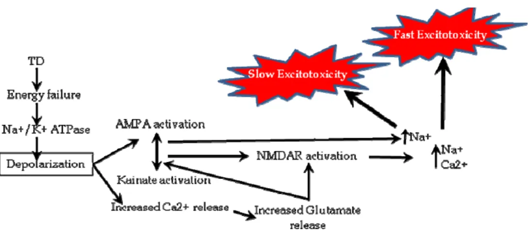
During TD, oxidative decarboxylation of pyruvate and α-ketoglutarate is inhibited, resulting in decreased ATP production, pyruvate accumulation and lactate production (Aikawa et al., 1984; Navarro et al., 2005). This net synthesis of lactic acid within the brain of thiamine-deprived animals is a phenomenon that has been well recognized for many years (Kinnersley and Peters, 1930; Holowach et al., 1968; McCandless and Schenker, 1968). Lactic acidosis induced by TD is type B (i.e. acidosis in the presence of good tissue perfusion and oxygenation), with a pH of 7 and blood lactate below15 mmol/l and is typically unresponsive to alkalinization (Chadda et al., 2002). Unless thiamine is rapidly supplemented, refractory acidosis and death can occur within twenty four hours. Areas of increased lactic acidosis are localized to brain regions that subsequently develop histological lesions (McCandless, 1982; Hakim, 1984; Munujos et al., 1993), and likely play a significant role in the pathophysiology in TD animals.
1.2.4 Disruption of membrane potential
Thiamine and its derivatives are crucial in stabilizing the resting membrane potential (Itokawa and Cooper, 1970; Fox and Duppel, 1975), thereby maintaining ionic balance and conduction of the action potential (Cooper et al., 1963). Chronic deprivation of thiamine impairs this function and the electrophysiological characteristics of the cell, which can lead to severe pathological consequences.
1.2.5 Blood-brain barrier alterations in TD
Integrity of the BBB is crucial for normal CNS function. The first report of BBB damage in TD was proposed by Scholz (1949) and was described in more detail by
Pentschew and Garro (1966) in which they suggested blood vessels and, in particular, capillary endothelial cells, to be the primary site of damage, proposing the term "system-bound dysoric encephalopathy" for WE. Since then, considerable evidence for BBB damage has been described, with disturbances localized to brain regions vulnerable to TD (Calingasan et al., 1995b), and including the presence of hemorrhagic lesions (Torvik, 1985, Vortmeyer and Colmant, 1988). Such a process may also contribute to previous reports of brain edema identified in both TD and in cases of WE (see below).
Impairment in oxidative metabolism plays a significant role in BBB breakdown in TD in which oxidative stress mediated by eNOS is involved (Beauchesne et al., 2009a), and is likely also the case for several neurodegenerative disease states. In addition, inflammatory processes occur in TD (see below) which are known to disrupt the BBB (Guenther and Neu, 1984). CNS pathologies often involve BBB disturbances in which astrocyte-endothelial cell interaction is abnormal, and astrocytes secrete transforming growth factor-β, which downregulates brain capillary endothelial expression of the fibrinolytic enzyme tissue plasminogen activator and the anticoagulant thrombomodulin (Tran et al., 1999). Indeed, several chemical agents circulating in plasma or secreted from cells associated with the BBB are capable of increasing brain endothelial permeability and impairing its transport and metabolic function (Kis et al., 2001). A number of studies have evaluated BBB integrity, both spatially and temporally in experimental TD and acute WE. For example, findings have revealed disruption of BBB integrity adjacent to the third ventricle, cerebral aqueduct and fourth ventricle in acute WE (Schroth et al., 1991), consistent with the location of histological lesions. Increased BBB permeability was also reported in whole brain (Warnock and Burkhalter, 1968) and in vulnerable brain
regions in TD (Manz and Robertson, 1972), with a likely factor contributing to this susceptibility being the cerebral energy deficit. Interestingly, amyloid beta peptide (Aβ) has been reported to increase neuronal membrane fluidity and lipid peroxidation (Avdulov et al., 1997), which may contribute to the observed BBB changes in TD. Additionally, Aβ has been shown to increase endothelial cell permeability to albumin (Blanc et al., 1997), a finding which might explain the leakage of this protein into the brain parenchyma during TD (Harata and Iwasaki, 1995). Furthermore, this effect was also shown to be reversible following treatment with antioxidants (Blanc et al., 1997). Since Aβ may also play a role in excitotoxicity and is capable of inducing apoptosis (Forloni et al., 1993; Loo et al., 1993), a feature also reported in TD (Matsushima et al., 1997), altogether, these findings suggest this peptide plays a role in increased BBB permeability and possibly neuronal cell death in TD. Recent evidence also indicates that BBB tight junction proteins such as occludin and associated scaffolding proteins are decreased, concomitant with increased matrix metalloproteinase-9 levels in TD (Beauchesne et al., 2009b). The underlying basis for these changes, however, remains currently unresolved.
1.2.6 Oxidative stress
Maintenance of an optimal redox environment is the primary requirement for proper cellular functioning, and this environment is preserved by enzymes that maintain a reduced state through normal energy metabolism. Disturbances in this redox state can cause toxic effects through the production of net reactive oxygen species (ROS), particularly in the mitochondria (Lin and Beal, 2006), that can damage vital components of the cell, including proteins, lipids, and DNA. This imbalance in ROS metabolism, or
development of oxidative stress is an important factor in the pathogenesis of TD (Gibson and Blass, 2007). Increased ROS production in TD can trigger cell membrane damage, including lipoperoxidation (Valko et al., 2007) and alterations in the functional integrity of ion channels and transporters. Persistent net ROS formation in TD can initiate a cascade of cell death pathways via intracellular messengers, e.g. intracellular caspase-3-mediated apoptosis. Development of oxidative stress also leads to disturbances in brain function, including an inhibition of glutamate uptake due to transporter protein nitrosylation following peroxynitrite formation (Volterra et al., 1994; Trotti et al., 1996; Hazell, 2007). Under conditions of oxidative stress, levels of heme oxygenase-1, endothelial nitric oxide synthase (eNOS), the inducible form of NOS (iNOS), intracellular adhesion molecule-1, and microglial activation are increased (Calingasan et al., 1999,2000; Gibson et al., 2000). Thus, oxidative stress can lead to profound neuropathological consequences in TD.
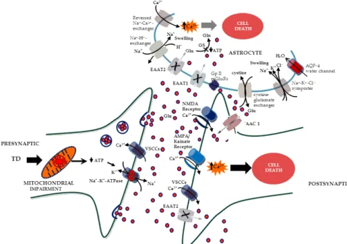
1.2.7 Excitotoxicity
Excitotoxicity is the pathological process by which nerve cells are damaged and killed by excessive stimulation of receptors for the excitatory neurotransmitter glutamate. The normal levels of glutamate approach 10 mmol/kg during synaptic transmission, while extracellular concentrations remain approximately 25 nM (Schousboe, 1981; Herman and Jahr, 2007). These low extracellular levels of glutamate are essential to ensure appropriate signal to noise for excitatory signaling and to limit excessive activation of glutamate receptors that can cause excitotoxicity (Choi, 1992; Conti and Weinberg, 1999). The only process known to actively clear extracellular glutamate is mediated by a family of Na+-dependent transporters also known as glutamate or excitatory amino acid
transporters (Schousboe, 1981; Danbolt, 2001). These transporters couple the movement of 3 Na+ ions and 1 H+ to the inward transport of glutamate, providing sufficient energy to maintain a transmembrane concentration gradient of up to one million-fold (Zerangue and Kavanaugh, 1996).
Excitotoxicity may be involved in spinal cord injury, stroke, traumatic brain injury, hearing loss (orototoxicity) and in neurodegenerative diseases such as, Alzheimer's disease, amyotrophic lateral sclerosis (ALS), Parkinson's disease, alcoholism or alcohol withdrawal, and Huntington's disease (Kim et. al., 2002; Hughes, 2009). Other common conditions that cause excessive glutamate concentrations around neurons are hypoglycemia (Camacho, 2006), status epilepticus (Fujikawa, 2005) and mitochondrial dysfunction (Jhala and Hazell, 2011). Consequences of excitotoxicity during TD are discussed in more detail in section 1.3
1.2.8 Inflammation
Cerebral inflammation is now recognized as a key component of the neurodegenerative process, and occurs e.g. in AD, PD, multiple sclerosis (Bojinov, 1971; Allen et al., 1981; Aisen and Davis, 1994), along with other neurological conditions such as stroke and brain trauma (Garcia, 1975; Mathew et al., 1994) Previous studies describing alterations in glial cell morphology in TD including evidence of swelling and the appearance of phagocytic vacuoles (Collins, 1967; Robertson et al., 1968) indicate that although neuronal damage is a feature of this disease process, glial elements are also profoundly affected. Evidence in support of the existence of an inflammatory process has been described in TD, including the early development of increased microglial reactivity (Todd and Butterworth, 1999), while production of pro-inflammatory cytokines in both
vulnerable and non-vulnerable regions of brain has been reported (Ke et al., 2006; Vemuganti et al., 2006; Karuppagounder et al., 2007). In a recent study, we demonstrated that in vulnerable brain regions in TD, inflammatory genes represent the largest functional group of transcripts upregulated (e.g. pro-inflammatory cytokines including IL-6, IL-18, TNF-α, AIF1, and osteopontin), interferons (IFNs), IFN-inducible proteins, and chemokines (Gro1, MCP-1, MIP-1α, and MIP-1β) (Vemuganti et al., 2006). Interestingly, many of these genes are also known to be expressed strongly in astrocytes during inflammation and may therefore be a contributing factor to astrocyte dysfunction in TD. In addition, many transcription factors known to control inflammatory gene expression such as Egr-1, c-EBP-β, c-EBP-δ, CPBP and Klf-4 are also upregulated following TD. During impaired oxidative metabolism, Egr-1 and c-EBP-β may play an important role in starting the inflammatory cascades. Furthermore, levels of these various inflammatory-related gene products in different brain regions may be an important determining factor for selective vulnerability in TD. Recent studies have also demonstrated that alcohol induces inflammatory responses in brain that include microglial activation and cytokine production (Qin et al., 2008; He and Crews, 2008). Such findings may also be relevant in the brains of cases of WE. Additional studies aimed at identifying how these expression changes are occurring in TD and in alcoholics are required to better understand the way in which the process of inflammation develops. Such investigations may also yield important details regarding the process of inflammation in neurodegenerative disease states, given the similarities of impaired oxidative metabolism between TD and these maladies.
1.2.9 Induction of immediate early genes
Immediate-early genes (IEGs) are induced rapidly and transiently in response to a wide variety of cellular stimuli. They represent a mechanism that is activated at the transcription level at a very early stage as a response to stimuli before any new proteins are synthesized. The earliest known and best characterized IEGs are fos, myc and c-jun, a group of genes homologous to retroviral oncogenes. Many of these transcription factors can bind to consensus sites on the promoter region of other genes, thus allowing regulation of downstream gene expression that may play a significant role in alterations in cell function that occur as a consequence of TD. Expression of IEGs has been found to be increased dramatically in association with cell death (Colotta et al. 1992; Dragunow et al. 1993; Estus et al. 1994), and in TD (Hazell et al., 1998c), and has provided additional support for an excitotoxic event, in which IEG induction is linked to membrane depolarization, L-type voltage-sensitive calcium channel activation, SCC activation, and subsequent loss of Ca2+ homeostasis (Morgan and Curran, 1986; Murphy et al., 1991), all features of this disorder.
1.3 GLUTAMATE-MEDIATED EXCITOTOXICITY
1.3.1 Cerebral vulnerability to glutamate mediated excitotoxicity
Glutamate is a principal excitatory neurotransmitter in the central nervous system and almost 90% of the synaptic connections in the brain are estimated to be glutamatergic. At chemical synapses, glutamate is stored in vesicles. Nerve impulses trigger release of glutamate from the pre-synaptic cell. In the opposing post-synaptic cell, glutamate receptors, such as the NMDA receptor, bind glutamate and are activated.
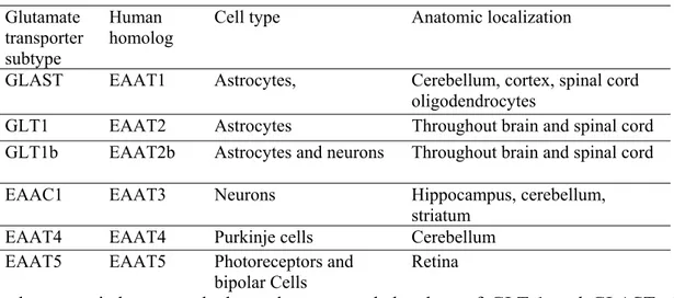
Because of its role in synaptic plasticity, glutamate is involved in cognitive functions like learning and memory in the brain. The form of plasticity known as long-term potentiation takes place at glutamatergic synapses in the hippocampus, neocortex, and other parts of the brain (Doble, 1999). Glutamate works not only as a point-to-point transmitter but also through spill-over synaptic crosstalk between synapses in which summation of glutamate released from a neighbouring synapse creates extrasynaptic signalling/volume transmission (Pellerin, 2005). Hence, it is the glutamate concentration in the surrounding extracellular fluid that determines the extent of receptor stimulation. Any disturbance in glutamate homeostasis may therefore have severe pathological consequences and may lead to glutamate excitotoxicity (Fig. 4). Thus, it appears that the cerebral vulnerability to glutamate excitotoxicity is associated with selective increase in extracellular glutamate concentration. Glutamate is constantly being released from cells and is continually being removed from the extracellular fluid by the action of high affinity Na+ dependent glutamate transporters or excitatory amino acid transporters. Five excitatory amino acid transporters cDNAs have been identified and cloned (EAAT1–5) thus far. The predominant glutamate transporters in the brain are GLAST and GLT-1, also identified in the rodent as glutamate/aspartate transporter (GLAST) and glutamate transporter-1 (GLT-1), respectively, which are primarily expressed in astrocytes, while EAAT3, EAAT4, and EAAT5 are found primarily in neurons. Downregulation of glutamate transporter followed by accumulation of glutamate in the extracellular fluid have been documented in chronic and debilitative neurological disorders of diverse etiology. Thus, any disturbance in the physiological processes implicated in the regulation of glutamate, namely glutamate release/transport/uptake and energy status, may increase the likelihood
of the brain to excitotoxic insult, i.e. making it vulnerable to glutamate mediated excitotoxic damage (Fig. 4).
1.3.2 Glutamate transport and release
Transport and release of excitatory neurotransmitter, glutamate, is an essential event in glutamate neurotransmission. Glutamate taken up by cells can be used either for metabolic purposes (protein synthesis, energy metabolism, ammonia fixation) or it can be reused as a transmitter. In nerve terminals, glutamate is transported into synaptic vesicles by vesicular glutamate transporters and subsequently released by exocytosis. It seems likely that glutamate is also released, to some extent, directly from cytosol (non-vesicularly) through plasma membrane proteins. In astrocytes, glutamate taken up from the extracellular fluid may be converted to glutamine which is released to the extracellular fluid, taken up by neurons and reconverted to glutamate inside neurons (Doble, 1999). Synaptic glutamate transmission requires two types of neurotransmitter transporters. First, vesicular neurotransmitter transporters remove transmitters from the cytosol and transport them into the lumen of secretory vesicles to allow exocytotic release (Moriyama and Omote, 2008). Four types of vesicular transporters have thus far been identified which include VAChT, VMATs, VGAT (or VIAAT) and vesicular glutamate transporters (VGLUTs). VGLUT1 and 2 are expressed in complementary subsets of glutamatergic neurons in the CNS. In contrast, the most recently identified isoform, VGLUT3, is co-expressed with VAChT and VMAT2 in a number of cholinergic and aminergic cell types (Fei et. al., 2008). Synaptic transporters then remove excess neurotransmitter from the synaptic cleft and transport them to the interior of the synaptic terminal for recycling. In addition to these transporters another essential group of proteins
involved in synaptic transmission are the complexions. They are small, cytosolic and highly charged molecules (molecular mass of approximately 15 kDa) that are localized in presynaptic nerve terminals. They bind to the neuronal SNARE (soluble N-ethylmaleimide-sensitive factor-attachment protein receptors) complex and are involved in regulating calcium-dependent neurotransmitter release (Hazell and Wang 2011). Current evidence suggests that loss of VGLUTs and complexins may destabilize synaptic terminal release of glutamate, contributing to dysfunction of circuitry with possible pathological consequences. Alterations in the function and/or expression of these proteins is a complicating factor which can contribute to the excessive sustained release of glutamate which may lead to excitotoxicity.
1.3.3 Glutamate receptors and excitotoxicity
Excessive release of glutamate in the synaptic cleft leads to activation of several types of pre and post-synaptic glutamate receptors. The consequent rise in intracellular calcium concentration may lead to mitochondrial dysfunction, generation of reactive oxygen species, and the activation of proteases, phospholipases, and endonucleases, leading to cell death.
1.3.3.1 Ionotropic glutamate receptors
There are three known types of ionotropic glutamate receptors based on their pharmacological properties; the N-methyl-D-aspartate (NMDA) receptor, α-amino-3-hydroxy-5-methyl-4-isoxazole propionic acid (AMPA) receptor, and the kainate receptor. The NMDA and AMPA/kainate receptors are all glutamate gated ion channels (conducting only Na+ or both Na+ and Ca2+). The AMPA receptors open readily upon glutamate exposure, but desensitize quickly and are of low affinity. In contrast, the
NMDA receptors have much higher affinities and are slowly inactivating. To be activated they need both glutamate binding and an already depolarized membrane. Additionally, NMDA receptors are highly permeable to calcium and distributed widely on CNS neurons and, are the major initiators of excitotoxicity (Fig. 4). Moreover, pretreatment with (+)-5-methyl-10, ll-dihydro- 5H-dibenzocyclohepten-5, lO-imine hydrogen maleate (MK-801 ), a noncompetitive antagonist of the NMDA type glutamate receptor, protects against TD-induced lesions and suggests that TD-induced neuronal loss, particularly within thalamus, may be mediated by glutamate excitotoxicity (Langlais and Zhang, 1993; Todd and Butterworth, 1998). However, activation of Ca2+permeable AMPA or kainate receptors can also trigger neuronal cell death. Antagonists of these receptors display a higher protective efficacy than NMDA receptor antagonists in some experimental neurodegenerative conditions. Thus, neuroprotective influence exerted by NMDA receptor antagonists, as well as by AMPA and kainate receptor antagonists following brain injury in animal models, including TD, further supports the notion that excessive glutamate receptor stimulation contributes to damage via an excitotoxic process.
1.3.3.2 Metabotropic glutamate receptors
Metabotropic glutamate receptors (mGluRs) are G-protein coupled receptors which produce their effects via signalling mechanisms involving phosphoinositide-dependent processes, cyclic AMP or protein kinase C. Recent studies have identified mGluRs as a way in which neural cells regulate the release of glutamate and its uptake. Three groups of mGluRs have been characterized to date. Group I mGluR agonists have been reported to cause a downregulation of the EAAT1 transporter, while the Group II
agonist DCG IV upregulates its expression. Group II mGluRs are found on both pre- and post-synaptic membranes as well as glial cells, are negatively coupled to cyclic AMP, and regulate glutamate release via presynaptic Group II autoreceptors. Group III mGluRs are also negatively coupled to cyclic AMP. TD results in decreased ATP levels, a source of cyclic AMP via activity of adenylate cyclase. Thus, it is conceivable that loss of glutamate transporter regulation occurs as a consequence of changes in activity of mGluRs due to the declining ATP status (Hazell, 2009).
1.3.4 Glutamate transporters
Of the five types of glutamate transporters cloned to date, considerable evidence indicates that GLT-1 contributes to excitotoxicity and neuronal death in a number of neurological disorders, including ischemic stroke, traumatic brain injury, and ALS. Previous studies indicate that extracellular glutamate concentration is increased in vulnerable brain regions in TD (Langlis and Zhang, 1993; Hazell et al., 1993). This effect of TD on glutamate transporter levels was also demonstrated in primary cultures of astrocytes (Hazell et al., 2003).
Table 2. Nomenclature and expression pattern of glutamate transporters Glutamate
transporter subtype
Human homolog
Cell type Anatomic localization
GLAST EAAT1 Astrocytes, Cerebellum, cortex, spinal cord
oligodendrocytes
GLT1 EAAT2 Astrocytes Throughout brain and spinal cord
GLT1b EAAT2b Astrocytes and neurons Throughout brain and spinal cord
EAAC1 EAAT3 Neurons Hippocampus, cerebellum,
striatum
EAAT4 EAAT4 Purkinje cells Cerebellum
EAAT5 EAAT5 Photoreceptors and
bipolar Cells
Retina
occurs in the cerebral cortex of alcoholic cases of WE (Hazell et. al., 2010). Based on the evidence it is likely that downregulation of glutamate transporters exacerbate glutamate mediated brain damage or excitotoxicity in TD. Focal accumulation of lactic acid is a classical feature of TD and one of the major consequences is gliotoxicity (Navarro et. al., 2005). Astrocytes are particularly sensitive to lactate. Thus, exposing them for extended periods to lactic acidosis leads to an inability of these cells to maintain ATP production (Bender et. al., 1997). The resulting decrease in ATP levels then causes a collapse of the ionic gradients across the astrocyte cell membrane due to suppression of Na+ /K+ ATPase activity, leading to movement of Na+ into the cell along its concentration gradient. Elevated intracellular Na+ concentration results in transport of glutamate from the astrocyte to the extracellular space. Moreover, astrocytic swelling due to glutamate uptake or excessive K+ spatial buffering can also lead to the depolarization of these cells, leading to release of glutamate via transporter reversal (Chan et. al., 2004; Danbolt 2001). The major consequence of this transporter reversal is an increase in extracellular glutamate concentration which is a major causative factor implicated in excitotoxicity (Danbolt 2001).
1.4 REGULATION OF GLUTAMATE TRANSPORTER
Glutamate is the major neurotransmitter of the excitatory signaling pathway in the brain; in addition, it is involved in changes in the protein repertoire through the activation of signaling cascades, which regulate protein synthesis at transcriptional and translational levels. Activity-dependent differential gene expression by glutamate is related to the activation of ionotropic and metabotropic glutamate receptors and its subsequent removal from the extra-synaptic space by Na+-dependent astrocytic glutamate transporter. Moreover, glutamate receptor stimulation is involved in processes of learning and memory as well as in other plastic changes in the CNS such as synapse induction and elimination during development. Excessive accumulation of extracellular glutamate and overactivation of glutamate receptors is associated with decreased expression and function of astrocyte glutamate transporters (Rothstein etal., 1996). In addition, a number of acute CNS diseases have been shown to be associated with astrocyte glutamate transporter dysfunction including CNS ischemia (Martin et al., 1997) and trauma (Yi and Hazell, 2006) as well as chronic neurodegenerative disorders such as Alzheimer’s disease (AD) (Masliah et.al., 1996), and ALS (Lin et al., 1998), hepatic encephalopathy (Knecht et al., 1997), epilepsy (Mathern et al., 1999; Tanaka et al., 1997) and TD/WE (Jhala et.al., 2011; Hazell et.al., 2001, 2003, 2010). Glutamatergic synaptic transmission is involved in many important brain functions and elevated concentrations of extracellular glutamate can cause severe excitotoxic damage to the receiving neurons. It is therefore crucial to maintain efficient glutamate uptake. Recent studies involving targeted gene disruption confirmed significance of glutamate transporters in maintaining glutamate homeostasis (Tanaka et al. 1997). Based on these studies it is critical for our understanding to study
the regulation of glutamate transporter for its use as a pharmaceutical target in the treatment of neurodegenerative diseases. The mechanisms of glutamate transporter regulation are not well defined, and little is known about the factors that are responsible for regulating protein expression and activity. Regulation of transporter protein can occur at multiple levels, including DNA transcription and protein translation or posttranslational modification, consequently it may affect glutamate transporter activity, localization and protein targeting. Thus, it is likely that a combination of all of these mechanisms is important for the regulation of glutamate transporters.
1.4.1 Transcriptional regulation 1.4.1.1 Neuron derived factors
Studies over the past years have identified numerous chemical factors that are responsible for the transcriptional regulation of glutamate transporters. Among these factors most important are those that are derived from neurons. Upregulation of both types of astrocytic glutamate transporters, GLT-1/EAAT2 and GLAST/EAAT1 by neuron derived factors were reported previously (Drejer et al. 1983; Gegelashvili et al. 1997; Schlag et al. 1998). In the absence of neurons, astrocytes maintain polygonal shapes and express only the GLAST transporter. When co-cultured with neurons, astrocytes exhibit more complex morphologies and show increased expression for GLT-1 (Swanson et al. 1997), thus suggesting that there are neuronal soluble factors that increase the levels and expression of glutamate transporter protein and mRNA. Although the soluble factors present in neuron conditioned media (NCM) have not been yet identified, progress have been made in identifying their signal transduction pathways. Previously it has been proposed that upregulation of glutamate transporters depends on the activation of p42/44
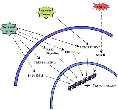
![Mot à mot, brin par brin : les suites [Nom préposition Nom] comme motifs](data:image/gif;base64,R0lGODlhAQABAIAAAP///wAAACH5BAEAAAAALAAAAAABAAEAAAICRAEAOw==)