Université de Montréal
Bcl-xL (S49) and (S62) sequential phosphorylation/dephosphorylation during mitosis prevents chromosome instability and aneuploidy
Par
Prasamit Saurav Baruah
Programme de biologie moléculaire Faculté de Médecine
Thèse présentée à la Faculté de Médecine En vue de l’obtention du grade de doctorat en
Biologie Moléculaire
Juin 2016
Université de Montréal Faculté de Médecine
Cette thèse intitulée :
Bcl-xL (S49) and (S62) sequential phosphorylation/dephosphorylation during mitosis prevents chromosome instability and aneuploidy
Présentée par : Prasamit Saurav Baruah
A été évaluée par un jury composé des personnes suivantes :
Président-rapporteur Directeur de recherche Membre du jury Examinateur externe Représentant de la doyenne
Abstract
An interesting feature of Bcl-xL protein is the presence of an unstructured loop domain between its α1 and α2 helices, a domain not essential for its anti-apoptotic function and absent in CED-9, ortholog protein in Caenorhabditis elegans. Within this domain, Bcl-xL undergoes dynamic phosphorylation and dephosphorylation at Ser49 and Ser62 during G2 and mitosis in human cancer cells. When these residues are mutated and proteins expressed in cancer cells, cells harbor mitotic defects, including chromosome mis-attachment, lagging, bridging and mis-segregation, events associated with chromosome instability and aneuploidy. To further analyze the effects of Bcl-xL Ser49 and Ser62 in normal cells, the present studies were performed in normal human diploid cells, and in vivo in Caenorhabditis elegans.
First, we studied normal human diploid BJ foreskin fibroblast cells expressing Bcl-xL(wild type), (S49A), (S49D), (S62A), (S62D) and the dual (S49/62A) and (S49/62D) mutants. Cells expressing S49 and/or S62 phosphorylation mutants showed reduced kinetics of cell population doubling. These effects on cell population doubling kinetics correlated with early outbreak of senescence with no impact on the cell death rate. Senescent cells displayed typical senescence-associated phenotypes including high-level of senescence-associated β-galactosidase activity, interleukin-6 secretion, tumor suppressor p53 and cyclin-dependent kinase inhibitor p21Waf1/Cip1 activation as well as γH2A.X-associated nuclear chromatin foci. Fluorescence in situ hybridization analysis and Giemsa-banded karyotypes revealed that the expression of Bcl-xL phosphorylation mutants in normal diploid BJ cells provoked chromosome instability and aneuploidy. These findings suggest that dynamic Bcl-xL Ser49 and Ser62 phosphorylation/ dephosphorylation cycles are important in the maintenance of chromosome integrity during mitosis in normal cells.
Second, we undertook experiments in Caenorhabditis elegans to understand the importance of Bcl-xL Ser49 and Ser62 in vivo. Transgenic worms carrying single-site S49A, S62A, S49D, S62D and dual-site S49/62A mutants were generated and their effects were analyzed in germlines of young adult worms. Worms expressing Bcl-xL variants showed decreased egg-laying and hatching, variations in the length of their mitotic regions and transition zones, chromosomal abnormalities at their diplotene stages,
and increased germline apoptosis. Some of these transgenic strains, particularly the Ser to Ala variants, also showed slight modulations of lifespan compared to their controls. The in vivo observations confirmed the importance of Ser49 and Ser62 within the loop domain of Bcl-xL in maintaining chromosome stability.
These studies could impact future strategies aiming to develop and identify compounds that could target not only the anti-apoptotic domain of Bcl-xL protein, but also its mitotic domain for cancer therapy.
Résumé
Une caractéristique intéressante de la protéine Bcl-xL est la présence d'un domaine en boucle non-structurée entre les hélices α1 and α2 de la protéine. Ce domaine protéique n'est pas essentiel pour sa fonction anti-apoptotique et absent chez CED-9, la protéine orthologue chez Caenorhabditis elegans. A l'intérieur de ce domaine, Bcl-xL subit une phosphorylation et déphosphorylation dynamique sur les résidus Ser49 et Ser62 en phase G2 du cycle cellulaire et lors de la mitose. Lorsque ces résidus sont mutés et les protéines exprimées dans des cellules cancéreuses, les cellules démontrent plusieurs défauts mitotiques liés à l'instabilité chromosomique. Pour analyser les effets de Bcl-xL Ser49 et Ser62 dans les cellules normales, les présentes études ont été réalisées dans des cellules diploïdes humaines normales, et in vivo chez Caenorhabditis elegans.
Dans une première étude, nous avons utilisé la lignée cellulaire de cellules fibroblastiques diploïdes humaines normales BJ, exprimant Bcl-xL (type sauvage), (S49A), (S49D), (S62A), (S62D) et les double (S49/62A) et (S49/62D) mutants. Les cellules exprimant les mutants de phosphorylation ont montré des cinétiques de doublement de la population cellulaire réduites. Ces effets sur la cinétique de doublement de la population cellulaire corrèle avec l'apparition de la sénescence cellulaire, sans impact sur les taux de mort cellulaire. Ces cellules sénescentes affichent des phénotypes typiques de sénescence associés notamment à haut niveau de l'activité β-galactosidase associée à la sénescence, la sécrétion d' interleukine-6, l'activation de p53 et de p21WAF1/ Cip1, un inhibiteur des complexes kinase cycline-dépendant, ainsi que la formation de foyers de chromatine nucléaire associés à γH2A.X. Les analyses de fluorescence par hybridation in situ et des caryotypes par coloration au Giemsa ont révélé que l'expression des mutants de phosphorylation de Bcl-xL provoquent de l'instabilité chromosomique et l'aneuploïdie. Ces résultats suggèrent que les cycles de phosphorylation et déphosphorylation dynamiques de Bcl-xL Ser49 et Ser62 sont importants dans le maintien de l'intégrité des chromosomes lors de la mitose dans les cellules normales.
Dans une deuxième étude, nous avons entrepris des expériences chez Caenorhabditis elegans pour comprendre l'importance des résidus Ser49 et Ser62 de Bcl-xL in vivo. Les vers transgéniques portant les mutations de Bcl-xL (S49A, S62A, S49D, S62D et
S49/62A) ont été générés et leurs effets ont été analysés sur les cellules germinales des jeunes vers adultes. Les vers portant les mutations de Bcl-xL ont montré une diminution de ponte et d'éclosion des oeufs, des variations de la longueur de leurs régions mitotiques et des zones de transition, des anomalies chromosomiques à leur stade de diplotène, et une augmentation de l'apoptose des cellules germinales. Certaines de ces souches transgéniques, en particulier les variants Ser/Ala, ont également montré des variations de durée de vie par rapport aux vers témoins. Ces observations in vivo ont confirmé l'importance de Ser49 et Ser62 à l'intérieur du domaine à boucle de Bcl-xL pour le maintien de la stabilité chromosomique.
Ces études auront une incidence sur les futures stratégies visant à développer et à identifier des composés qui pourraient cibler non seulement le domaine anti-apoptotique de la protéine Bcl-xL, mais aussi son domaine mitotique pour la thérapie du cancer.
Contents
Abstract iii
Résumé v
Contents vii
List of figures and tables x
Abbreviations xii
Nomenclature convention xvi
Acknowledgements xvii
1. Introduction 1
1.1 Cell cycle, senescence and cell death: a brief overview 1
1.2 Bcl-2 family of proteins in mammalians 4
1.2.1 Structure of anti-apoptotic Bcl-xL protein 4 1.2.2 Structure and importance of the loop domain of Bcl-xL 6 1.2.3 Bcl-2 protein family at interface with the cell cycle 8 1.3 The cell cycle in mammalians: regulation at interphase 11
1.3.1 Cyclin-dependent kinases and cyclin-dependent kinase inhibitors 11
1.3.2 G1-S phase transition 12
1.3.3 G2-M phase transition 13
1.4 The cell cycle in mammalians: mitosis regulation 14
1.4.1 Chromosome - microtubule attachment 16
1.4.1.1 The kinetochores 16
1.4.1.2 The KMN network 17
1.4.2 Activation of the spindle assemble checkpoint 18
1.4.2.1 Bub protein kinetochore recruitment 19
1.4.2.2 Mad1 and Mad2 kinetochore localization 20
1.4.2.3 The Mad2 template model 20
1.4.2.4 Phosphorylation control 22
1.4.2.5 Cdc20 under control 24
1.4.3 Silencing of the spindle assemble checkpoint 24 1.4.3.1 Correct kinetochore-microtubule attachment stops mitotic
checkpoint complex production 24
1.4.3.2 Inactivation of spindle assembly checkpoint re-engagement 27
1.4.4 Mitotic exit and cytokinesis 28
1.4.4.1 RhoGTPase and Myosin II 29
1.4.4.2 Chromosome passenger complex 30
1.4.4.3 Membrane trafficking and remodeling during cytokinesis 31
1.5 Cellular senescence 32
1.5.1 Replicative senescence 33
1.5.2 Premature senescence 34
1.5.3 Senescence-associated phenotypes and markers 35 1.5.3.1 Senescence-associated Beta-Galactosidase activity 35 1.5.3.2 Senescence-associated secretory phenotype 36 1.5.3.3 Senescence-associated heterochromatin foci 39
1.5.3.4 γ-H2A.X as DNA damage foci 40
1.6 Aneuploidy 41 1.6.1 Failure of spindle assembly checkpoint and aneuploidy 43
1.6.2 Failure of cytokinesis and aneuploidy 46
1.6.3 DNA damage, aneuploidy and senescence 47
1.7 The C. elegans experimental model 48
1.7.1 The anatomy and development of C. elegans 48
1.7.2 Germ cell specification 51
1.7.3 Germ line proliferation and maintenance 54
1.7.4 Regulation of mitotic and meiotic progression in the germ line 55 1.7.5 Physiological germ line apoptosis in C. elegans 57 1.7.6 DNA damage and apoptotic pathway in C. elegans 57 2. Rationale of the thesis and contribution of authors 62 3. Bcl-xL (S49) and (S62) dynamic phosphorylation/dephosphorylation
during mitosis prevents chromosome instability and aneuploidy in
human normal diploid fibroblasts 64
3.1 Summary 65
3.2 Introduction 66
3.3 Results 68
3.4 Conclusion and discussion 71
3.5 Materials and methods 75
3.6 Disclosure of conflicts of interest 77
3.7 Acknowledgements 77
3.8 Bibliography 90
4. Expression of human Bcl-xL(Ser49) and (Ser62) mutants in Caenorhabditis
elegans causes germline defects and aneuploidy 94
4.1 Summary 95
4.2 Introduction 96
4.3 Results 97
4.4 Conclusion and discussion 100
4.5 Materials and methods 102
4.6 Disclosure of conflicts of interest 104
4.7 Acknowledgements 104
4.8 Bibliography 116
5. Discussion and perspectives 120
5.1 Interplay of Bcl-xL in the cell cycle 120
5.2 Expression of Bcl-xL (Ser49) and (Ser62) mutants leads to cellular
senescence in BJ fibroblasts: association with the p53/p21 pathway 121 5.3 Expression of Bcl-xL (Ser49) and (Ser62) mutants leads to cellular
senescence in BJ fibroblasts: association with other markers 126 5.4 Ser49 and Ser62 of Bcl-xL loop domain contributes to the
maintenance of chromosome stability in BJ cells 129 5.5 Expression of Bcl-xL (Ser49) and (Ser62) mutants affects germline
development in C. elegans 130 5.6 Expression of Bcl-xL (Ser49) and (Ser62) mutants leads to aneuploidy
and cellular apoptosis in C. elegans 132
5.7 Expression of Bcl-xL Ser62 and Ser49 mutants affects life span
in C. elegans 135
5.8 Future perspectives 136
5.8.1 Bcl-xL Ser62 and MCC complexes for SAC activation and resolution 136 5.8.2 Bcl-xL Ser49 and Ser62 and its interaction with cytoplasmic
dynein protein 138
5.8.3 Bcl-x Ser49 and membrane remodelling and trafficking during cytokinesis 139
5.8.4 Bcl-xL and mouse embryonic development 140
5.8.5 Bcl-xL and cell fate 141
6. Summary of major findings 143
List of figures and tables
1. Introduction
Figure 1: Schematic view of cellular response and fate to DNA damage 3
Figure 2: Comparison of domain structures of Bcl-2 family members 6
Figure 3: Bcl-xL structure. 7
Figure 4: Schematic representation of Bcl-xL phosphorylation during mitosis 10
Figure 5: Eukaryotic cell cycle phases with respective cyclin-Cdk complexes and inhibitors 13
Figure 6: Progression through mitosis 15
Figure 7: Organization of the KMN network 18
Figure 8: Mad2-template model of MCC production at unattached kinetochores 21
Figure 9: Speculative model of the regulation of kinetochore–microtubule binding through outer kinetochore phospho-regulation by Aurora-B and B56-PP2A 23
Figure 10: The spindle assembly checkpoint delays mitotic progression 25
Figure 11: Error correction and the spindle assembly checkpoint at different stages during mitosis 27
Figure 12: Pathways inducing senescence 38
Figure 13: Genomic and chromosome instability 42
Figure 14: Aneuploidy due to SAC failure during mitosis 45
Figure 15: Anatomy of C. elegans adult hermaphrodite 49
Figure 16: Anatomy of the germ line in adult hermaphrodite and male 50
Figure 17: Germ line specification during the embryogeneis in C. elegans 53
Figure 18: DTC niche maintains GSCs through Notch signalling 54
Figure 19: Interplay between FBF1/2 and GLD-1 determines the fate of germ line cells 56
Figure 20: DNA damage responses at the germline 59
Figure 21: Direct and indirect pathway of apoptosis in C. elegans 61
3. Bcl-xL (S49) and (S62) dynamic phosphorylation/dephosphorylation during mitosis prevents chromosome instability and aneuploidy in human normal diploid fibroblasts Table 1 : Chromosomal aberrations in control BJ cells and BJ cells expressing Bcl-xL (wt) and Bcl-xL phosphorylation mutants 78
Figure 1: Effect of Bcl-xL and Bcl-xL phosphorylation mutant expression on cell population doubling of BJ cells 79
Figure 2: Effect of Bcl-xL and Bcl-xL phosphorylation mutant expression on outbreak of senescence in BJ cells 81
Figure 3: Effect of Bcl-xL and Bcl-xL phosphorylation mutant expression on chromosome stability and aneuploidy in BJ cells 83
Figure 4: Effect of Bcl-xL and Bcl-xL phosphorylation mutant expression on senescence-associated biomarkers in BJ cells 85
Figure S1: Kinetics of cell population doubling of control BJ cells
and BJ cells expressing Bcl-xL phosphorylation mutants 87 Figure S2: Correlation between aneuploidy and senescence-associated
biomarkers in control BJ cells and BJ cells expressing Bcl-xL
phosphorylation mutants 88
Figure S3: Bcl-xL somatic mutations found in human tumours
and short genetic variations 89
4. Expression of human Bcl-xL Ser49 and Ser62 mutants in Caenorhabditis elegans causes germline defects and aneuploidy
Table 1: Vector design and transgenic strains 105
Figure 1: Expression of Bcl-xL (wt) and Bcl-xL variants in transgenic worms 106 Figure 2: Effects of Bcl-xL (wt) and Bcl-xL variants on C. elegans
progeny fecundity 108
Figure 3: Effects of Bcl-xL (wt) and Bcl-xL variants on mitotic region
and transition zone in in C. elegans gonads 109
Figure 4: Effects of Bcl-xL (wt) and Bcl-xL variants on C. elegans
chromosome stability and aneuploidy 111
Figure 5: Effects of Bcl-xL (wt) and Bcl-xL variants on gerlime apoptosis 112 Figure 6: Effects of Bcl-xL (wt) and Bcl-xL variants on C. elegans lifespan 113 Figure S1: Effects of Bcl-xL (wt) and Bcl-xL variants on the gonads 114 Figure S2: RNAi experiments reversed the effects of Bcl-xL variants on C. elegans 115
6. Summary of major findings
Figure 22: Summary of major findings in normal human BJ fibroblasts 143
Figure 23: Summary of major findings in C. elegans 144
Abbreviations:
Apaf-1 Apoptotic peptidase activating factor 1 APC/C Anaphase promoting complex/cyclosome AKT commonly known as Protein kinase B (PKB) Arf Alternate reading frame
Atm Ataxia telangiectasia mutated
Atr Ataxia telangiectasia and Rad-3 related protein B56-PP2A B56 regulatory subunit of PP2A
Bad Bcl-2 associated death promoter protein Bak Bcl-2 homologous antagonist killer Bax Bcl-2 associated X protein
Bcl-2 B-cell lymphoma 2 protein Bcl-b B-cell lymphoma B protein Bcl-g B-cell lymphoma G protein Bcl-rambo B-cell lymphoma Rambo Bcl-xL B-cell lymphoma extra long Bcl-xES B-cell lymphoma extra short Bcl-x B-cell lymphoma X
Bcl-w B-cell lymphoma W
Bfl-1/A1 Synonym for Bcl-2 related protein A1 BH Bcl-2 homology domain
BHRF-1 BamH1 restricted fragment 1
Bid BH-3 interacting domain death agonist Bik Bcl-2 interacting killer protein
Bim Commonly called for protein Bcl-2 like protein 11 BMP Bone morphogenic protein
Bmf Bcl-2 modifying factor
Boo/Diva Bcl-2 homologue of ovary/ Death inducer binding to vBcl-2 53BP-1 p53 binding protein 1
Bok/Mtd Bcl-2 related ovarian killer/ Matador Brca1/2 Breast cancer protein 1/2
BubR1 Budding uninhibited by benzimidazoles related protein 1 Bub3 Budding uninhibited by benzimidazoles 3 homolog
C Celsius
CARD Caspase recruitment domain
Ccl Chemokine ligand
Cdc20 Cell division cycle protein 20 Cdc42 Cell division cycle protein 42 Cdk Cyclin dependent kinase CED-9 C. elegans cell death protein 9 CEP-1 C. elegans p53 like-1
Cenp-E Centromere protein E
Chmp3 Charged multivesicular protein 3 Chk1/2 Checkpoint kinase1/2
Cki Cyclin-dependent kinase inhibitor protein CPC Chromosome passenger complex
CRISPR/Cas9 Clustered regulatory interspaced short palindromic repeats/ CRISPR associated protein 9
Cxcl8 Chemokine (C-X-C motif) ligand-1 Dad-1 Defender against apoptotic death-1 DAPI 4',6'-diamidino-2-phénylindole DDR DNA damage response
DNA Dexyribonucleic acid
DNA –SCAR DNA segments with chromatin alterations reinforcing senescence
DNMT DNA methyl transferases
Drp Dynamin related protein DSB Double-strand break
DTC Distal tip cell
Ect2 Epithelial cell transforming sequence 2 ECM Extracellular matrix
EGL-1 Egg laying defective -1
Ercc Excision repair cross-complementing ERK Extracellular signal-regulated kinases
ESCRT Endosomal sorting complex required for transport machinery
FBF Fem-3 binding factor
FISH Flourescent in-situ hybridization
FRAP Fluorescence recovery after photobleaching GAP GTPase activating protein
GEF Guanine exchange factor GFP Green fluorescent protein GIN Genomic instability
GLD Germ line defective
GLP-1 Glucagon like peptide-1 GLD-2 PAP GLD-2 poly (A)-polymerase
GM-Csf Granulocyte macrophage colony-stimulating factor
GSC Germline stem cell
GTP Guanine nucleotide phosphate
h hour
HAT Histone acetyltranferase
HDAC Histone deacetylase
Ha-Ras Harvey rat sarcoma
γ-H2A.X H2A histone family member X phospho-serine 139 H3K9-me3 histone-3- lysine 9 trimethyl
HmgA High-mobility group A Hp-1 Heterchromatin protein 1
HR Homologous recombination
Hrk Harakiri Bcl-2 interacting protein HUS-1 Hydroxyurea sensitive-1
ICD-1 Inhibitor of cell death-1 Igf Insulin-like growth factor
IgfBPs Igf-binding proteins Il Interleukin
Il-6R Il-6 receptor
IF Immunofluorescence
InsR Insulin receptor
IP Immunoprecipitation
IR Ionizing radiation
Jnk c-Jun N-terminal kinase
Kif14 Kinesin-like protein 14
KMN Knl-1/Mis12/Ndc80 complex
KSHV Kaposi sarcoma associated herpes virus LAG-1 Longevity assurance gene 1
Mad2 Mitotic arrest deficient 2
Mapk Mitogen-activated protein kinase Mcak Mitotic centromere-associated kinesin MCC Mitotic checkpoint complex
Mcl-1 Myeloid leukemia cell differentiation protein 1 Mdm2 Mouse double minute 2 homolog
MEF Mouse embryonic fibroblast MES Maternal-effect sterile
MgcRacGAP RACGAP1 gene encoding Rac GTPase-activating protein
min minute
Mip-3α Macrophage inflammatory protein 3α Mklp2 Mitotic kinesin like protein 2 phosphatase Mlcp Myosin light chain phosphatase
Mlck Myosin light chain kinase Mmp Metalloproteinase
MOF Males absent on the first protein (D. melanogaster) MosSCI Mos1-mediated Single Copy Insertion
MPS-1 Monopolar spindle
Mre-11 Meiotic recombination 11
MRN MRE11, RAD50, NBS1 complex
mRNA messenger ribonucleic acid
MRT Mortal germline-2
MT Microtubule
mTor Mammalian target of rapamycin NF-κβ Nuclear factor kappa B
NGM Nematode growth medium
NMR Nuclear magnetic resonance NOEs Nuclear Overhauser effects Nos-3 Nitric oxide synthase protein 3
Noxa Latin word for Damage/ Phorbol-12-myristate-13-acetate-induced protein 1
NWGR Asp-Trp-Gly-Arg motif
OIS Oncogene-induced senescence PAI-1 Plaminogen activator inhibitor-1
PCAF P300/CBP-associated factor PCD Programmed cell death
PDK-1 3-Phosphoinositide dependent protein kinase PIE-1 Pharynx and intestine in excess protein 1 Plk1 Polo like kinase 1
PML Promyelocytic leukaemia
Pmp-3 Plasma membrane proteolipid protein 3 PP1 Protein phosphatase 1
PP2A Protein phosphatase 2A pRb Retinoblastoma
PRC1 Protein regulator of cytokinesis 1 PS Premature senescence
PSR Phosphatidylserine receptor Pten Phosphatase and tensin homolog
Puma p53 upregulated modulator of apoptosis
Ras Rat sarcoma
RE Recycling endosomes
RhoA Ras homolog gene family, member A
RNAseq RNA sequencing
ROCK Rho-associated protein kinase ROS Reactive oxygen species
RT-PCR Reverse transcriptase polymerase chain reaction RS Replicative senescence
SA-β-Gal Senescence-associated β-galactosidase SAC Spindle assembly checkpoint
SAHF Senescence-associated heterochromatin foci SAPK Stress-activated protein kinases
SASP Senescence-associated secretory phenotype
SMAD Small body size Mothers Against Decapentaplegic (D. melanogaster)
s second
SEC Securin
SKY Spectral karyotype
SOG Short gastrulation
SSB Single-strand break
TIF Telomere dysfunction-induced foci TGF- β Transforming growth factor-β
TGF- βR1 Transforming growth factor-β receptor 1 TM Transmembrane domain
TUNEL Terminal deoxynucleotidyl transferase dUTP nick end labeling VEGF Vascular endothelial growth factor
VP16 Synonym for etoposide
wt wild-type
XPF Xeroderma Pigmentosum Group F-Complementing Protein YFP Yellow fluorescent protein
Nomenclature convention
Human genes and mRNAs are with ITALIC scripts (example BCLX) Human proteins are with Normal scripts (example Bcl-xL)
C. elegans genes and mRNAs are with italic scripts (example ced-9) C. elegans proteins are with NORMAL scripts (example CED-9)
Mouse genes and mRNAs are with Italic scripts (example Bclx) Mouse proteins are with Normal scripts (example Bcl-xL)
Genes from other species (yeast, virus) are with Italic scripts (example Cdc8)
Proteins from other species (yeast, virus) are with NORMAL scripts (example BHRF1)
Protein domains are with NORMAL scripts (example BH1) Protein complexes are with NORMAL scripts (example APC/C)
Acknowledgements
First and foremost I will like to thank God for his continuous blessings and guidance throughout this project. I’m very much grateful beyond words towards my supervisor, Dr. Richard Bertrand without whom I would never have either got this opportunity or this research program. He has been always there relentlessly and guiding me throughout in every ups and downs of my doctorate career. I forward my heartful gratitude to Myriam Beauchemin who has been with me in every step of this journey and has given me so much love and affection making my working environment a stress less ambience. I have learnt and gathered much experience after entering this laboratory.
My family has been a big support in the completion of this project and without them I can hardly see myself here. Special thanks my friends Redwane Boukharfane, Ahmed Mohammed Abdel Latiff, Macha Samba Mondonga, Abdel Hamid Bekri, Anne Calvé, Julie and Yasmin who have always helped and motivated me in this adventure.
I would like to thank studentship support from Faculté des études supérieures de l’Université de Montréal, the Fondation de l’institut de Cancer de Montréal and Centre de Recherche du Centre hospitalier de l’Université de Montréal for their financial aid and support.
1. Introduction
1.1 Cell cycle, senescence and cell death : A brief overview
The term cell cycle refers to the orderly biological process where one cell will generate 2 daughter cells through the duplication of their genetic material and cell division. In eukaryotes the cell cycle is divided into specific phases, gap phase 0 (G0), gap phase 1 (G1), DNA synthesis phase (S), gap phase 2 (G2), and mitosis (M)1-4
. To ensure that the cells pass accurate copies of their genomes on to the next generation, evolution has overlaid the core cell cycle machinery with a series of surveillance pathways termed cell cycle checkpoints5,6,7
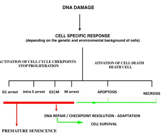
. DNA damage including single nucleotide damage, base pair mismatch, DNA single-strand breaks (SSB) and double-strand breaks (DSB), chromosome misattachment and missegregation during mitosis represent global and serious threats to genomic and chromosome stability which will rapidly induce complex pleiotropic cell responses paired to cell cycle checkpoints and repair mechanisms1,8-10
. The overall function of these checkpoints in response to damaged or abnormally structured DNA is to slow down and halt cell cycle progression, thereby allowing time for appropriate repair mechanisms to correct the genetic lesions and/or structural aberrations before they are passed on to the next generation of daughter cells. At their most proximal signaling elements, these complex machineries contain, sensor proteins or protein complexes that scan chromatin for partially replicated DNA, DNA errors, DNA strand breaks, or chromosome misattachment and missegregation, and translate these derived stimuli into biochemical signals that will modulate the functions of specific target proteins11. These mechanisms first promote cell cycle arrest, DNA repair and proper chromosome alignment and segregation, but can also promote irreversible cellular senescence or cell death12
. The repair mechanisms correct minor irregularities during a temporary cell cycle halt, whereas more deleterious defects are believed to result in the induction of cellular senescence or cell death. Defects in those signalling cascades and/or repair mechanisms combined with errors initiating cellular senescence or cell death could yield to mutations and/or aneuploidy leading to genomic and/or chromosome instability 13,14
Cells can enter into an irreversible cell cycle arrest termed cellular senescence. The process of cellular senescence was first described more than 50 years ago by Hayflick and Moorehead as an irreversible cell cycle arrest of human fibroblasts that lost their proliferative capacity5,15,16
. It was later found that telomeres, necessary for chromosome integrity and proper cell division, were gradually depleted to a threshold level within 40– 45 generations, which triggered the induction of senescence. This threshold was termed as the Hayflick limit17
. This natural process was named replicative senescence (RS), which differs from premature senescence (PS), an accelerated mechanism that occurs in response to extrinsic or intrinsic stress stimuli. These include DNA damage, disrupted chromatin organization, increased oncogenic signalling, increased replicative stress, treatment with chemotherapeutic drugs or irradiation18
and oxidative stress19,20
. Cellular senescence is a safeguard limiting the proliferative competence of cells in living organisms and can act as a potent tumor suppressor mechanism for normal cells 21
. Cell death is often associated with apoptosis22
, a morphologically distinct form of physiological and programmed cell death, explicitly described through many years of research22,23
. An understanding of apoptosis in mammalian cells was first achieved by research in the nematode Caenorhabditis elegans (C. elegans)24
. Apoptosis has since been widely accepted as the primary mode of programmed cell death (PCD), which genetically eliminates predetermined cells from an organism during development. The process is also active in adult organisms as a homeostatic mechanism to maintain cell populations in tissues. Apoptosis also occurs as a defense mechanism such as in immune reactions or when cells are damaged in association with diseases, noxious agents or deregulation of cellular processes25
. Programmed necrosis or necroptosis and, in some contexts, autophagy are often considered as two others forms of PCD, easily distinguished by their morphological differences26
. Apoptosis, or type I PCD, described by Kerr et al.22
is characterized by cell shrinkage, nuclear disassembly associated with chromatin condensation and fragmentation, dynamic membrane blebbing and loss of adhesion to neighbors or to extracellular matrix. Biochemical changes include chromosomal DNA cleavage into internucleosomal fragments, phosphatidylserine externalization and a number of intracellular substrate cleavages by specific proteolysis27,28
formation of autophagosomes, which plays a crucial pro-survival role in cell homeostasis. It is required during periods of starvation or stress due to growth factor deprivation but in some contexts also leads to a form of cell death29, 30-33
. Type III PCD termed programmed necrosis or necroptosis, involves cell swelling, organelle dysfunction and cell lysis34-36
. Thus, PCD may play an important role during preservation of tissue homoeostasis and elimination of damaged cells; this has profound effects on malignant tissues37
.
The intimate link between the cell cycle, cellular senescence and cell death with diseases including cancer initiation and development and tumor responses to cancer treatment is getting clearer as research progresses, but it is very far from being completely understood38-42
. A schematic view of these concepts is shown in Figure 1.
Figure 1: Schematic view of cellular response and fate after DNA damage (adapted from Wang et al., 2011 43).
The first sections of the Introduction (1.2 to 1.6) focus on mechanisms in mammalian cells, whereas the last section (1.7) is devoted to C. elegans, the second model used for these studies.
1.2 Bcl-2 family of proteins
BCL2 was the first anti-death gene discovered in mammals44
, a milestone with far reaching implications for tumor biology. BCL2 was discovered because of its involvement in t(14;18) chromosomal translocations observed in non-Hodgkin’s lymphomas44,45
. Multiple members of Bcl-2 family of apoptosis regulating proteins have been identified since, including mammalian anti-apoptotic proteins (Bcl-2, Bcl-xL, Mcl-1, Bcl-xES, Bcl-B, Bcl-w, Bfl-1/AMcl-1, Boo/Diva), structurally similar pro-apoptotic proteins (Bax, Bak, Bok/Mtd, Bcl-xS, Bcl-rambo, Bcl-gL) and several structurally diverse pro-apoptotic interacting proteins that operate as upstream agonists or antagonists, called the BH3only proteins (Bad, Bik, Bid, Bim, Noxa, Puma, Hrk, Bnip1 -3, Bmf, Mcl-1s, Bcl-gS, Spike)46
. Proteins of the Bcl-2 family play central roles in cell death regulation and are capable of regulating diverse cell death mechanisms that encompass apoptosis, necrosis and autophagy47,48
, and thus are found undoubtedly altered in many cancers and leukemia49-52
. Apart from their well-studied roles in controlling apoptosis, members of the Bcl-2 family of proteins also interface with the cell cycle53-69
, DNA repair pathways70-73
and membrane remodelling mechanisms74,75
, pathways which are well separated from their roles in apoptosis53-55,75-77.
1.2.1 Structure of anti-apoptotic Bcl-xL protein
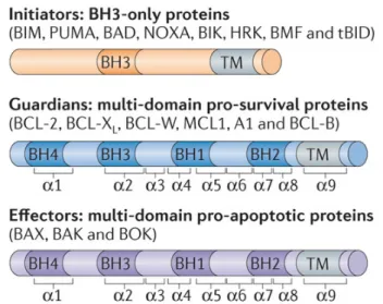
The pro-survival family of Bcl-2 proteins has been divided into two sub-classes based on the presence of one or more of Bcl-2 homology (BH) regions (Fig. 2). Four of these regions (BH1-4) of sequence homology have been identified, and each Bcl-2 family members contains at least one of them78,79. Several members of the pro-survival subclass, such as Bcl-2, Bcl-xL, Bbl.-w, and the CED-9 protein from C. elegans, possess all four BH regions. Others, such as Mcl-1, the BHRF1 protein from Epstein–Barr virus, and KSHV- Bcl-2 from Kaposi sarcoma virus, only possess strong sequence homology in the BH1, BH2, and BH3 regions.
The first published structure of a Bcl-2 family member was that of human Bcl-xL determined by X-ray crystallography and nuclear magnetic resonance (NMR) spectroscopy. It showed that the overall structure of Bcl-xL consists of nine α–helices connected by loops of varying lengths. Bcl-xL adopts a globular structure; it consists of two central, primarily hydrophobic α-helices (α5 and α6), which are surrounded by amphipathic helices: α3 and α4 and by α1, α2 and α7. A 60-residue loop connecting helices α1 and α2 are flexible and non-essential for anti-apoptotic activity80
. The signature “NWGR” sequence directly precedes α5. In Bcl-xL, this region appears to play both an important structural and functional role. Structurally, the tryptophan residue makes extensive hydrophobic contacts with residues in α7 and α8. The arginine residue also plays a key functional role in the binding of Bcl-xL to pro-apoptotic proteins and peptides such as Bax and Bak. The Bcl-2 family of proteins share homology domains BH1 and BH2 and mutations in these regions in either Bcl-2 or Bcl-xL abrogates the anti-apoptotic activity and block the heterodimerization with other members of the Bcl-2 family (e.g., Bax and Bak) that promote apoptosis81,82,83
. BH1, BH2 and BH3 are in close proximity and form an elongated hydrophobic cleft in Bcl-xL, the site for interaction with death-promoting BH3-only proteins. The BH3 region is involved in activity of the death promoting proteins84-86
. The BH3 amphipathic helix of BH3‐only proteins binds the hydrophobic groove of pro‐survival proteins predominantly by the insertion of four hydrophobic residues (h1–h4) along one face into hydrophobic pockets in the groove, and by the formation of a salt bridge between a conserved BH3 Asp residue and a conserved Arg residue in the BH1 domain of the pro‐survival proteins87-89
. Structural studies have shown that the BH3 binding groove of the pro-survival Bcl-2 family members has considerable plasticity90,91
, which probably contributes to their ability to associate with multiple distinct BH3 domains. Besides the BH regions, many of the Bcl-2 family members possess a carboxy-terminal hydrophobic domain, which is predicted to be responsible for membrane localization92,93
Figure 2: Comparison of domain structures of Bcl-2 family members. All Bcl-2 family of proteins contains at least one of the Bcl-2 homology (BH) domains; BH1, BH2, BH3 and BH4. They also possesses a Transmembrane (TM) domain. The BH3 only proteins contain only one, BH3 domain for their pro-apoptotic functions. (diagram adapted from Peter E. Czabotar et. al 201479).
The sequence homology between Bcl-xL and other Bcl-2 members suggests similar structural folds. The arrangement of α-helices in Bcl-xL is reminiscent of the membrane translocation domain of bacterial toxins, in particular diphtheria toxin and the colicins94
.
1.2.2 Structure and importance of the loop domain of Bcl-xL
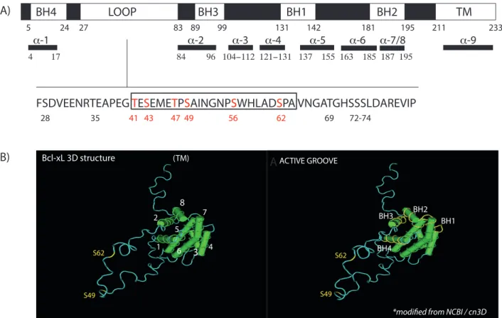
An interesting feature of the Bcl-xL protein is the presence of a long loop between α1 and α295
(Fig. 3). This loop is largely unstructured as evidenced by the lack of electron density for residues 28-80 and the lack of medium and long range nuclear Overhauser effects (NOEs) for these residues78
. In addition, this region has highly variable amino acid sequence among Bcl-2 family members. This loop domain has been shown to be the site of some post-translational modifications affecting the activity of both Bcl-xL and Bcl-296
. For example, interleukin-3 (Il-3) or erythropoietin treatment of NSF/N1.H7 cells induced the phosphorylation of Ser70, resulting in the inactivation of Bcl-297
. Mutant proteins with Ser to Ala mutation80
or deletion of the loop domain together98
was able to inhibit PCD better than the wild type protein. In contrast, proteolytic cleavage of the Bcl-2 loop at Asp34 by caspase-3 converts it from an anti-apoptotic to a pro-apoptotic protein99,100
Figure 3: Bcl-xL structure. A) Bcl-xL contains BH1, BH2, BH3 and BH4 domains, a COOH-terminus hydrophobic transmembrane domain (TM) and an unstructured loop domain (LOOP), between BH4 and BH3. The amino acid sequence of the flexible loop domain is indicated. A region of the loop domain previously identified as important for Bcl-xL cell cycle functions is highlighted in the boxed region53
. Amino acids that have been mutated (Thr/Ala, Ser/Ala) and studied in a series of functional assays are highlighted in red101-103
(adapted from Wang et al 201143). B) Visualization of the 3D structure of Bcl-xL, with the annotated α-helices, BH domains, S49 and S62 (modified from the National Center of Biotechnology Information (NCBI)/ cn3D Web site.)
However, compared to the full-length protein, Bcl-xL loop deletion mutants tend to display a similar ability to inhibit apoptosis and do not show significant alterations in their ability to bind pro-apoptotic proteins53,95,98
. There is growing evidence indicating that Ser62 of Bcl-xL is highly phosphorylated in cells exposed to microtubule inhibitors, and a few protein kinases have been proposed to phosphorylate Bcl-xL(Ser62) in microtubule inhibitor-exposed cells104-109
. Previous work in our laboratory has revealed that two serine residues within the unstructured loop domain of Bcl-xL, Ser49 and Ser62, undergo
FSDVEENRTEAPEG TESEMETPSAINGNPSWHLADSPA VNGATGHSSSLDAREVIP
28 35 41 43 47 49 56 62 69 72-74 5 24 27 83 89 99 131 142 181 195 211 233 BH4 LOOP BH1 BH2 TM 4 17 84 96 104−112 121−131 137 155 163 185 187 195 α-1 α-2 α-3 α-4 α-5 α-6 α-7/8 α-9 BH3 BH4 BH3 BH2 BH1 S62 S49 ACTIVE GROOVE
*modified from NCBI / cn3D
S49 S62 1 2 4 6 (TM) Bcl-xL 3D structure 3 7 5 8 A) A B)
dynamic phosphorylation/dephosphorylation events during cell cycle progression101-103 . The function of the unstructured loop domain within Bcl-xL remains elusive, and is the subject of this work.
1.2.3 Bcl-2 family proteins interface with cell cycle
Numerous studies have revealed links between some Bcl-2-like family members, cell cycle progression and cell cycle checkpoint regulation. First, Bcl-2 has been shown to slow entry from the quiescent G0 into the G1 phase of the cell cycle in multiple cell lineages from transgenic mice. In contrast, Bcl2-/- knockout cells enter S-phase more
quickly108,110
. More recently, phosphorylated forms of Bcl-2 also have been found to co-localize in nuclear structures and on mitotic chromosomes, revealing the importance of phosphorylation events for Bcl-2 protein localization during cell cycle progression111. Mcl-1, another Bcl-2 homologue known to function as an anti-apoptotic protein112
, inhibits cell cycle progression through the S phase of the cell cycle. The cell cycle regulatory function of Mcl-1 is partially mediated through its interaction with proliferating cell nuclear antigen, a cell cycle regulator that is crucial in DNA replication53,113
. Others have reported that a proteolytic fragment of Mcl-1 regulates cell proliferation via its interaction with cyclin-dependent kinase 1 (Cdk1/Cdc20)114
and that Mcl-1 is essential in Atr-mediated Chk1 phosphorylation106
. Others have discerned the involvement of Bid, a BH3-only protein with pro-apoptotic activity, at the intra-S phase checkpoint under replicative stress and in response to DNA-damaging agents. This function of Bid is mediated through its phosphorylation at Ser78 and Ser61/64 by the DNA-damage signaling kinase Atm 115,116
.
Previous studies from our laboratory reported that Bcl-xL, an anti-apoptotic Bcl-2 family member, not only counteracts BH3-only protein-mediated cell death signals after DNA-damaging treatment, it also stabilizes the G2 cell cycle checkpoint and favours the establishment of premature senescence in surviving cells after DNA topoisomerase I (camptothecin) and II (VP16) inhibitor exposition53
. Bcl-xL co-localizes with Cdk1/Cdc2 in nucleolar structures and binds to Cdk1/Cdc2 during the G2 checkpoint, whereas its overexpression stabilizes G2 arrest and premature senescence in surviving cells after DNA damage. Interestingly, Bcl-xL potently inhibits Cdk1/Cdc2 kinase activities in
vitro. In in vitro kinase assays using recombinant Bcl-xL protein, this effect was reversed by the addition of a synthetic peptide corresponding to the 41st
to 60th
amino acids, a region rich in Ser- and Thr- putative phosphorylation residues within the flexible loop domain of Bcl-xL. Furthermore, a deletion mutant of this region (Bcl-xLΔP3) did not alter the anti-apoptotic function of Bcl-xL, but impeded its effect on Cdk1/Cdc2 activities and on the G2 checkpoint after DNA damage53
. Bcl-xL is phosphorylated on Ser62 at the loop domain during normal cell cycle progression and DNA-damage induced G2 arrest by Plk1 and Mapk9/Jnk2102
. Phosphorylated Bcl-xL(Ser62) accumulates in nucleolar structures including nucleoli and Cajal bodies during the stabilization of DNA damage-induced G2 arrest and co-localizes with Cdk1/Cdc2 avoiding unwanted mitosis during DNA damage102
.
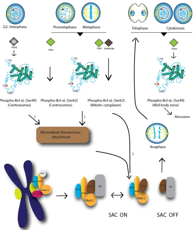
During mitosis, Bcl-xL(Ser62) is strongly phosphorylated by Plk1 and Mapk14/Sapkp38α at prometaphase, metaphase and the anaphase boundary, while it is dephosphorylated at telophase and cytokinesis103
. Phospho-Bcl-xL (Ser62) localizes in centrosomes with γ-tubulin, and in the mitotic cytosol with some spindle-assembly checkpoint (SAC) signaling components, including Plk1, BubR1 and Mad2. In taxol- and nocodazole-exposed cells, phospho-Bcl-xL(S62) also binds to Cdc20- Mad2- BubR1- and Bub3-bound complexes, while the phosphorylation mutant Bcl-xL(S62A) does not103 (Fig. 4).
In parallel, Bcl-xL undergoes cell cycle-dependent phosphorylation on Ser49, which accumulates in centrosomes during the G2 cell cycle checkpoint, particularly during DNA damage-induced G2 arrest 101
. Bcl-xL(Ser49) is rapidly dephosphorylated at early mitotic phases (prometaphase, metaphase, anaphase) and is rephosphorylated during telophase/cytokinesis by Plk3. Phospho-Bcl-xL(S49) is found in association with microtubule-associated dynein motor proteins and at the mid-zone body during telophase/cytokinesis101
(Fig. 4).
In tumor cells, expression of the phosphorylation mutants xL(S62A), Bcl-xL(S49A) or dual Bcl-xL(S49/62A) has no effect on apoptosis, but leads to an increased number of cells harbouring mitotic defects103
. These defects include multipolar spindles, chromosome lagging and bridging, and cells with micro-, bi- or multi-nucleated cells, and
Figure 4: Schematic representation of Bcl-xL phosphorylation during the progression of mitosis. Question marks (?) indicate that the exact mechanisms are still unknown (modified from Wang et al 2014103
). cells that fail to resolve and complete mitosis103
. Together, these observations indicated that during mitosis, Bcl-xL(S49) and (S62) phosphorylation/dephosphorylation dynamics impact on chromosome stability, mitosis resolution and cytokinesis completion, at least in tumour cells101-103
1.3 The cell cycle : regulation at interphase
1.3.1 Cyclin-dependent kinases and cyclin-dependent kinase inhibitors
Proper progression through the cell cycle is monitored by checkpoints that sense possible defects during DNA synthesis and chromosome segregation. During interphase, activation of these checkpoints induces cell cycle arrest, which is controlled by interplay modulation of cyclin dependent kinases (Cdks) and their associated cyclins. Cell cycle arrest at these checkpoints allows the cell to repair defects, thus preventing transmission of damage to the daughter cells117
. Cdks are the catalytic subunits of a family of mammalian heterodimeric serine/threonine kinases, best characterized in the control of cell cycle progression. Cdks were first implicated in cell cycle control based on pioneering work in yeast, in which Cdc genes were identified including Cdc8 in the budding yeast S. cerevisiae and Cdc2 in the fission yeast S. pombe, and were found to promote transitions between different cell cycle phases through its interactions with various regulatory cyclin subunits118-121
. Cyclins are synthesized and destroyed at specific times during the cell cycle, regulating kinase activity of Cdks in a timely manner. Soon, homologs of CDC2 were identified in human cells 122
by their ability to complement yeast mutants123
. Subsequently, CDK2 was discovered because of its ability to complement Cdc8 S. cerevisiae mutants124-127
. Currently more than 20 members of the Cdk family each characterized by a conserved catalytic core made of an ATP binding pocket, a PSTAIRE-like cyclin binding domain and an activating T-loop motif. Cyclins belong to a remarkably diverse group of proteins classified solely on the existence of a cyclin box that mediates binding to Cdk128
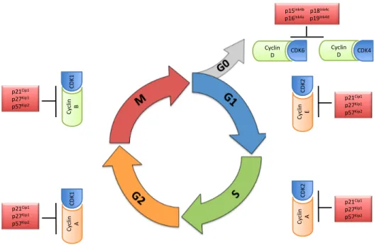
. Cdk activities are restrained by another class of proteins, the cyclin-dependent kinase inhibitors (Cki). Cki are subdivided into two families based on their structure and Cdk specificity. Ink4 proteins, including Ink4A, Ink4B, Ink4C and Ink4D129
; primarily target Cdk4 and Cdk6. The Cip/Kip family composed of p21, p27 and p57129
are more promiscuous and broadly interfere with the activities of cyclin D, E, A and B dependent kinase complexes130
. Cki have been shown to block the proliferation of adult stem cells in multiple tissue types. Loss of Cki may expand the stem cell population, possibly contributing to the development of specific tumours.
1.3.2 G1-S phase transition
After cytokinesis is completed, the newly generated cells can either continue cell division or stop proliferating. If cells are deprived of growth factors prior to the G1 checkpoint, they exit into a state of quiescence known as G0. Those cells that continue proliferating advance to the G1 phase of the new cycle (Fig. 5). According to the classical model for the mammalian cell cycle, specific Cdk-cyclin complexes are responsible for driving the various events known to take place during interphase in a sequential and orderly fashion. Progression through G1 is mainly regulated by Cdk4, Cdk6 and Cdk2 and their regulatory cyclins130,131
. At the beginning of G1, the mitogenic signaling induces synthesis of the D-type cyclins (D1, D2 and D3) and possibly the proper folding and transport of Cdk4 and/or Cdk6 to the nucleus and the activation of the latter. These Cdk-cyclin complexes phosphorylate members of the retinoblastoma (Rb) protein family; pRb, p107 (RbL1) and p130 (RbL2) at their unique phoshorylation sites. The retinoblastoma protein (pRb) and the pRb-related p107 and p130 comprise the 'pocket protein' family of cell cycle regulators. These proteins are best known for their roles in restraining the G1-S transition through the regulation of E2f-responsive genes. pRb and the p107/p130 pair are required for the repression of distinct sets of genes, potentially due to their selective interactions with E2fs that are engaged at specific promoter elements132
. Inactivation of pocket proteins allow for the expression of the E-type cyclins (E1 and E2) which bind and activate Cdk2133,134
. Cyclin E- cdk2 complexes further phosphorylate these pocket proteins, leading to their complete inactivation134,135
. Another kinase, Cdk3 might also participate in inactivation of pRb.
Cyclin E-Cdk2 activity is thought to be essential for initiating DNA replication by facilitating loading of the Mcm chromosome maintenance proteins onto origins of replication. Once cells enter S-phase, cyclin E-cdk2 complexes need to be silenced to avoid the re-replication of DNA136
. Rapid degradation of cyclin E is carried about by Scf-Fbxw7 ubiquitin ligase followed by its subsequent cleavage by the proteasome. In addition, cyclin E-cdk2 phosphorylates its own inhibitor p27, thereby facilitating the degradation of this inhibitor by the proteasome136
. Inactivation of pRb also activates transcription of A-type and B-type cyclins. Cyclin A-cdk2 is required for proper completion and exit from S phase. S phase proteins also include upstream regulators of
cyclin A (pRb), transcription factors (E2f1, B-Myb), protein involved in DNA replication (Cdc6, Hssb, Mcm4), DNA repair (Brca1, Ku70), histone deposition and nucleosome assembly (Hira)137
, ubiquitin mediated proteolysis (hHR6A and Cdc20) and cell cycle checkpoints (p53, p21Cip1
, Mdm2)130 .
Figure 5: Eukaryotic cell cycle phases with respective cyclin-Cdk complexes and inhibitors. The Cdk-cyclin complexes regulate the cell cycle in terms of its entry from one phase to another apart from the checkpoint proteins. Cyclin D-Cdk4/6 complex stimulates the initiation of G1 phase and the start of the cell cycle. Increasing levels of cyclin E-Cdk2 triggers the onset of S phase towards the end of G1 phase. Then, Cyclin A- Cdk2 regulates the completion of S phase and entry into G2, where cyclin B-Cdk1 is involved. The level of cyclin B increases initially and decreases at the end of M phase, followed by a decrease in Cdk1. (Diagram modified from Moghadam et al., 2011138
)
1.3.3 G2-M phase transition
At the end of the G2 phase, B-type cyclins associate with Cdk1(cdc2), the master regulatory kinase that controls the entry into mitosis. Cdk1 is only active at the G2/M border and becomes inactive as cells enter the anaphase stage of mitosis139,140
. During G2, mammalian cyclin B1/Cdk1 complexes are held in an inactive state by phosphorylation of Cdk1 at two negative regulatory sites; Thr14 and Thr15, catalyzed by Myt1 and Wee1
p21Cip1& p27Kip1& p57Kip2& Cy clin & B& CDK 1& p21Cip1& p27Kip1& p57Kip2& Cy clin & A& CDK 1&
p15Ink4b&&&&p18Ink4c&&& p16Ink4a&&&&&&p19Ink4d&
Cyclin&
D& CDK6& Cyclin&D& CDK4&
Cy clin & E& CDK 2& p21Cip1& p27Kip1& p57Kip2& Cy clin & A& CDK 2& p21Cip1& p27Kip1& p57Kip2&
kinases respectively, when it is bound to cyclin B1141-146
. Cdc25 phosphatases dephosphorylate these sites for the activation of Cdk1. Mammalian cells have three Cdc25 phosphatases, Cdc25A, B and C, which appear to have some level of specificity for different cyclin/Cdk complexes along the cell cycle. Studies indicate that Cdc25A regulates G1/S and G2/M transitions, whereas Cdc25B and Cdc5C are involved in intra-S and G2/M regulation140,147-153
. Entry into mitosis absolutely requires progressive accumulation of active cyclin B1/Cdk1(cdc2) complexes in the nucleus. Cyclin B1/Cdk1(cdc2) kinase activity is therefore highly organized to coordinate and trigger different mitotic events. The initial activation of cyclin B1/Cdk1(cdc2) complexes occurs about 20 to 25 minutes before nucleolar disassembly and nuclear breakdown154,155
. After these events, cyclin B1/Cdk1(cdc2) rapidly reaches its maximum activity to promote mitosis.
1.4 The cell cycle : mitosis regulation
Mitosis can be divided into five distinct phases: prophase, prometaphase, metaphase, anaphase and telophase (Fig. 6). During prophase, chromosomes condense into highly compacted rigid bodies for physical segregation of sister chromatids into the daughter cells156
. Centrosomes increase the assembly rate of dynamic microtubules and move apart to form a bipolar spindle. During prometaphase chromosomes successively attach to the mitotic spindle microtubules via their kinetochores, multi protein structures that assemble on centromeric chromatin157
. Chromosomes align at the metaphase plate along the spindle equator with sister chromatids, the two identical copies of a chromosome, facing opposite poles158
. Once all sister kinetochores are attached to microtubules originating from opposite spindle poles, mitotic exit initiates by cleavage of the cohesion rings that hold sister chromatids together159
. In anaphase sister chromatids are then segregated towards opposite spindle poles.
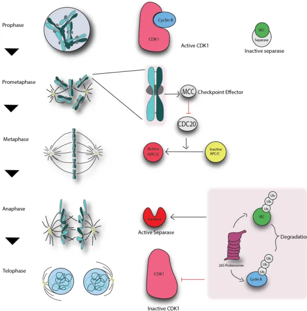
Figure 6: Progression through mitosis. Mitosis proper in general involves four major stages- Prophase, metaphase, anaphase and telophase. The stages are shown by schematic of the mitotic spindle and chromosomes. Sister chromatids, the two identical copies of a chromosome, are separated at the end of the mitosis into two equal daughter cells. (diagram inspired from Cheeseman and Desai, 2008157
)
In telophase, many mitotic changes revert back to the interphase state; chromosomes decondense and the nuclear envelope reassembles around two individual nuclei. Finally, cytokinesis physically splits the cytoplasm to form the two new daughter cells. To ensure smooth progression of the cell cycle, cell cycle checkpoints constantly monitor the molecular mechanistics of cell division.
Monitoring the order and fidelity of chromosome alignment and segregation through mitosis and meiosis is largely achieved by the actions of two checkpoints during mitosis: the spindle assembly checkpoint (SAC) and the mitosis exit network (MEN). The SAC functions in metaphase to prevent premature separation of sister chromatids at anaphase
160-162
. The MEN acts at the end of telophase and control cytokinesis and cell division itself 163. Accurate chromosome segregation is essential for genome inheritance and cellular fitness. Lethality or aneuploidy results when chromosomes fail to segregate during mitosis. Aneuploidy leads to aberrant gene dosage and exposes detrimental recessive mutations, potentially causing birth defects and promoting cancer cell proliferation164,165
. Accurate segregation is achieved by linking sister chromatids after replication, which is mediated by spindle microtubules that attach to chromosomes at the kinetochores.
1.4.1 Chromosome - microtubule attachment 1.4.1.1 The kinetochores
The kinetochore is a hierarchical protein assembly composed of nearly 100 proteins that links centromeric DNA to spindle microtubules and thereby couples forces generated by microtubule dynamics to power chromosome movement. Core components of the kinetochore is established by the constitutive centromere associated network (CCAN)166 and the Knl1-Mis12-Ncd80 (KMN) protein complex167
, which bind centromeric DNA and microtubules, respectively. These networks are conserved across eukaryotes, with additional contributions from species-specific auxiliary DNA and microtubule binding proteins. Regulatory proteins at the kinetochore safeguard against erroneous segregation and thereby increase the fidelity of mitosis in two ways. First, attachments on bi-oriented kinetochore pairs are selectively stabilized, whereas erroneous attachments are destabilized and eliminated. This allows for another opportunity for bi-orientation. Second, unattached kinetochores are the primary signal to activate the SAC. The competing need for speed and fidelity in chromosome segregation are integrated mainly at the kinetochore. The KMN network is an essential and conserved complex of proteins that constitutes the core microtubule binding activity at the kinetochore and is a platform for SAC signaling. In addition to mediating chromosome spindle attachment, the kinetochore also plays an essential role in relaying microtubule binding status to the SAC to delay exit from metaphase and chromosome segregation.
1.4.1.2 The KMN network
The kinetochore localized KMN network is composed of Knl1 (Kinetochore null protein 1), four subunits of Mis12 (Mis-segregation 12) and four subunits of Ndc80/Hec1 (Nuclear division cycle 80) (Fig. 7). The Ndc80 complex is a heterotetramer comprising Ndc80/Hec1, nuclear filamentous 2 (Nuf2), spindle pole component 24 (Spc24) and Spc25. The site where kinetochores are assembled is determined by the presence of a modified histone H3 or Cenp-A in humans, within nucleosomes at the periphery of each sister centromere. The KMN network associates with kinetochores in prophase and disappears from kinetochores in telophase168
. Heterodimers of Spc24-Spc25 and Ndc80/Hec1-Nuf2 interact via coiled coil domains and assemble into a coiled like structure with distinct functional domains at each end169-172
. The globular domains of the Ndc80/Hec1-Nuf2 heterodimer fold into a calponin homology domain, which mediates microtubule binding167,173,174
. The Spc24-Spc25 heterodimer globular domains are essential for kinetochore targeting of the Ndc80/Hec1 complex, as they directly bind to the Mis12 complex175
and CCAN components176
. To couple chromosome movement to microtubule dynamics, an electrostatic interaction between the basic amino terminal tail of the Ndc80/Hec1 protein and the acidic E-hook of tubulin confers affinity172,173,177
. The complex then binds to microtubules by recognizing both α-tubulin and β-tubulin at the inter- and intra-tubulin interfaces177
. The Ndc80/Hec1 complex binds to the microtubule every 4 nm space, acting as a sensor allowing it to detach near depolymerizing microtubule ends.
Knl1 has a microtubule binding activity, which enhances the binding of the KMN network with microtubules in vitro167
. The Mis12 complex function as an inter-complex scaffold that links the KMN network to the centromeric DNA via direct association with the CCAN protein CenpC178,179
. The Mis12 complex also bridges Knl1 and Ndc80 complex at the kinetochores175
.
A number of other proteins within and at the periphery of the kinetochore outer domain depend on the presence of members of the KMN network for their kinetochore localization. These include MT-associated proteins in the proximity of kinetochore MT plus ends and members of the SAC160,180
. Current understanding of how KMN networks promote kinetochore function is limited and requires further work.
Figure 7: Organization of the KMN network. The KMN network consists of KNL1, NDC80 and MSL12. The four subunits of the MSL12 complex bridges KNL1 and the NDC80 complex to the constitutive centromere associated network (CCAN) and centromeric DNA. (diagram inspired from Emily A. Foley 2013181)
1.4.2 Activation of the spindle assemble checkpoint
In 1991, two independent screens identified various genes, mutation of which bypassed the ability of wild type S. cerevisiae cells to arrest in mitosis in the presence of spindle poisons182,183
. The genes which are conserved across eukaryotes, include the human Ser/Thr kinases multipolar spindle protein 1 (Mps1) and budding uninhibited by benomyl 1 (Bub1), as well as the non-kinase components including mitotic arrest deficient 1 (Mad1), Mad2, Bub3 and the likely pseudo-kinase Bub1 related (BubR1)182-184
. These genes are collectively involved in a pathway that is active in prometaphase and which prevents the premature separation of sister chromatids185,186
. This pathway constitutes the spindle assembly checkpoint (SAC). These proteins delay the activation of Cdc20, a cofactor of the E3 ubiquitin ligase known as anaphase promoting complex/cyclosome (APC/C)187,188
. The APC/C is a master regulator of anaphase entry189 . A mitotic checkpoint complex (MCC) that contains three SAC proteins, Mad2, BubR1/Mad3 and Bub3, as well as Cdc20 acts as a SAC effector. The MCC binds the
APC/C and seems to render it unable to exercise its ubiquitin-ligase activity on securin and cyclin B190-196
. Besides MCC, other core SAC components include Mad1, Bub1, Mps1 and Aurora-B. These proteins are required to amplify the SAC signal and the rate of MCC formation197
. The SAC inhibits the APC/C functions by inactivation of Cdc20 through the MCC complex198
.
The key step in MCC formation is conformational activation of Mad2 from the free ‘open’ form (O-Mad2) to the Cdc20-bound ‘closed’ form (C-Mad2)198,199
. This conversion is a catalytic process, occurring through the association of soluble O-Mad2 with kinetochore bound C-Mad2. Mad1 is the receptor for C-Mad2 at the kinetochore, distinct from Cdc20-bound C-Mad2, which facilitates Mad2 conformational conversion. The kinetochore at this point promotes Mad2 conversion through hierarchical recruitment of SAC proteins. This cascade seems to consist of kinases Aurora-B and Mps1 at top, followed by recruitment of the Bub1-Bub3 complex, then by the recruitment of BubR1-Bub3, and finally by recruitment of a heterotetramer composed of Mad1 and Mad2200-204
.
1.4.2.1 Bub-related protein kinetochore recruitment
Recently it has been established that core kinetochore protein Knl1 recruits Bub1, BubR1 and Bub3205-208
, although complex recruitment isn’t clearly understood. Bub1, a protein kinase, and BubR1, a pseudokinase in vertebrates, contain catalytic domains that are universally required for the checkpoint and are important for kinetochore bi-orientation209,210
. Bub1 and BubR1 bind to Bub3 through a Bub3-binding domain also known as GLEBS domain. Bub1 interacts via its TPR motif with the KI motif on Knl1 of the kinetochore204,205
. Mps1 kinase activity stimulates Bub1 localization and checkpoint activation and Mps1 mediated phosphorylation of Thr residues on the MELT-like motifs of Knl1, which is required for Bub1 kinetochore localization206-208
. Crystallography and biochemical studies have shown that Bub3 binding to Knl1 is the key step in localizing Bub1-Bub3 to kinetochores211
. In contrast, BubR1 localization depends on Bub1 but not through Bub3-KNL1 binding. It is suggested that Bub1 directly recruits BubR1 through dimerization212-214
1.4.2.2 Mad1 and Mad2 kinetochore localization
Although Bub1-Bub3 localization is required for the checkpoint, its localization does not always correlate with checkpoint activation. For instance, some Bub1 is retained on early anaphase kinetochores203,215,216
and Bub1 but not Mad1 is present on kinetochores bound to the sides of microtubules, which do not signal the checkpoint217
. Bub1 is a key component in localizing Mad1 to the kinetochore via its RLK motif218
. Another Mad1 co-receptor Rod-Zwilch-Zw10 (RZZ) is thought to play role in Mad1 localization219
. Knl1 and its constituent binding partner Zwint are required to localize RZZ220,221
and RZZ localization may be regulated through Aurora-B dependent phosphorylation of Zwint222
. RZZ is required for stable Mad1 localization in human cells. The kinetochore dynamics of Mad1 consists of two roughly equal sized pools: a more stably bound pool and a mobile, high turn-over pool223. Mad2 when bound to Mad1 adopts the C-Mad2 conformation and doesn’t seem to dissociate from Mad1 during checkpoint activation.
Mad1-C-Mad2 accounts for the more stable kinetochore pool of Mad2. C-Mad2 bound to Mad1 is the kinetochore receptor for O-Mad2224-226
. Therefore, the mobile and immobile fractions of kinetochore Mad2 consist, respectively, of rapidly cycling C- and O-Mad2 and the Mad1-Mad2 receptor.
1.4.2.3 The Mad2 template model
How kinetochores promote MCC formation is not entirely clear. Fluorescence recovery after photobleaching (FRAP) experiments revealed that some of the checkpoint proteins are stably bound to unattached kinetochores (Bub1, Mad1 and a pool of Mad2), whereas other checkpoint components turnover more rapidly (BubR1, Mps1, Bub3, a pool of Mad2 and Cdc20) supporting the idea that unattached kinetochores catalyze the formation of a diffusible checkpoint inhibitor201,223
. The existence of C-Mad2 and O-Mad2 lead to the O-Mad2 template model227
(Fig. 8). The fundamental principle is that O-Mad2 can dimerize with C-O-Mad2, which induces a conformational change from O-O-Mad2 to C-Mad2, thereby binding to Cdc20. Unattached kinetochores stably bind a tetrameric Mad1:C-Mad2 complex226,228
and thus, unattached kinetochores can serve as template for continuous conversion of cytosolic O-Mad2 molecules. This model explains how a single unattached kinetochore can generate an efficient checkpoint response. However, it also
predicts that the checkpoint signal spreads in the cytoplasm: C-Mad2:Cdc20 could then serve as template and hence uncouple checkpoint signaling from unattached kinetochores. Amplification of C-Mad2 away from kinetochores might be prevented by several mechanisms. First recruitment of O-Mad2 to Mad1:C-Mad2 depends on Mps1 activity229
, a process that might be restricted to the kinetochore environment. Second, in the cytosol the dimerization interface of C-Mad2 is blocked when bound to p13/comet,
a protein with structural similarity to Mad2230
. Third, to form functional inhibitory MCC, C-Mad2:Cdc20 forms a complex with the Bub3:BubR1 where binding to Mad3 has been shown to block the dimerization interface of Mad2231
.
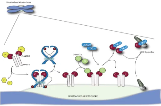
Figure 8: Mad2-template model of MCC production at unattached kinetochores. In the cytoplasm C-Mad2 forms a tetrameric complex with Mad1, but dimerization with O-Mad2 is blocked by p31/comet. Upon Mps1 phosphorylation Mad1:C-Mad2 binds to unattached kinetochores. This releases p31/comet, and together with Mps1 activity allows cytosolic O-Mad2 to dimerize with C-Mad2. This initiates a conformational change and enables the formation of C-Mad2:Cdc20, which subsequently assembles with the Bub3:BubR1 complex to form the MCC (diagram inspired from Lara-Gonzales et al., 2012232).