HAL Id: hal-03003382
https://hal.archives-ouvertes.fr/hal-03003382
Submitted on 13 Nov 2020
HAL is a multi-disciplinary open access
archive for the deposit and dissemination of
sci-entific research documents, whether they are
pub-lished or not. The documents may come from
teaching and research institutions in France or
abroad, or from public or private research centers.
L’archive ouverte pluridisciplinaire HAL, est
destinée au dépôt et à la diffusion de documents
scientifiques de niveau recherche, publiés ou non,
émanant des établissements d’enseignement et de
recherche français ou étrangers, des laboratoires
publics ou privés.
Inhibitor ofMycobacterium tuberculosisMycolic Acid
Methyltransferases
Julien Vaubourgeix, Fabienne Bardou, Fanny Boissier, Sylviane Julien,
Patricia Constant, Olivier Ploux, Mamadou Daffé, Annaïk Quémard, Lionel
Mourey
To cite this version:
Julien Vaubourgeix, Fabienne Bardou, Fanny Boissier, Sylviane Julien, Patricia Constant, et al..
S-Adenosyl-N-decyl-aminoethyl, a Potent Bisubstrate Inhibitor ofMycobacterium tuberculosisMycolic
Acid Methyltransferases. Journal of Biological Chemistry, American Society for Biochemistry and
Molecular Biology, 2009, 284, pp.19321 - 19330. �10.1074/jbc.m809599200�. �hal-03003382�
S-Adenosyl-N-decyl-aminoethyl, a Potent Bisubstrate Inhibitor
of Mycobacterium tuberculosis Mycolic Acid Methyltransferases
□SReceived for publication, December 19, 2008, and in revised form, March 26, 2009 Published, JBC Papers in Press, May 13, 2009, DOI 10.1074/jbc.M809599200
Julien Vaubourgeix‡§, Fabienne Bardou‡§, Fanny Boissier‡§1, Sylviane Julien‡§, Patricia Constant‡§, Olivier Ploux¶, Mamadou Daffe´‡§, Annaïk Que´mard‡§2, and Lionel Mourey‡§3
From‡CNRS, Institut de Pharmacologie et de Biologie Structurale, De´partement Me´canismes Mole´culaires des Infections Mycobacte´riennes, 205 Route de Narbonne, F-31077 Toulouse, the§Universite´ de Toulouse, Universite´ Paul Sabatier, Institut de Pharmacologie et de Biologie Structurale, F-31077 Toulouse, and the¶Laboratoire de Biochimie des Micro-organismes: Enzymologie, Me´tabolisme, et Antibiotiques, Ecole Nationale Supe´rieure de Chimie de Paris, CNRS UMR 7573, F-75231 Paris, France
S-Adenosylmethionine-dependent methyltransferases (Ado-Met-MTs) constitute a large family of enzymes specifically transferring a methyl group to a range of biologically active mol-ecules. Mycobacterium tuberculosis produces a set of paralo-gous AdoMet-MTs responsible for introducing key chemical modifications at defined positions of mycolic acids, which are essential and specific components of the mycobacterial cell envelope. We investigated the inhibition of these mycolic acid methyltransferases (MA-MTs) by structural analogs of the AdoMet cofactor. We found that S-adenosyl-N-decyl-amin-oethyl, a molecule in which the amino acid moiety of AdoMet is substituted by a lipid chain, inhibited MA-MTs from Mycobac-terium smegmatis and M. tuberculosis strains, both in vitro and in vivo, with IC50values in the submicromolar range. By
con-trast, S-adenosylhomocysteine, the demethylated reaction product, and sinefungin, a general AdoMet-MT inhibitor, did not inhibit MA-MTs. The interaction between Hma (MmaA4), which is strictly required for the biosynthesis of oxygenated mycolic acids in M. tuberculosis, and the three cofactor analogs was investigated by x-ray crystallography. The high resolution crystal structures obtained illustrate the bisubstrate nature of S-adenosyl-N-decyl-aminoethyl and provide insight into its mode of action in the inhibition of MA-MTs. This study has potential implications for the design of new drugs effective against multidrug-resistant and persistent tubercle bacilli.
One-third of the world population is infected with the tuber-cle bacillus, Mycobacterium tuberculosis, and tuberculosis kills one person every 20 s. The inhaled pathogenic bacilli are taken up by phagocytosis by pulmonary macrophages, which, together with lymphocytes and dendritic cells, form granulo-mas. The bacilli persist in the granuloma until their reactiva-tion, dissemination into the lungs, and the triggering of disease. The natural resistance of persistent tubercle bacilli to drugs and the emergence of multidrug-resistant and extensively
drug-re-sistant M. tuberculosis strains are two main concerns in the treatment of the disease. A survey carried out by the Centers for Disease Control and Prevention and the World Health Organi-zation between 2000 and 2004 reported that 20% of 17,690 M.
tuberculosisisolates from 49 countries were multidrug-resist-ant, and 2% were extensively drug-resistant (1). The develop-ment of new drugs effective against persistent and drug-resis-tant bacilli has therefore become a priority.
The thick lipid-rich envelope of the Mycobacterium genus is characterized by the presence of mycolic acids (MAs),4
very long chain (C60–C90)␣-alkylated -hydroxylated fatty acids (2).
MAs are the major components of the mycomembrane (3, 4) lipid bilayer, which plays a key role in both the architecture and permeability of the mycobacterial envelope. The MA biosyn-thetic pathway is essential for mycobacterial survival. MAs are generated by Claisen condensation between two fatty acyl chains as follows: the very long meromycoloyl chain (C40–C60)
and a shorter saturated chain (C22–C26) (2). The different types
of MAs are defined by the presence of decorations introduced at proximal and distal positions of the meromycolic chain (Fig. 1A) by a family of paralogous S-adenosylmethionine-depend-ent methyltransferases (AdoMet-MTs), the mycolic acid meth-yltransferases (MA-MTs). These chemical modifications are known to be important for the pathogenicity, virulence, and persistence of M. tuberculosis. For example, the cis-cyclopro-pane introduced at the proximal position of␣-MAs by PcaA has an impact on the persistence of the tubercle bacillus within infected organisms (5). Furthermore, the keto and methoxy groups, with a vicinal methyl ramification at the distal position of oxygenated MAs, play a role in M. tuberculosis virulence in the mouse model of infection (6) and have recently been reported to be involved in host-pathogen interplay. Indeed, oxygenated MAs have been shown to modulate IL-12p40 pro-duction by macrophages (7) and to trigger the in vitro differen-tiation of monocyte-derived macrophages into foamy macro-phages, which house the bacillus in a dormant state, within granulomas (8). Oxygenated MA biosynthesis requires the
□S The on-line version of this article (available at http://www.jbc.org) contains
supplemental Figs. S1–S3 and Table 1.
1Present address: Institut de Biochimie et Ge´ne´tique Cellulaires, UMR 5095
CNRS-Universite´ Victor Segalen Bordeaux 2, 33077 Bordeaux Cedex, France.
2To whom correspondence may be addressed. Tel.: 33-561-175-576; Fax:
33-561-175-994; E-mail: annaik.quemard@ipbs.fr.
3To whom correspondence may be addressed. Tel.: 33-561-175-436; Fax:
33-561-175-994; E-mail: lionel.mourey@ipbs.fr.
4The abbreviations used are: MA, mycolic acid; CFAS, cyclopropane
fatty-acid synthase; CTAB, cetyltrimethylammonium bromide; DMSO, di-methyl sulfoxide; MIC, minimum inhibitory concentration; MA-MT, mycolic acid methyltransferase; SADAE,
S-adenosyl-N-decyl-amin-oethyl; AdoMet, S-adenosyl-L-methionine; AdoMet-MT,
S-adenosyl-methionine-dependent methyltransferase; AdoHcy, S-adenosyl-L
-homocysteine; MES, 4-morpholineethanesulfonic acid.
THE JOURNAL OF BIOLOGICAL CHEMISTRY VOL. 284, NO. 29, pp. 19321–19330, July 17, 2009 © 2009 by The American Society for Biochemistry and Molecular Biology, Inc. Printed in the U.S.A.
at BIBL SANTE on July 10, 2009
www.jbc.org
Hma (MmaA4) methyltransferase (Fig. 1B), as demonstrated by the absence of the oxygenated form in an M. tuberculosis hma knock-out mutant (6, 9). These results suggest that the enzymes responsible for adding the decorations to MAs, including oxy-genated groups in particular, may be relevant pharmacological targets for the development of new antituberculous drugs (10). Based on the essential role played by MA-MTs in the phys-iopathology of tuberculosis, several studies have investigated the possible inhibition of this family of enzymes. A recent study revealed that the antituberculous drug thiacetazone and its chemical analogs inhibited MA cyclopropanation at concentrations in the micromolar range (11). Another study, based on mixtures of crude extracts of heat-inactivated mycobacteria and recombinant Escherichia coli
overproduc-ing MA-MTs, suggested that the incorporation of [3H]AdoMet into
growing meromycolic chains is inhibited by a high concentration (1 mg/ml, i.e. 2.6 mM) of S-adeno-syl-L-homocysteine (AdoHcy) or sinefungin (12), the demethylated reaction product and a natural structural analog of AdoMet, respectively (Fig. 2). By contrast, AdoHcy and sinefungin are strong inhibitors of other AdoMet-MTs in
vitro, including the cyclopropane fatty-acid synthase (CFAS) from
E. coli(Kiof 30 and 0.22M,
respec-tively) (13, 14). However, they are active only against the isolated enzyme, whereas S-adenosyl-N-de-cyl-aminoethyl (SADAE), a mole-cule in which the amino acid moiety of AdoMet is substituted by a lipid chain (Fig. 2), is active against CFAS both in vitro (Ki,app⫽ 6M) and in
vivo(complete inhibition at 150M) (15). The broad screening of possi-ble inhibitors of MA-MTs with an in
vitromini-assay poses a major chal-lenge, as these enzymes most likely use very long meromycolic chains as substrates. In this context, the simi-larity between CFAS and Hma in terms of their sequences (31% sequence identity) and substrates may be useful, as it suggests that SADAE may inhibit MA-MTs (15).
We report here our investigations of the interactions between Hma and SADAE, as compared with those between Hma and AdoHcy or sinefungin, and the potential impact of these interactions on the activi-ties of Hma and other MA-MTs and mycobacterial growth. Our high resolution crystallographic charac-terization of the Hma-SADAE interaction illustrates the bisub-strate nature of the ligand, which is strongly correlated with its strong inhibitory properties.
MATERIALS AND METHODS
Strains and Cultures—M. tuberculosis H37Rv was grown at 37 °C in Middlebrook 7H9 broth (Difco) supplemented with 0.2% (w/v) glycerol. Mycobacterium smegmatis mc2155/
pMV261 and M. smegmatis mc2155/pMV261::hma (mmaA4,
Rv0642c) strains were grown in the same medium supple-mented with 10g/ml kanamycin and, in some cases, albumin/ dextrose/catalase. These recombinant strains were prepared as described previously (16).
FIGURE 1. A, structures of MAs from M. tuberculosis and M. smegmatis. D, distal position; P, proximal position. Enzymes involved in the introduction of decorations on the meromycolic chain are indicated. B, proposed
reaction scheme for the introduction of oxygenated groups. m⫽ 17, 19; n, unknown; X, unknown carrier.
at BIBL SANTE on July 10, 2009
www.jbc.org
Preparation of Cell Wall Extracts—Freshly grown bacteria were washed with 10 mMpotassium phosphate buffer, pH 7.0, and resuspended in 50 mMpotassium phosphate buffer, pH 7.0, plus 3 mM-mercaptoethanol (buffer A). Bacteria were lysed by cell disruption, by two passages through a One-shot Cell Dis-rupter (Constant Systems Ltd.) at 0.8 kbar. The crude extracts were centrifuged for 15 min at 3,000⫻ g and at 4 °C. The very dense layer at the surface of the supernatant, consisting of cell wall and membrane fragments (17), was removed, resuspended in buffer A, and homogenized with a syringe (needle size 0.7⫻ 30 mm). The protein concentration of the extracts was deter-mined with the DC method (Bio-Rad) for insoluble proteins, after denaturation of the proteins by boiling in 0.5NNaOH at 100 °C for 10 min.
Evaluation of Ability of AdoMet Analogs to Inhibit MA Bio-synthesis in Vitro—MA biosynthesis was assayed in vitro in a reaction medium containing 100 mM potassium phosphate buffer, pH 7.0, 3 mMMgCl2, 7 mMKHCO3, 7 mMATP, 0.7 mM
CoASH, and 500g of cell wall extract proteins in a total vol-ume of 750l. Mixtures were first incubated in the presence or absence of AdoMet analogs (i.e. SADAE (10M), dissolved in dimethyl sulfoxide (DMSO) such that the maximum final sol-vent concentration in the reaction medium was 5% (v/v); AdoHcy (1 mM) and sinefungin (3 mM), evaluated either with or without DMSO) for 15 min at 37 °C, and we then added 50M of either [1-14C]acetate for MA biosynthesis assays or [CH
3 -14C]AdoMet for MA-MT activity assays. Reaction mixtures
were incubated at 37 °C for 60 min; the reactions were stopped
by saponification, and the MA profiles were analyzed by TLC (see below).
The apparent IC50value of SADAE was determined by meas-uring the rate of MA biosynthesis in the presence of various concentrations of inhibitor (from 10 pMto 10M), using the same procedure as that described above, and calculated by fit-ting the data by nonlinear regression using the program Graph-Pad Prism 4.02.
[1-14C]Acetate (specific activity, 56.60 mCi/mmol) and
[CH3
-14C]AdoMet (specific activity, 50.43 mCi/mmol) were
purchased from PerkinElmer Life Sciences. Sinefungin (Mr⫽ 381.4 g/mol) and AdoHcy (Mr⫽ 384.4 g/mol) were obtained
from Sigma, and SADAE (Mr⫽ 466.65 g/mol) was obtained
from the Institut de Chimie des Substances Naturelles (Gif sur Yvette, France).
Evaluation of the in Vivo Effects of SADAE on MA Bio-synthesis—SADAE (50 and 90M) was added to broth cultures of M. smegmatis in the exponential growth phase. The cultures were then incubated at 37 °C for 90 min before the addition of 6.8M[1-14C]acetate. Cells were incubated for 3 or 6 h and then harvested. For M. tuberculosis cultures, also in the expo-nential growth phase, SADAE was added at concentrations of 25, 50, and 90M. Cultures were incubated at 37 °C for 7 h before the addition of [1-14C]acetate and then for a further
24 h before the harvesting of whole cells for MA content characterization.
Extraction and Analysis of MAs—Reaction media containing cell wall extracts or harvested whole bacteria were saponified by incubation with a mixture of 40% KOH (w/v)/methoxyethanol (1:7) for 3 h at 110 °C in a screw-capped tube (18). The suspen-sions were acidified by adding an equal volume of 20% H2SO4,
and fatty acids were extracted with diethyl ether. The neosyn-thesized and radiolabeled MAs were methylated and analyzed by TLC on Silica Gel 60 (Macherey-Nagel) run in dichlo-romethane for M. smegmatis strains and in petroleum ether/ diethyl ether (9:1) (five runs) for M. tuberculosis (18), followed by phosphorimaging (Variable Mode Imager Typhoon TRIO, Amersham Biosciences).␣-MAs were purified by preparative TLC on Silica Gel 60, and fractionated on AgNO3-impregnated
silica gel TLC plates developed with dichloromethane (9, 19). The total labeled and unlabeled lipids in the extracts were visu-alized by spraying with molybdophosphoric acid (10% in etha-nol) and charring. The radiolabeled spots were identified based on the Rfvalues of the fatty and mycolic acids visualized by the
latter method and by comparison with previous data (16, 19).
Inhibition of Mycobacterial Growth—The susceptibility of M.
smegmatisstrains or of M. tuberculosis H37Rv to SADAE was evaluated by determining the minimum inhibitory concentra-tion (MIC). We used a colorimetric microassay based on the reduction of 3-(4,5-dimethylthiazol-2-yl)-2,5-diphenyltetrazo-lium bromide (Sigma) to formazan by metabolically active cells (20 –22). Briefly, serial 2-fold dilutions of the inhibitor were prepared in 100l of Middlebrook 7H9 broth base (Difco) and dispensed into 96-well microtiter plates, and to each well we added 100l of M. tuberculosis or M. smegmatis suspension (diluted in 7H9 broth to an A600of 0.02). Plates were incubated for 6 days at 37 °C, and 3-(4,5-dimethylthiazol-2-yl)-2,5-diphe-nyltetrazolium bromide was added (50l of a 1 mg/ml
solu-FIGURE 2. Structure of AdoMet and of the AdoHcy, sinefungin, and
SADAE analogs. at BIBL SANTE on July 10, 2009
www.jbc.org
tion). Plates were incubated for a further 24 h, and solubiliza-tion buffer (dimethylformamide/SDS 20% (w/v), 1:2) was added to each well. Absorbance was measured at 570 nm. The MIC was determined as the lowest concentration of SADAE yielding 100% inhibition of bacterial growth (the absorbance value obtained for untreated bacilli was taken as a growth control). Ethambutol- and isoniazid-treated cultures were used as posi-tive controls.
X-ray Crystallography of Complexes of Hma with AdoMet Analogs—Crystals of apo-Hma were obtained from the purified His6-tagged protein, as described previously (16). Crystals of the complex with AdoHcy were prepared by cocrystallization in the presence of 2 mMcofactor product. The crystals were cryo-cooled in a stream of nitrogen gas at 100 K after immersion for 3 min in the crystallization solution supplemented with 20% (v/v) glycerol. For preparation of the complex with sinefungin, crystals of the apo-form were soaked in a solution containing both the cryoprotectant and 50 mMsinefungin for 2 min. The complex with SADAE was prepared by soaking crystals of the apo-form three times in freshly prepared drops of 5 mMSADAE in crystallization conditions and 5% (v/v) DMSO, for a total soaking time of 15 min. A fourth soaking was then carried out during 3 min in a drop containing the same solution supple-mented with 20% glycerol. All diffraction data were collected at multistation beam-line ID14 of the European Synchrotron Radiation Facility (Grenoble, France).
The CCP4 suite (23), as implemented in the graphical user interface (24), was used for crystallographic calculations. X-ray diffraction data were processed with MOSFLM (25) and scaled
with SCALA (26). The structures of the complexes were solved, using the previously determined crystallographic coordinates of Hma in complex with AdoMet (16) (Protein Data Bank code 2FK8) and discarding non-protein atoms. All structures were con-structed manually inA weighted electron density maps (27), using Coot (28). Restrained refinements were obtained with the REFMAC5 program (29), using a bulk solvent correction based on the Babinet principle and minimizing a maxi-mum likelihood target function. Solvent molecules were automati-cally added as neutral oxygen atoms with wARP (30) and Coot. Ligands were added at the end of refinement after water attribution. The coordi-nates of AdoHcy and sinefungin (adenosylornithine) were obtained from the Hetero-compound Infor-mation Center Uppsala (HIC-Up) (31). The energy-minimized coordi-nates and refinement library for SADAE were generated with the PRODRG server (32). TLS parame-ters were refined, using a single group for the whole molecule (33), resulting in a similar improvement of the R and Rfreevalues.
Protein-ligand interactions were defined with HBPLUS (34). Figs. 7–9 were produced using PyMOL (35).
RESULTS
Action of AdoHcy, Sinefungin, and SADAE on MA Biosynthe-sis in Vitro—We first tested the effects of the three putative inhibitors on MA-MT activities in cell wall extracts of M.
smeg-matis/pMV261 and M. smegmatis/pMV261::hma. With these bacterial extracts, the in vitro biosynthesis of MAs can be fol-lowed in the presence of a radiolabeled marker, [1-14C]acetate.
The neosynthesized and radiolabeled MAs were extracted, meth-ylated, and then analyzed by TLC and phosphorimaging. SADAE almost totally inhibited the biosynthesis of epoxy-MAs in M.
smeg-matis/pMV261 and that of epoxy-, keto-, and hydroxy-MAs in M.
smegmatis/pMV261::hma (Fig. 3A), at a concentration similar to the Kiof CFAS, 10M(15). By contrast, concentrations of the
structural analogs AdoHcy and sinefungin 100 –300 times higher than the effective concentration of SADAE had no significant impact on these metabolic pathways (Fig. 3A).
A potentially specific effect of SADAE on the various species of␣-MAs was investigated further after separation on AgNO3
-impregnated TLC plates. Interestingly, in the treated cell wall extracts of both transformed and untransformed M. smegmatis strains, cis,cis-diethylenic␣-MA (␣5) labeling essentially
per-sisted (Fig. 3B), whereas the labeling of cis,cis or cis,trans-dicy-clopropanic (␣1), cis-cyclopropanic,trans-ethylenic (␣2), and
cis,trans-diethylenic (␣4) MAs was undetectable and that of the
FIGURE 3. Effect of AdoMet analogs on MA biosynthesis and MA-MT activities in vitro. A, MA biosynthesis
assays with M. smegmatis/pMV261 and M. smegmatis/pMV261::hma cell wall extracts and AdoHcy (1 mM),
sinefungin (3 mM), or SADAE (10M). Neosynthesized MAs were labeled by incubation with [1-14C]acetate.
Nonradiolabeled reference lipids were visualized by spraying molybdophosphoric acid (left and right lanes).
B, [1-14C]acetate-labeled␣-MA methyl esters were scraped off the plate shown in A and separated by TLC on
AgNO3-impregnated plates. C, MA-MT activity assays with [CH3
-14C]AdoMet labeling. All TLC runs were
per-formed with dichloromethane, and radiolabeled spots were detected by phosphorimaging and identified by
comparison of their Rfwith those of known lipids. F, fatty acid methyl esters; K, keto-; E, epoxy-; H, hydroxy-MA
methyl esters.␣1, cis,trans- or cis,cis-dicyclopropanic;␣2, cis-cyclopropanic,trans-ethylenic;␣3,
cis-cyclopropan-ic,cis-ethylenic;␣4, cis,trans-diethylenic;␣5, cis,cis-diethylenic MA methyl esters. at BIBL SANTE on July 10, 2009
www.jbc.org
cis-cyclopropanic,cis-ethylenic MA (␣3) was weaker (Fig. 3B
and Fig. 1A). These data strongly suggest that SADAE inter-fered with the activity of MA-MTs. We tested this hypothesis by carrying out a similar experiment with [CH3
-14C]AdoMet,
which is specific for AdoMet-MT activity. In these conditions, only MAs bearing a cyclopropane (␣-MA subpopulation in M.
smegmatis(19)) or a methyl branch (oxygenated MAs or␣1- to
␣4-MAs) were radiolabeled in untreated cell wall extracts
because they incorporated14C-methyl groups (Fig. 3C).
Radio-activity was also detected in methyl-branched fatty acids, cor-responding to tuberculostearic acid (10-methylstearic acid) and its homolog 10-methylpalmitic acid (Fig. 3C and supple-mental Fig. 1). As expected, almost no labeling of MAs or fatty acids was observed in the presence of SADAE (Fig. 3C).
The apparent IC50value of SADAE was determined by
meas-uring the rate of oxygenated MA biosynthesis over a large range of inhibitor concentrations. The IC50values for keto-MA and
for epoxy-MA synthesis were 0.065⫾ 0.013M(0.031⫾ 0.006 g/ml) and 0.713 ⫾ 0.036 M(0.333⫾ 0.017g/ml),
respec-tively. The submicromolar IC50values reflect the high potential
of SADAE to inhibit MA-MTs.
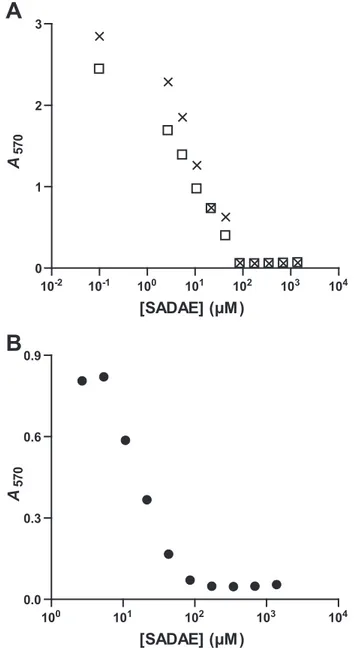
SADAE Inhibits the Growth of M. smegmatis and M. tuberculosis—The failure of most antibiotics to permeate the exceptionally impermeable mycobacterial cell wall is a major cause of poor whole-cell potency and a lack of susceptibility to drugs in mycobacteria (36). We also assessed the impact of SADAE on the growth of mycobacterial species by determin-ing MICs for M. smegmatis/pMV261, M. smegmatis/ pMV261::hma, and M. tuberculosis. The mycobacterial cells were grown in microplates, and serial 2-fold dilutions of SADAE were added during the exponential growth phase. The final counts of bacteria were determined by measuring the metabolism of a tetrazolium salt into a colored compound, formazan, detected at 570 nm. Curves showed growth inhibi-tion at similar concentrainhibi-tions for both M. smegmatis and M.
tuberculosis, corresponding to MIC values of about 100M(47
g/ml) (Fig. 4). Thus, SADAE is able to reach targets within the bacilli and to stop mycobacterial growth.
Action of SADAE on MA-MTs in Vivo—We investigated whether MA-MTs were particularly accessible to SADAE and could be considered among its targets in vivo, by evaluating the effect of this compound on the biosynthesis of MAs in intact mycobacteria. We first treated M. smegmatis/pMV261 and M.
smegmatis/pMV261::hma cultures at (100 M) or below (50
M) the MIC, for 90 min, and then added [1-14C]acetate. Cul-tures were incubated for 3 or 6 h, and the lipids were then extracted. TLC analysis showed that SADAE almost totally inhibited the biosynthesis of epoxy-, keto-, and hydroxy-MAs at the MIC (Fig. 5A andsupplemental Fig. 2). We then carried out similar experiments on M. tuberculosis. Bacteria were treated with SADAE concentrations of 25–90Mfor 7 h, and
were then labeled with [1-14C]acetate for 24 h (Fig. 5B). The
biosynthesis of oxygenated MAs was strongly inhibited. Rela-tive radiolabeling levels for these MAs were up to 86% lower than those in the absence of SADAE, whereas␣-MA and regu-lar size fatty acid levels remained stable (Fig. 5D).
We investigated the effects of SADAE treatment on the structure of␣-MAs in more detail by purifying these
com-pounds and analyzing them by argentation TLC (Fig. 5C). Untreated M. tuberculosis produced dicyclopropanated MAs (␣1⬘), but the profile of compounds produced was radically
changed by SADAE treatment. SADAE strongly inhibited the synthesis of␣1⬘-MAs (86% inhibition at 90M) and induced the
accumulation of several new species with a much smaller Rf
(Fig. 5E). The migration of these compounds corresponded to that of mono- and di-ethylenic␣-MAs (␣2⬘ and␣3⬘,
respec-tively) (9, 19). The appearance of unsaturated␣-MAs probably results from the inhibition of PcaA and MmaA2, which intro-duce cis-cyclopropane rings into␣1⬘-MAs, and the inhibition of
Hma and MmaA2/CmaA2, which catalyze the biosynthesis of
cis/trans-cyclopropanated oxygenated MAs (Fig. 1). These data collectively demonstrate that SADAE also inhibits MA-MTs with cyclopropane synthase activity.
In conclusion, SADAE concentrations equivalent to the MIC inhibit MA-MTs in vivo in both M. smegmatis and M. tuberculosis.
FIGURE 4. Determination of the MIC values of SADAE. A, M. smegmatis/ pMV261 (crosses) and M. smegmatis/pMV261::hma (open squares). B, M.
tuberculosis.
at BIBL SANTE on July 10, 2009
www.jbc.org
For oxygenated groups, the activities of Hma in MA biosynthesis in M. tuberculosis and of uncharacterized enzymes in the synthesis of M. smegmatis epoxy-MAs were at least impaired, whereas the effects of SADAE on the activity of MmaA3, the MA-MT cata-lyzing the introduction of a methyl group downstream from the Hma reaction (Fig. 1B), remained unclear.
Determination and Comparison of the Three-dimensional Structures of Hma in Complex with AdoHcy, Sinefungin, and SADAE—We investigated the reasons for this unique effect of SADAE on MA-MT function not observed with AdoHcy and sinefungin by determining the high resolution crystal structure of the complexes formed between Hma and the three ligands. Crystals of His6-tagged Hma prepared in 50 mMMES, 50 mM
NaCl, pH 6.5, were grown from the purified protein, using var-ious amounts of polyethylene glycol 3350 over a broad pH range, as reported previously (16). Complexes with the cofactor analogs were obtained either by cocrystallization or through
soaking experiments. SADAE is not directly soluble in water, and DMSO was therefore required for its solubilization, as for many organic compounds. We used con-centrations of inhibitor no greater than 5 mM to minimize the final concentration of DMSO. At this concentration, a single soaking for a few minutes gave insufficient density for the ligand. By contrast, SADAE binding was achieved by iterative soakings, each time in a new drop. Complete data sets were collected for all three complexes, from single crystals at a maximum resolution of 2.2 to 2.35 Å. Initial model building was based on the known three-dimensional struc-ture of Hma in complex with the cofactor substrate (16), taking into account only the protein atoms. Refinement was run smoothly. We first modified the protein, then introduced water molecules, and finally assigned the compounds whose position and orientation were obvious from the electron den-sity maps (supplemental Fig. 3). Refinement statistics are summa-rized in Table 1. All structures have ⬎91% of the residues in the most favored region of the Ramachand-ran plot and none in the disallowed region, as defined by PROCHECK (37).
The overall structure of the Hma protein in complexes with AdoHcy, sinefungin, and SADAE is very similar to that of Hma-AdoMet (16), giving root mean square deviation (r.m.s.d.) values after superimposition of 0.5 Å/281 C-␣ atoms, 0.5 Å/281 C-␣ atoms, and 0.6 Å/270 C-␣ atoms for the complexes with AdoHcy, sinefungin and SADAE, respectively. All three structures display characteristic folding of residues 158 –160 as 310helix1
(the secondary structure numbering used is from Ref. 16), a marker of AdoMet-MT-cofactor complex formation. As already reported for Hma-apo and Hma-AdoMet (16), there are missing residues at the N terminus, which starts at residue number 21 for AdoHcy and sinefungin, and residue 27 for SADAE. Residues 186 –187 (within helix␣2), residues 263–264 (making a short con-nection between helices␣4 and ␣5), and residue 301 at the C ter-minus are also missing in the Hma-SADAE complex ( supplemen-tal Fig. 3). It has previously been reported that helix ␣B, encompassing residues 105–120, is translated along its axis toward the cofactor binding cleft upon AdoMet binding (16). In the three complexes described here, this helix occupies the same position as in Hma-apo. Thus, helix ␣B seems to display dynamics of
FIGURE 5. Inhibition of MA-MT activities in vivo. Bacteria in culture were treated with various concentrations
of SADAE, and the neosynthesis of mycolic acids in vivo was followed by [1-14C]acetate labeling. A, M.
smegmatis/pMV261::hma. Only points after 6 h of incubation are displayed for reasons of clarity; similar profiles
were obtained after 3 h of incubation. B and C, M. tuberculosis. C,␣-MA methyl esters scraped off the plate
shown in B and separated by TLC on an AgNO3-impregnated silica gel plate. D and E, relative labeling
intensi-ties for the various molecular species assessed on plates B and C, respectively. All runs were performed with dichloromethane (A and C) or petroleum ether/diethyl ether (9:1) (five times) (B), and radiolabeled spots were detected by phosphorimaging. F, fatty acid methyl esters; K, keto-; E, epoxy-; H, hydroxy-; M, methoxy-MA
methyl esters. ␣1⬘, dicyclopropanated;␣2⬘, monoethylenic-monocyclopropanated; ␣3⬘, diethylenic␣-MA
methyl esters.
at BIBL SANTE on July 10, 2009
www.jbc.org
unknown function. Other regions displaying structural variations (deviations exceeding r.m.s.d. values when in comparison with the structure of Hma-AdoMet) are confined to the N-terminal end, the␣2-␣3 motif, and ␣4-␣5, all of which contribute to the embel-lishment pattern of MA-MTs.
Fine Description of the Complexes—The ligands used in this study bear a common adenosine core and specific non-nucleo-side appendages in the C-5⬘ position, the specific appendages of
AdoHcy and sinefungin at this position being similar (Fig. 2). AdoHcy and sinefungin bind to Hma at the same position and in a similar bent conformation as observed for Hma-AdoMet (16) and for other MA-MTs (38). Indeed, all three ligands are posi-tioned on Hma with the ribose lying at the apex of the-sheet contribut-ing to the AdoMet-MT canonical fold, whereas the adenine is sand-wiched on one side of the-sheet, between the tip of strand1, helix 1, and the loop between strand 3 and helixC, with the amino acid portion pointing toward the N ter-minus of helix␣A on the other side of the-sheet (Fig. 6). The superim-position of AdoHcy and sinefungin with AdoMet in Hma complexes gave r.m.s.d. values of 0.2 Å for 26 and 25 common atoms, respec-tively. Specific hydrogen bonds and hydrophobic contacts between both the common adenosine core and the amino acid appendage and protein residues are mostly conserved (Fig. 7, A and B, andsupplemental Table 1).
In the Hma-SADAE complex, the interactions of the adenine and sugar moieties with the protein are also conserved (Fig. 7C and supplemental Table 1), and the r.m.s.d. obtained after
superimpo-FIGURE 6. Three-dimensional structure of the complexes formed between Hma and AdoHcy, sinefungin,
and SADAE. A, ribbon representation of Hma (coordinates from the Hma-AdoHcy complex) showing helices
and-strands of the conserved AdoMet-MTs core in cyan and red, respectively. Helices of the MA-MT
embel-lishment pattern are shown in green. The three ligands were superimposed and are shown as sticks with carbon atoms of the adenosine core in yellow and nitrogen, oxygen, and sulfur atoms in blue, red, and green, respec-tively. The carbon atoms of the decylaminoethyl group of SADAE are shown in navy. B, perpendicular view. The van der Waals surfaces of SADAE atoms are also depicted with dots.
TABLE 1
Data collection and refinement statistics
Crystal
Hma-AdoHcy Hma-sinefungin Hma-SADAE
Data collection
Beam line ID14-2 ID14-1 ID14-4
Space group P3121 P3121 P3121
Unit cell dimensions, Å a⫽ b ⫽ 57.34, c ⫽ 203.98 a⫽ b ⫽ 56.78, c ⫽ 202.79 a⫽ b ⫽ 57.43, c ⫽ 205.08
Resolution limits,aÅ 28.7 to 2.2 (2.32 to 2.20) 27.3 to 2.3 (2.42 to 2.30) 20.0 to 2.35 (2.48 to 2.35)
No. of measured reflections 69,191 (9536) 91,988 (13,557) 73,932 (11,193)
No. of unique reflections 20,193 (2844) 17,694 (2532) 16,765 (2422)
Completeness, % 98.4 (97.8) 99.8 (100.0) 98.2 (99.4) Rsym, % 4.7 (30.1) 6.2 (32.6) 4.6 (32.7) I/I 9.1 (2.4) 8.1 (2.3) 10.1 (2.4) BWilson, Å 2 44.1 43.8 60.2 Refinement statistics Resolution range,aÅ 28.7 to 2.2 (2.26 to 2.20) 27.3 to 2.3 (2.36 to 2.30) 20.0 to 2.35 (2.41 to 2.35)
No. of residues/total expectedb 281/318 281/318 270/318
Missing residues tag, 4–20 tag, 4–20 tag, 4–26, 186–187, 263–264, 301
No. of protein atoms 2256 2235 2081
No. of water molecules 63 67 45
No. of ligand atoms 26 27 32
No. of heteroatoms 2345 2329 2158
No. of reflections work/testa 19,162/1031 16,737/897 15,905/849
Crystallographic R factor/Rfree
a 0.202/0.250 (0.241/0.294) 0.183/0.229 (0.205/0.263) 0.211/0.243 (0.236/0.280)
r.m.s.d. bond lengths, Å 0.018 0.017 0.014
r.m.s.d. bond angles, ° 1.674 1.558 1.439
r.m.s.d. planarity, Å 0.114 0.104 0.106
Mean temperature factor, Å2
Main chainc 56.7 55.6 87.0
Side chainc 58.4 56.8 86.2
Solvent 55.5 52.1 86.5
Ligand 55.3 46.7 88.7
aThe numbers in parentheses are for the highest resolution shell.
bData were derived by taking into account the changes brought to the sequence. cFull B factors are given, including the contribution of TLS parameters.
at BIBL SANTE on July 10, 2009
www.jbc.org
sition with AdoMet is 0.12 Å for 19 common atoms. By contrast, the ali-phatic tail, which is borne by the C-5⬘ atom of SADAE, runs in the opposite direction to the amino acid counter-parts of AdoMet/AdoHcy and sine-fungin. The decylaminoethyl group points toward the center of the embellishment pattern of MA-MTs (Fig. 6), in a binding pocket delineated by residues Tyr-42 (between the N-terminal helices␣Y and ␣Z), Glu-146 (in the loop between strand4 and helix1), Glu-149 and His-150 (helix 1), Ser-178 (strand 5), Ile-204 (helix␣3), Phe-209 (at the C terminus of helix␣3), Leu-214 (in the loop between helices␣3 and ␣E), Tyr-241 and Leu-245 (helix␣4), and Tyr-274, Cys-278, and Phe-282 (helix␣5). The lipophilic group of SADAE is tightly sequestered but nonetheless makes a few van der Waals/hydro-phobic interactions with the protein, through carbon atoms located at both ends of the tail, and a single hydrogen bond between the nitrogen atom of the tail and the main chain oxygen atom of Glu-146 (Fig. 7C and supple-mental Table 1). Two different con-formations are found in equal propor-tions at the very tip (the last two carbon atoms) of the aliphatic chain of SADAE, indicating that its orienta-tion is not restricted. As described at the end of the previous section, SADAE binding seems to induce per-turbations in the embellishment pat-tern of MA-MTs, with electron den-sity less well defined in some regions. Moreover, some protein residues are shifted when the ligand is present in the binding pocket. These residues correspond, in order of increasing displacement when compared with other structures, to Tyr-42 (0.7 Å), Tyr-274 (0.9 Å), His-150 (1.2 Å), Ile-204 (1.2 Å), and Phe-209 (1.5 Å). Local rearrangements in the immedi-ate surroundings of SADAE also seem to be accompanied by small ampli-tude medium range effects on the loop between helices1 and ␣D, the N terminus of␣D, the last two turns of␣3, and the first two turns of ␣5.
The three-dimensional structure of Hma-SADAE shows that the lipid moiety binds in a U-shaped
confor-FIGURE 7. Binding of AdoHcy, sinefungin, and SADAE to Hma. Stereo image of the chemical environment of AdoHcy (A), sinefungin (B), and SADAE (C). The three ligands and the residues defining the corresponding binding sites in Hma are shown and labeled. Nitrogen, oxygen, and sulfur atoms are shown in blue, red, and
green, respectively. Carbon atoms are shown in light blue for the protein and yellow for AdoHcy, sinefungin, and
SADAE. The van der Waals surfaces of interacting atoms (listed insupplemental Table 1) are also shown. Water
molecules are shown as red spheres. Hydrogen bonds are represented by black dotted lines.
at BIBL SANTE on July 10, 2009
www.jbc.org
mation, with the 10-carbon alkyl chain partly superimposed on the alkyl chains of the two cationic detergents, didecyldimeth-ylammonium bromide and cetyltrimethdidecyldimeth-ylammonium bromide (CTAB), which have been crystallized with CmaA2 (38) and MmaA2, respectively, and were suggested to define the sub-strate-binding pocket (38) (Fig. 8A). The nitrogen atom of the 2-N-decylaminoethyl group of SADAE is found at approxi-mately the same position as the positively charged quaternary nitrogen atom of CTAB/didecyldimethylammonium bromide, with a distance between the two atoms (calculated on the basis of the nonoptimized C-␣ superimposition of the structures) of about 1.4 Å (Fig. 8A). However, the nitrogen atom of SADAE, which is likely to be mostly charged under our crystallization conditions (i.e. pH 6.5), most probably mimics the positively charged carbon atom of the high energy carbocation inter-mediate, as it is closer to the CE atom of the AdoMet methyl group and at good distance for the nucleophilic attack by the water molecule hydrogen-bonded to the carboxylate group of Glu-146 in the active site of the Hma-AdoMet complex (16) (Fig. 8B).
DISCUSSION
Two structural analogs of the AdoMet cofactor, its demethyl-ated product AdoHcy and sinefungin, which has antibiotic activ-ity, have been reported to inhibit prokaryotic and eukaryotic Ado-Met-dependent methyltransferases (39 – 41) and the CFAS from
E. coli(13, 14). Given their broad effect, we decided to evaluate the ability of these two molecules to inhibit MA-MTs, with the aim of rationally improving their potency and specificity. Both molecules interacted with the Hma MA-MT from M. tuberculosis, as shown by crystallography, but were found to have no significant effect on the biosynthesis of MAs in vitro.
By contrast, SADAE, in which the amino acid part of AdoMet analogs is replaced by 2-N-decylaminoethyl, strongly inhibited MA modification. This was demonstrated in vitro, using both the wild-type and a recombinant M. smegmatis strain into which the M. tuberculosis hma gene had been introduced. Our
data clearly revealed an absence of neosynthesis of the naturally occurring epoxy- and cyclopropanated or methyl-branched ␣-MAs, and of hydroxy- and keto-MAs, following the treat-ment of cell wall extracts from these strains with SADAE. Thus, SADAE can simultaneously target the whole range of enzymes responsible for introducing chemical groups at both the proxi-mal and distal positions of these MAs. This action is similar to that of the antituberculous prodrug thiacetazone, although thi-acetazone has been shown to target only the MA-MTs involved in MA cyclopropanation (11). SADAE also inhibited MA-MTs
in vivoand stopped the growth of both M. smegmatis and M.
tuberculosis. The concentrations almost totally inhibiting MA-MT activities in vivo correlate with the MIC values (100 M). Thus, the bacteriostatic effect of SADAE is linked to com-bined inhibition of the pool of MA-MTs within bacilli, includ-ing at least Hma, PcaA, MmaA2, and CmaA2 (Fig. 1A). The inactivation of a single MA-MT gene slows but does not abolish the in vitro multiplication of mycobacteria (6), but the total absence of chemical group introduction into the meromycolic chains may result in the death of the mycobacteria or, at least, an inability to divide. However, we cannot rule out the potential inhibition of other targets by SADAE.
The three-dimensional structure of the complex formed between Hma and SADAE shows that the ligand sits both in the cofactor-binding site and the putative substrate-binding pocket. More precisely, the adenosine moiety of SADAE exactly replaces that of the cofactor, whereas the lipid chain is deeply buried in the hydrophobic environment provided by the resi-dues lining the substrate-binding pocket. Thus, the potent inhi-bition mediated by SADAE seems to be linked to its ability to compete with both the cofactor and the substrate. This finding is consistent with the inhibition kinetics of the E. coli CFAS enzyme, suggesting that SADAE behaves as a noncompetitive bisubstrate inhibitor able to bind to both cofactor- and sub-strate-binding sites, in which it competes with AdoMet for binding to the free enzyme and with lipids for binding to the
FIGURE 8. Active-site architecture of MA-MTs. A, chemical environment of SADAE in Hma and of AdoHcy and a cationic lipid, as observed in the structure of MmaA2-AdoHcy-CTAB (Protein Data Bank code 1TPY). B, superimposition of SADAE and AdoMet from the Hma-AdoMet complex (Protein Data Bank code 2FK8). Color code is as follows: Hma, light gray carbon atoms; MmaA2, dark gray carbon atoms; SADAE, yellow carbon atoms; AdoHcy and CTAB, orange carbon atoms; AdoMet, cyan carbon atoms. The Hma-AdoMet active site water molecule discussed in the text is represented as a red sphere. Residue numbering: Hma/MmaA2.
at BIBL SANTE on July 10, 2009
www.jbc.org
binary enzyme-cofactor complex (15). SADAE was a more effective inhibitor of MA-MTs (0.1⬍ IC50(M)⬍ 1) than of
E. coliCFAS (IC50⫽ 10M) (15) in vitro.
The bisubstrate nature of the SADAE inhibitor was unequiv-ocally demonstrated for Hma through the structure of the com-plex between these two molecules. Given the strong conserva-tion of the sequence and structure of the substrate-binding pocket, we anticipate that this effect might be generalized to all MA-MTs, thus accounting for the remarkable inhibition effect observed in vivo and in vitro. We and others have argued that the use of such a molecule, with pleiotropic effects on several MA-MTs, would greatly decrease the probability of bacilli developing a resistance phenotype (11, 16, 38) and would there-fore be a good starting point for the development of a new antituberculous drug. Furthermore, MA-MTs contribute to the virulence and persistence of the tubercle bacillus (5, 6), and to pathogen-induced immunomodulation of the host (7, 8, 42). Thus, in addition to inhibiting bacterial multiplication, SADAE may rep-resent a first step toward the design of antituberculous molecules that would allow the elimination of persistent bacilli. The stronger inhibition observed for SADAE than for AdoHcy or sinefungin probably derived from large favorable entropy, because of the hydrophobic effect forcing the lipophilic tail out of the solvent and into the hydrophobic substrate-binding cavity of MA-MTs. As recently suggested (43), a modest gain in the enthalpy contribution to the thermodynamic signature of the molecule could bring sig-nificant benefits in terms of binding affinity. Current works in our laboratory are dedicated to lead optimization of the adenosine core, using a fragment-based approach (44).
Acknowledgments—We thank Nawel Slama, Audrey Noguera, and Romain Galy for technical assistance. We thank Vale´rie Guillet, Lau-rent Maveyraud, and Samuel Tranier for assistance with data collec-tion and the scientific staff of the European Synchrotron Radiacollec-tion Facility (Grenoble, France) for excellent data collection facilities. We also thank Franc¸oise Viala for help in preparing the figures.
REFERENCES
1. Center for Disease Control and Prevention (2006) MMWR Morb. Mortal
Wkly Rep. 55,301–305
2. Marrakchi, H., Bardou, F., Lane´elle, M. A., and Daffe´, M. (2008) in The
Mycobacterial Cell Envelope(Daffe´, M., and Reyrat, J. M., eds) pp. 41– 62, American Society for Microbiology, Washington, D. C.
3. Hoffmann, C., Leis, A., Niederweis, M., Plitzko, J. M., and Engelhardt, H. (2008) Proc. Natl. Acad. Sci. U.S.A. 105, 3963–3967
4. Zuber, B., Chami, M., Houssin, C., Dubochet, J., Griffiths, G., and Daffe´, M. (2008) J. Bacteriol. 190, 5672–5680
5. Glickman, M. S., Cox, J. S., and Jacobs, W. R., Jr. (2000) Mol. Cell 5, 717–727
6. Dubnau, E., Chan, J., Raynaud, C., Mohan, V. P., Lane´elle, M. A., Yu, K., Que´mard, A., Smith, I., and Daffe´, M. (2000) Mol. Microbiol. 36, 630 – 637 7. Dao, D. N., Sweeney, K., Hsu, T., Gurcha, S. S., Nascimento, I. P., Ro-shevsky, D., Besra, G. S., Chan, J., Porcelli, S. A., and Jacobs, W. R. (2008)
PLoS Pathog. 4,e1000081
8. Peyron, P., Vaubourgeix, J., Poquet, Y., Levillain, F., Botanch, C., Bardou, F., Daffe´, M., Emile, J. F., Marchou, B., Cardona, P. J., de Chastellier, C., and Altare, F. (2008) PLoS Pathog. 4, e1000204
9. Dinadayala, P., Laval, F., Raynaud, C., Lemassu, A., Laneelle, M. A., Laneelle, G., and Daffe, M. (2003) J. Biol. Chem. 278, 7310 –7319 10. Barry, C. E., 3rd, Lee, R. E., Mdluli, K., Sampson, A. E., Schroeder, B. G.,
Slayden, R. A., and Yuan, Y. (1998) Prog. Lipid Res. 37, 143–179
11. Alahari, A., Trivelli, X., Gue´rardel, Y., Dover, L. G., Besra, G. S., Sacchettini, J. C., Reynolds, R. C., Coxon, G. D., and Kremer, L. (2007) PLoS ONE 2, e1343 12. Yuan, Y., Mead, D., Schroeder, B. G., Zhu, Y., and Barry, C. E., 3rd (1998)
J. Biol. Chem. 273,21282–21290
13. Wang, A. Y., Grogan, D. W., and Cronan, J. E., Jr. (1992) Biochemistry 31, 11020 –11028
14. Smith, D. D., Jr., and Norton, S. J. (1980) Biochem. Biophys. Res. Commun.
94,1458 –1462
15. Guianvarc’h, D., Guangqi, E., Drujon, T., Rey, C., Wang, Q., and Ploux, O. (2008) Biochim. Biophys. Acta 1784, 1652–1658
16. Boissier, F., Bardou, F., Guillet, V., Uttenweiler-Joseph, S., Daffe´, M., Que´-mard, A., and Mourey, L. (2006) J. Biol. Chem. 281, 4434 – 4445 17. Lacave, C., Que´mard, A., and Lane´elle, G. (1990) Biochim. Biophys. Acta
1045,58 – 68
18. Daffe´, M., Lane´elle, M. A., Asselineau, C., Levy-Frebault, V., and David, H. (1983) Ann. Microbiol. 134, 241–256
19. Laval, F., Haites, R., Movahedzadeh, F., Lemassu, A., Wong, C. Y., Stoker, N., Billman-Jacobe, H., and Daffe´, M. (2008) J. Biol. Chem. 283, 1419 –1427 20. Hansen, M. B., Nielsen, S. E., and Berg, K. (1989) J. Immunol. Methods 119,
203–210
21. Gomez-Flores, R., Gupta, S., Tamez-Guerra, R., and Mehta, R. T. (1995)
J. Clin. Microbiol. 33,1842–1846
22. Sankar, M. M., Gopinath, K., Singla, R., and Singh, S. (2008) Ann. Clin.
Microbiol. Antimicrob. 7,15
23. Collaborative Computational Project Number 4 (1994) Acta Crystallogr.
D Biol. Crystallogr. 50,760 –763
24. Potterton, E., Briggs, P., Turkenburg, M., and Dodson, E. (2003) Acta
Crystallogr. D Biol. Crystallogr. 59,1131–1137
25. Leslie, A. G. W. (1987) in Proceedings of the Daresbury Study Weekend:
Computational Aspects of Protein Crystal Data Analysis(Helliwell, J. R., Machin, P. A., and Papiz, M. Z., eds), pp. 39 –50, Science and Engineering Research Council, Daresbury Laboratory, Warrington, UK
26. Evans, P. R. (1993) in Proceedings of the CCP4 Study Weekend: Data
Col-lection and Processing (Sawyer, L., Issacs, N., and Burley, S., eds) pp. 114 –122, Science and Engineering Research Council, Daresbury Labora-tory, Warrington, UK
27. Read, R. J. (1986) Acta Crystallogr. A 42, 140 –149
28. Emsley, P., and Cowtan, K. (2004) Acta Crystallogr. D Biol. Crystallogr. 60, 2126 –2132
29. Murshudov, G. N., Vagin, A. A., and Dodson, E. J. (1997) Acta Crystallogr.
D Biol. Crystallogr. 53,240 –255
30. Perrakis, A., Sixma, T. K., Wilson, K. S., and Lamzin, V. S. (1997) Acta
Crystallogr. D Biol. Crystallogr. 53,448 – 455
31. Kleywegt, G. J. (2007) Acta Crystallogr. D Biol. Crystallogr. 63, 94 –100 32. Schu¨ttelkopf, A. W., and van Aalten, D. M. (2004) Acta Crystallogr. D Biol.
Crystallogr. 60,1355–1363
33. Winn, M. D., Isupov, M. N., and Murshudov, G. N. (2001) Acta
Crystal-logr. D Biol. CrystalCrystal-logr. 57,122–133
34. McDonald, I. K., and Thornton, J. M. (1994) J. Mol. Biol. 238, 777–793 35. DeLano, W. L. (2002) The PyMOL Molecular Graphics System, DeLano
Scientific, San Carlos, CA
36. Sacchettini, J. C., Rubin, E. J., and Freundlich, J. S. (2008) Nat. Rev.
Micro-biol. 6,41–52
37. Laskowski, R. A., MacArthur, M. W., Moss, D. S., and Thornton, J. M. (1993) J. Appl. Crystallogr. 26, 283–291
38. Huang, C. C., Smith, C. V., Glickman, M. S., Jacobs, W. R., Jr., and Sac-chettini, J. C. (2002) J. Biol. Chem. 277, 11559 –11569
39. McCammon, M. T., and Parks, L. W. (1981) J. Bacteriol. 145, 106 –112 40. Zheng, S., Hausmann, S., Liu, Q., Ghosh, A., Schwer, B., Lima, C. D., and
Shuman, S. (2006) J. Biol. Chem. 281, 35904 –35913
41. Osborne, T. C., Obianyo, O., Zhang, X., Cheng, X., and Thompson, P. R. (2007) Biochemistry 46, 13370 –13381
42. Rao, V., Fujiwara, N., Porcelli, S. A., and Glickman, M. S. (2005) J. Exp.
Med. 201,535–543
43. Freire, E. (2008) Drug Discov. Today 13, 869 – 874
44. Carr, R. A., Congreve, M., Murray, C. W., and Rees, D. C. (2005) Drug
Discov. Today 10,987–992
at BIBL SANTE on July 10, 2009
www.jbc.org
143 199 COOCH3 CH3 H3C M = 312 g/mol
M
M-43
5 C16:1 C16 C17 C18:1C18 C19 C14 C12 74 87 143 199 269 312 50 100 150 200 250 300 350m/z
10 15 20Time (min)
Abundance
Abundance
5000000 500000 400000 300000 200000 100000 4000000 3000000 2000000 1000000A
B
E
α
'
α
F
-
+ DMSO + SADAE 50 µM 90 µM 3h 6h 3h 6h 3h 6h 3h 6hα5 α3 α2 α4 αB η1 301 21 α5 α3 α2 α4 αB η1 301 21 α5 α3 α4 αB η1 300 188 185 265 262 27