Gene transfection is a technique of deliberately introducing DNA into cells through the membrane. The cold atmospheric plasma CAP is potentially a new alternative, safe and damage-free technique. It can lead to a transient permeabilization of the cell membrane allowing processes of gene transfection in which DNA and cells are both exposed to fluxes of active plasma species (electrons, ions, and neutral radicals). The mechanisms of more particularly membrane poration are far to be clear and controlled. Therefore, the aim of this thesis is to numerically study the mechanisms of plasma-induced membrane permeabilization using a specific micro-air plasma. More precisely, is to develop and exploit a specific Monte Carlo poration model. This model is aimed to simulate the pore formation of few nm of width through cell membranes when irradiated by micro-air plasma. This developed model requires a prior input data on the density of charged particles and the temperature of gas and electrons. Thus, an experimental characterisation by OES of the micro-air corona discharge is performed. Rotation temperature was determined (between 700K to 2350K) even though under our non-equilibrium conditions Tg remains ~300K. OES also has given the space
variation from the high voltage tip to the grounded plate of vibration temperatures (between 3000K up to about 6500K) and Te (about 6.75 eV down to 3.4 eV near the plate). A magnitude around 1015cm-3 for the
electron and ion densities have been also determined. Moreover, knowing that there are no literature simulations devoted to membrane permeabilization and pore formation when impacted by plasma actives species, we developed for the first time in literature a specific Monte Carlo poration model. In this framework, we assumed each plasma species (electrons, ions, and neutral radicals) as a super-particle grouping a large number of particles. The species fluxes were estimated from a plasma reaction kinetic model and OES study. The membrane layers were assumed as a simple membrane model superposing four layers of phospholipids and proteins. Each layer was constituted by a succession of super-sites subjected to specific super-processes (recombination, reflection, activation of a site, opening, etc). For an accurate exploitation of our model, the estimation of the probability of occurrence of the whole considered super-processes is absolutely necessary. Thus, a large parametric study is conducted. The aim is to evaluate the effects of the initial simulation parameters as well as the magnitude of the occurrence probabilities of each reaction process on pore formation. Several important results are emphasized. First, energetic electrons play a main role on site activations and openings due their strong anisotropy in the forward direction. In addition, due to their lower energy close to background gas, reflection processes due to ions, and radicals, have shown their role to widen and deepen the pore dimensions. Overall, it is more particularly shown that the initial particle number Np is the most efficient parameter of the membrane poration. We observed a direct correlation between Np and the exposure time of the cell membrane to the microplasma. This means that Monte Carlo poration model is an interesting tool of the prediction of the optimal exposure time versus the input data of the low-temperature plasma parameters, the cell membrane structure and the needed pore sizes. Under the specific chosen simulation conditions coming from the parametric study, it is shown a dynamics of formation of membrane pores having dimensions pore (diameters~10nm) compatible for the gene transfection. Our Monte Carlo simulation results are qualitatively validated from a first comparison with the measured transfected efficiency of DNA plasmid and the surviving cell rate in the case of mouse fibroblast cells. The present Monte Carlo method is, therefore, a very promising tool for a better understanding of the plasma gene transfection mechanisms.
RÉSUMÉ
La transfection est le processus de transfert de gènes (ADN) dans des cellules. L’utilisation des plasmas froids à la pression atmosphérique est un excellent vecteur pour la transfection de gènes. Cela peut conduire à une perméabilisation temporaire de la membrane cellulaire permettant ainsi le processus de transfection de gènes, dans lequel l’ADN et les cellules sont exposées aux flux des espèces actives du plasma (électrons, ions et radicaux neutres). Cependant beaucoup de questions restent sans réponse notamment sur les mécanismes de transfection par plasma, en particulier de formation de pores et de perméabilisation de la membrane par interactions avec les espèces actives du plasma. Ainsi, nous avons développé un modèle Monte Carlo simulant la formation de pores de quelques nm de largeur sous l’effet d’un microplasma d’air. Ce model nécessite a priori des données d’entrées sur la densité des espèces chargées et la température du gaz et des électrons. C'est pourquoi nous avons aussi effectué une caractérisation expérimentale par spectroscopie d'émission optique OES de la micro décharge couronne. On a estimé les températures rotationnelles de plusieurs espèces variant entre (700K-2350K) même si dans nos conditions de plasma hors équilibre la température du gaz demeure ~300K. Les variations spatiales de la température vibrationnelle Tvib et des électrons Te le long de l’espace inter-électrode (de la pointe vers l’électrode de
masse) ont aussi été estimées (Tvib:3000K-6500K et Te:6.75 eV-3.4eV). Les densités des ions et des
électrons ont été déterminées et valent environ 1015 cm-3. Par ailleurs, sachant qu’il n’existe dans la
littérature aucune modélisation consacrée à la perméabilisation de la membrane et la formation de pore par interactions avec les espèces actives du plasma, nous avons développé pour la première fois dans la littérature un modèle spécifique de simulation Monte Carlo pour la poration. Chaque espèce du plasma (électrons, ions, neutres radicaux) est considérée comme une macro-espèce (ou super-particule) représentant un grand nombre de particules. La proportion des espèces du plasma arrivant sur la membrane est estimée à partir de leurs flux, calculés à l'aide d’un modèle de cinétique réactionnelle et par mesures spectroscopiques. La membrane est supposée comme une simple structure multicouche de phospholipides et protéines. Les interactions avec les couches membranaires sont considérées comme étant des super-processus (recombinaison, réflexion, activation, ouverture). Une probabilité d'occurrence de chacun de ces super-processus est assignée à chaque super-particule sur la base d’une étude paramétrique. Le but est d'évaluer les effets des paramètres de simulation initiaux ainsi que l’effet des probabilités d'occurrence de chaque processus sur la formation de pores. Plusieurs résultats importants ont été obtenus. Les électrons jouent un rôle principal sur l’activation et l’ouverture des sites dus à leur forte anisotropie dans la direction avant. Malgré les faibles énergies, proche de celle du gaz, des ions et des radicaux, leur processus de réflexion est déterminant pour élargir et approfondir les dimensions des pores. Il a été montré que le nombre initial de particules NP est le paramètre qui contrôle le plus efficacement la formation de pores. De plus,
nous avons observé une corrélation directe entre NP et la durée d'exposition de la membrane cellulaire au
plasma. Dans les conditions actuelles de simulation, on a obtenu une dynamique de formation de pores avec des dimensions (diamètres~10nm) compatibles pour la transfection de gènes. Les résultats de simulation Monte Carlo ont été qualitativement validés par une comparaison préliminaire avec les mesures des taux de transfection d’ADN et de survie de cellules fibroblaste de souries. La méthode de Monte Carlo développée dans ce travail représente un outil très prometteur pour une meilleure compréhension des mécanismes de transfection de gènes par plasma.
Acknowledgments
This Ph. D thesis was carried out in the framework of NESSIE exchanges program in a partnership with the Graduate School of Science and Engineering Ehime University (Matsuyama, Japan) and University Paul Sabatier (Toulouse, France). Moreover, it was under an agreement of an international join supervision between Ehime University and Université Paul Sabatier. More precisely this thesis was achieved within the Electrical Energy Conversion Laboratories (EECL) and Laboratoire de Plasma et Conversion d’Energie (LAPLACE) under the supervision, respectively, of Masafumi JINNO, Professor at Ehime University (Matsuyama, Japan) and Mohammed YOUSFI, C.N.R.S Professorship (LAPLACE, Toulouse, France). Moreover it was achieved within the collaboration with Institut des Matériaux de Nantes Jean Rouxel (IMN), Université de Nantes (Nantes, France).
Foremost,I would like to express my sincere gratitude to my advisor Prof. Masafumi JINNO for his continuous guidance and critical supervision throughout this work. More specially, my deepest thanks go to my advisor Prof. Mohammed YOUSFI for his patience, motivation, enthusiasm, and immense
knowledge. His guidance helped me in all the time of research and writing of this thesis. I could not have imagined having a better advisor and mentor for my Ph. D study.
My sincere thanks also go to Dr. Hideki MOTOMURA, Associate Professor, who gives me access to the laboratory and research facilities and for the conduction and co-operation in experiments. Without they precious support it would not be possible to conduct this research.
I also would like to express my sincere gratitude to Ahmed RHALLABI, Professor at Université de Nantes France, for his active implication in simulation part of this thesis, and without which, this work would not present the numerical simulation aspects that it contains. I would like to express my deep gratitude for all that he taught me and I enormously thank him for time that he invested, for his great patience and to make itself available for my defense. A warm thank to the members of Institut des Matériaux de Nantes Jean Rouxel IMN for their collaboration and cordial reception during my two stays. These stays were essential for the elaboration of the Monte Carlo Poration Model.
In addition, a thank you to Dr. Spiros KITSINELIS, to introduce me to Optical Emission Spectroscopy (OES). His assistance during my first experiments on OES measurements was invaluable. I also appreciate the help and assistance of Dr. Yoshihisa IKEDA, Assistant professor at Ehime University.
I am very grateful to Patricia BERTONCINI, lecturer at Université de Nantes (Nantes, France) and Anne TALNEAU, Research Director at C.N.R.S (Marcoussis, France), to have agreed to be reviewers of my thesis and for their critical reading of the manuscript and valuable comments.
I should like to express my thanks to Kazunori KADOWAKI and Satoshi SHIMOMURA, Professors at Ehime University and Georges ZISSIS, Professor at Université Paul Sabatier, for the honor that they made me while agree to examine this work and to make itself available for my defense.
I thank my fellow labmates : Melissa MAULOIS, Joseph-Marie PLEWA, Florian JUDÉE, Jérémy-Marie PLEWA, Julie CHAUVIN, Maeva COURREGE, Zoe LAFOREST, in LAPLACE laboratory and Yasuhito SONE, Hidetoshi MURAKAMI, Youhei IKEDA, in EECL laboratories, for the stimulating discussions and for all the fun we have had during my thesis preparation. Special thanks to all my friends from all over the word: Hanan ALAWADHI, Fred NELSON, Yousif ELSAMANI, Bernard KANO, Cindy KARINA, Alina BISTA, Lydie LAREXSISTE, Ikbal MARGHAD, for all the good moments spent together. In particular, I am grateful to Yuko MATSUSHITA of General Affairs Section Faculty of Engineering at Ehime University for her assistance, her support and her good mood throughout my stays in Japan.
Last but not least, I would like to thank my family: my husband, my sweetheart son Abdel-Karim (Bébé thèse), my parents and my sister, for their strong supports and the enormous motivation that they brought to me throughout these three years of thesis in order to undertake as well as possible this Ph. D thesis.
TABLE OF CONTENTS
GENERAL INTRODUCTION ... 1
CHAPTER 1: BIBLIOGRAPHIC OVERVIEW ON GENE TRANSFECTION AND CONTEXT 1.1 Introduction ... 9
1.2 Overview on gene transfection ... 11
1.2.1 Applied fields of gene transfection ... 12
1.2.2 Description of different methods of gene transfection ... 14
1.2.2.1 Viral method ... 14
1.2.2.2 Chemical method ... 15
1.2.2.3 Physical method ... 18
1.2.2.4 Non-equilibrium cold atmospheric plasma discharge ... 21
1.3 Non-thermal plasmas at atmospheric pressure for gene transfection ... 21
1.3.1 Mechanisms of plasma gene transfection ... 22
1.3.1.1 Cell charging (plasma direct effect) ... 22
1.3.1.2 Chemical lipid peroxidation (indirect plasma effect) ... 23
1.3.2 Overview on plasma setups used for gene transfection in literature ... 23
1.3.3 Overview on the different experimental setups tested at Ehime University for gene transfection ... 30
1.3.4 Comparative study on the different experimental setups tested at Ehime University for gene transfection: Transfection efficiency and cells viability ... 34
1.4 Parametric investigation on gene transfection efficiency using air microplasma for different protocols ... 38
1.4.1 Plasma treatment protocols ... 39
1.4.2 Discussion on plasma transfection protocols and effects ... 41
1.5 Conclusion ... 43
CHAPTER 2: EXPERIMENTAL CHARACTERIZATIONS BY OPTICAL EMISSION SPECTROSCOPY OF AIR MICROPLASMA USED FOR GENE TRANSFECTION
2.1 Introduction ... 53
2.2 Experimental setup used for plasma spectroscopy measurements ... 55
2.3 Results and discussion ... 57
2.3.1 Determination of rotational temperature Trot ... 57
2.3.2 Estimation of Trot from N2(SPS) and N2+(FNS) emission spectra ... 58
2.3.2.1 Estimation of Trot from OH ultraviolet band system ... 62
2.3.3 Determination of vibration temperature from N2+(FNS) spectra at 388.4 and 391.4 nm………..64
2.3.4 Determination of electron temperature Te from N2 (SPS) and N2+ (FNS) emissions ………..66
2.3.4.1 Reactions involved in creations and losses of upper levels of N2(SPS) and N2+(FNS) emissions ... 68
A. Electron impacts ... 69
B. Radiative decays ... 70
C. Quenching ... 70
2.3.4.2 Relations between N2 (C) and N2+ (B) densities and rates of creations and losses………..70
2.3.4.3 Relations between N2(C) and N2+(B) densities and spectra intensities ... 71
2.3.5 Determination of densities of plasma charged particles ... 76
2.3.5.1 Electron density ηe from Hα line ... 76
A. Instrumental broadening ... 79
B. Doppler broadening ... 80
C. Collisional broadening ... 82
D. Stark broadening ... 83
2.3.5.2 Estimation of Nitrogen ion density fromN2 (SPS) and N2+ (FNS) spectra 85 2.4 Conclusion ... 89
2.5 References ... 92
CHAPTER 3: DESCRIPTION OF MONTE CARLO MODEL OF MEMBRANES PORATION BY PLASMA SPECIES FLUXES 3.1 Introduction ... 97
3.2 Overview on the numerical modeling of cold plasma interaction with cells membrane and tissues ... 99
3.2.1 MD works on electroporation of cell membranes for gene transfection ... 99
3.2.2 Reactive MD simulations of the interactions between plasma species and biomolecules ... 105
3.2.2.1 Reactive MD simulations for reactive species interacting with lipids .... 105
3.2.2.2 Reactive MD simulations for reactive species interacting with peptidoglycan ... 107
3.2.2.3 Reactive MD simulations for interaction of reactive species with DNA 109 3.2.2.4 Reactive MD simulations for interaction of reactive species with liquids ………109
3.3 Monte Carlo poration model of cell membranes for application to plasma gene transfection ………..111
3.3.1 Descriptions of input data and Monte Carlo model ... 111
3.3.1.1 Estimation of fluxes of plasma species impacting the cell membrane .... 111
3.3.1.2 Reaction processes with cell membrane ... 115
3.3.1.3 Membrane structure and discretization domain ... 118
3.3.2 Description of Monte Carlo poration model ... 119
3.3.2.1 Flowchart of the Monte Carlo poration model ... 119
3.3.2.2 Identification of the targeted super-particle neigbours ... 121
3.4 Conclusion ... 123
3.5 References ... 124
CHAPTER 4: RESULTS AND DISCUSSION ON MONTE CARLO SIMULATIONS OF MEMBRANES PORATION BY GASEOUS PLASMA SPECIES 4.1 Introduction ... 129
4.2 Computational simulation conditions ... 130
4.3 Reaction probabilities in the case of each super-particle ... 132
4.4 Results and discussion ... 135
4.4.2 Effect of electron energy distribution ... 137
4.4.3 Effect of the reflection processes in the case of ions, and radicals ... 141
4.4.4 Role of the processes of electron opening of an activated site ... 143
4.4.5 Effect of the initial particle number Np ... 144
4.5 Experimental validation of the Monte Carlo stochastic simulations of the membrane poration ... 145
4.5.1 Simulation and experimental conditions ... 145
4.5.2 Correlation between particle number and plasma irradiation exposure time 146 4.6 Conclusion ... 149
4.7 References ... 150
1
GENERAL INTRODUCTION
The transfection of nucleic acids into cells through their membranes is one of the most valuable and frequently used tools of biological science. Transfection methods are used for a range of applications, including gene function studies, modulation of gene expression, and production of recombinant proteins. The transfection techniques which are commonly used to introduce a foreign gene into host cells can be classified into three groups. In the first group, methods that make use of genetically engineered viruses, particular viruses have been selected as gene delivery vehicles because of their capacities to carry foreign genes and their ability to efficiently deliver these genes associated with efficient gene expression. In general, the achieved transfection efficiencies in primary cells and cell lines are high. However, only cell types carrying a viral specific receptor can be transferred. The first step in the infection cycle of a virus is the interaction between the virus and a cellular receptor on the surface of a target cell, resulting in the fusion of the viral and the cellular membrane. Cells which are not carrying such a receptor cannot be infected by the virus. Other limitations of viral gene transfer are the time consuming and laborious production of vectors, elevated laboratory costs due to higher biosafety level requirements, limitation of insert size, variability in infection potencies of the generated virus particle preparations and possible immunogenic reaction. For the second group, there are chemical methods or methods that rely on carrier molecules to overcome the cell membrane barrier. In fact, there are no chemical reactions taking place between the carrier molecule and the nucleic acid or any cellular component. The principle consists of the interaction of negatively charged nucleic
2 acids with positively charged carrier molecules, like polymers or lipids, enabling the nucleic acid to come into contact with the negatively charged membrane components and incorporating the gene into the cell by endocytosis and later releasing it into the cytoplasm. This transfection technique is able to transfect a wide range of cell types (mainly adherent cell lines) with high efficiency, and relatively low costs. Additionally, it offers advantages like the successful delivery of DNA of all sizes, delivery of RNA and protein, as well as the applicability to use this technique for both transient and stable protein production. Despite these advantages, there are several drawbacks, including low efficiencies in most primary cells, as well as its cytotoxicity and its dependence on cell division. In the case of the third group, physical methods or methods that deliver nucleic acids directly to the cytoplasm or nucleus by physical or mechanical means and without the usage of foreign substances like lipids. Generally, it is based on the application of an electric field (electroporation) or acoustic field (sonoporation) or magnetic field (magnetoporation) or even photon (laser) to destabilize the membrane. The electroporation is a frequently used physical gene transfer method. The high voltage pulses of electricity applied to the cells create a potential difference across the membrane, as well as charged membrane components, and induces temporary pores in the cell membrane for DNA entry. With this physical method, it is possible to transfect large DNA fragments and the efficiencies achieved in cell lines are good. Unlike the chemical reagents, there is no reagent-induced cytotoxicity towards the cells. However, the drawbacks are low efficiency in primary cells and high mortality rates caused by the high voltage pulses or by only partially successful membrane repair. The technique requires fine tuning and optimization for duration and strength of the pulse for each type of cell used. As a consequence of the compromise between efficiency and mortality, usually 50% of the cells are lost. Thus, not all transfection methods can be applied to all types of cells or experiments, and there is wide variation
3 with respect to the achieved transfection efficiency, viability, level of gene expression etc. Thus the development of a new method for safe and high efficiency gene transfer is an important subject in medical and biological fields. The reader interested by more details on these different methods used for gene transfection can consult the references given in chapter 1.
The cold atmospheric plasma irradiation is potentially a new safe and damage-free gene transfection method. It can lead to a transient permeabilization of the cell membrane allowing processes of gene transfection in which DNA and cells are both exposed to fluxes of active plasma species (electrons, ions and neutral radicals) and also to the plasma-induced electric field. The mechanisms leading to membrane permeabilization during plasma species/cell interactions are cell charging, lipid peroxidation and the well-known membrane electroporation provided a high enough plasma-induced electric field. However, the mechanisms of more particularly membrane poration are far to be clear and controlled. Therefore, the aim of this thesis is to study the mechanisms of plasma-induced membrane permeabilization using a specific air microplasma that is also characterized in the framework of this thesis. This aim is achieved by numerically simulate the membrane permeabilization and pore formation when the cells are impacted by the active species of the micro air plasma.
The thesis is divided into four chapters that follow this general introduction.
The first chapter is devoted to a bibliographical synthesis including the various existing or potential applications of gene transfection in the biomedical domain. Thereafter, the different common main transfection methods, as well as the new ones based on atmospheric plasma irradiation are described. In the second part of the first chapter, I begin by a bibliographic overview on the different developed and used plasma setups for gene transfection. This is aimed to underline that the cold plasma irradiation is an effective tool for gene transfection. However, one of the most
4 serious problems of the plasma transfection method is a difficulty in keeping simultaneously both high transfection efficiency and low cell damage. Another serious problem is the reproducibility: even if the plasma generation conditions such as applied voltage, waveform, frequency, etc. are identical, the transfection efficiency varies shot by shot. To solve this problem, the researchers of electrical engineering department of Ehime University have evaluated four different plasma configurations: arc plasma discharge, an atmospheric pressure plasma jet (APJ) using helium carrier gas and equipped with 4 jets, a dielectric barrier discharge (DBD) plasma using also helium flow, and last an air microplasma discharge. Thus in the continuation of this first chapter, I describe the different experimental setups used for each plasma source. This is followed by some important results coming from a comparative study between rates of plasmid transfection and cell viability obtained in the case of each plasma setup. Finally, the most promising, safe and high efficiency plasma configuration, which is the micro air plasma discharge, will be focused on. For this purpose, an experimental parametric investigation on the involved process and gene transfection mechanism by using this reliable air microplasma discharge as well as the most important obtained results will be discussed. Through all of that, I will discuss the state of the knowledge of the various mechanisms involved in the plasma gene transfection. This thus allows the reader to a better understand the motivations of the present research work which are aimed to more precisely develop and exploit a specific Monte Carlo poration model. This model is aimed to simulate the pore formation of few nanometre of width through cell multilayer membranes when irradiated by the air microplasma discharge generated in ambient air.
The second chapter is devoted to the spectroscopic characterizations of the air microplasma. Such plasma characteristics are needed as input data for the developed Monte Carlo poration model that requires the a priori knowledge of more particularly the fluxes and the energy of the main
5 plasma species arriving to the cell membrane. This concerns more particularly the knowledge of the density of charged particles (electrons and N2+ ion), and the temperatures of gas Tg and
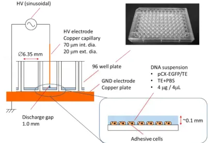
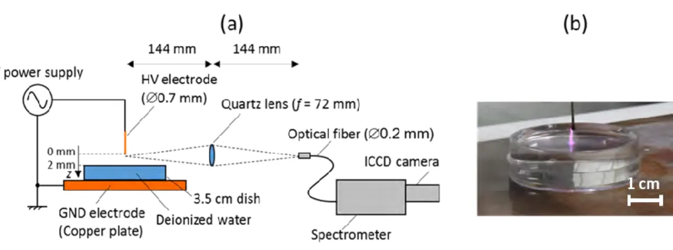
electrons Te. In fact, the micro air plasma used for gene transfection is a corona discharge generated
in ambient air from the tip of a pulsed high voltage microtube placed 2 mm in front of a petri dish containing deionized water and set over a grounded copper plate. The rotational temperature Trot
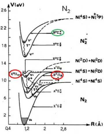
is estimated from comparison of synthetic and experimental spectra of OH(A-X), N2+(FNS: 0, 0)
at 391.4 nm, and N2(SPS: 0, 0) at 337nm. Based on N2+(FNS: 0, 0) and N2+(FNS: 1, 1) head bands
spectra at 391.4 nm and 388.4 nm. The vibrational temperature Tvib is estimated along the plasma
axis between the tip electrode up to the grounded plate. Moreover, the electron temperature Te is
estimated from an interesting approach based on the experimental ratio of the closest nitrogen emission spectra of N2+(FNS: 0, 0) at 391.4nm and N2(SPS: 2, 5) at 394.3 nm. This is based on
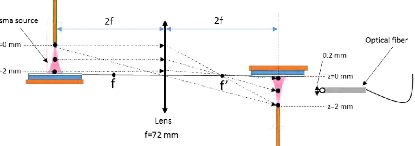
one hand a balance equation between creations and losses of excited upper levels of these two UV spectra and on the other hand on the electron impact rates of the creation of these upper levels calculated from a solution of multi-term Boltzmann equation. Then using the measured Hα spectra, electron density ηe has been estimated from Stark broadening versus the inter-electrode position.
Last, the spatial variation along z axis of the nitrogen ion density has been determined from the relative intensities of the same close wavelength spectra (N2(SPS: 2, 5) at 394.3 nm and N2+(FNS:
0, 0) at 391.4 nm). The present experimental plasma characteristics are used to better understand the mechanisms and the processes involved during plasma gene transfections when using the Monte Carlo poration model developed in the present work.
The third chapter begins with a review on the progress made so far on the numerical modeling of cold plasma interactions with cells and tissues. I underlined that there are no literature simulations devoted to membrane permeabilization and pore formation when impacted by plasma
6 actives species. This is why I developed for the first time in literature a specific Monte Carlo poration model based on the concepts of super-particles interacting with super-sites of the membrane following super-reaction processes. The present Monte Carlo poration model is aimed to statistically simulate, at a global (or macro) scale, the pore formation during atmospheric pressure plasma interactions with cell membranes. In the framework of this simulation model, each plasma species was assumed as a super-particle grouping a large number of particles. Three kinds of plasma active species are considered: electrons, ions and neutral radicals, with fluxes estimated from a plasma reaction kinetic model and the OES study conducted in chapter 2. The membrane layers were assumed as a simple membrane model superposing four layers of phospholipids and proteins. Moreover, each layer was constituted by a succession of super-sites subjected to specific super-processes (recombination, reflection, activation of a site, opening) during the membrane impacts by the plasma super-particles.
The last chapter (chapter 4) is first devoted to a description of the computational simulation conditions as for instance the initial particle number, the simulation domain geometry and the fraction of the initially activated sites. The different occurrence probabilities of each super-process between a given plasma super-particle and a membrane super-site that are considered in the Monte Carlo model are also defined and described. These reaction probabilities are selected in a suitable way based on some biophysical considerations; as for example the magnitude of the layer density (either protein or lipid layer), the plasma super-particle type and the energy magnitude of the electrons. In addition, the super-site state is also considered as a factor determining the choice of reaction probabilities. Furthermore, for an accurate exploitation of our Monte Carlo poration model, the good estimations of the different probabilities of occurrence of the whole considered super-processes are absolutely necessary. This is why a large parametric
7 study is conducted in the second part of chapter 4. This is aimed to evaluate the effects of the initial simulation parameters as well as the magnitude of the occurrence probabilities of each reaction super-process on the cell membrane permeabilization and pore formation. More precisely, I discuss the effects of the initial incidence angle of ions, and radicals, the effect the energy distribution of the electrons, as well as the effects of reaction as for instance the role of the reflection process in the case of ions, and radicals, and the importance of the electron energy and processes in the super-site opening process. This parametric study enabled to underline several important and original results. Moreover, a comparison with the measured transfected efficiency of DNA plasmid and the surviving cell rate in the case of mouse fibroblast cells are used to discuss the validation of the present Monte Carlo poration model.
Last, these discussions and analyses of Monte Carlo simulations of membrane poration using air microplasma generated at atmospheric pressure are followed by a general conclusion summarizing the main results obtained in the framework of this thesis and giving some future orientations to the present research thesis.
9
BIBLIOGRAPHIC OVERVIEW ON GENE
TRANSFECTION AND CONTEXT
1.1 Introduction
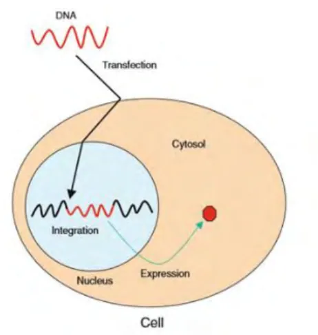
As underlined in the general introduction, Gene transfection, the process of inserting using a vector a foreign genetic material, such as DNAs and RNAs, into cells, has origins as far back as the 1950s [1]. Transfection is one of the most valuable and frequently used tools of biological science. It is an important tool used in studies investigating gene function and the modulation of gene expression, thus contributing to the advancement of basic cellular research and drug discovery. The insertion of DNA into a cell enables the expression of proteins using the cells own machinery [2] in the aim to correct a cellular dysfunction, to modify genetic inheritance in order to treat pathology, to provide a new cellular function or to manufacture programmed cells able to grow and to become designed organs [3] (cf. Figure 1-1). Various approaches, such as viral, chemical and physical methods with non-viral vectors, have been developed to make gene delivery safer and more efficient [4]. However all these reliable methods are limited to a few experimental systems and can have known drawbacks. Therefore, the development of a new safe and damage-free technique of gene transfection is an interesting complementary technique to the existing ones. To this purpose, gene transfection based on low-temperature atmospheric pressure plasmas can lead to a transient permeabilization of the cell membrane allowing processes of gene transfection.
10 Nevertheless, one of the most important issues for the plasma irradiation technique when used for gene transfection is to find an optimal relationship between transfection efficacy and cell damages. In fact, under the condition of high transfection rates, the cells can also be damaged. Thus in the aim to optimize this trade-off relationship, several types plasma sources with different designs and configurations as well as gene transfection protocols were developed. Moreover, the mechanisms and the processes of plasma gene transfection, more precisely membrane poration are far to be clear and controlled. Therefore fundamental understanding on plasma-induced membrane permeabilization is required for further progress in the field of plasma gene transfection.
The aim if this first chapter will be, in first part, to make a short bibliographical synthesis including the various applied and potentially applied fields of gene transfection in the biomedical domain. Thereafter, the different common main transfection methods as well as the new ones based on atmospheric plasma irradiation will be reviewed, while detailing the main advantages and the drawbacks of each method.
In the second part of the chapter, on the one hand, a bibliographic overview on the various developed and used plasma setups for gene transfection in literature will be reviewed. On the other hand a detailed overview on the plasma setups developed in Ehime University (arc plasma discharge, an atmospheric pressure plasma jet (APJ) using helium carrier gas and equipped with 4 jets, a dielectric barrier discharge (DBD) plasma using also helium flow, and last an air microplasma), will be described, followed-up by some important results derived from a comparative study based on transfection and cells viability rates obtained in the case of each plasma setup. Finally, the most promising, safe and high efficiency plasma configuration, which is the air microplasma discharge, will be focused on. To this aim, an experimental parametric investigation on the involved process and gene transfection mechanism by using this reliable
11 microplasma discharge as well as the most important obtained results will be discussed based on different gene transfection protocols. Through all of that, it is discussed the state of knowledge of the various mechanisms imply in the plasma gene transfection, and the importance of their better understanding to make further improvements and progresses in gene transfection technique.
Figure 1-1 Schematic diagrams of gene transfections principle by using virus vector. Foreign DNA (red-wave) is delivered to nucleus by passage through the cell and nuclear membranes. Foreign DNA is integrated into the host genome (black-wave) and expressed sustainably [5].
1.2 Overview on gene transfection
Genes have long been considered as medicines [6]. During the past decade, the field of gene therapy has grown into a dynamic and exciting interdisciplinary research that not only impacts on the development of innovative cures but also provides significant added value by applying gene transfection tools to study and manipulate biological systems. It is widely anticipated that improved gene transfection approaches will yield new treatments and cures of a wide range of human diseases [7]. There are some of the most urgent unmet medical needs, such as hereditary immune deficiencies, neurodegenerative and neuromuscular disorders, hemophilia, cardiovascular disease, congenital blindness, infectious diseases and cancer [4]. In the absence of effective drugs or alternative therapies, the advances in gene therapy technology represent the best hope for the many patients and families that are blighted by these various diseases.
12
1.2.1 Applied fields of gene transfection
The diverse applications of gene transfection are categorized under four strategies:
The gene replacement therapy for monogenic diseases: Many human genetic diseases are clearly defined by a single gene defect, and gene replacement is a straightforward approach to treating these diseases. Not surprisingly, the majority of the most advanced clinical gene therapy development falls into the category of gene replacement, because the conceptually simple design leads to extensive investigation. Gene replacement can take place either directly in vivo or through ex vivo cell therapy [8]. The promising gene replacement clinical trials involve treating of respiratory diseases [9][10], leukodystrophies (degeneration of the white matter in the brain)[11], life threatening inflammation in pancreas [12] and blood disorders. Taken together, these human gene replacement applications have pioneered the gene therapy field.
Gene addition for complex disorders and infectious diseases: In contrast to a single gene defect underlying monogenic diseases, the combination effects of multiple genes and environmental factors cause complex disorders such as cancer and heart diseases rendering gene replacement not feasible for these disorders. Complex disorders are often common, and represent the most urgent unmet medical needs. In addition to genetic disorders, infectious diseases also debilitate or kill a large population worldwide. Therefore, significant efforts of gene therapy development have been devoted to these diseases [8]. Including heart disease [13], cancer [14], cardiovascular diseases [15] and neurodegenerative diseases [16], as well as infectious diseases. Indeed gene therapy vaccines are being developed and trialed for tackling infectious diseases, including tuberculosis [17], malaria[18], HIV [19] and influenza [20].
Gene expression alteration targeting RNA: RNA can be an intermediate (e.g. messenger RNA) or final (e.g. microRNA) gene product, with diverse functions in biology and disease. Due to the
13 versatile roles of RNA molecules in controlling gene expression, gene therapy strategies specifically designed to target RNA or to produce effector RNA molecules deserve close attention [8]. Here, two commonly utilized gene therapy strategies based on RNA biology; Gene knockdown by RNA interference (RNAi)[10][21][22] and Reprogramming messenger RNA (mRNA) [23].
Gene editing to introduce targeted changes in host genome: The potential of genome editing in human gene therapy applications is evident, as a number of successful studies in human cells and model organisms have been reported [24][25]. However, the specificity of editing needs to be rigorously tested to address possible off-target effects before widespread clinical use [26]. In addition, editing efficiency needs to be further improved to meet therapeutic needs, particularly in in vivo gene therapy settings that require gene editing in an enormous number of cells throughout
an organ or the whole body.
Furthermore, the manipulation of gene expression is a core technique in more research areas such as drug development and tissue engineering [27][28]. However, although the gene transfer field is rapidly progressing, is still far from providing enough tools for the treatment of the previously mentioned diseases. Moreover, many gene transfer technologies still remain highly unexplored both experimentally and theoretically [29]. The most pressing issue that the field of gene therapy has to address is the development of efficient and safe gene delivery methods. Indeed, one of the main difficulties with gene transfection is how to effectively and safely deliver the genetic material to different cells, organs and tissues. For instance, this is achieved by using gene delivery vehicles, namely vectors which carry the material into the cell, and it can be categorized into two classes: DNA (non-viral) vectors and viral vectors [7][30]. However side effects such as the carcinogenesis and leukemia occurred by the gene therapy using the virus vector, it is not in practical use. And, in the gene therapy using the DNA vector, it is also not in use as a gene therapy because demerits such
14 as cytotoxicity, low gene transfection and integration efficiency are exist. To be in practical use of gene therapy, it is necessary to develop the safe and high efficiency gene transfection method in substitution for viral vector method.
1.2.2 Description of different methods of gene transfection
Transfection can be accomplished using many developed methods, for instance, biological methods based on the virus as delivery vector of the nucleic acid. As well as , the common main methods for non-viral delivery, which are broadly classified into chemical and physical methods [5]. Many types of genetic material, including plasmid DNA, siRNA, proteins, dyes, and antibodies, may be transfected using any of these methods. However, a single method cannot be applied to all types of cells; transfection efficiencies and cytotoxicity may vary dramatically and depend on the method, cell type being utilized, and types of experiments being performed [5]. Therefore, to obtain high transfection efficiencies, low cell toxicity, minimal effects on normal physiology, and be easy to use and reproducible, all relevant factors should be considered for planning and selecting the appropriate transfection method.
1.2.2.1 Viral method
The most commonly used method in clinical research is virus-mediated transfection. This process named transduction can be applied to several biological models and represents the most efficient approach to achieve good gene expression levels in many cells [31]. The use of viral vectors was employed as early as the late 1970’s to express functional mRNA and protein [32]. Many mammalian viruses have been explored as gene delivery vectors [33][34] [35]. Thus, virus-mediated transfection is highly efficient and it is easy to achieve sustainable transgene expression in vivo owing to the viral nature of integration into the host genome for example, retrovirus murine
15 in humans [36]. MLV integrates its DNA into the host genome and the integrated DNA is expressed in the host. The integrated MLV DNA replicates as the host genome does. Consequently it segregates into daughter cells, which enables sustainable transgene expression. The major drawbacks of virus-mediated transfection are immunogenicity and cytotoxicity. Introduction of a viral vector may cause an inflammatory reaction and an insertional mutation, because viral vectors integrate into the host genome randomly, which may disrupt tumor suppressor genes, activate oncogenes, or interrupt essential genes [9]. Another disadvantage of this method is that a virus package has limited space for a foreign gene to keep infectivity as well as the high costs due to biosafety requirements[5][37]. For these reasons, much effort has been made to develop non-viral transfection methods even though virus-mediated transfection is highly effective and easy to use [5].
1.2.2.2 Chemical method
Chemical transfection methods are the most widely used methods in contemporary research and were the first to be used to introduce foreign genes into mammalian cells [38]. These methods commonly use compounds such as cationic polymer (one of the oldest chemicals used), calcium phosphate and cationic lipid (the most popular method), to name a few [38][39]. The elementary principle of chemical methods is similar. Positively charged chemicals make nucleic acid/ chemical complexes with negatively charged nucleic acids. These positively charged nucleic acid/chemical complexes are attracted to the negatively charged cell membrane. The exact mechanism of how nucleic acid/chemical complexes pass through the cell membrane is unknown but it is believed that endocytosis and phagocytosis are involved in the process [5]. Transfected DNA must be delivered to the nucleus to be expressed and again the translocation mechanism to the nucleus is not yet known[5].
16 Calcium phosphate represents the oldest and most inexpensive chemical method for transfecting nucleic acids. It has been a popular transfection method since its introduction in the early 1970s by Graham and van der Eb [40]. Furthermore, the technique is easy to master, it is effective with many types of cultured cells, and it can be used for both transient and stable transfection of a variety of cultured cell types. However, calcium phosphate co-precipitation is prone to variability due to its sensitivity to slight changes in pH, temperature, and buffer salt concentrations, and can be cytotoxic to many types of cell cultures, especially of primary cells. In addition, it is unsuitable for in vivo transfer of nucleic acids to whole animals, and it shows relatively poor transfection efficiency compared to other chemical transfection methods such as lipid-mediated transfection [41].
Cationic lipid-mediated transfection is one of the most popular methods for introducing foreign genetic material into cells. Although first generation of lipid-based transfection reagents relied on artificial liposomes that could envelop nucleic acids and then fuse with the cell membrane to deposit their cargo inside [42], newer cationic lipid-based reagents spontaneously form condensed nucleic acid-cationic lipid reagent complexes via electrostatic interactions between the negatively charged nucleic acid and the positively charged head group of the synthetic lipid reagent. These complexes are believed to be taken up by the cell through endocytosis and then released in the cytoplasm. Once in the cell, transfected DNA is translocated to the nucleus to be expressed by a yet unknown mechanism [43][44]. The advantages of cationic lipid-mediated transfection are the ability to transfect a broad range of cell lines with high efficiency, and its ability to deliver DNA of all sizes, as well as RNA and proteins. In addition, this method can be applied to both stable (Stable viral delivery systems that provide long-term, mid-level production of protein or gene expression) and transient expression (express the foreign gene without its integration into their
17 genome, for a finite period of time, usually several days, after which the foreign gene is lost through cell division or other factors). Moreover, unlike other chemical methods, it can be used for in vivo transfer of DNA and RNA to animals and humans[30]. The main drawback of cationic lipid-mediated transfection is the dependence of transfection efficiency on the cell type and culture conditions, requiring the optimization of transfection conditions for each cell type and transfection reagent [30].
Another chemical method based on cationic polymers, which differ from cationic lipids in that they do not contain a hydrophobic moiety and are completely soluble in water. Although they differ dramatically in their degree of transfection efficiency and cytotoxicity. Among the cationic polymers used for the transfection of cultured mammalian cells, DEAE-dextran was one of the first studied [30]. This reagent has been successfully used to deliver nucleic acids into cells for transient expression and short-term expression analyses but is not suitable for stable transfection studies [45]. The primary drawback of the DEAE- dextran system is the cytotoxicity. Therefore, the effects of concentration and exposure times need to be determined before transfection for individual cell lines. Other synthetic cationic polymers have been used to transfer DNA into mammalian cells. The polymers include protamine, intact and fractured polyamidoamine dendrimer [46][47][48], more recently, polyethylenimine (PEI) [49][50] which appears to be better than cationic liposomes at delivering DNA across the nuclear envelope [51][52].
Despite that these methods have merits of relatively low cytotoxicity, no mutagenesis, no extra-carrying DNA, and no size limitation on the packaged nucleic acid, the transfection efficiency of chemical methods is largely dependent on factors such as nucleic acid/chemical ratio, solution pH, and cell membrane conditions, so the process results in low transfection efficiency, especially in vivo, compared with virus-mediated methods [31].
18
1.2.2.3 Physical method
The physical transfection methods are the most recent methods. It uses diverse physical tools to deliver nucleic acids [5]. It depends neither on viral vehicles and their receptors nor on biochemical structures or features of target cell membranes required for uptake of a vehicle-DNA complex [30]. These methods directly deliver nucleic acids into the cytoplasm or the nucleus of the cell. I can cite for instance, direct micro injection, biolistic particle delivery, laser-based transfection, electroporation [53], sonoporation [54], and other methods using for instance magnetic field to destabilize the cell membrane. Finally, I can also cite, the novel transfection method using cold gas plasma [55] [56].
Briefly, the direct micro injection delivers nucleic acids into the cytoplasm or the nucleus [57], one cell at a time by means of a fine needle; therefore, this method is limited to ex vivo applications such as the transfer of genes into oocytes to engineer transgenic animals or the delivery of artificial chromosomes [57]. Although direct microinjection is nearly 100% efficient, it demands considerable technical skills, is extremely labor-intensive, and often causes cell death. Moreover, this method is not appropriate for studies requiring the transfection of large number of cells.
Moreover, the relatively new physical method of gene transfer is biolistic particle delivery, also known as particle bombardment technique [58]. It involves projecting of microscopic heavy-metal particles (often gold or tungsten) coated with nucleic acids into recipient cells at high velocity using a ballistic device (i.e., “gene gun”)[59][60]. Biolistic particle delivery can be used to transiently transfect dividing and non-dividing cells in culture as well as cells in vivo, and it is often used for genetic vaccination and agriculture applications [61]. While this technique is reliable and fast, it requires costly equipment, causes physical damage to the samples, and necessitates a large number of cells due to high mortality.
19 Furthermore, the laser-mediated transfection, also known as phototransfection, laserfection, or optoporation, uses a laser pulse to transiently permeabilize the cell membrane [62][63]. When the laser induces a pore in the membrane, the osmotic difference between the medium and the cytosol facilitates the entry of nucleic acids or other desired substances in the medium (ions, small molecules, proteins, semiconductor nanocrystals, etc.) into the cell. Advantages of laser-mediated transfection include high transfection efficiency and the ability to make pores at any location on the cell. However, the method requires an expensive laser-microscope system and the cells have to be attached to a substrate.
In addition to the methods mentioned above, electroporation is the most widely used physical delivery technology [30]. The exact mechanism is unknown, but it is supposed as shown in Figure 1-2 [64] that a short electrical pulses at an optimized voltage and lasting only a few microseconds to a millisecond generates an electrical field, which disturbs the cell membrane capacitance and creates transient membrane pores through which small particles can pass into cells (further details on the mechanisms of electroporation of cell membranes in chapter 3, sub-section 3.2.1. This technology was initially developed for in vitro DNA delivery but has expanded to include transfection of other nucleic acids (e.g., oligos, mRNA, siRNA, and miRNA), drug delivery, cell-cell fusion (i.e., electrofusion), and membrane protein insertion (i.e., electro-insertion)[5].
Indeed, electroporation can be an effective and efficient alternative to chemical transfection. Its main advantage consists on its applicability for transient and stable transfection of all cell types. In fact, it provides a robust and universal approach for transfecting various cell types including bacterial, mammalian, yeast, and plant cells, with any type of nucleic acid [5]. Furthermore, because electroporation is easy and rapid, it is able to transfect a large number of cells in a short
20 time once optimum electroporation conditions are determined [5]. However, the major drawback of electroporation is substantial cell death caused by high voltage pulses and only partially successful membrane repair, requiring the use of greater quantities of cells compared to chemical transfection methods [30]. The high cell mortality can be minimized through optimization of experimental conditions and may be counterbalanced by increased transfection efficiencies: Conditions similar to chemical transfection (e.g., nucleic acid amount and cell density) or parameters unique to electrical methods (e.g., voltage and pulse types).
Figure 1-2 DNA vaccination and schematic representation of the effect of electroporation at the cell membrane level [64].
As already emphasis and with a burgeoning interest in transfection, particularly for therapeutic purposes, advances in the field of transfection are spurred by the need to increase efficiency, to broaden the range of transfectable target cell or tissue types, and to address specific workflow requirements. Therefore, the improvement of these current techniques of gene transfection or the development of new safe and damage-free techniques is in demand.
21
1.2.2.4 Non-equilibrium cold atmospheric plasma discharge
The emergence of plasma medicine field proves that the non-equilibrium plasmas are able to initiate, promote, control, and catalyze various complex behaviors and responses in biological systems. For instance apoptosis, cell detachment [65], and cell permeabilization [66]. More importantly, it showed that the non-thermal plasma effects can be tuned to achieve various desired medical purposes, especially in medical sterilization and treatment of different kind of skin diseases, such as wound healing, tissue regeneration and gene transfection [67][57]. Indeed, a method of interesting efficiently of transferring numerous selected molecules into various cells by using cold gas plasma was patented by Miyoshi et al. [68] in 2002 and was then reported by Ogawa et al.[55] . The plasma, which is generated by discharge contains a large number of charged and
uncharged particles such as ions, electrons, and radicals and could easily provide an appropriate impact on the samples and mediate gene transfer as well as electroporation [57][67][69]. On the one hand, it was shown that the plasma can transfer the genes even into primary neuronal cells without a loss of function of the cells, into which electroporation cannot sufficiently do it [57]. On the other hand, since this technique is free from adverse effect associated with viruses, there is no risks compared to the others methods mentioned above [69].
1.3 Non-thermal plasmas at atmospheric pressure for
gene transfection
As already highlighted, gene transfection induced by low-temperature plasmas can be an interesting alternative to the previous conventional methods. It was once shown in the literature that the plasma irradiation leads to a transient permeabilization of the cell membranes allowing gene transfection (ref Ogawa et al. [55], Sakai et al.[56] Leduc et al. [65][70], Nakajima et al.[66], Jinno et al. [67] [69] [71] [72] [73] and Sasaki et al. [74]) in which DNA and cells are
22 both exposed to fluxes of active plasma species (such as electrons, ions and neutral radicals) and also to plasma-induced electric field [66]. Thus, the non-thermal plasmas are already well-known for their gene transfer ability; however, the mechanisms of this gene transfection are under debate. Short and long-living active species and radicals produced by plasma, and bombardment by charged particles are all listed as potential candidates for cell membrane poration and permeabilization.
1.3.1 Mechanisms of plasma gene transfection
The mechanisms leading to membrane permeabilization during plasma species/cell interactions are partly evoked in the literature [70] as twofold mechanisms: cell charging (direct plasma effect) and lipid peroxidation (indirect plasma effect). Obviously, the well-known membrane electroporation could be the third mechanism provided high enough plasma-induced electric field is present.
1.3.1.1 Cell charging (plasma direct effect)
In fact, in physical cell charging, the electrons accumulated on the cell membrane can generate Coulomb forces at the membrane surface high enough to deform the cell from its ellipsoidal form to a spherical form. This deformation of the cells could create a shear stress high enough to partially disrupt the wall of the cell thus creating transient pores. Experiments on poration indicate that direct plasma exposure is effective for gene transfection [56]. It is also evoked the possibility of a significant role of electric charging in this poration phenomena. Anyway, the contribution of cell surface charging is not really quantified. This is why cell detachment and poration in interactions with actives plasma species should be further investigated using numerical models of the plasma membrane poration.
23
1.3.1.2 Chemical lipid peroxidation (indirect plasma effect)
The active species of the plasma reaching the cell membrane can react with the lipid bilayer and ultimately enable creation of transient pores. The hydroxyl radical OH is the most probable active species to initiate lipid peroxidation. Indeed, OH is known to react rapidly with organic molecules which is the pathway leading to lipid peroxidation. The propagation of the lipid peroxidation leads to the cross-linking of the fatty acid side chain which can lead to formation of transient pores [65].
In the aim to investigate experimentally the potentiality to use of non-thermal plasmas generated at atmospheric pressure for the gene transfection applications, different plasma devices in terms of configuration, generation, and irradiation of plasma have been used for gene transfection. An overview of these different existing plasma setups in literature as well as those tested at Ehime University, with the corresponding obtained results is an essential step and it is presented in the following sub-section.
1.3.2 Overview on plasma setups used for gene transfection in
literature
In 2005, Ogawa et al. [55] followed by Sakai et al. [56] presented a novel transfection method for eukaryotic cells using a type of atmospheric pressure discharge plasma jet. The plasma generation and irradiation was carried out as follows: a high air flow (around 90 slm) provided by an air pump, blown-off between electrodes powered by a pulsed high frequency (20 kHz) generating, on the downstream side, a U-shaped active gas plasma jet that impacts the sample mixtures of cells and plasmid placed under the electrodes (See Figure 1-3). The height (H) of the electrodes is adjusted to 22mm from the dish bottom so that the lower end of the flare can reach the target cells. The turntable under the electrodes was designed to rotate at 2 rounds per second
24 during electric discharges, and the distance between the two electrodes was adjusted to the half diameter of the 6 cm dish so that the whole cells on the sample dish could be homogenously exposed to the gas plasma during transfection.
Figure 1-3 Schematic diagram of the plasma generation and the treatment of the cultured cells with the plasma generator [56].
The experimental conditions of the considered cells (HeLa-S3 and other cell lines), the transfected DNA (pEGFP-C1 plasmid at a concentration of 0.5µg/µl), the solution used for cell and DNA immersion (phosphate buffered saline PBS), the method of cell evaluation (fluorescence microscopic observation and the flow cytometry analysis) are already described elsewhere [56]. The obtained results (cf. Figure 1-4) establish an optimum exposure time around (1–3s) of the treated cells to the plasma irradiation with a favorable transfection efficiencies (17.8-21.6%) and relatively low cell mortalities (0.65-2.86%). Indeed, the results of transfection clearly show that the cells became transiently permeable for plasmid DNA during the plasma exposure, suggesting that the plasma-mediated transfection may involve similar mechanisms that accounts for electroporation.
25 Figure 1-4 Transfection efficiencies with various lengths (0–5 s) of plasma exposures at the DNA concentration of 0.5µg/µl.Filled diamonds, transfection efficiency; and open squares, mortality rate. TE, transfection efficiency [56].
Leduc et al., in 2009 [70] used another plasma jet, referred to atmospheric pressure glow discharge torch (APGD-t). This last, is formed by the injection of a plasma-forming helium gas (at 0.5 slm) inside an annular space defined by a central capillary electrode (connected to RF power supply at 13.56 MHz) and a quartz confinement tube itself coated with a conductive paste (see Figure 1-5). The end of the nozzle is positioned 3mm away from the bottom of the Petri dish containing a mixture of cells (HeLa cells) and plasmid (hrGFP-II-1 at a concentration of 0.05 µg/µl). A motorized X–Y platform is used to move the sample and treat each plate in a selected pattern. The obtained results, show that the APGD-t is capable of including temporary cell permeabilization allowing a local transfection efficiencies as high as 35%. Moreover, the maximum radius of macromolecules able to enter into HeLa cells following a plasma treatment, was evaluated below 6.5 nm. On the other hand, it was show that no degradation occurs when the
26 plasmid DNA suspended in culture media is plasma-treated at the operating conditions leading to cell permeabilization [65].
Figure 1-5 Picture of the APGD-t mounted over a Petri dish placed on the X–Y motorized platform and schematic drawing [70].
There is also low frequency (at 2.5kHz pulsed) plasma jet used by Nakajima et al., in 2010 [66]. It’s generated by argon gas (at 2 slm) in an outer glass tube with a stainless wire electrode covered with another glass tube and the outer glass tube is covered by a grounded mesh electrode. The gap between the tip of the generator and the surface of the agar medium or the water surface was 20 mm (Figure 1-6).
Figure 1-6 A schematic of the experimental setup for plasma exposure and Photograph of the generated plasma torch [66].
27 The plasma jet was exposed to the mixture of E. coli strain MV1184 cells and plasmid pUC19 DNA, and the transfection was clearly demonstrated [66].
Moreover, we can also refer to different types of dielectric barrier discharge DBD (ref Leduc et al., in 2010 [65] and Sasaki et al., 2014 [74]). Figure 1-7 shows both the photography and
schematic drawing of the DBD plasma sources used by Leduc et al. [65]. It is based on a helium carrier gas flow (at 1 slm) passing through a mesh electrode and reaching to a glass plate placed over grounded electrode. The mesh electrode is powered by an AC high-voltage (10 kHz) (cf. Figure 1-7). On the other hand, it was found that based on the typical configuration of the used plasma sources, the plasma exposure to cells can be direct (DBD [65]) or indirect (APGD-t [70]). A comparison between both plasma sources was conducted in the aim to assess the possible negative effects of direct and indirect plasma treatment of mammalian cells (HeLa cells ATCC CCL-2) and naked DNA (hrGFP-II-1 at a concentration of 0.05 µg/µl)[65]. It was found that, the direct plasma treatment using DBD caused an oxidative stress to the cells, and both the direct and indirect plasma sources were able to fragment naked DNA. No lipid peroxidation was found in the treated samples [65]. From the obtained results we conclude that the plasmas may have unintended negative effects on cells which require a deep understanding of the mechanism of plasma-biological surface interactions.
28 In the aim to investigate the mechanism of gene transfer by plasma irradiation, Sasaki et al., in 2014 [74] used another non-equilibrium atmospheric pressure plasma jet (APPJ). The last, is based on a helium carrier gas flow (at 3 slm) crossing a glass tube wrapped by two copper cylindrical electrodes that are powered by an AC high-voltage (10 kHz) (See Figure 1-8).
Figure 1-8 Schematic of the experimental setup [74].
The generated DBD plasma flows out from the nozzle quartz glass tube. The living cells in suspension consisting of genes (fluorescent dye YOYO-1) are irradiated with the plasma. The distance between the downstream electrode and the edge of the glass tube is defined as dg = 73mm
and the distance between the edge of the glass tube and the surface of the cell suspension as dair = 5mm. The cells after direct plasma irradiation under various conditions (suspension volume
l= 100 or 150µL and plasma irradiation time td=0–60s) are evaluated by simultaneous analysis of
transfection efficiency and cell viability. As a result (see Figure 1-9), the efficiency has a maximal value at a short plasma irradiation time (3–5s) while maintaining a very high cell viability, and the volume of irradiated cell suspension changes the time dependence of the efficiency. Indeed, the no monotonically increase of the efficiency predicted a mechanism of gene transfer based on a
29 competition between various factors such as reactive oxygen species ROS and electric field stimulation [74].
Figure 1-9 (a) Transfection efficiency ƞ (%) of YOYO-1 and (b) cell viability (%) as a function of plasma irradiation time td (s) when the final volume of cell suspension l is 100 or 150µL [74].
From this overview, one notes that one of the most important issues for the plasma irradiation technique when used for gene transfection is a trade-off relationship between transfection efficacy and cell damages. In fact, under the condition of high transfection rate, the cells can also be damaged [65]. Another most important issue is the transfection reproducibility [55][56]. For a given plasma source, even if the operating parameters of the power supply used to generate the plasma (e.g. applied voltage, waveform, frequency, etc.) are chosen identical, the transfection rates can vary significantly shot by shot [69][67]. Since the size and the spatial position of the plasma irradiations changes randomly in time, in some trials the genes and the cells are heavily damaged and reach necrosis or apoptosis by being exposed to the high density part of plasma, while in some other trials, the plasma irradiations do not reach the cells and transfection is not efficient. It was assumed that these important issues can be associated more particularly to the plasma instability [69]. In order to solve such instability, several types of plasma sources with
30 different designs and configurations were tested from 2002 until 2011 at Ehime University. An overview on these different tested plasma setups as well as the obtained results issue from a comparative study, are presented in the sub-section 1.3.3.
1.3.3 Overview on the different experimental setups tested at
Ehime University for gene transfection
Four different types of plasma sources were developed and used at Ehime University: A an arc plasma discharge, an atmospheric pressure plasma jet (APJ) using helium carrier gas and equipped with 4 jets, a dielectric barrier discharge (DBD) plasma using also helium flow, and last an air microplasma discharge [69] [73].
The arc plasma configuration was identical to Ogawa et al..[55], which have a custom made HV power supply (Pearl Kogyo) for discharge initiation, (see Figure 1-10 (a)). The output voltage was pulse modulated sinusoidal waveform (20 kHz). The modulation frequency range was 10200 Hz (typically 25 Hz) and its duty ratio range was 0100%. The amplitude voltage could be set up to 15 kV. Argon or helium gas can be supplied to the discharge space with controlled flow rate using a MFC (mass flow controller, Advanced Energy, FC7710C). Two copper wire electrodes (diameter 1 mm) were attached in line on the bottom of a ceramic head (25 mm wide 22 mm deep 30 mm high). The head had a vertical hole (cross section of 4.3 8.3 mm2) for working gas supply. A 3.5 cm dish was on a rotation stage. The distance between the electrode and the sample solution in the dish was set at 12 mm. A flare-like plasma was initiated between the electrodes and the dish, as shown in the bottom of Figure 1-10 (a). The plasma irradiation area on the sample solution was about 30 mm in diameter, although it was perturbed because of waving of the flare.
![Figure 1-2 DNA vaccination and schematic representation of the effect of electroporation at the cell membrane level [64]](https://thumb-eu.123doks.com/thumbv2/123doknet/2180516.10487/30.918.260.691.411.708/figure-vaccination-schematic-representation-effect-electroporation-membrane-level.webp)
![Figure 1-3 Schematic diagram of the plasma generation and the treatment of the cultured cells with the plasma generator [56]](https://thumb-eu.123doks.com/thumbv2/123doknet/2180516.10487/34.918.249.691.243.479/figure-schematic-diagram-plasma-generation-treatment-cultured-generator.webp)
![Figure 1-5 Picture of the APGD-t mounted over a Petri dish placed on the X–Y motorized platform and schematic drawing [70]](https://thumb-eu.123doks.com/thumbv2/123doknet/2180516.10487/36.918.216.733.197.424/figure-picture-mounted-petri-motorized-platform-schematic-drawing.webp)
![Figure 1-12 The cell viability (left) and the transfection efficiency (right) for four different plasma configurations (arc plasma jet, APJ, DBD plasma, and microplasma) [73]](https://thumb-eu.123doks.com/thumbv2/123doknet/2180516.10487/46.918.117.803.130.387/figure-viability-transfection-efficiency-different-plasma-configurations-microplasma.webp)