The prion-like properties of the mutant huntingtin
protein: demonstration in in vitro and in vivo systems
Thèse
Maria Masnata
Doctorat en neurobiologie
Philosophiæ doctor (Ph. D.)
The prion-like properties of the mutant huntingtin
protein: demonstration in in vitro and in
vivo systems
Thèse
Doctorat en neurobiologie - Philosophiæ Doctor (Ph. D.)
Maria Masnata
Sous la direction de:
Dre Francesca Cicchetti
Résumé
La maladie de Huntington (MH) est une maladie neurodégénérative autosomique dominante qui affecte environ 3 à 8 personnes sur 100 000 dans le monde. La MH est causée par une mutation du gène HTT, lequel code pour la protéine huntingtine (HTT). Cette mutation consiste en une expansion de 35 répétitions CAG à même l'exon 1 du gène et aboutit à une répétition de la séquence polyglutamine (polyQ) au sein du segment N-terminal de la protéine HTT. Les personnes atteintes de MH développent des troubles moteurs, cognitifs et psychiatriques sévères, principalement à l'âge adulte. L'âge d’apparition des symptômes est généralement inversement proportionnel au nombre de répétitions CAG bien que la symptomatologie varie considérablement d'un patient à l'autre. L'origine de la MH est associée à l'expression de la protéine HTT mutée (mHTT) qui, en raison de son expansion polyQ, adopte une conformation pathogène et s'accumule en petits et/ou gros agrégats cytotoxiques. Bien que l'on suppose que ces événements soient responsables de la neurodégénérescence, le mécanisme sous-jacent aux voies physiopathologiques menant à l'apparition de la maladie et à la mort neuronale est toujours à l'étude. Un nombre croissant d'observations suggère que la mHTT possède des capacités de type prion, c’est-à-dire une aptitude à recruter des protéines endogènes normales et les corrompre afin de créer des agrégats toxiques; capacités qui sont également connues pour d'autres protéines, notamment l'amyloïde, la tau et la a-synucléine, associées à diverses maladies neurodégénératives. Nous avons émis l'hypothèse que le mHTT se propage de cellule en cellule et qu'elle se comporter tel un prion, contribuant à influencer le développement de la MH. Afin de tester cette hypothèse, des fibrilles synthétiques de mHTT ont été administrées à plusieurs lignées cellulaires ainsi qu'à des souris de différentes souches génétiques. Suite à une période d'incubation, les effets des fibrilles de mHTT sur la viabilité cellulaire, le comportement animal et les caractéristiques neuropathologiques ont été examiné. Nous avons ainsi observé que les fibrilles de mHTT provoquaient la mort cellulaire et des changements morphologiques des cellules cultivées, tandis qu’elles induisaient un phénotype comportemental transitoire de la maladie chez des souris saines. Les fibrilles de mHTT pouvaient également exacerber les déficits moteurs, anxieux et cognitifs de type MH dans un modèle de souris huntingtonien. Ainsi, notre étude suggère que la mHTT extracellulaire peut se propager de cellule à cellule et, une fois recrutée au sein de la cellule, provoquer des
changements pathologiques. À la lumière de ces observations, nous croyons que la mHTT extracellulaire pourrait représenter une cible attrayante pour le développement de futures stratégies thérapeutiques. De surcroît, la plupart des traitements en études cliniques sont conçus pour cibler le gène HTT dans le but de diminuer l'expression de la protéine, ignorant la quantité importante de mHTT déjà présente dans le système à l'âge adulte. Par conséquent, une thérapie combinatoire ciblant, à la fois l'expression de mHTT et la mHTT extracellulaire préexistante, pourrait se révéler une voie prometteuse pour le traitement de la MH.
Abstract
Huntington’s disease (HD) is an autosomal dominant neurodegenerative disease that affects approximately 3 - 8 people in 100,000 individuals worldwide. HD is caused by a mutation in the HTT gene, which codes for the protein huntingtin (HTT), consisting of an expansion of 35 CAG repeats in the exon 1 of the gene and resulting in the elongated polyglutamine (polyQ) stretch at the N-terminal fragment of the protein HTT. Individuals who suffer from HD develop severe motor, cognitive and psychiatric impairments, which primarily manifest in adulthood. The onset of the disease is usually inversely proportional to the CAG repeat expansion, however, HD comes with a high variability of symptoms. HD is also associated with the expression of the mutated HTT (mHTT) protein. The mHTT protein adopts a pathogenic conformation, which accumulates in small and/or large cytotoxic aggregates. Although these events are suspected to contribute to neurodegeneration, the exact mechanisms underlying the pathophysiological pathways leading to disease onset and neuronal death are still under investigation. A growing body of evidence suggests that mHTT possesses prion-like capacities – the capacity to spread between cells and seed disease – a phenomenon associated with other proteins such as amyloid, tau and a-synuclein, all involved in various neurodegenerative diseases. We hypothesized that mHTT propagates in a non-autonomous manner and behaves in a prion-like fashion to influence the onset and severity of HD. To address this, exogenous synthetic mHTT fibrils were administered to several cell lines and to mice of different genetic backgrounds. Following an incubation period, the effects of mHTT fibrils on cellular viability, animal behavior and neuropathological features were examined. We observed that mHTT fibrils provoked cell death and morphological changes in cultured cells, induced transient HD-related behavioral phenotypes in healthy mice and exacerbated motor, anxiety-like and cognitive deficits in an HD mouse model. Our study suggests that extracellular mHTT can propagate between cellular elements and once uptaken, trigger pathological changes. In light of these observations, we believe that extracellular mHTT could represent an appealing target for future therapeutic strategies. Current disease-modifying treatments tested in the clinic are designed to target the HTT gene to decrease the expression of the protein, overlooking the mHTT load outside of the cell boundaries and/or which has accumulated in the system prior to the application of gene silencing/editing. Hence, a combinational therapy addressing both
the intracellular and extracellular expression of mHTT could serve as a more global treatment of HD.
Table of contents
Résumé ... ii
Abstract ... iv
Table of contents ... vi
List of figures ... viii
List of tables ... ix List of abbreviations ... x Acknowledgements ... xiii Avant-propos ... xiv Introduction ... 1 1. Historical background ... 1 2. Symptomatology ... 2 3. Neuropathology ... 3 4. Genetics ... 5 5. Treatment ... 6 5.1 Pharmacological treatments ... 6 5.2 Non-pharmacological treatments ... 7 5.3 Experimental treatments ... 8 6. HD animal models ... 13 7. HD, a proteinopathy ... 16
8. The Huntingtin protein ... 17
9. The mutant huntingtin protein ... 19
10. Prions and prion-like proteins ... 24
11. Evidence for mHTT prion-like behavior in vitro and putative spreading mechanisms ... 26
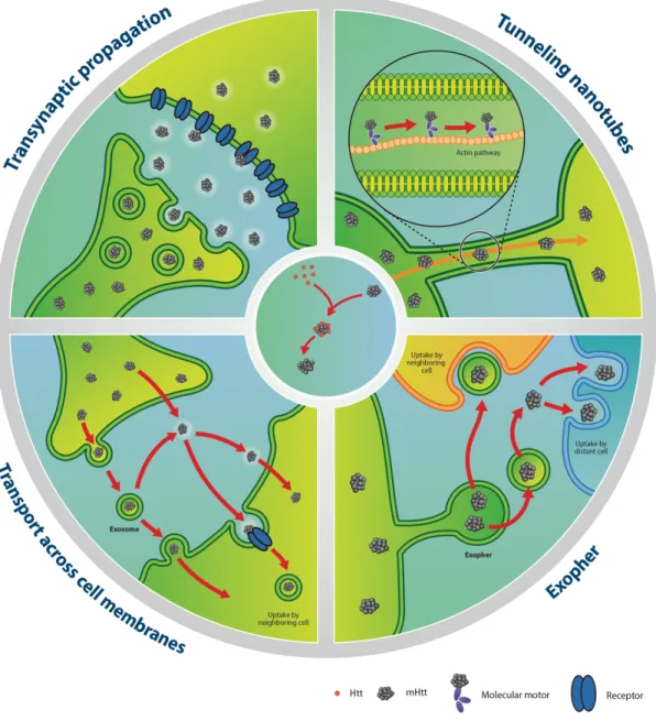
11.1 Transneuronal/transsynaptic propagation ... 26
11.2 Tunneling nanotubes ... 28
11.3 Exosomes and exophers ... 29
11.4 Endocytosis ... 30
11.5 Direct penetration of plasma membranes ... 32
11.6 Additional mechanisms ... 33
12. Evidence for mHTT prion-like behavior in vivo ... 39
Chapter 1: Demonstration of prion-like properties of mutant huntingtin fibrils ... 44
in both in vitro and in vivo paradigms ... 44
1.1 Résumé ... 46
1.2 Abstract ... 47
1.3 Introduction ... 48
1.4 Materials and methods ... 49
1.4.1 Recombinant HTTExon1Q25 and Q48 fibrils ... 49
1.4.2 Cell lines and culture conditions ... 50
1.4.3 Animals ... 54
1.4.4 Behavioral assessment ... 56
1.4.5 Post-mortem analyses ... 59
1.4.6 Statistical analysis ... 61
1.5 Results ... 62
1.5.1 Uptaken mHTTExon1 fibrils of human origin are toxic to multiple cells lines .. 62
1.5.2 Fibrils can recruit WT HTT into aggregates ... 64
1.5.3 Intracerebral injection of HTTExon1Q48 fibrils induces cognitive deficits and anxiety-like behavior in WT mice ... 65
1.5.4 Changes in the staining patterns of endogenous HTT in adult WT mice ... 66
1.5.5 Injection of HTTExon1Q48 fibrils precipitates disease in the R6/2 mouse model of HD ... 67
1.5.6 Exogenous HTTExon1Q48 fibrils colocalize with endogenous mHTT in R6/2 mice ... 69
1.5.7 Peripheral injection of mHTTExon1 fibrils initiates an immune response ... 71
1.6 Discussion ... 72
1.7 Acknowledgements ... 76
1.8 Contribution ... 76
1.9 Figures ... 78
Conclusions and perspectives ... 91
2.1 Results ... 91
2.2 Contribution to the field ... 92
2.3 Technical considerations and limits ... 99
2.4 Perspective ... 101
List of figures
Introduction
Figure 1. HD symptomatology ... page 3 Figure 2. HD neuropathology ... page 4 Figure 3. HD genetics ... page 6 Figure 4. Schematic representation of mHTT aggregation models ... page 21 Figure 5.Protein aggregation and the prion-like propagation ... page 23 Figure 6. Putative mechanisms of mHTT spreading and seeding capacities ... page 38
Chapter 1
Figure 1.1. Uptake of fibrillar HTTExon1Q25 and Q48 by different cell types ... page 78 Figure 1.2. Seeding of endogenous HTT by exogenous HTTExon1Q48 fibrils ... page 79 Figure 1.3. Manifestation of behavioral impairments in adult WT mice injected with
HTTExon1Q48 fibrils ... page 80 Figure 1.4. Post-mortem identification of HTTExon1Q25 and Q48 fibrils in adult WT mice ... page 81 Figure 1.5. Precipitation of behavioral phenotype in R6/2 mice following injection of HTTExon1Q48 fibrils ... page 82 Figure 1.6. Colocalization of HTTExon1Q48 fibrils and endogenous mHTT in R6/2 mice
... page 83 Figure 1.7. Toxic effects of exogenous HTTExon1Q48 fibrils on multiple cell lines page 84 Figure 1.8. Absence of motor impairments in WT adult mice injected with HTTExon1Q48 fibrils ... page 85 Figure 1.9. Examples of behavioral measures not affected in R6/2 mice following injection of HTTExon1Q48 fibrils ... page 86 Figure 1.10. Colocalization of HTTExon1Q48 fibrils with endogenous mHTT in R6/2 mice at 4 weeks of age ... page 87 Figure 1.11. Colocalization of HTTExon1Q48 fibrils with ubiquitin ... page 88 Figure 1.12. Changes in staining patterns of endogenous HTT following injection of
HTTExon1Q48 fibrils ... page 89 Figure 1.13. Development of an immune response following intravenous injection of
HTTExon1Q25 and Q48 fibrils in adult WT mice ... page 90
Conclusion
List of tables
Introduction
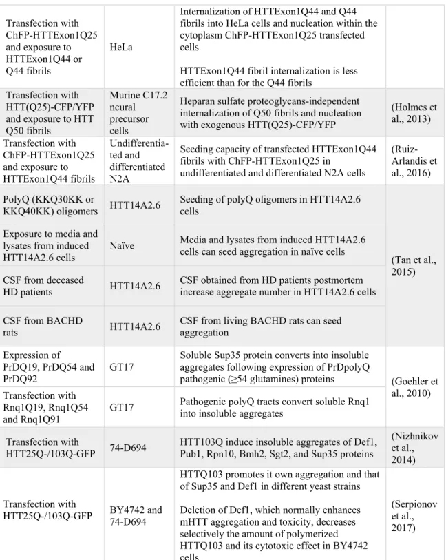
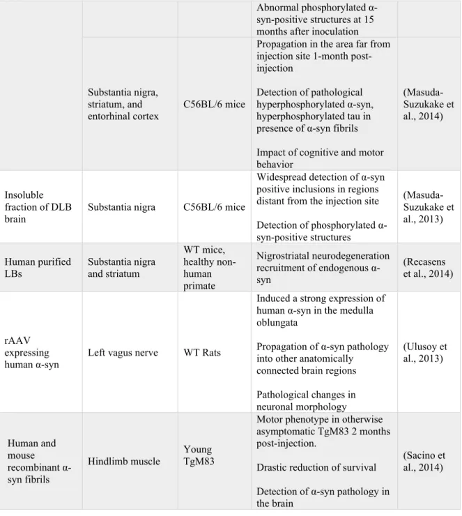
Table 1. Most used HD mouse models ... page 15 Table 2. In vitro evidence of mHTT spreading capacities ... page 34 Table 3. In vitro evidence of mHTT seeding capacities ... page 36 Table 4. Experimental evidence for the in vivo prion-like behavior of causative protein of neurodegenerative diseases ... page 40
Conclusion
List of abbreviations
α-syn, α-synuclein Aβ, Amyloid beta
AAV, Adenoassociated vector AD, Alzheimer’s disease
AGD, Argyrophilic Grain disease AP, Antero-posterior
APP, Amyloid precursor protein ASO, Antisense oligonucleotide B, Baseline
BAC, Bacterial artificial chromosome
BACHD, Bacterial artificial chromosome (BAC) transgenic rat/mouse model of
Huntington’s disease
BDNF, Brain-derived neurotrophic factor
BiFC, Bimolecular fluorescence complementation assays BoNT, Botulinum toxin
BSA, Bovine serum albumin
CAS9, CRISPR associated protein 9 CBD, Corticobasal degeneration CFP, Cyan fluorescent protein CGNs, Cerebellar granule neurons ChFP, mCherry fluorescent protein CJD, Creutzfeldt-Jakob disease CNS, Central nervous system CSF, Cerebrospinal fluid
Cryo-ET, Cryo-electron tomography
CRISPR, Clustered regularly interspaced short palindromic repeats CTRL, Control
CTX, Cortex
CTX M1, Primary motor cortex D, Day
DARPP32, Dopamine- and cAMP-regulated neuronal phosphoprotein DBS, Deep brain stimulation
Def1, RNA polymerase II degradation factor 1 DHA, Docosahexaenoic acid
DLB, Dementia with Lewy bodies DMD, Duchenne muscular dystrophy DNA, Deoxyribonucleic acid
Drd2, Dopamine receptor D2 DV, Dorso-ventral
EGFP, Enhanced green fluorescent protein ENK, Enkephalin
ESCs, Embryotic stem cells
Ethyl-EPA, Eicosapentaenoic acid F, Female
FDA, Food and Drug Administration FITC, Fluorescein isothiocyanate GFP, Green fluorescent protein GLT1, Glutamate transporter 1
GPe, External segment of the globus pallidus GPi, Internal segment of the globus pallidus HA-tag, Human influenza hemaglutinin tag HAP1, Huntingtin-associated protein 1 HD, Huntington’s disease
HD143F, Human fibroblast derived from Huntington’s disease patient carrying 143
polyglutamine repeats
hGFP neurons, Human GFP positive neurons HPC, Hippocampus
HPT, Hypothalamus HR, Hour
HRS, Hours
HSPG, Heparan sulfate proteoglycans HTT, Huntingtin
IBs, Inclusion bodies
iGABA, Induced pluripotent stem cell derived GABA neurons iPSC, Induced pluripotent stem cells
JHD, Juvenile Huntington’s disease kDa, Kilo Dalton
KI, Knock-in LBs, Lewy bodies LNs, Lewy neurites LV, Lateral ventricle m, Month M, Male
MAP2, Microtubule-associated protein 2 mHTT, Mutant huntingtin
ML, Medio-lateral mRNA, Messenger RNA MSNs, Medium spiny neurons
mTOR, Mammalian target of rapamycin N-terminal, Amino-terminal
N-terminus, Amino-terminus NFTs, Neurofibrillary tangles NLS, Nuclear localization signals NSCs, Neural stem cells
NTs, Neuropil treads PD, Parkinson’s disease PFA, Paraformaldehyde PI3-kinase, Phosphatidylinositol-3-kinase PolyQ, Polyglutamine PrD, Prion domain
Pub1, Nuclear and cytoplasmic polyadenylated RNA-binding protein Q, Glutamine
rAAV, Recombinant Adeno-Associated Virus RNA, Ribonucleic acid
SMA, Spinal muscular atrophy
SNP, Single nucleotide polymorphism SNr, Substantia nigra
SP, Substance P STR, Striatum
Sup35, Yeast eukaryotic release factor 3 Tg, Transgenic
TNTs, Tunneling nanotubes V1, Venus protein half 1 V2, Venus protein half 2 W, Week
WT, Wild-type
YAC, Yeast artificial chromosome YFP, Yellow fluorescent protein
Acknowledgements
My sincere thanks go to my research supervisor, Dr. Francesca Cicchetti for introducing me and guiding me in my doctoral studies. With her scientific knowledge, hard-working and quick-thinking skills, she will always be a role model for my future career and personal life. I also want to express my utmost gratitude to all the members of Dr. Francesca Cicchetti’s team, professionals and students, that I had the pleasure and luck to work with. Each one of them contributed to my professional and personal growth in a fashion that I will always be grateful for.
I would like to acknowledge the organizations that sponsored my graduate studies providing me awards and scholarships. My greatest appreciation goes to the Huntington Study Group, which provided me with their New Member Award to attend their 25th meeting in Houston,
USA, in 2018; and to the Bourse d'excellence du Centre thématique de recherche en Neurosciences (CTRN), the Bourse de la Fondation du CHUL and the Bourse Formation de doctorat FRQS which funded my project and my Ph.D. studies.
To conclude, my greatest appreciation goes to the members of my doctoral committee, Dr. Roger Barker, Dr. Abid Oueslati, Dr. David Gosselin for their availability in evaluating my thesis as well as the president of the jury Dr. Martin Lévesque.
Avant-propos
During my Ph.D., I investigated the prion-like behavior of the mutant huntingtin protein (mHTT), which resulted in a publication in Acta Neuropathologica. More specifically, the work presented in Masnata et al. examined the pathophysiological effects of the inoculation of exogenous human fibrillar mHTT (Q48) and huntingtin (HTT Q25) N-terminal fragments in three different cell lines as well as animal models. Additionally, I conducted a thorough literature search on the capacity of mHTT to spread in in vitro settings, which resulted in the publication of the review article “The evidence for the spread and seeding capacities of the mutant huntingtin protein in in vitro systems and their therapeutic implications” in Frontiers
in Neuroscience (Masnata and Cicchetti, 2017). I also contributed to the research projects of
some of my colleagues that aimed to uncover the prion-like behavior of mHTT (Maxan et al., 2020, Neurobiology of Disease; Rieux et al., 2020, Molecular Psychiatry).
In the last years of my doctorate studies, my interest shifted to understanding the contribution of the tau protein to HD pathology, work that is currently being pursued in Dr. Cicchetti’s lab. I wrote an additional review article on the topic entitled “Targeting Tau to treat clinical features of Huntington's disease” (Masnata et al., 2020, Frontiers in Neurology).
List of peer-reviewed publications
Peer-reviewed publications
Masnata M*, Sciacca G*, Maxan A*, Bousset L*, Denis H, Lauruol F, David L, Saint-Pierre
M, Kordower J, Melki R., Alpaugh M, Cicchetti F. Demonstration of prion-like properties of mutant huntingtin fibrils in both in vitro and in vivo paradigms. Acta Neuropathologica, doi: 10.1007/s00401-019-01973-6.
Maxan A, Sciacca G, Alpaugh M, Tao Z, Breger L, Dehay B, Ling Z, Qin C, Cisbani G,
Masnata M, Salem S, Lacroix S, Oueslati A, Bezard E, Cicchetti F. Use of adeno-associated
virus-mediated delivery of mutant huntingtin to study the spreading capacity of the protein in mice and non-human primates. Neurobiology of Disease, 10.1016/j.nbd.2020.104951.
Rieux M*, Alpaugh M*, Saint-Pierre M, Sciacca G, Masnata M, Denis HL, Lévesque SA, Herrmann F, Bazenet C, Garneau AP, Isenring P, Truant R, Oueslati A, Gould PV, Ast A, Wanker EE, Lacroix S & Cicchetti F. Shedding a new light on Huntington’s disease: how blood can both propagate and ameliorate disease pathology. Molecular Psychiatry, 10.1038/s41380-020-0787-4.
*Equally contributing authors.
Review articles
Masnata M*, Salem S*, de Rus Jacquet A, Anwer M, and Cicchetti F. Targeting tau to treat
clinical features of Huntington's disease. Frontiers in Neurology, doi: 10.3389/fneur.2020.580732.
Masnata M and Cicchetti F. The evidence for the spread and seeding capacities of the mutant
huntingtin protein in in vitro systems and their therapeutic implications. Frontiers in
Neuroscience, doi: 10.3389/fnins.2017.00647
Introduction
1. Historical background
Since the Middle Ages, there have been oral and written reports describing "dancing disorders", what we now more commonly refer to as chorea (the Greek word for dance). Around the year 1000, involuntary, jerking and twitching movements were interpreted as consequences of curses and demonic possession (Vale and Cardoso, 2015). Hundreds of years later, this belief persisted and women persecuted during the witch hunt in Salem, Massachusetts, in the 1690s, were in fact affected by similar diseases (Vale and Cardoso, 2015). In the XIX century, the American physician George Huntington became interested in motor abnormalities and dementia in individuals of his community. He spent his life observing and collecting evidence in troubled middle-aged individuals who presented a family history of such disturbances. In 1872, his observations were published in Medical and
Surgical Reporter of Philadelphia in manuscript entitled “On Chorea”, where he described
the disease that was later named after him (Huntington, 2003).
Further progress in the field of Huntington’s disease (HD) would come during the HD Centenary Celebration in New York City, USA, when the work of the Venezuelan doctor Americo Negrette sparked the interest of the conference attendees. His report described an unusual cluster of HD-affected individuals in the geographically isolated village near the city of Maracaibo, Venezuela. Collecting testimonies from locals, he attempted to reconstruct the historical origin of HD and identify the patient zero, which he later confirmed as a European who had moved in the area in the 1860s (Bhattacharyya, 2016).
Under the leadership of the geneticist Nancy Wexler, a team of researchers travelled to Lake Maracaibo, intending to uncover the mutation that led to the development of HD. The study lasted 20 years during which blood samples and information on thousands of HD individuals were collected and shared globally between the members of the “Huntington's Disease Collaborative Research Group” (Wexler, 2012). Researchers and physicians were provided information for the identification of the causative gene. This extraordinary effort was brought by the Huntington's Disease Collaborative Research Group to achieve one of the most
important milestones in the history of HD: the isolation of the mutation responsible for the disease (MacDonald et al., 1993).
2. Symptomatology
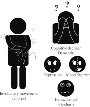
HD affects approximately 3 - 8 per 100,000 people worldwide, with a higher prevalence in western Europe, North America and Australia, where it reaches 5 - 13 people per 100,000 (Baig et al., 2016). HD is also known as Huntington’s chorea, a definition that accurately describes its most characteristic motor features: spasms affecting the voluntary muscles (Figure 1). In adulthood, these involuntary movements occur in concomitance with other motor abnormalities such as dystonia, rigidity, akinesia and bradykinesia (Phillips et al., 2008). Notably, some HD patients, even prior to showing motor disturbances, display progressive cognitive impairments (Papp et al., 2011) that impact activities of daily living and that can progress into severe dementia (Peavy et al., 2010). Behavioral changes, such as mood disorders, depression, delusion, hallucinations and psychosis (Figure 1) commonly accompany the motor and cognitive features, troubling HD patients who, after 15 – 20 years, lose their autonomy and become bedbound. In the last years of the disease, death occurs due to heart failure (Abildtrup and Shattock, 2013) or aspiration pneumonia (Heemskerk and Roos, 2012). Notably, there is a wide variability of signs and symptoms within the HD population. Some gene carriers manifest mostly with motor disturbances and minimal mood or cognitive dysfunctions, while others suffer from cognitive and mood changes with limited motor symptoms (Hahn-Barma et al., 1998). Even though an inverse correlation exists between CAG repeat and the severity of symptoms, it does not fully explain the diversity of clinical profiles nor the progression patterns that are unique to each person (Andresen et al., 2007). A clear example of this is reported in a study in which monozygotic twins showed HD clinical phenotype 7 years apart, reinforcing the concept that disease onset is not exclusively determined by genetic factors (Friedman et al., 2005).
Although HD is a disease that mostly affects the central nervous system (CNS), it can also cause weight loss (Djoussé et al., 2002), skeletal muscle atrophy (Zielonka et al., 2014), testicular atrophy (Selvaraj et al., 2020) and cardiac problems that commonly lead to death (Abildtrup and Shattock, 2013).
Figure 1. HD symptomatology. HD patients manifest
with an array of motor, cognitive and psychiatric dysfunctions. Some of the most common motor symptoms consist of uncontrolled twerking movements defined as chorea. Cognitive symptoms, such as memory loss, learning difficulties, trouble in accomplishing the daily routine can degenerate into dementia. Psychiatric symptoms include depression, mood disorders, hallucinations and psychosis. Abbreviation: HD, Huntington’s disease. Illustration made by Maria Masnata.
HD can also affect children (juvenile HD, JHD), who, in contrast to adults, do not always exhibit severe chorea or involuntary movements. Instead, juvenile cases are mainly affected by tremors and rigidity (Quarrell et al., 2013). Furthermore, a typical feature of JHD is the tendency to develop epileptic seizures, especially if the disease manifests before 10 years of age (Cloud et al., 2012; Gambardella et al., 2001). Similarly to adults, JHD individuals display cognitive impairments and behavioral changes, which can result in increased aggressiveness, apathy and poor scholastic performances (Ribaï et al., 2007). The progression of JHD is rather fast and the life expectancy does not exceed 10 to 15 years following the appearance of symptoms (Quarrell et al., 2013).
3. Neuropathology
The basal ganglia - a group of subcortical brain structures located deep in the brain - are the target of massive and progressive neurodegeneration in HD (Vonsattel et al., 1985). The basal ganglia span over two main brain subdivisions: the forebrain and the midbrain. The forebrain includes the dorsal striatum (caudate nucleus and putamen), the ventral striatum (nucleus accumbens and olfactory tubercle), the globus pallidus and the ventral pallidum. The substantia nigra and the diencephalon are found in the midbrain. Motor control and executive and cognitive functions are generated by these cerebral structures (Helie et al., 2013). In HD
patients, the caudate nucleus and the putamen are the first structures affected by neurodegenerative processes (Vonsattel et al., 1985).
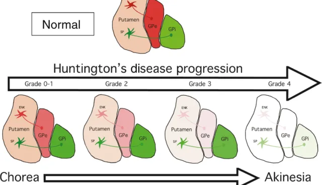
Within the striatum, the medium spiny neurons (MSNs) are the most vulnerable cellular elements. MSNs are GABAergic neurons, which give rise to two distinct pathways: the striatonigral (direct pathway) and the striatopallidal (indirect pathway) (Smith et al., 1998). The striatonigral pathway projects to the substantia nigra pars reticulata (SNr) or the internal segment of the globus pallidus (GPi) (Smith et al., 1998). The striatopallidal pathway projects to the external segment of the globus pallidus (GPe) (Gerfen, 1992). In the early stages of disease, the MSNs of the striatopallidal pathway, which also express dopaminergic D2 and enkephalin (ENK) receptors, are the first to degenerate (Crossman, 1987) (Figure 2). This is associated with the onset of involuntary movements (Crossman, 1987) (Figure 2). As the disease progresses, the MSNs of the striatonigral pathway, which express dopaminergic D1 receptors and substance P (SP), start dying and other motor dysfunction, such as rigidity and akinesia, begin to manifest (Berardelli et al., 1999) (Figure 2).
Figure 2. HD neuropathology. Schematic illustration of HD striatal degeneration according to Vonsattel
individuals mostly depict jerking and twitching movements (chorea). From grade 2 to 4, progressive loss of the ENK-GPe and SP-GPi projections is observed and is accompanied by progressive akinesia. At Grade 4, almost 95% of neurons have degenerated and the GPi, GPe and putamen are characterized by severe atrophy. Abbreviations: ENK, Enkephalin; GPe, External segment of the globus pallidus; GPi, Internal segment of the globus pallidus; SP, Substance P. Source (Reiner and Deng, 2018).
As HD progresses, other cerebral structures are affected by the neurodegenerative processes. For instance, the cortex is the second most affected forebrain structure (Cudkowicz and Kowall, 1990). More specifically, the large pyramidal cortical projection neurons, located in layers V and VI that project to the striatum, are the most vulnerable neuronal populations (Cudkowicz and Kowall, 1990; Hedreen et al., 1991). These neurons are part of the primary motor cortex, and their death arises in concomitance with motor impairments (Thu et al., 2010). Overtime, HD-associated neurodegeneration targets other areas such as the hippocampus (Begeti et al., 2016; Spargo et al., 1993; van den Bogaard et al., 2011). Neuronal death in the CA1 region (Spargo et al., 1993) and subsequent overall hippocampal atrophy (van den Bogaard et al., 2011) have been linked with the occurrence of cognitive impairments, in particular deficits in learning task (Begeti et al., 2016).
4. Genetics
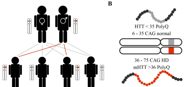
Classified as an autosomal dominant neurodegenerative disease (Figure 3A), HD is caused by a mutation in the HTT, or IT15, gene. HTT is located on the short arm of chromosome 4 and codes for the protein huntingtin (HTT). The mutation consists of the insertion of more than 35 repetitions of the CAG trinucleotide in the exon 1 of the HTT gene (MacDonald et al., 1993) (Figure 3B). While 36 to 39 repeats are associated with reduced penetrance and an uncertain outcome, more than 40 CAG repeats result in HD onset (Myers, 2004). Moreover, the increase in the number of CAG repeats is a common feature during the transmission of genetic material (Maat-Kievit et al., 2001). Thus, if one of the parental chromosomes has 27 – 35 CAG repeats, a CAG expansion can occur, leading to HD-affected offspring (Maat-Kievit et al., 2001). The expanded (>35) CAG triplets encode a polyglutamine (polyQ) sequence in the mutant huntingtin protein (mHTT), which causes cytotoxicity that leads to neurodegeneration (Vonsattel and DiFiglia, 1998).
Figure 3. HD genetics. Schematic of the genetic transmission of the non-mutated (gray) and mutated (red) HTT
gene (A) and of the chromosome 4 with non-mutated and mutated alleles and respective encoded proteins (B). The non-mutated HTT gene, with less than 36 CAG repeats, encodes for the HTT protein (< 35 polyQ), while the mutated HTT gene, with more than 36 CAG, encodes for mHTT (> 36 polyQ). Abbreviations: HD, Huntington’s disease; HTT, Huntingtin; mHTT, Mutant huntingtin; PolyQ, polyglutamine. Illustration made by Maria Masnata.
5. Treatment
5.1 Pharmacological treatments
HD patients typically receive treatments to alleviate their symptoms, i.e. motor or mood disorders. To treat motor problems, more specifically involuntary movements (chorea), the drugs Tetrabenazine (Xenazine) and Deutetrabenazine (Austedo) are most often prescribed (Huntington Study Group, 2006; Yero and Rey, 2008). Tetrabenazine is well tolerated and ameliorates chorea when taken over extended periods of time (Fasano et al., 2008), however, it also causes a number of side-effects which include sedation, drowsiness, fatigue, insomnia, depression, suicidal thoughts, akathisia, anxiety and nausea (Huntington Study Group, 2006; Jankovic and Beach, 1997).
While selective serotonin reuptake inhibitors, such as citalopram (Celexa), fluoxetine (Prozac) and sertraline (Zoloft), are used to alleviate depression and are comparatively safe and efficient (De Marchi et al., 2001; Rowe et al., 2012), the treatment of psychosis is more challenging. Atypical antipsychotics, including risperidone (Risperdal), quetiapine (Seroquel) and olanzapine (Zyprexa), induce motor disturbances while typical antipsychotic,
such as haloperidol (Haldol) and fluphenazine (Prolixin), suppress involuntary movements but in turn, can cause dystonia and rigidity (Unti et al., 2017).
At the moment, there are no available treatments to specifically improve cognition. Several candidates, approved by the Food and Drug Administration (FDA), have been tested in clinical trials, including Memantine (Beister et al., 2004; Cankurtaran et al., 2006) Lithium (Vestergaard et al., 1977) and Atomoxetine (Strattera) (Beglinger et al., 2009). However, none of these treatments have shown to significantly ameliorate cognitive deficits in HD (Beglinger et al., 2009; Beister et al., 2004; Cankurtaran et al., 2006).
The quest to find new symptomatic treatments, while exploring disease-modifying strategy, is still ongoing (Barker and Mason, 2019). For instance, a phase III clinical trial named PROOF-HD has just been launched (Teva Pharmaceutical Industry, 2016). This study investigates the drug Pridopidine (TV-7820), which is a dopamine stabilizer and an agonist of the sigma 1 receptor. It was initially thought to improve motor deficits in HD patients, but recent clinical trials reported just a trend towards amelioration of motor deficits (Prilenia, 2020), which was interpreted as sufficient evidence to pursue its evaluation.
5.2 Non-pharmacological treatments
5.2.1 Cell replacement therapy
Following the early success of cell transplantation in PD patients (Lindvall and Björklund, 2004), HD patients have also been considered candidates for cell replacement therapy. To this day, seven open-label trials were carried out to establish safety, tolerability and efficacy in this patient population. Trials were performed on a small number of patients with mild to advanced HD and yielded mild transient symptom improvements and a lack of long-term benefits (Bachoud-Lévi et al., 2000; Barker et al., 2013; Capetian et al., 2009; Gallina et al., 2010; Hauser et al., 2002; Kopyov et al., 1998; Maxan et al., 2018; Rosser et al., 2002). Post-mortem studies reported that the cellular grafts presented a progressive disease-like neuronal degeneration and an increase of microglial activation (Cicchetti et al., 2009; Maxan et al., 2018). Furthermore, mHTT aggregates, were detected within the neurons, infiltrating immune cells and extracellular matrix inside the boundaries of the grafted tissues (Cicchetti et al., 2014; Maxan et al., 2018). More recently, an open label study recruited a large cohort
of patients (67 patients) to specifically evaluate the motor score between grafted and non-grafted patients (Assistance Publique - Hôpitaux de Paris, 2017). However, no motor improvements were reported, possibly due to graft rejection (alloimmunization). The authors suggested that the implantation of cell–derived neural precursors, instead of fetal human cells, could represent a better option to prevent graft rejection (Bachoud-Lévi and on behalf the Multicentric Intracerebral Grafting in Huntington’s Disease Group, 2020).
5.2.2 Deep brain stimulation
Deep brain stimulation (DBS) is a treatment that consists of surgically implanting a device that delivers electrical signals to specific brain areas. This signal regulates the activity of the neuronal circuits responsible for motor abnormalities and can be controlled and modified according to the patients' needs. This technique was initially adopted to treat PD patients, showing significant improvement in motor symptoms (e.g. rigidity, tremors, bradykinesia, gait disturbances and problems with balance) (Deuschl et al., 2006; Williams et al., 2010). In HD patients, DBS of the internal segment of the globus pallidus has been proposed and tested on a small number of patients. Studies showed improvement of chorea, no effect on dystonia or cognition, but worsening of bradykinesia and rigidity (Delorme et al., 2016; Gonzalez et al., 2014; Velez-Lago et al., 2013; Wojtecki et al., 2015). Taken together, these results suggest that DBS could be beneficial for the treatment of chorea in HD patients, although long-term effects should be investigated.
5.3 Experimental treatments
5.3.1 Gene silencing and gene editing
Since HD is caused by a single genetic mutation, gene silencing and gene editing deserve serious consideration. Gene silencing through antisense oligonucleotide (ASO) consists of promoting mRNA degradation or blocking its translation, inducing a decrease in the synthesis of the target-protein. ASOs’ potential for the treatment of HD was explored in preclinical trials with consistent reports of a high success rate in lowering mHTT and improving behavioral deficits in Hu97/18, an HD mouse model that fully expresses the human HTT, and YAC128 mice, an HD transgenic (Tg) mouse model overexpressing human full-length mHTT (Southwell et al., 2018; Stanek et al., 2013). Silencing of HTT with the
ASO drug IONIS-HTTRx is now under investigation in clinical trials (Tabrizi et al., 2019). IONIS-HTTRx is delivered intrathecally and aims to lower both normal HTT and mHTT in patients with early manifest HD. Results collected thus far show that ASO efficiently reduces mHTT CSF levels without serious side-effects (Mullard, 2019). However, treatment has not shown improvements in neurological outcomes, and no differences were observed between placebo-treated concerning cognitive and psychiatric symptoms or functional capacity (Smith and Tabrizi, 2019). IONIS-HTTRx is currently under investigation in the phase III trial “GENERATION HD1” (Hoffmann-La Roche, 2020).
While IONIS-HTTRx indiscriminately targets normal and mutated HTT, selective ASOs, such as WVE-120101 and WVE-120102, have now been trialed. This approach has been developed by WAVE Life and 2 trials are underway: PRECISION-HD1 recruited adult patients with early manifest HD who carry a targeted SNP rs362307 (SNP1), while PRECISION-HD2 was geared towards HD patients who carry an SNP rs362331 (SNP2) (Wave Life Sciences Ltd., 2020a, 2020b). The phase III reported reduced mHTT CSF levels by 12.4%, a much lower percentage in comparison to the one reported by IONIS-HTTRx, which reached 40 to 60% decrease in mHTT CSF content (Tabrizi et al., 2019). Higher doses are now being administered to the participants of the PRECISION-HD trials.
Preventing HTT synthesis by inhibiting translation could also be achieved using microRNAs. microRNAs are non-coding ribonucleic acid fragments that modulate gene expression. UniQure has developed the drug AMT-130, an adenoviral vector carrying a microRNA that non-selectively targets the HTT gene. As observed in the pre-clinical context, AMT-130 induced a dose-dependent HTT decrease and improvements in cognitive and anxiety-like behavior of Hu128/21 mice (Caron et al., 2020). The company has recently started testing AMT-130 in phase I and II clinical trials (UniQure Biopharma B.V., 2020).
The genome editing technique CRISPR/Cas9 was investigated in preclinical studies to address the pathological mutation of the HTT gene. CRISPR/Cas9 is a system of gene editing developed naturally by bacteria, which, in case of viral infections, can cut the viral DNA and prevent the virus from multiplying to attack the host. CRISPR/Cas9 has been tested and shown to inhibit the expression of mHTT aggregates in the striatum of KI140 mice,
knock-in HD mice that contaknock-ins 140 CAG repeats knock-in the exon 1 of the HTT gene, successfully reducing the expression of mHTT and ultimately improving motor coordination and strength (Yang et al., 2017).
Gene silencing and gene editing are considered some of the most promising therapeutic approaches for the treatment of genetic disorders such as HD. For instance, in 2016, new ASO therapies were approved by the FDA for the treatment of the monogenic neuromuscular disorders Duchenne muscular dystrophy (DMD) and spinal muscular atrophy (SMA) (Commissioner, 2020a, 2020b). However, the long-term consequences and side effects of genome engineering are still unknown. It is therefore critical to investigate and closely monitor future clinical studies that target the HTT gene (Barker et al., 2020).
5.3.2 Immunotherapies
HD is characterized by abnormal hyper-activation of the immune system (Ciccocioppo et al., 2020). It has been established that the presence of pathogenic misfolded aggregates in the brain triggers a chronic immune response, mainly via activation of microglia, which in turn, release pro-inflammatory mediators including oxygen- and nitrogen-derived free radicals. This leads to progressive neuroinflammation, disruption of the integrity of the blood brain barrier and neuronal damage (Ciccocioppo et al., 2020; Sweeney et al., 2018). In the early stages of disease, reactive microglia have been detected in the neostriatum, cortex and globus pallidus, and their accumulation in the cortex and striatum directly linked to neuronal functionality and loss (Pavese et al., 2006; Sapp et al., 2001).
In recent years, several compounds have been identified as possible treatments to restore the physiological immune response in the brains of HD patients. Several of these drugs had been yielded promising results in animal studies and a selection went on to be tested in the clinic. Some of the drugs that reached phase II-III clinical trials are briefly discussed below:
• Pepinemab is an anti-SEMA4D monoclonal antibody developed by Vaccinex and the Huntington Study Group. Pepinemab’s epitope is SEMA4D, a multifunctional transmembrane protein that regulates multiple neuroinflammation processes (Fisher et al., 2016). Intraperitoneal injections of the anti-SEMA4D antibody improved central and peripheral pathology as well as cognitive and anxiety-like behavior in YAC128
mice (Southwell et al., 2015). The inhibition of SEMA4D prevents astrogliosis, which would otherwise likely cause neuronal damage (Fisher et al., 2016; Southwell et al., 2015). Recently, Pepinemab was tested in the phase II SIGNAL clinical study, which recruited late prodromal or early manifest HD patients. Although the study did not fulfill the pre-established secondary outcomes (brain metabolic activity and behavioral improvements), a trend towards cognitive improvements, such as an amelioration in memory tasks and planning ability, was observed (“Top-line results of phase 2 SIGNAL study in Huntington’s disease support potential for cognitive benefit of Pepinemab |Vaccinex, Inc.,” n.d.). On the basis of these achievements, the FDA has conferred to Pepinemab the designation of “Orphan Drug” and “Fast Track” for the treatment of HD.
• Eicosapentaenoic acid (Ethyl-EPA) or Miraxion, a ω-3 fatty acid commonly used to treat hypertriglyceridemia, has been associated with anti-inflammatory properties. When administered to R6/1 mice - a transgenic model expressing exon 1 of the human
HTT gene with approximately 114 CAG repeats - it significantly improved motor
coordination and locomotor activity (Clifford et al., 2002), possibly by regulating the structure of the plasma membrane (Clifford et al., 2002), reducing oxidative stress (Puri et al., 2005) or inhibiting apoptosis (Murck and Manku, 2007). Ethyl-EPA was further investigated in the TREND-HD study by Amarin Neuroscience Ltd and the Huntington Study Group. It reached phase III, but failed to show clinical improvements, except for a small cohort of patients with a CAG repeat length shorter than 45 (Puri et al., 2005). After 12 months of treatment, these patients showed mild improvements on motor scores (Huntington Study Group TREND-HD Investigators, 2008). However, an additional clinical studiy detected no significant improvements in motor deficits in mild-to-moderate HD patients following 6 months of treatment with Ethyl-EPA (Ferreira et al., 2015).
• Laquinimod is an immunomodulator initially studied to treat multiple sclerosis. When tested in the R6/2 mouse model - a transgenic mouse which expresses exon 1 of the human HTT gene with approximately 125 CAG repeats - it reduced motor deficits and striatal pathology (Ellrichmann et al., 2017). Laquinimod’s mechanism of action is
unclear, but it seems to exert an anti-inflammatory action, reducing free radical damage (Ellrichmann et al., 2017). It is also thought to increase levels of brain-derived neurotrophic factor (BNDF), a pro-survival factor essential for the proper activity of the striatal neurons (Ellrichmann et al., 2017). The phase II trial for Laquinimod, named LEGATO-HD, was carried out by TEVA Pharmaceuticals for a total of 13 months. Ultimately, LEGATO-HD revealed that the compound did not meet the primary endpoints of the study, which consisted in the amelioration of baseline motor deficits (Teva Branded Pharmaceutical Products R&D, Inc., 2020).
Immunotherapies for HD have been explored since the early 2000s with inconsistent results between animal and human studies. For instance, minocycline, a tetracycline antibiotic, improved motor performances and extended life expectancy of R6/2 mice, seemingly by inhibiting apoptosis and decreasing oxidative stress and microglial activation (Chen et al., 2000; Tikka and Koistinaho, 2001). However, the absence of symptom improvements in mild to moderate functionally-impaired HD patients halted further clinical investigations (Huntington Study Group DOMINO Investigators, 2010). Modulating neuroinflammation is a challenging task, especially considering the complexity of its intricate pathways. Targeting specific elements, as does Pepinemab, could help avoid unwanted side effects and better predict overall results. Furthermore, immunotherapies could be excellent candidates in combinational therapies to tackle different neuropathological aspects of HD.
5.3.3 Antibody treatment
For several neurodegenerative disorders similar to HD, active (injection of antigens that would induce the organism’s own immune response) or passive (injection of ready-made antibodies) immunization therapies have been tested in clinics with variable outcomes (Alpaugh and Cicchetti, 2019). For instance, while immunotherapies against Aβ have brought mixed results (Schilling et al., 2018), tau immunotherapies are now gaining momentum (Congdon and Sigurdsson, 2018). Either anti-tau active or passive immunization has been demonstrated to be safe and to significantly improve AD pathology and cognitive deficit (Congdon and Sigurdsson, 2018). For HD, antibody-based therapies are still in early stage of development and no compounds have yet been translated to the clinic.
Active immunization against mHTT has been evaluated by injections of plasmids containing mHTT N-terminal fragment with 103 polyQ administered to R6/2 mice (Miller et al., 2003). Treated animals promptly developed an immune response against the antigen and, despite no effect being detected with respect to the number of mHTT aggregates, the treatment improved the diabetic phenotype of these mice (Miller et al., 2003). Other studies have tested antibodies (passive immunization) which target extracellular mHTT, i.e. the caspase-6 cleaved mHTT fragments enzyme (aa586)) (Bartl et al., 2020). This antibody, developed by AFFiRiS and referenced to as mAB C6-17, efficiently prevented cell-to-cell mHTT propagation in in vitro systems, decreasing by more than 90% the uptake of HTTExon1Q103 by healthy acceptor cells (Bartl et al., 2020). Antibodies directed against intracellular mHTT (intrabodies) have also been developed (Amaro and Henderson, 2016). For example INT41, which specifically targets proline-rich region of HTT, was administered to R6/2 mice using a recombinant AAV and shown to induce a reduction of mHTT aggregates within the striatum (Amaro and Henderson, 2016).
Research in the field of immunotherapy has made significant progress in the development of treatments for AD and PD (Alpaugh and Cicchetti, 2019), and is now being considered for HD (Denis et al., 2019). Antibody-based therapies have the distinct advantage of being highly specific to the selected epitopes and can target intracellular or extracellular mHTT both in the periphery and CNS. The antibodies’ low molecular weight makes them suitable for nanocarrier-based delivery approaches to control the rate of antibody release, reduce antibody degradation and prevent unwanted host immune response (Wagh and Law, 2013). Based on the concept that mHTT is frequently found outside the cell boundary (Cicchetti et al., 2014; Drouin-Ouellet et al., 2015; Tan et al., 2015; Wild et al., 2015), active and passive immunization strategies targeting extracellular mHTT are attracting interest (Denis et al., 2019).
6. HD animal models
Animal models allow the study of pathophysiological and behavioral changes in disease-engineered living organisms and they represent one of the most powerful resources to study novel therapeutic strategies. The development of more relevant models of HD followed the discovery of the HTT gene mutation, with the elaboration of transgenic mouse models that
expressed exon 1 of the human HTT gene, known as the R6 line (Mangiarini et al., 1996). Four R6 mice were generated, two of which, the R6/1 and R6/2, are still commonly used in HD research for their disease-like phenotype. The two models slightly differ in terms of their behavioral and neuropathological characteristics (Mangiarini et al., 1996). The R6/1 mice express 114 CAG repeats, develop a motor behavior at 15–21 weeks of age, and mHTT aggregates at 2 months of age (Table 1). On the other hand, the R6/2 model is notorious for developing an early-onset HD phenotype (Table 1). R6/2 mice express 144 to 150 CAG repeats and show the first signs of motor and cognitive deficits starting from 3 weeks of age. The appearance of intranuclear mHTT aggregates begins soon after birth and proceeds aggressively (Mangiarini et al., 1996).
Shortly after the arrival of the R6 mice, another transgenic mouse model was developed which, in contrast to the R6, expresses the full-length human HTT (Hodgson et al., 1999). It was generated using a yeast artificial chromosome (YAC) containing a full-length human
HTT with expanded polyQ repeats (Hodgson et al., 1999) (Table 1). The YAC128 mice
depict motor deficits at 4 months of age (Menalled et al., 2009) and cognitive deficits at 8 months of age (Van Raamsdonk et al., 2005). Accumulation of nuclear mHTT aggregates starting from 6 months of age is associated with decreased striatal volume and selective striatal neurodegeneration (Hodgson et al., 1999; Slow et al., 2003). As the YAC128, the BACHD model was generated using a bacterial artificial chromosome (BAC), expressing the human full-length human HTT gene (Table 1). BACHD mice are characterized by progressive motor impairments starting at 2 months of age (Menalled et al., 2009), while mHTT aggregates are detected after 6 months of age (Gray et al., 2008).
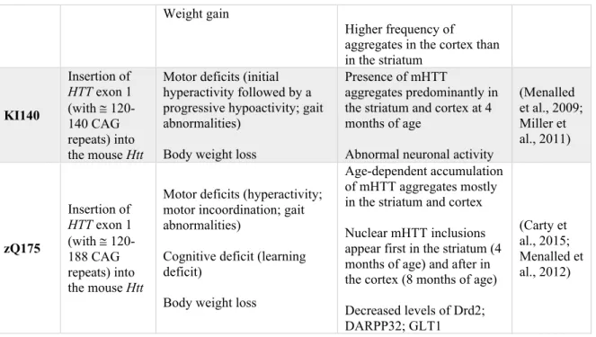
Chimeric knock-in (KI) models, expressing the human exon 1 of the HTT gene with CAG expansion, were also engineered to model HD. The KI140 mouse was designed by inserting the human HTT exon 1, with approximately 120-140 CAG repeats, into the mouse Htt (Menalled et al., 2003) (Table 1). The mice manifest complex motor deficits, with initial hyperactivity at 1 month of age followed by a progressive decrease in locomotor activity starting from 4 weeks (Menalled et al., 2009). Numerous mHTT aggregates, predominantly in the striatum and cortex, are detectable starting from 4 months of age (Menalled et al., 2003) (Table 1). The zQ175 mouse model, which originates from the KI140, expresses the
human HTT exon1 with 188 CAG repeats and slightly differs in behavioral phenotype. It displays motor and learning deficits starting at 6 months of age (Menalled et al., 2012). It additionally develop an age-dependent accumulation of mHTT aggregates mostly in the striatum and cortex. Nuclear mHTT inclusions are visible in the striatum (4 months of age) and in cortex (8 months of age) (Menalled et al., 2012).
Model Genetic manipulation Behavioral Changes Neuropathology References
R6/1 Expression of exon 1 of the human HTT gene containing 116 CAG Motor performance abnormalities at 15–21 weeks of age
Body weight loss
Comparatively short live span (32–40 weeks)
Reduced brain volume by 18 weeks of age
Neuronal atrophy in absence of overall neuronal loss Deposition of mHTT aggregates starting from 2 months of age (Mangiarini et al., 1996) R6/2 Expression of exon 1 of the human HTT gene containing 144-150 CAG
Motor symptoms starting from 4-6 weeks of age (clasping, loss of motor coordination, stereotypic involuntary movements, shaking, reduced locomotor activity)
Cognitive symptoms from (impaired short and long-term and working memory; early-onset learning deficit) Anxiety-like behavior (tendency to stay in the dark) Epileptic seizure
Body weight loss Short live span (12 – 18 weeks)
Reduced brain volume, particularly of the striatum Progressive neuronal atrophy with neuronal loss at 12 weeks of age
Extended mHTT aggregates deposition staring at day 1 post-birth (Carter et al., 1999; Mangiarini et al., 1996; Menalled et al., 2009) YAC128 Expression of the human HTT with 128 CAG repeats
Motor deficit (loss of motor coordination; initially mild hypoactive then hypokinetic) Weight gain
Accumulation of nuclear mHTT aggregates starting from 6 months of age Decreased brain weight and striatal volume starting from 12 months of age (Hodgson et al., 1999; Menalled et al., 2009; Slow et al., 2005, 2003) BACHD mice Expression of the human HTT with 97– 98 CAG repeats
Motor deficit (loss of motor coordination at 4 weeks of age; hypoactivity)
Anxiety-like behavior (higher preference for the dark)
Brain atrophy starting from 12 months (loss of cortical and striatal volume)
Diffuse nuclear accumulation of mHTT aggregates starting from 6 months of age
(Gray et al., 2008; Menalled et al., 2009)
Weight gain
Higher frequency of
aggregates in the cortex than in the striatum KI140 Insertion of HTT exon 1 (with @ 120-140 CAG repeats) into the mouse Htt
Motor deficits (initial hyperactivity followed by a progressive hypoactivity; gait abnormalities)
Body weight loss
Presence of mHTT
aggregates predominantly in the striatum and cortex at 4 months of age
Abnormal neuronal activity
(Menalled et al., 2009; Miller et al., 2011) zQ175 Insertion of HTT exon 1 (with @ 120-188 CAG repeats) into the mouse Htt
Motor deficits (hyperactivity; motor incoordination; gait abnormalities)
Cognitive deficit (learning deficit)
Body weight loss
Age-dependent accumulation of mHTT aggregates mostly in the striatum and cortex Nuclear mHTT inclusions appear first in the striatum (4 months of age) and after in the cortex (8 months of age) Decreased levels of Drd2; DARPP32; GLT1 (Carty et al., 2015; Menalled et al., 2012)
Table 1. Most commonly used HD mouse models. Abbreviations: DARPP32, Dopamine- and
cAMP-regulated neuronal phosphoprotein; Drd2, Dopamine receptor D2; GLT1, Glutamate transporter 1; HTT, Huntingtin; mHTT, Mutant huntingtin. Table made by Maria Masnata.
7. HD, a proteinopathy
HD, as Alzheimer’s disease (AD) and Parkinson’s disease (PD), belongs to the family of neurodegenerative diseases defined as proteinopathy. Proteinopathies are caused by proteins that adopt an abnormal structural configuration, assemble into progressively more complex structures and eventually form insoluble aggregates (Soto and Pritzkow, 2018). This process induces cellular damage and ultimately cell death. Each proteinopathy is associated with one or more proteins that actively contribute to neuropathological changes. For instance in AD, amyloid precursor protein (APP) as well as tau misfold and aggregate respectively into senile plaques and neurofibrillary tangles (NFTs) or neuropil threads (NTs) (Serrano-Pozo et al., 2011). In PD, the distinctive histopathological signatures are Lewy bodies (LBs), aggregates mostly composed of the protein a-synuclein (α-syn) (Spillantini et al., 1998). In HD, the acknowledged player in neurodegeneration is mHTT, whose polyQ expansion induces abnormal protein folding and aggregation which confer a toxic gain of function to the protein (DiFiglia et al., 1997). The aggregation process and putative gain of function mechanisms of mHTT will be discussed in detail in the following paragraphs (section 9).
Recently a growing body of evidence has further indicated that mHTT may not be the sole protein involved in HD and that tau could also contribute to pathology at both genetic and molecular levels (Gratuze et al., 2016). At the genetic level, although no tau (MAPT) mutations have yet been reported in HD patients (Moss et al., 2017), individuals with H2
MAPT haplotype manifest with a more severe cognitive decline (Vuono et al., 2015).
Furthermore, altered exon 10 splicing of MAPT has been identified in HD patients, resulting in an increased expression of 4R tau isoforms (Fernández-Nogales et al., 2014). Because of its increased concentration, 4R tau was detected into nuclear rod structures within the striatum of HD patients (Fernández-Nogales et al., 2014). At the histopathological levels, increased levels of soluble and insoluble hyperphosphorylated tau (NFTs and NTs), mainly in the putamen and cortex, (Caparros-Lefebvre et al., 2009; Davis et al., 2014; St-Amour et al., 2018; Vuono et al., 2015) can be observed. Taken together, emerging evidence suggests that tau might take part in HD neurodegenerative processes.
8. The Huntingtin protein
HTT is a large (350 kDa) protein encoded by the HTT gene. The HTT gene is widely expressed among metazoans, thus highly conserved among vertebrates (Baxendale et al., 1995; Li et al., 1999; Tartari et al., 2008). In higher vertebrates, HTT is expressed in embryos and is essential for survival, since the depletion of the HTT mouse homolog (Hdh−/−) induces
embryonic death (Duyao et al., 1995; Nasir et al., 1995; Zeitlin et al., 1995). In humans, HTT is highly expressed in the brain, testes, cardiovascular system, skeleton and digestive tract (Strong et al., 1993). In the brain, HTT is expressed by both neurons and glial cells (Landwehrmeyer et al., 1995). Within neurons, HTT is frequently found associated with other proteins in the nucleus as well as in the cytoplasm, bound to the cytoplasmic membrane, organelles or vesicles (Saudou and Humbert, 2016). HTT is involved in several physiological functions, such as transcriptional regulation, axonal trafficking, endocytosis and cellular death (DiFiglia et al., 1995). The following paragraphs describe the currently known HTT functions and how mHTT may interfere with them.
• Transcription. Transcription, the copy of genetic information from DNA to RNA, is the first of the two key elements of gene expression. This is followed by translation, the transfer of genetic information from RNA to proteins (Kornberg, 2007). HTT, as a
transcriptional regulator, is responsible for the conversion of DNA to mRNA (Saudou and Humbert, 2016). In particular, WT HTT binds to numerous transcription factors that are involved in the transcription of hormones (Steffan et al., 2000), tumor suppression (Steffan et al., 2000) and inflammation agents (Takano and Gusella, 2002). On the other hand, mHTT can sequester transcriptional regulators and impair the transcription of various proteins, such as the BDNF. For example, in the cortex of YAC72 mice, mHTT expression is associated with reduced BDNF production, which results in neuronal death (Zuccato et al., 2001).
• Axonal transport. Axonal transport is a cellular mechanism responsible for the movement of organelles, such as mitochondria or synaptic vesicles, or molecules, including lipids and proteins, from the soma towards the synaptic terminal (anterograde transport) (Maday et al., 2014). This process also helps molecules to be transported from the axon to the soma, where they can be digested (retrograde transport) (Maday et al., 2014). HTT is an essential component of the machinery involved in axonal transport, where it acts as a regulatory factor for vesicular transport. HTT binds to Huntingtin-associated protein 1 (HAP1), which mediates HTT interactions with kinesin (anterograde transport) and with dynein and its cofactor dynactin (retrograde transport) (Block-Galarza et al., 1997). HTT facilitates the axonal transport of vesicles containing various proteins and receptors, including BDNF, autophagosomes, endosomes and lysosomes (Caviston and Holzbaur, 2009; Gauthier et al., 2004; Saudou and Humbert, 2016). In cultured striatal neurons transfected with the HTTExon1Q120, mHTT has been shown to accumulate within axonal projections, hindering the axonal transport (Li et al., 2001).
• Endocytosis. Endocytosis is a cellular mechanism that controls various cellular functions such as internalization and recycling of plasma membrane components/ligands as well as the uptake and degradation of macromolecules and extracellular particles. HTT regulates endocytic pathways, including the clathrin-mediated endocytosis (Metzler et al., 2001) and the endosomal trafficking (Pal et al., 2006). In the immortalized striatal neuronal progenitors expressing Q111 (STHdhQ111/Q111), mHTT interferes with endocytic processes, perturbing cellular
homeostasis and consequently inducing accumulation of intracellular cholesterol and relocation of membrane proteins (Borgonovo et al., 2013; Trushina et al., 2006). • Autophagy. Autophagy, the cellular machinery responsible to degrade protein and
organelles, is governed by several proteins including HTT, which, in turn, regulates autophagy through numerous and complementary mechanisms. For instance, HTT is involved in the axonal transport of autophagosomes (Wong and Holzbaur, 2014) and binds p62, an autophagy receptor, to control autophagy (Rui et al., 2015). While the expansion of the polyQ stretch in mHTT compromises the normal autophagy functioning (Martin et al., 2015), reducing mHTT promotes autophagy and increases the longevity of KI140 HD mice (Zheng et al., 2010).
• Apoptosis. Programmed cell death is a multistep process, which is modulated by an enzymatic reaction involving caspases. HTT promotes cell survival by inhibiting the activation of the pro-apoptotic enzymes caspase-3 and -9 (Rigamonti et al., 2000) In contrast, the expression of truncated mHTT with expanded polyQ (Q60 and Q150), as tested in N2A cells, is linked to increased activation of caspases-1, -3 and -9 (Chen et al., 2000; Jana et al., 2001; Ona et al., 1999). Notably, inhibiting caspase-1, -3 and -9 in the CNS extends the life span of R6/2 mice (Chen et al., 2000).
9. The mutant huntingtin protein
As a result of the CAG expansion in the HTT gene, mHTT presents an elongated polyQ stretch at the N-terminal region (Sieradzan et al., 1999). As WT HTT, mHTT is also ubiquitously expressed and both proteins share similarities and differences in intracellular location, activity and degradation processes (Strong et al., 1993). While WT HTT is predominantly observed in the axons, mHTT is more often detected in the cell bodies (Gourfinkel-An et al., 1997). In particular, mHTT is observed in the soma of striatal, cortical and thalamic neurons and its concentration correlates with the length of the CAG repeat expansion (Gourfinkel-An et al., 1997).
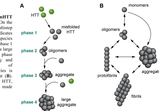
The elongated polyQ stretch at the N-terminus of the mHTT protein confers the propensity to misfold and aggregate (Sieradzan et al., 1999). Several models have been developed to explain the aggregation process. One of them is the “multistep aggregation model”, which
suggests that mHTT aggregation occurs in consecutive steps (Ossato et al., 2010). Another widely accredited model sustains that mHTT aggregation is a dynamic process, where small and large species of mHTT assemble and disassemble concurrently (Legleiter et al., 2010; Soto and Pritzkow, 2018) (Figure 4).
This multistep aggregation process is divided into 4 main phases: in phase 1, soluble misfolded monomers accumulate; in phase 2, soluble misfolded monomers give rise to the formation of small oligomers; in phase 3, the increasing load of structurally abnormal monomers and oligomers triggers the nucleation stage and in phase 4, the nucleation stage concludes with the formation of insoluble inclusion bodies (IBs) (Ossato et al., 2010) (Figure
4A). Importantly at this final stage, WT HTT is recruited into the IBs through its N-terminus
(Ossato et al., 2010), contributing to the depletion of WT HTT and compromising the roles of HTT (HTT loss of function) (Rajan et al., 2001).
Figure 4. mHTT aggregation models. On the
left (A), the multistep aggregation model indicates that more complex species are formed starting at phase 1 until the formation of a large insoluble aggregate in phase 4. The interchanging and dynamic formation of different mHTT species is illustrated on the right (B). Abbreviation: HTT, huntingtin. Illustration made by Maria Masnata.
In contrast to the multistep model, other studies suggest that the aggregation process is not a linear sequence of events, but a dynamic series of conformational changes and protein bindings that occur in parallel, or simultaneously (Legleiter et al., 2010; Soto and Pritzkow, 2018) (Figure 4B). Oligomers, in particular, are the most dynamic species and they are in equilibrium with smaller mHTT forms, such as monomers, or with more complex ones, such as fibrils. Furthermore, oligomers are capable of permanently aggregating into IBs (Soto and Pritzkow, 2018). Notably, all studies agree that the aggregation process depends on the polyQ length; the longer the polyQ strain, the quicker the aggregation process (Arrasate et al., 2004; Drombosky et al., 2018; Legleiter et al., 2010).
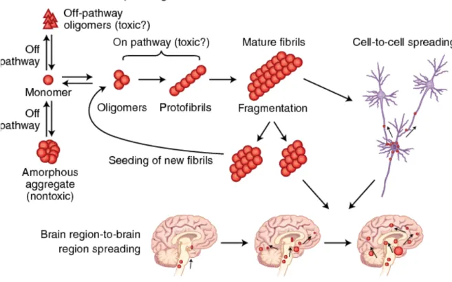
Another debated issue is the identification of the mHTT pathogenic entity among large aggregates, soluble oligomers, fibrils and soluble monomers (Ast et al., 2018; Caughey and Lansbury, 2003; DiFiglia et al., 1997; Pieri et al., 2012; Scherzinger et al., 1997) (Figure 5). Initially, the end-products of mHTT aggregation – the large insoluble IBs – were considered the main culprits in neuronal death (Davies et al., 1997; DiFiglia et al., 1997). This hypothesis was based on post-mortem observations of R6/2 mice brains which revealed that the deposition of nuclear IBs, similar to those identified in HD brains, appeared in concomitance with behavioral deficits (Davies et al., 1997; DiFiglia et al., 1997). However, in vitro models in which mHTT expression is induced by adenoviral (Dong et al., 2012) or lentiviral vectors (Zala et al., 2005), have revealed that cortical neurons accumulate a significant number of
IBs, but with no clear toxic consequences. In contrast, striatal neurons develop significant morphological changes that are accompanied by the loss of neurofilaments and ultimately cell death, despite the fact that mHTT aggregates are rarely seen within these cells (Dong et al., 2012; Zala et al., 2005). In agreement with these findings, post-mortem human tissue shows elevated concentration of IBs in the cortex rather than in the striatum, suggesting that IBs localization and density does not associate with cell death (Gutekunst et al., 1999). Some studies have suggested that large aggregates can be protective, while soluble mHTT was instead to be blamed for cytotoxicity (Arrasate et al., 2004; Arrasate and Finkbeiner, 2012). This hypothesis was supported by in vitro work using primary culture of rat striatal neurons transfected with N-terminal fragment of HTTExon1Q72 or Q103, where it was observed that the neurons with larger IBs survived longer than neurons with smaller mHTT aggregates (Arrasate et al., 2004). Several other in vitro experiments confirmed that cell survival was independent of IBs, but correlated with the presence of soluble mHTT (Lajoie and Snapp, 2010; Takahashi et al., 2008). Furthermore, in mice expressing the human mHTT N-truncated form, the widespread deposition of mHTT aggregates does not associate with behavioral changes (Slow et al., 2005), while in mice expressing the full-length human mHTT, motor and cognitive deficits occur despite scarce mHTT aggregates (Slow et al., 2005). Collectively, in vitro and in vivo evidence indicates that IBs are unrelated to pathological outcomes, and could instead signify a coping response to pathogenic stressors (Arrasate and Finkbeiner, 2012).
Unlike IBs, mHTT soluble oligomers are currently considered culprits of HD cytotoxicity (Saudou et al., 1998; Takahashi et al., 2008). However, determining how oligomers induce cytotoxicity is particularly challenging due to their propensity to rapidly shift conformation and form larger aggregates or disaggregate into monomers. Some fluorescence microscopy techniques have proven useful to study the process of oligomer formation. For instance, they provided evidence that, in living cells, mHTT oligomers induce a higher death rate than insoluble mHTT aggregates (Herrera et al., 2011; Lajoie and Snapp, 2010; Takahashi et al., 2008) and could be used as a tool to test novel therapeutic approaches to tackle toxic derivates of the mHTT aggregation process (He et al., 2020). Although the cytotoxicity of oligomers has been demonstrated, a study that performed a direct comparison between syntenic