Domains and Maturation Processes That Regulate the Activity of
ADAMTS-2, a Metalloproteinase Cleaving the Aminopropeptide of
Fibrillar Procollagens Types I-III and V
Alain Colige‡, Florence Ruggiero§, Isabel Vandenberghe¶, Johanne Dubail‡, Frederic Kesteloot‡, Jozef Van Beeumen¶, Alain Beschin||, Lea Brys||, Charles M Lapière‡, and Betty Nusgens‡
From the
‡Laboratory of Connective Tissues Biology, Center of Biomedical Integrative
Genoproteomics, University of Liège, B-4000 Sart Tilman, Belgium,
¶Laboratorium voor
Eiwitbiochemie en Eiwitengineering, Universiteit Gent B-9000, Gent, Belgium,
||
Laboratorium voor Cellulaire en Moleculaire Immunologie, Department of Cellular and
Molecular Interactions, Flanders Interuniversity Institute for Biotechnology, Vrije Universiteit
Brussels, 1050 Brussels, Belgium, the
§Institut de Biologie et Chimie des Protéines, UMR
CNRS 5086, IFR128 BioSciences, 69367 Lyon cedex 7, France
Processing of fibrillar collagens is required to generate collagen monomers able to self-assemble into elongated and cylindrical collagen fibrils. ADAMTS-2 belongs to the "A disintegrin and metalloproteinase with
thrombospondin type 1 motifs" (ADAMTS) family. It is responsible for most of the processing of the aminopropeptide of type I procollagen in the skin, and it also cleaves type II and type III procollagens. ADAMTS are complex secreted enzymes that are implicated in various physiological and pathological processes. Despite accumulating evidence indicating that their activity is regulated by ancillary domains, additional information is required for a better understanding of the specific function of each domain. We have generated 17 different recombinant forms of bovine ADAMTS-2 and characterized their processing, activity, and cleavage specificity. The results indicated the following: (i) activation of the ADAMTS-2 zymogen involves several cleavages, by pro-protein convertases and C-terminal processing, and generates at least seven distinct processed forms; (ii) the C-terminal domain negatively regulates enzyme activity, whereas two thrombospondin type 1 repeats are enhancer regulators; (iii) the 104-kDa form displays the highest aminoprocollagen peptidase activity on procollagen type I; (iv) ADAMTS-2 processes the aminopropeptide of α1 type V procollagen homotrimer at the end of the variable domain; and (v) the cleaved sequence (PA) is different from the previously described sites ((P/A)Q) for ADAMTS-2, redefining its cleavage specificity. This finding and the existence of multiple processed forms of ADAMTS-2 strongly suggest that ADAMTS-2 may be involved in function(s) other than processing of fibrillar procollagen types I-III.
Collagens types I-III are synthesized as precursors (procollagens) formed by a central triple helical collagen domain extended by propeptides at both extremities. Removal of the C-propeptide,1 by BMP-1 and related enzymes, and the N-propeptide, by aminoprocollagen peptidases (ADAMTS-2, -3, or -14), is required to generate collagen monomers able to assemble spontaneously into elongated and cylindrical collagen fibrils. It has been shown in vitro and in vivo that ADAMTS-2 is the enzyme responsible for most of the
aminoprocollagen type I processing in the skin (1-3), although it was suggested that an essential function of 3 is the processing of aminoprocollagen type II in cartilage (4). A closely related enzyme, ADAMTS-14, also displays aminoprocollagen peptidase activity (5). More recently, it has been shown that ADAMTS-2 is also able to process aminoprocollagen type III in vitro (6). Type V collagen is a minor fibrillar collagen. Among
1 The abbreviations used are: C-propeptide, C-terminal propeptide; ADAMTS, A disintegrin-like and metalloproteinase with
thrombospondin type I repeats; N-propeptide, N-terminal propeptide; BMP-1, bone morphogenic protein-1; DMEM, Dulbecco's modified Eagle's medium; TSPI, thrombospondin-like type I repeat; PNP domain, C-terminal domain specific to aminoprocollagen peptidases (ADAMTS-2, -3 and -14); mAb, monoclonal antibody; HA, hemagglutinin; PNGase F, peptide:N-glycosidase F; NEM, N-ethylmaleimide; PMSF, phenylmethylsulfonyl fluoride; DTT, dithiothreitol.
the four different individual collagen type V chains, α1 is the most abundant and ubiquitous and can be found as a homotrimer or as a heterotrimer in association with type V or type XI a chains (7,8). Its maturation from procollagen to collagen is reported to be different from what is observed for procollagens I-III, because the C-propeptide can be cleaved by furin (9, 10) and/or by BMP-1 (11, 12) depending on the chain type. In addition, the N-propeptide does not seem to be processed by aminoprocollagen peptidases. Instead, BMP-1 cleavage has been reported between the proline/arginine-rich protein domain and the variable domain of the α1 chain (9) and between the small and the large collagenous domain of α3 chain (10).
ADAMTS proteases are complex secreted enzymes that contain a reprolysin-type pro-metalloproteinase domain attached to ancillary domains with highly conserved structures, including at least one thrombospondin type 1 repeat (13). As for matrix metalloproteinases and ADAMs, the control of their enzymatic activity can be exerted at multiple points, including transcription, translation, zymogen activation, and inhibition by specific natural inhibitors. For most ADAMTS enzymes, the N-terminal domain is cleaved by furin or a related enzyme (6, 14). In addition, proteolytic processing in the ancillary domains of some ADAMTS has been reported (15), increasing or inhibiting their enzymatic activity (16). However, the implication of the various domains for enzyme activity and substrate specificity remains largely unknown. Similarly, additional studies are required for a better understanding of the mechanisms of ADAMTS activation and for the identification of enzymes responsible for ADAMTS processing.
In this work, various forms of bovine recombinant ADAMTS-2, either full size, lacking specific domains, or mutated at specific sites, were produced. They have been used to determine the specific role of individual domains for aminoprocollagen type I peptidase activity, to characterize the activation process of the
zymogenform of ADAMTS-2, and to evaluate the enzymatic activity of ADAMTS-2 on aminoprocollagen type V.
MATERIALS AND METHODS
Restriction enzymes, Pwo DNA polymerase, hygromycin B, FuGENE 6 transfection reagent, α2-macroglobulin, and anti-HA rat antibody were from Roche Diagnostics; ligase (DNA Ligation kit version 2) was from Takara Biomedicals (Shiga, Japan). Competent bacteria (XL-Gold® Ultracompetent Cells) and QuickChange™ XL site-directed mutagenesis kit were from Stratagene (Cedar Creek, TX). pCEP4 vector, DMEM culture medium, and DM1™ competent cells used for preparation of plasmid requiring BclI digestion were from Invitrogen. Fetal bovine serum was from Cambrex (Verviers, Belgium); the ECL detection system was from Amersham
Biosciences, and the anti-FLAG™ M2 mouse monoclonal antibody used for Western blotting was from Sigma. The gon-1 cDNA was the kind gift of J. Kimble (17).
Creation of the Various Expression Vectors and Cell Transfection— For the creation of construct 1, a vector containing the bovine ADAMTS2 cDNA (18) was digested at the NotI and HindIII sites (Fig. 1A). The released insert was then subcloned in a pCDNA3 vector containing a modified multiple cloning site. The 3'-end of the cDNA was PCR-amplified with Pwo DNA polymerase using primer A as forward primer
(CTACAAGGACGCCTTCAGCCTCT-3') and primer B containing NheI- and XbaI-specific sequences at its 5'-end as reverse primer (5'-CTCTTCTAGATTAGCTAGCGAACTTTCCGAGCAT-CTCTTTCTTC-3'). After HindIII/XbaI digestion, the PCR product was ligated downstream from the NotI/HindIII insert. The construct was then linearized at the NheI site in order to introduce a double-stranded adaptor (sense,
5'-CTAGCGATTATAAAGATGACGATG-ACAAATAA-3', and reverse, 5'-CTAGTTATTTGTCATCGTCAT-CTTTATAATCG-3') coding for an M2-FLAG (DYKDDDDK) followed by a TAA stop codon. Because of its design, ligation of the adaptor restored an NheI site only at its 5'-end. Finally, the construct was digested (NotI/XbaI), and the insert containing the M2-FLAG at its 3'-end was ligated in the NotI/NheI site of a modified pCEP4 vector. Constructs 2-5 were obtained by replacing the HindIII/NheI cassette of construct 1 by PCR products amplified using primer A as forward primer and primers
C(5'-CTCTTCTAGATTAGCTAGCTAGCCAC-TGGACCACGTAGCTCT-3'), D CTCTTCGCTAGCAGGGCA-CAGCTCGCGGTTGCA-3'), E CTCTTCGCTAGCGGAGCAC-TCCTGTGGGTTGCA-3'), or F
(5'-CTCTTCTAGATTAGCTAGC-CTCATAGCCCACAGAGTCGTCTT-3') as reverse primers. Construct 6 was created by using a QuickChange™ XL site-directed mutagenesis kit and two mutagenic primers (M, 5'-GTTCGTGGTGGCCC-ACGCGACTGGCCATGTGCTGG-3', and N,
5'-CCAGCACATGG-CCAGTCGCGTGGGCCACCACGAAC-3') changing the GAG codon for Glu of the Zn2+-binding catalytic site (HETGH) into a GCG codon for Ala. For constructs 8-10, the EcoRI/HindIII fragment of construct 1 was removed and replaced by PCR products amplified using J (5'-GGATCTCAAACATCTTGATGTAACCA-3') as reverse primer and primers G CACAGAATTCCATGCTGCCGACGACGACTA-CAAC-3'), H
(5'-CACAGAATTCACGCTGAACCACGAGGACG-GCTT-3 ') as forward primers, each of them containing an EcoRI site at their 5'-end. For construct 7, the vector containing construct 8 was linearized at the EcoRI site, allowing the ligation of an EcoRI-digested PCR product amplified with primers K (5'-TTTGGCCGAGACCTG-CACCTGC-3') and L (5'-CACAGAATTCCATACTCCGCCTG-GAGCTGTTGA-3') containing an EcoRI site at their 5'-end. Primer L was also designed in order to change the sequence coding for a furin cleavage site
(RRRMRR) into a sequence coding for an unrelated amino acid sequence (FEMSRR). For construct 11, the BclI/HindIII fragment of construct 1 was removed. After incubation with Pwo DNA polymerase and dNTP in order to generate blunt ends, the vector was self-ligated. This introduced a large deletion but did not change the open reading frame of the remaining coding sequence. For constructs 12 and 13, construct 1 was digested by HindIII/NheI or BcII/HindIII, respectively, and the deleted sequences were replaced by corresponding domains amplified from gon-1 cDNA using primers O
(5'-CACACAG-CTAGCTCTTGGACATGGAATTCTGTTACATTC-3')and
PC-ACACAAAGCTTGGTTATAACGAAGTAATGAAGATTCCA-3'), or Q CACACAAAGCTTTCCTTGCTCATTAAATGTTCCTTT-GA-3') and R
(5'-CACAGGATCCTGTCCAACATCATGACGTTG-CAATC-3'). Construct 14 coding for ADAMTS-14 was as described earlier (5), and constructs 15-17 were obtained by exchanging the homologous coding sequences of
ADAMTS2 and ADAMTS14 by restriction enzyme digestion and ligation. Additional constructs were also created
by introducing, in-frame, a double-stranded adaptor (forward, AGCTATATCCTTACGATGTTCCTGACTATGCTA-3'; reverse,
5'-AGCTTAGCATAGTCAGGAACATCGTAAGGATAT-3') coding for an HA-FLAG (YPYDVPDYA) in the HindIII site of 9 vectors (1, 2, 5-7,14 -17). These modified constructs (not shown) will be designated in the paper as constructs 1A, 2A, 5A to 7A, and 14A to 17A. This 9-amino acid insertion does not modify the level of expression or the enzymatic activity of the various forms of the recombinant enzymes.
The various expression vectors were used to transfect different cell lines (HT 1080 fibrosarcoma cells, WI26 immortalized lung fibroblasts, COS, Balb, MCF7, SaOS2 osteosarcoma cells, 293EBNA epithelial kidney cells) by using FuGENE 6 according to the manufacturer's recommendations. Stable cell lines were then selected in DMEM culture medium supplemented with 10% fetal calf serum and 200 µg/ml hygromycin.
Cell Culture and Treatment—Stably transfected cells expressing the various constructs were cultured in DMEM supplemented with fetal calf serum and hygromycin. At confluence, the growth medium was removed, and the cells were maintained in DMEM alone or containing specific chemicals or inhibitors at the indicated concentrations. After 24 or 48 h, the different media were collected, and the cells were scraped and extracted for 2 h in extraction buffer (50 mM Tris, pH 7.5; 500 mM NaCl; 2 mM CaCl2; 25 mM NEM; 1 mM PMSF). After centrifugation, the cell extracts were collected (cell extract 1), and the pellets were solubilized in SDS-PAGE denaturation buffer containing 100 mM DTT (cell extract 2).
For correlation studies comparing the levels of each individual band of enzyme to the aminoprocollagen peptidase activity, 293 cells expressing various constructs were cultured for 24 h in medium supplemented with 0.1 or 5% fetal calf serum alone (control conditions) or containing additional chemicals (EDTA at 0.04,0.2, or 1 mM; ZnCl2 at 16 or 80 µM; CuCl2 at 16 or 80 µM; heparin at 1, 5, or 25 µg per ml; decanoyl-RVKR (furin inhibitor) at 10, 20, or 40 µM; L-arginine at 25, 50, or 100 mM).
Compartmentalization Studies—Dermatosparactic calf fibroblasts, cells that do not synthesize ADAMTS-2, were grown to confluence (duplicate culture, 24-wells culture plates) in DMEM supplemented with 10% fetal calf serum. Cells were then incubated at 4 °C in 400 µl of medium containing or not heparin (5 µg/ml) and recombinant purified ADAMTS-2 (10 µg/ml). After 6 h, medium was removed, and the cell layer was denatured in Laemmli sample buffer containing 0.1 M DTT. Western blot analysis using mAb23 was then performed to determine the proportion of ADAMTS-2 bound to the cell layer.
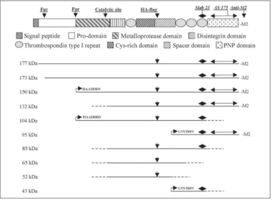
FIGURE 1. Creation of the various forms of recombinant ADAMTS-2. A, localization of the introduced or
endogenous restriction sites is illustrated above the schematic representation of ADAMTS2 cDNA. Position of the primers used for the creation of the various constructs and their 5' to 3' orientation are illustrated by arrows. B represents the schematic domain organization of ADAMTS-2. The localization of the Zn2+-binding catalytic site and potential cleavage sites by furin [Fur) are illustrated. Position of epitopes recognized by the different antibodies and size of the various domains, expressed by amino acid number, are also indicated. *, the cleavage site of the signal peptide was not exactly determined. From amino acid sequence analysis, cleavage is expected to occur 30-40 amino acids downstream of the initial methionine residue. Accordingly, the exact size of the prodomain could not be determined. **, HA FLAG coding sequence was introduced in the Hindlll restriction site of constructs 1, 2, 5-7, and 14-16. These HA-flagged constructs are designated as constructs 1A, 2A, 5A-7A, and
14A-17A in the text. Domain structure organization of the various forms of ADAMTS-2 is illustrated in C. Dotted lines represent internal lacking sequences and arrowheads localization of point mutations. For chimeric
enzymes 12 and 13, domains of GON-1 are in black, and arrows represent position of the gon-1-specific oligonucleotides used to create the constructs. Constructs coding for 14 [construct 14) or ADAMTS-2/ADAMTS-14 chimeric enzymes [constructs 15-17) were created by restriction enzyme digestion and ligation. Domains originating from ADAMTS-14 are in black.
Large Scale Purification of Enzyme—For large scale purification of recombinant enzymes, 100 Petri dishes of stably transfected cells were grown in DMEM supplemented with 10% fetal calf serum and hygro-mycin. At confluence, medium was removed and replaced by serum-free DMEM containing soybean trypsin inhibitor (40 µg/ml), heparin (5 µg/ml), and ZnCl2 (80 µM). After 48 h, the conditioned medium was collected, concentrated, and loaded on a 25-ml concanavalin A-Sepha-rose column pre-equilibrated in buffer A (50 mM Tris, pH 7.5; 1M NaCl; 2 mM CaCl2). After washing with buffer A, elution was carried out in the same buffer containing 500 mM
α-methyl-D-mannoside. Fractions containing the recombinant enzyme were then dialyzed against buffer B (50
mM Tris, pH 7.5; 0.2 M NaCl; 2 mM CaCl2) and loaded on a 5-ml heparin-Sepharose column. After washing in buffer B, elution was performed in buffer A.
Antibody Preparation and Characterization—AS175 was obtained by immunizing a rabbit with recombinant ADAMTS-2 purified by SDS-PAGE. Although the immunization was performed with a full-length protein, the antiserum was specific for the C-terminal domain only, as determined by Western blotting using the various recombinant enzymes. For production of monoclonal antibody, mice were immunized with active and not denatured recombinant ADAMTS-2. Among the tested hybridoma, one (mAb23) produced an antibody (IgG2b) able to recognize native and denatured ADAMTS-2. By using the various recombinant enzymes, it was
determined that this antibody is specific for the last TSPI repeat of ADAMTS-2. Further analysis showed that AS175 and mAb23 do not block the aminoprocollagen peptidase activity of ADAMTS-2 and do not display cross-reactivity with ADAMTS-3 and -14.
Characterization of the Various Recombinant Enzymes—The electrophoretic pattern of the various
recombinant enzymes present in the conditioned media and in the two cell layer-associated fractions (see above) was determined by Western blotting analysis using antibodies specific for the M2- or HA-FLAG, the last TSPI repeat (mAb23), or the C-terminal domain (AS 175) of ADAMTS-2. The various samples of conditioned medium and cell extracts were also assayed for their aminoprocollagen peptidase activity (see below). In order to identify which forms of the enzyme were responsible for the processing of procollagen substrates, a correlation was calculated between the activity measured in the various samples and the relative abundance of each individual form of recombinant enzymes.
Protein Deglycosylation—A crude cell extract containing all the ADAMTS-2 forms detected during our study was incubated with PNGase F or neuraminidase according to manufacturer's protocol. The electrophoretic pattern of ADAMTS-2 before and after treatment was determined by Western blotting using mAb23. FIGURE 2. Evaluation of recombinant ADAMTS-2 synthesis and activity at increasing time points after
transfection. A 293EBNA cells collected 2-26 days after transfection by construct 1 were analyzed by Western blotting using anti M2-FLAG antibody. B, samples analyzed on A were further characterized by using mAb23, an antibody specific for the 4th TSPI repeat. C aminoprocollagen type peptidase activity of the corresponding cell extract was also determined and is expressed in counts/min of processed substrate. This assay allowed accurate and linear evaluation of enzyme activity in the range of 0-1500 cpm of processed substrate. A striking discrepancy was observed between the amount of ADAMTS-2 and the level of enzymatic activity.
Protein Sequencing—For the determination of the N-terminal sequence of the various ADAMTS-2 polypeptides, a highly purified preparation of recombinant enzyme (construct 1) was dialyzed against 1 M ammonium acetate, concentrated by lyophilization, and migrated in a pre-run 7% acrylamide/piperazine
diacrylamide gel in 50 mM Tris borate buffer, pH 8.3, containing 0.1% SDS and 0.1 mM thioglycolic acid. After transfer on a polyvinylidene difluoride membrane (in Tris (50 mM)/borate; pH 8.5) and Coomassie Blue staining (in 40% methanol), the different bands were collected and submitted to six cycles of Edman degradation. For type V collagen sequencing, recombinant type V pNcollagen (19) was incubated for 18 h with highly purified recombinant ADAMTS-2 and processed as described above. Determination of the N-terminal sequence was performed only for the polypeptide generated by ADAMTS-2 digestion.
Purification of Type V Collagen from Bovine Skin—Skin, collected from fetal calves at the third trimester stage,
was ground at liquid nitrogen temperature and homogenized with an Ultra Turrax (8000 rpm) in washing buffer (50 mM Tris-HCl, pH 7.5; 0.25 M sucrose; 2.5 mM NEM; 1 mM PMSF; and 2 mM EDTA). After centrifugation (20,000 x g), the pellets were collected, and the washing procedure was repeated once. Pellets were then
suspended and rotated for 18 h at 4 °C in extraction buffer (50 mM Tris-HCl, pH 7.5; 2.5 mM NEM;1 mM PMSF; and 2 mM EDTA) containing 0.15 M NaCl. After centrifugation, supernatants were collected, and pellets were further sequentially extracted in the same buffer containing 1 M NaCl and in 0.1 M acetic acid. The various supernatants were dialyzed in 0.1 M acetic acid containing 0.7 M NaCl. After centrifugation (20,000 X g), a large amount of type I collagen was present in the pellets, whereas type V collagen remained in solution. Supernatants were then further dialyzed (0.1 M acetic acid, 1.2 M NaCl). After centrifugation, type V collagen was recovered in the pellet. Even in the most enriched fractions, type V collagen was recovered in low amounts (0.5 to 2% of total protein content, as judged after SDS-PAGE and Coomassie Blue staining), the most abundant protein being type I collagen.
Inhibition of ADAMTS-2 Activity by α2-Macroglobulin—ADAMTS-2 (~ 10 ng, as determined by Coomassie
Blue staining after SDS-PAGE) was preincubated for 1 h at 37 °C with α2-macroglobulin (25 units/ml, 1 unit (40 µg) inhibiting 9.1 µg of trypsin in a standard assay) at varying concentrations (0.025 to 0.4 units). The
aminoprocollagen type I was then added, and the peptidase activity was evaluated (see below). The bait and trap mechanism of inhibition of ADAMTS-2 by α2-macroglobulin, resulting in an electrophoretic band shift, was investigated by Western blotting using anti-HA-FLAG antibody. Two types of samples were used. In the first series, purified ADAMTS-2 was incubated alone or with α2-macroglobulin (0.1 unit) in the absence or presence of EDTA. The second series consisted of enzyme (constructs 1A, 2A, or 5A) secreted in culture medium supplemented with 10% fetal calf serum as a source for α2-macroglobulin.
Procollagen Processing in Vitro—The aminoprocollagen peptidase activity of ADAMTS-2 was determined by using either 14C-labeled pNI collagen as substrate (20) or by evaluating the level of processing of unlabeled substrate by Western blotting. Briefly, purified bovine pNI (2 µg) or recombinant pNV homotrimer (0.2 µg) (19) was incubated at 26 °C with the various enzyme preparations as described previously (20). After 18 h, the samples were denatured and submitted to electrophoresis and were either stained with Coomassie Blue or transferred on polyvinylidene difluoride membrane. For Western blotting experiments, the pattern of collagen polypeptide was determined by using rabbit antiserum specific for type I or pepsinized type V (NOVOTEC, Lyon, France) collagen.
RESULTS
Expression of Recombinant Enzyme—Various cell lines were trans-fected with expression construct 1 (Fig. 1) or empty vector as control. Within a few days after transfection, large amounts of recombinant ADAMTS-2 were produced by most cell lines, as determined by Western blotting analysis of the cell lysate using an anti-M2 FLAG antibody, but only very low levels of aminoprocollagen peptidase activity could be detected (not shown). Because 293 cells were identified as the cells producing the largest amount of active recombinant enzyme, other cell lines were not further used. During the selection process, the amount of recombinant enzyme progressively decreased, as judged from Western blotting analysis using anti-M2 FLAG antibody (Fig. 2A). Most surprisingly, enzymatic activity was not similarly altered (Fig. 2C). A moderate increase of activity was first observed, followed by a small decrease and a sharp increase after 3 weeks of selection. These data suggested that a maturation process was required for full activity. This hypothesis was confirmed by Western blotting analysis (Fig. 25), using a monoclonal antibody specific for the 4th TSPI repeat.
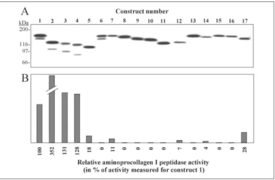
In order to determine which domains of ADAMTS-2 are involved in the regulation of its activity, various recombinant variants (Fig. 1, constructs 2-17) were produced. In a first set of experiments, recombinant enzyme lacking either the PNP domain (construct 2) or the PNP domain and one, two, or three TSPI repeats (constructs 3-5) were created. In a second set, the second potential cleavage site by furin was mutated (construct 7), or various portions of the region at the border between the pro-domain and the metalloproteinase domain were deleted (constructs 8-10). As negative controls, enzyme mutated at the catalytic site (construct 6) or lacking the central domains (construct 11) was also produced. Finally, as a third set of constructs, chimeric enzymes were produced by replacing domains of ADAMTS-2 by the corresponding domains of GON-1, a Caenorhabditis
elegans ADAMTS (constructs 12 and 13), or of ADAMTS-14, an enzyme closely related to ADAMTS-2 but
displaying only very low aminoprocollagen peptidase activity (constructs 15-17). All the recombinant proteins were synthesized, as determined by the detection of the M2-FLAG by Western blotting (Fig. 3A). The presence of a second low molecular weight band for constructs 1-4 and its absence for constructs 7-10 strongly suggested that ADAMTS-2 was processed at the furin cleavage site. However, no processing was observed for constructs
5,11-13, and 14-17. As an additional observation, we showed that the M2-FLAG at the C terminus of the various recombinant proteins was progressively degraded and that the level of degradation varied from one recombinant protein to another, preventing reliable and reproducible quantifications of the recombinant protein by Western blotting using the anti-M2 FLAG antibody (not shown). Because no other antibody was available to detect all 17 variants, aminoprocollagen peptidase activity was investigated in samples corresponding to a similar number of cells. Removal of the PNP domain (construct 2) significantly increased the enzyme activity (Fig. 35) as compared with the wild type enzyme (construct 1). On the contrary, removal of the fourth and the second TSPI repeats (constructs 3 and 5) repressed the enzymatic activity as compared with the activity measured for construct 2. As expected, no activity was detected for negative control constructs lacking the central domains (construct 11) or mutated at the catalytic site (construct 6). Mutating the potential cleavage site by furin (construct 7) strongly repressed, but did not abolish, the aminoprocollagen peptidase activity. Removal of the adjacent domains (constructs 8 -10) completely suppressed the activity. The use of chimeric enzyme indicated that significant levels of activity were obtained only when the metalloprotease domain and/or the other central domains of ADAMTS-2 are present (compare constructs 12,13, and 17 to 14-16). However, the presence of the C-terminal domains (spacer, TSPI 2nd, 3rd, and 4th, and the PNP domain) is also important for activity because their replacement by the corresponding domains of GON-1 (compare construct 12 to construct 2) or ADAMTS-14 (compare construct 17 to construct 1) resulted in a reduced activity.
FIGURE 3. Western blotting analysis of the various recombinant enzymes and evaluation of their
aminoprocollagen type I peptidase activity. A extracts of 293EBNA cells stably transfected with constructs 1-17
were analyzed by Western blotting using anti-M2 FLAG antibody. Differences in the electrophoretic mobility were in good correlation with both the expected protein length and the glycosylation sites that are essentially found in the C-terminal sequence of ADAMTS-2. B, aminoprocollagen peptidase activity in each cell extract was quantified and expressed in % of the activity measured for the recombinant wild type form of ADAMTS-2 (construct 1).
Processing of ADAMTS-2—The existence of a complex activation process for ADAMTS-2 was suggested by data in the literature about a few other ADAMTS and by our preliminary observations. To characterize the mechanisms involved in the ADAMTS-2 processing, various stably transfected 293 cells were cultured in different conditions. Cell layers and conditioned media were then analyzed for the presence of ADAMTS-2 fragments by Western blotting using four antibodies (AS175, anti-M2 FLAG, anti-HA FLAG, and mAb23; see Fig. 1B). In a first series of experiments, cells expressing the full-length enzyme (construct 1A) were used. The anti-HA antibody detected six major bands (at 173, 150, 132,104, 85, and 52 kDa) in the cell layer (Fig. 4A, lane
1). In the conditioned medium, the pattern was similar except for the absence of the 173-kDa band and the
presence of two additional bands around 65 kDa. By using the mAb23 antibody, the overall patterns were similar except for the presence of a barely detectable 95-kDa product and the absence of low molecular bands (Fig. 4A,
(at 173, 150, and 132 kDa) were also detected, but the 104-kDa band was not (Fig. 4A, lane 4). The 95-kDa product was found in association with the cell layer, and a low molecular weight product was recovered only in the conditioned medium. The pattern was similar with the anti-M2 FLAG antibody, except for the absence of immunoreactive high molecular weight product in the culture medium and for the relative intensity of bands in the cell layer (Fig. 4A, lane 4). These two observations are most probably related to a specific degradation of the M2 FLAG.
PNGase F deglycosylation resulted in the gel shift of most ADAMTS-2 forms (Fig. 4B, compare lanes 1 and 2). Most interestingly, the number of bands detected before and after PNGase F treatment did not change,
demonstrating that the various bands represent individual processed forms of ADAMTS-2 and not different levels of glycosylation of a more restricted number of products. Neuraminidase treatment did not alter the electrophoretic mobility of any bands.
In order to determine whether the processing of the recombinant ADAMTS-2 observed in 293 cells is relevant to physiological processing in vivo, the electrophoretic pattern of enzyme purified from calf skin was examined using mAb23 (Fig. 4C). Prominent bands at 104 and 85 kDa were detected in skin extracts and fainter bands at 150, 132, 115, and 65 kDa. All these fragments, except the 115 kDa, were also observed for the recombinant enzyme.
FIGURE 4. Western blotting analysis of the various processing forms of ADAMTS-2. A, cells stably
transfected with construct 1A were grown to confluence in culture. Samples of cell layer (C) and conditioned medium (M) were collected and analyzed by immu-noblot using four different antibodies (HA, mAb23, anti-M2, and AS175). Apparent deduced molecular sizes of ADAMTS-2 fragments are reported. Large differences in the electrophoretic pattern were observed. The 173-kDa band, detected by the four antibodies, was not found in the culture medium, suggesting that this product is either intracellular or strongly bound to the cell surface. The almost absence of product reacting with the anti-M2 FLAG in the culture medium probably illustrated the rapid degradation of either the ADAMTS-2 C-terminal end or the M2-FLAG itself. The most obvious difference was the presence of a prominent 104-kDa product observed only with anti-HA and mAb23, demonstrating that this product does not contain most of the PNP domain. B, extracts of cells expressing wild type recombinant ADAMTS-2 were treated with PNGase F (lane 1) or neuraminidase (lane 3). Electrophoretic mobility of the various bands was compared with samples incubated in identical condition without enzymes (lanes 2 and 4, respectively). C, the patterns of ADAMTS-2 purified from conditioned medium of cells expressing construct 1A or from fetal bovine skin (see "Materials and Methods" for culture conditions and purification procedures) were compared by Western blotting using mAb23 antibody specific for the fourth TSPI repeat (see Fig. 1). The two different exposure times allow the visualization of abundant (104 and 85 kDa) and minor (150, 132, and 65 kDa) processing forms present in both purified preparations. Only one product, with an estimated size of 115 kDa (*), was observed in the ADAMTS-2 preparation purified from fetal bovine skin. D, cells expressing construct 1A were cultured (35-mm dishes) in DMEM supplemented with 10% fetal calf serum. At confluence, medium was changed and replaced by 2 ml of DMEM alone (control) or supplemented with EDTA (200 µM), ZnCl2 (80 µM),
CuCl2 (80 µM), heparin (5 µg/ml), decanoyl-RVKR furin inhibitor (40 µM), or L-Arg (50 mM). After 48 h, the
conditioned media were collected, and the cell layer was extracted in a buffer containing 1 M NaCl. Samples, corresponding to 1/50 of the cell layer (C) and 1/100 of the conditioned medium (M), were analyzed by Western blotting using anti-HA-FLAG antibody. Deduced molecular sizes of the ADAMTS-2 processing forms are indicated on the right of the panels. On this blot, the bands at 177 and 173 kDa in the presence of the RVKRfurin inhibitor appear as a broad band. They can be individually identified on Fig. 5A
Treatment with various chemicals changed the relative ratio of each individual band (Fig. 4D). As an example, treatment of cells with EDTA significantly increased the amount of the 132-kDa band but strongly repressed the accumulation of the 104-kDa product (Fig. 4D, compare lanes 1 and 3). In the presence of heparin, all the ADAMTS-2 products were released from the cell layer compartment and accumulated in the culture medium (Fig. 4D, compare lanes 9 and 10). As another example, use of decanoyl-RVKR, a furin inhibitor, decreased the amount of the 104-kDa products and caused the accumulation of two high molecular products (177 and 173 kDa), most probably illustrating inhibition of processing at the two identified potential cleavage sites by enzymes of the furin family. Finally, when the culture was supplemented with L-Arg, the band pattern partially mirrored the pattern observed in presence of heparin (Fig. 4D, compare lanes 10 and 14).
To investigate further the mechanisms that generated the different ADAMTS-2 fragments and the reason why some recombinant enzymes do not display aminoprocollagen peptidase activity (Fig. 35), cell lines transfected with other constructs (6A, 7A, 14A, 15A, and 16A). Most surprisingly, a faint band was observed with an apparent molecular size of 153 kDa (Fig. 5A, see arrow B), suggesting that, in the absence of cleavage by furin, another enzyme might process ADAMTS-2 a few amino acids upstream of the cleavage site by furin. As observed for control construct 1A, EDTA and decanoyl-RVKR promoted the accumulation of the 132- or the 177-kDa product, respectively. Other bands appeared fuzzy Cells expressing constructs 14A to 17A (ADAMTS-14 and chimeric recombinant proteins constructed from part of ADAMTS-2 and -(ADAMTS-14) were also analyzed (Fig. 55). As compared with the wild type construct 1A, the electrophoretic pattern obtained with cells expressing these constructs was markedly different. ADAMTS-14 (construct 14A) consisted of one major (165 kDa) and four minor (in the range of 95-130 kDa) products, both in control and EDTA-treated cells. With constructs 15A and 16A, which did not display any significant aminoprocollagen peptidase activity (Fig. 3), the electrophoretic pattern consisted of one main band and was not altered by culture conditions. At the opposite, the chimeric enzyme 17A (composed of the N terminus of ADAMTS-2 fused to the C-terminal domains of ADAMTS-14) displayed enzyme activity (Fig. 3) and was actively processed (Fig. 5B, lanes 5 and 10).
FIGURE 5. Comparison of the various processing forms obtained from mutated recombinant ADAMTS-2 and chimeric enzymes. A, cells stably transfected with construct 1 A, 6A, or 7A (respectively corresponding to
recombinant wild type, mutated at the Zn2+-binding catalytic site or at the second cleavage site by furin) were cultured as described for Fig. 4D. Only data obtained from cell layer extracts are provided. In control culture conditions, a strong accumulation of the 150-kDa product was observed for ADAMTS-2 mutated at the catalytic site (compare lanes 1 and 2). In addition, the electro-phoretic mobility of most of the low molecular weight products is altered (compare the 104-kDa product in lane 1 with the product labeled by arrow A in lane 2). For ADAMTS-2 mutated at the cleavage site by furin (construct 7A, lane 3), bands at 150 and 104 are absent and replaced by products migrating slower (arrows B and C, respectively). In the presence of furin inhibitor [lanes 7-9), relative accumulation of high molecular weight products (173 and 177 kDa) was observed. B, pattern of ADAMTS-2 (1A), ADAMTS-14 (14A), and chimeric enzymes (15A-17A) are compared in control and EDTA-treated culture conditions. Processing products, observed by Western blotting using anti-HA antibody, are not observed for chimeric enzymes 15A and 16A. Intensity of products migrating at 104 kDa (lane 1) or at a close molecular size (lanes 2 and 5) can be correlated to the relative aminoprocollagen peptidase activity illustrated in Fig. 3.
Determination of ADAMTS-2 Cleavage Sites—Characterization of the various ADAMTS-2 products was performed by analyzing their electrophoretic pattern, their immunoreactivity with the various antibodies, and by determination of their N-terminal sequence. The high molecular size band (177 kDa) detected only in the presence of furin inhibitor (Fig. 5A, lanes 7-9) likely represents the full size enzyme after removal of the signal peptide. The relative amount of the 173-kDa product was also increased in the presence of the furin inhibitor, whereas the amount of the 150- and the 104-kDa product was decreased (Fig. 4D, lanes 11 and 12), suggesting that these two latter products are generated by processing of the 173-kDa product at the second furin cleavage site. Amino acid sequencing was performed on the most abundant forms of ADAMTS-2 purified from serum-free conditioned medium and separated by SDS-PAGE (Fig. 6). The N-terminal sequence of the 150- and 104-kDa products was determined (HAADDDY) and corresponded to the sequence found immediately downstream of the RRRMRR furin recognition sequence, confirming our hypothesis. Sequencing of the 132-kDa product was also performed, but no clear sequence was obtained because this band seemed to contain two or more
polypeptides. This band can be detected by Western blotting using anti-M2 FLAG antibody (Fig. 4A). Therefore, the difference in size as compared with the 150-kDa product results only from the use of a different processing site at the N terminus. Based on the ADAMTS-2 amino acid sequence, this suggests that the 132-kDa form lacks most of the metalloproteinase domain, including the catalytic site. The N-terminal sequence of the 95- and the 43-kDa products revealed a LNVDDNE sequence identical to a sequence found at the end of the "spacer" domain. Ambiguous sequences were obtained from other products.
FIGURE 6. SDS-PAGE analysis of purified ADAMTS-2. ADAMTS-2 (construct 1) was purified from
serum-free conditioned culture medium (see "Materials and Methods"). After electrophoresis and Coomassie Blue staining, the most abundant products correspond to ADAMTS-2 forms detected by Western blotting (see Fig. 4A). This demonstrates the high level of purity of the ADAMTS-2 preparation used for amino acid sequencing.
Identification of Active Forms of ADAMTS-2—In order to identify which form(s) was responsible for the aminoprocollagen peptidase activity, we tried to isolate individual polypeptides of ADAMTS-2. However, no pure fraction could be obtained because of co-purification problems (not shown). As an alternative approach, cells expressing the full-length enzyme (Fig. 1, construct 1A) were cultured in various conditions (see "Materials and Methods") to induce modifications of the ratio of the different ADAMTS-2 fragments (Fig. 4D and data not shown). The enzyme activity was then measured, and the individual bands were semi-quantified by Western blotting using AS175 and anti-HA antibodies. A correlation study comparing the relative intensity of each band to the overall aminoprocollagen I processing activity demonstrated that products at 173, 132, and 95 kDa and smaller did not process aminoprocollagen I (R ≤ 0.1) (not shown). On the contrary, a good correlation was found between the enzyme activity and the intensity of products at 150 kDa (R2 = 0.65) and 104 kDa (R2 = 0.81) (Fig.
7A). Because the aminoprocollagen peptidase assay is not linear at highest enzyme activity, the shape of the correlation curve for the 104-kDa product probably reflects the saturation process of the assay.
The serum-free conditioned medium of cells expressing constructs 1A, 2A, and 5A was analyzed, by Western blotting and enzymatic assay, in order to characterize further the role of both the 2nd TSPI repeat and the PNP domain (Fig. 7B). The most prominent band recovered from the conditioned medium of cells expressing construct 2A migrates slightly faster than the 104-kDa band of construct 1A (Fig. 7B, lane 2).
FIGURE 7. Identification of processed forms of ADAMTS-2 displaying aminoprocollagen peptidase activity.
A, cells expressing construct 1A were cultured (35-mm dishes) in DMEM supplemented with 10% fetal calf serum. At confluence, medium was changed and replaced by 2 ml of DMEM alone (control) or supplemented with EDTA (40, 200, or 1000 µM), ZnCl2 (16 or 80µM), CuCl2 (16 or 80µM), heparin (1, 5, or 25 µg/ml),
decanoyl-RVKRfurin inhibitor (10, 20, or 40 µM), or L-Arg (25, 50, or 100 mM). After 48 h, the conditioned media were collected, and the cell layer was extracted in a buffer containing 1 M NaCI. Samples, corresponding to 1/50 of the cell layer and 1/100 of the conditioned medium, were analyzed by Western blotting using anti-HA-FLAG and AS175 antibodies. The relative intensity of each band (see Fig. 4, A and D) was determined by scanning densitometry and correlated with enzyme activity in the corresponding sample using aminoprocollagen type I as substrate. Significant correlation was found only with the 150-and 104-kDa products. The highest R2 values were obtained by linear and logarithmic regression for the 150- and 104-kDa forms, respectively, B, enzyme forms secreted in the conditioned culture medium of cells expressing constructs 1A, 2A, and 5A were characterized and quantified after Western blotting using anti-HA antibody. C, for construct 1 A, only the forms at 150 and 104 kDa have aminoprocollagen peptidase activity (see ,A). For cells 2 A and 5 A, there is only one active form at 102 and 72 kDa, respectively, allowing calculation of specific activity. The two products at 102 (construct 2A) and 104 kDa (construct 1A) differ only by the presence of few amino acids at the C terminus of the 104-kDa form. Based on the hypothesis that these two products have a similar aminoprocollagen peptidase activity, specific activity of the 150-kDa product was calculated.
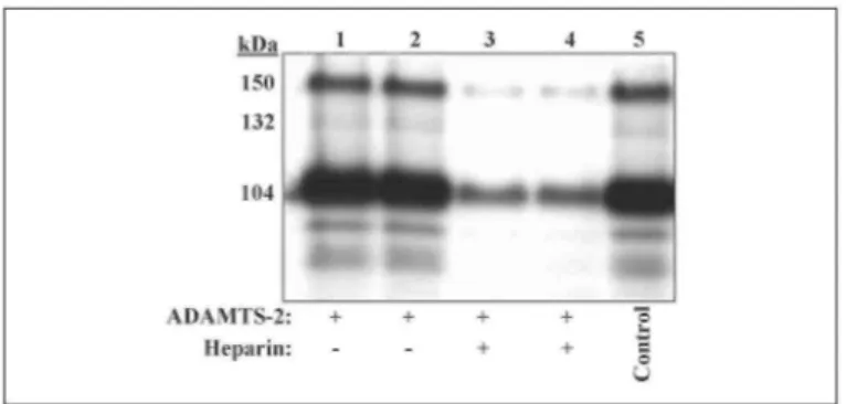
FIGURE 8. Compartmentalization of active forms of ADAMTS-2. Dermatosparactic calf fibroblasts were
incubated in culture medium containing 10 µg/ml ADAMTS-2 alone (lanes 1 and 2) or ADAMTS2 and heparin (5 µg/ml) (lanes 3 and 4). After 6 h, medium was removed, and the cell layers were solubilized before Western blotting analysis using mAb23. The electrophoretic pattern obtained from duplicate cultures (lanes 1 and 2) illustrated the reproducibility of this technique, because both the total amount of enzyme and the relative abundance of the various products are identical. Moreover, the ration of the 104- and 150-kDa products is similar to the ratio determined for the purified enzyme before incubation (lane 5). The presence of heparin (lanes 3 and 4) similarly affected the binding of all the enzyme forms.
FIGURE 9. Autocatalytic processing of ADAMTS-2. Cells expressing construct 6A were cultured alone (lane
1) or co-cultured with control cells (lane 2) or cells expressing the wild type construct (lane 3). Enzyme recovered from the cell layer was analyzed by Western blotting using anti-HA-FLAG antibody, preventing detection of any product resulting from the processing of the wild type enzyme (lane 4).
This suggests that the 104-kDa product is generated by a proteolytic cleavage occurring in the beginning of the PNP domain, a few amino acids downstream of the C-terminal end of product 2A. Based on the hypothesis that these two products have a similar enzymatic activity, it was calculated that the 150-kDa product is 3-4-fold less active than the 104-kDa form lacking the PNP domain (Fig. 7C). Similarly, it was determined that the product at 72 kDa (construct 5A, lacking the three C-terminal TSPI repeats and the PNP domain) is about 4-fold less active than the 104-kDa product.
Compartmentalization of Active Forms of ADAMTS-2—Purified ADAMTS-2 (Fig. 8, lane 5, control) was added in the culture medium of dermatosparactic calf fibroblasts. After 6 h of incubation, 25% of the total amount of enzyme was bound to the cell layer (not shown). The ratio of the 150- and 104-kDa products recovered from the cell layer (Fig. 8, lanes 1 and 2) was identical to the ratio in the purified preparation before incubation on cells (Fig. 8, lane 5). Moreover, heparin efficiently inhibited the binding of both products (Fig. 8
lanes 3 and 4), again suggesting a similar compartmentalization.
Autocatalytic Processing of ADAMTS-2—Cells expressing inactive HA-flagged protein 6A were cultured alone or in co-culture with empty vector transfected cells or with cells expressing full-length active enzyme without HA-FLAG (construct 1). Western blotting analysis with anti-HA FLAG antibody was then performed to specifically identify processed forms of the inactive protein 6A (Fig. 9). When cultured alone or co-cultured with control cells (Fig. 9, lanes 1 and 2), products at 173 and 150 kDa were the two most abundant forms. In the presence of cells expressing the "wild type" active enzyme (Fig. 9, lane 3), no product at 104 kDa was detected, but a strong accumulation of the 132-kDa product was observed. These bands resulted from the processing of protein 6A, because the antibody failed to detect any product associated with a cell layer of cells expressing the construct 1 only (Fig. 9, lane 4).
Other Substrates of ADAMTS-2—α2-Macroglobulin inhibits most proteinases by physical entrapment, upon
cleavage within a bait region. To determine whether ADAMTS-2 can cleave and be inhibited by α2
-macroglobulin, we examined the aminoprocollagen processing of ADAMTS-2 after its preincubation with α2 -macroglobulin. ADAMTS-2 (~10 ng, as determined by Coomassie Blue staining after SDS-PAGE) was preincubated with α2-macroglobulin at varying concentrations (0.025 to 0.4 units). The aminoprocollagen
substrate was then added, and the peptidase activity was evaluated. The observed dose-response inhibition of the activity (Fig. 10A) was in good agreement with previous data about ADAMTS-4 (21).
FIGURE 10. Inhibition of aminoprocollagen peptidase activity by α2-macroglobulin. A, purified ADAMTS-2
was incubated at 37 °C with increasing amount of α2-macroglob-ulin (25 units/ml; 1-16 µl) or bovine serum
albumin as negative control. After 1 h, the aminoprocollagen type I substrate was added, and ADAMTS-2 activity was measured. Preincubation with bovine serum albumin did not alter the enzymatic activity. On the contrary, addition of α2-macroglobulin strongly inhibited aminoprocollagen peptidase activity. B, purified
ADAMTS-2 was incubated alone (lane 1) or with α2-macroglobulin in the absence (lane 2) or presence (lane 3)
of EDTA, an inhibitor of ADAMTS-2 activity. The band shift observed only in the presence of α2-macroglobulin
and active enzyme (lane2) illustrated the bait and trap mechanism of inhibition. C, conditioned medium of cells expressing constructs 1 A, 2A, and 5A was analyzed by Western blotting using anti-HA antibody. The large amount of α2-macroglobulin present in the fetal calf serum is responsible for the band shift observed with the
three forms of recombinant enzyme.
FIGURE 11. α1 type V procollagen processing. A, purified fraction of homotrimeric recombinant α1(V)
collagen synthesized by 293 cells (19) contained aminoprocollagen (pN-α1) chains and a degradation product (TH-α1) migrating as pepsinized α1(V) (lane 1). In the presence of purified recombinant ADAMTS-2, an intermediate band was observed (pNδ-α1V) (lane2), suggesting a cleavage in the "variable" domain. This processing product was absent in the presence of EDTA used as ADAMTS-2 inhibitor {lane 3). Type V collagen purified from fetal calf skin was also analyzed (lane 4). Only two bands were visualized, migrating at sizes corresponding to pNδ-α1 and α2V. B, schematic representation of specific cleavage sites by ADAMTS-2 and other α1 type V collagen processing enzymes. PARP, proline/arginine-rich protein domain.
Purified ADAMTS-2 was also incubated alone (Fig. 10B, lane 1) or with α2-macroglobulin in the absence (lane
2) or the presence (lane 3) of EDTA, an inhibitor of ADAMTS-2 activity. The band shift observed only in the
presence of α2-macroglobulin and active enzyme (Fig. 10B, lane 2) as illustrated by the bait and trap mechanism of inhibition. In presence of serum, which contains large amounts of α2-macroglobulin, recombinant enzymes 1A, 2A, and 5A secreted in the conditioned culture medium were efficiently trapped, as demonstrated by the accumulation of large amount of high molecular weight products (Fig. 10C).
In order to verify the processing activity on a more specific potential substrate, purified ADAMTS-2 was incubated with aminoprocollagen type V, a fibrillar collagen that is N-terminally processed in vivo. Recombinant
α1 type V collagen homotrimer synthesized by 293 cells was used (20). After separation on SDS-PAGE (Fi.
11A, lane 1), this collagen appeared as two bands corresponding to aminoprocollagen V (pN-α1(V)), containing all the N-terminal domains (proline/arginine-rich protein domain, the variable region, and the short triple helix), and to a degradation product (TH-α1(V)) consisting of the full-length triple helix domain. After incubation with active ADAMTS-2, another product of intermediate size was observed (Fig. 11A, lane 2), indicative of a cleavage in the variable domain of type V pNcollagen. This pattern was similar in the absence or presence of DTT. The N-terminal sequence of this product was determined (ANQDTIYE) and was identical to a sequence located at the C-terminal end of the variable domain (SEIG-PGMPANQDTIYE). This defines a new cleavage site for ADAMTS-2 (PA versus the (A/P)Q described in procollagens I-III) in a different three-dimensional context (the variable domain rather than a sequence located between two collagen domains, Fig. 11B). As control for the in vivo relevance of this cleavage site, type V collagen purified from fetal calf skin was analyzed by Western blotting (Fig. 11A, lane 4). A product presenting the same electrophoretic mobility as the pNδ-α1(V) was observed together with another band consisting of the α2V chain. More interestingly, no other product was detected, indicative of the absence of α1V processed at the published BMP-1 cleavage site (Fig. 11B). The moderate increase of the amount of TH-α1V, or of a product of similar size, after incubation with ADAMTS-2 (Fig. 11A, lane 2) was not always observed and therefore was not investigated further.
FIGURE 12. Characterization of the various forms of ADAMTS-2. Characterization of the N- and C-terminal
sequences and the domain structure of the different ADAMTS-2 products was performed by analyzing
complementary data from electrophoretic mobility, Western blotting, and microsequencing. Estimated molecular sizes are reported on the left. The presence of specific epitopes and N-terminal sequence, when determined, is indicated on the schematic representation of each form. Dotted lines illustrate regions where processing is supposed to be performed but was not unambiguously confirmed by protein sequencing.
DISCUSSION
ADAMTS are complex enzymes containing numerous well defined domains. Recent studies (16, 22, 23) indicate that post-translational processing can alter ADAMTS-4 activity and specificity. Cleavage of recombinant ADAMTS-2 has also been reported (6). However, the relevance to the in vivo situation and the involvement of the processing for the regulation of ADAMTS-2 activity were not investigated.
Requirement of the Individual Domains for Aminoprocollagen Peptidase Activity—Recent studies have investigated the implication of some domains for the enzymatic activity of ADAMTS-1 and -4 (16, 24-26). In our work, we systematically evaluated the function of several domains of ADAMTS-2 by investigating the aminoprocollagen peptidase activity of the various forms of recombinant enzyme, either wild type, mutated, or lacking specific sequences. Removal of the PNP domain strongly and reproducibly increased the enzyme activity, as compared with the full-size enzyme. This suggested a negative regulatory function for this domain, either directly, by interfering with the catalytic activity of the metalloproteinase domain, or indirectly, by altering the recognition and the binding of the substrate. On the contrary, the second and fourth TSP1 repeat were positive regulators because their removal decreased the aminoprocollagen peptidase activity. Most interestingly, form 5 (lacking the three last TSP1 repeats and the C-terminal domain) was still significantly active, suggesting that full activity is provided by cooperation between several domains. Most ADAM and ADAMTS enzymes are synthesized as a "pro-" zymogen form that has to be cleaved by furin or related enzymes to display full activity. When the potential cleavage site by furin was mutated, only low but still significant aminoprocollagen peptidase activity was measured, confirming the involvement of furin cleavage for the regulation of ADAMTS-2 activity but suggesting also that either an alternative processing pathway is able to activate the pro-ADAMTS-2 or that the full size pro-enzyme can display activity as described for ADAMTS-13 (27). Recombinant enzymes lacking different sequences between the pro- and the metalloproteinase domains were efficiently synthesized and secreted but exhibited no activity. This result suggests that the pro-domain is not only responsible for the repression of enzyme activity but is also essential for the correct folding of the protein as observed for other enzymes (28).
It was demonstrated, by evaluation of the aminoprocollagen peptidase activity of forms 1-5, that the C-terminal part of ADAMTS-2 is required for full enzyme activity, probably for an efficient recognition and binding of the substrate (see above). In order to verify whether substrate specificity is also dictated by these domains or, on the contrary, by the metalloprotease domain itself or other domains lying in the central part of ADAMTS-2, chimeric enzymes were produced by domains swapping with either GON-1, a C. elegans ADAMTS, or
ADAMTS-14 (5), an enzyme closely related to ADAMTS-2 but exhibiting only low aminoprocollagen peptidase activity in vitro. Among the different chimeric enzymes, only the form 17 (containing the N-terminal part of ADAMTS-2 fused to the C-terminal part of ADAMTS-14) exhibited high aminoprocollagen peptidase activity, although lower than activity measured with the wild type form 1.
These data altogether suggest the following: (i) the pro-domain is required for the correct folding of ADAMTS-2 but has to be cleaved by furin for full enzyme activity; (ii) the PNP domain acts as a negative regulator for aminoprocollagen processing; (iii) the central domains of ADAMTS-2 are essential for aminoprocollagen type I processing; (iv) TSP1 repeats 2 and 4 are required for full enzyme activity, illustrating probably their
involvement in substrate recognition or binding; and (v) TSP1 repeats of other ADAMTS, even of the closely related 14, are not able to fulfill completely the function of the corresponding domains of ADAMTS-2, suggesting finely regulated cooperative interactions between the various domains of the enzyme.
Characterization of the ADAMTS-2 Maturation Products—The characterization of the various maturation
products was performed by integrating various complementary data (Fig. 12). The 177-, 173-, and 150-kDa products start, respectively, at the end of the signal peptide, at the first or the second furin cleavage site, and extend to the M2-FLAG (Fig. 12). No clear sequence was obtained for the 132-kDa product, but it possibly corresponds to the 118-kDa band reported to also contain two individual products, generated by processing in the metalloprotease domain (6). Size discrepancy between the two studies may be related to differences in the level of glycosylation, due to the fact that the two enzymes are from different origins (bovine and human) and to slight differences in the conditions used for SDS-PAGE analysis. Sequence of the N terminus of the 104-kDa product demonstrated a cleavage at the second cleavage site by furin, as for the 150-kDa product. Because the 104-kDa product is not recognized by antiserum AS175 specific for the C terminus of ADAMTS-2, it strongly suggests that it is produced by removal of most of the PNP domain, a region that was shown to negatively regulate activity of ADAMTS-2 (see above). This is further confirmed by SDS-PAGE analysis showing a similar size for the 104-kDa product and the most abundant form of protein 2A (102 kDa), lacking the PNP domain. The 95- and 43-kDa products are generated by cleavage at an identical site, at the end of the spacer domain. Based on the low activity displayed by recombinant enzyme form 5, this processing results probably in the production of an enzyme lacking significant aminoprocollagen peptidase activity. A mechanism of inactivation of
aminoprocollagen peptidase activity or the release of biologically active TSP1 repeats are potential functions of ADAMTS-2 processing at this site. Localization of other fragments was essentially deduced from Western blotting data experiments because these fragments were retained during the purification process, assuming also the presence of at least one of the C-terminally located TSP1 repeats that are required for efficient purification using heparin-Sepharose chromatography (data not shown; see Ref. 29).
TABLE ONE
Comparison of sequences corresponding to the end of the α1 type V N-terminal prodomain in various species The arrows ( ↓ ) indicate the site of cleavage by ADAMTS-2 in human recombinant α1 type V homotrimer and in identical or similar sequences in other species. The position of the cleavage site relative to the short triple helix, which is composed of 17 GXY repeats, and to main triple helix, is highly conserved. Localization and sequence of the potential cleavage sites between the two triple helical domains (A-Q) are also strictly conserved, aa indicates amino acids.
Human, mouse, rat and hamster
GPGMP ↓ ANQDT, 33 aa, 17 GXY repeats; 21 aa, AQESQAQ, 12 aa, central triple helix Chicken (Gallus gattus)
GPGMP ↓ ANQDT, 33 aa, 17 GXY repeats; 21 aa, AQEAQAQ, 12 aa, central triple helix In order to determine whether the maturation process observed for recombinant enzyme produced in 293 cells is relevant to the in vivo situation, active enzyme was purified from calf skin and characterized by Western blotting. The most prominent form of native enzyme has an estimated molecular mass of 104 kDa, thus migrating with the same mobility as the most abundant recombinant form. Products at 150, 132, and 85 kDa were also observed in both preparations, whereas another fragment, 118 kDa, was only present in the calf skin preparation. These observations suggested that the in vitro processing of recombinant ADAMTS-2 as observed in 293 cells is highly similar to the in vivo situation, indicating that this cell line represents a valuable model to
study the regulation of ADAMTS-2 maturation. Because 293 cells and calf skin fibroblasts are not expected to express a common panel of highly specific enzymes, this multiple processing is likely to be performed by ubiquitous enzymes or by an autocatalytic process.
ADAMTS-2 processing was further investigated in vitro by using cells expressing recombinant enzyme mutated at the catalytic site. The catalytically inactive ADAMTS-2 accumulated as a 150-kDa form with no 104-kDa form. Because the point mutation introduced in the catalytic site is not expected to modify the three-dimensional structure of the enzyme, this observation strongly suggested that the 104-kDa form results largely from an autocatalytic processing of the 150-kDa form. Because many activation processes require "co-factors" or are operated at the cell surface, a co-culture experiment was set up to investigate this hypothesis. In these conditions, a wild type active enzyme promotes a strong accumulation of the 132-kDa product from inactive protein 6A. However, the 104-kDa product was not detected. These data suggest that the generation of the 132-kDa product is an intermolecular autocatalytic process that probably occurs at the cell surface, whereas the 104-kDa form would result from an intramolecular cleavage. Alternatively, the possibility of an intermolecular processing that would be performed only during the secretion process, preventing its detection by the co-culture model, cannot be ruled out.
Identification of ADAMTS-2 Maturation Products Displaying Enzymatic Activity—Selective purification of each individual maturation product could not be efficiently performed because they displayed similar properties during the purification procession. To overcome this problem, cells expressing the wild type ADAMTS-2 were cultured in conditions known to modify the relative abundance of the various forms of the enzyme that were correlated to the enzyme activity. Inverse correlation, suggestive of an inhibitory function, was not found for any product. The only significant positive correlations were established with the 150-kDa form (linear regression, R2 = 0.65) and the 104-kDa form (logarithmic regression, R2 = 0.81). The specific activity of
the two products was measured. It indicated that the 104-kDa form is 3-4-fold more active than the 150-kDa product, again illustrating the inhibitory function of the PNP domain. These results are in agreement with other data. For example, the shape of the correlation curve of the 104-kDa product (Fig. 7A) probably illustrates saturation of the aminoprocollagen peptidase assay. Because this assay is linear only in presence of low enzymatic activity, this provides indirect evidence that the 104-kDa form is responsible for most of the activity. This is also suggested by our observations showing that active enzyme purified from skin is essentially
recovered in a 104-kDa form.
Compartmentalization of Active Forms of ADAMTS-2—A previous study (22) demonstrated that removal of the C-terminal spacer domain of ADAMTS-4 affected its compartmentalization. In this study, the binding properties of the 104- and 150-kDa products were found to be similar, both in absence or presence of heparin, demonstrating that the most critical domain for efficient immobilization is not located in the PNP domain. Preliminary analysis (not shown) of the various forms of ADAMTS-2 truncated at the C terminus (constructs 1-5) suggests that efficient binding both to the cell layer and to the heparin-Sepharose matrix is largely mediated by the second TSPI repeat.
Identification of Additional Substrates—Activity of ADAMTS-2 is strongly influenced by the
three-dimensional structure of the substrate. For example, aminoprocollagen processing is inhibited in vitro and in vivo when the native triple helical structure of type I procollagen is altered (20, 30). To investigate whether
ADAMTS-2 is able to process other structurally unrelated proteins, we used the α2-macroglobulin broad spectrum substrate. Cleavage of the bait region of α2-macroglob-ulin induces a large conformational change, resulting in the entrapment and the inhibition of the cleaving enzyme. Such a mechanism has been described for several types of proteases, including ADAM and ADAMTS (21, 31). Incubation of ADAMTS-2 with α2 -macroglobulin dose-dependently inhibits aminoprocollagen peptidase activity, demonstrating that ADAMTS-2 can cleave substrates other than fibrillar collagens. Moreover, preliminary results obtained using recombinant enzymes 2A and 5A suggest that the C-terminal domains do not regulate the cleavage of α2-macroglobulin by ADAMTS-2.
Type V collagen is a quantitatively minor fibrillar collagen that participates in the regulation of type I collagen fibrillogenesis. It is most widely found in vivo as an [α1(V)] 2 α2(V) heterotrimer, but other forms are also present such as an [α1(V)]3 homotrimer or cross-type heterotrimers composed of type V and type XI chains (for review, see Ref. 7). Characterization of the processing of the aminopropeptide of type V procollagen chains has led to confusing and sometime conflicting results (9,32-35), possibly because of the existence of different cleavage sites and tissue- or cell-dependent diversity in the level of processing. Moreover, extraction and purification of native type V collagen are low yield processes, hampering the characterization as an intact molecule and explaining why most recent works have been performed by using recombinant type V collagen (9
-12). Although the consensus sequence for cleavage by ADAMTS-2 (AQ) is present at the expected localization as compared with procollagen types I-III (between the short and the long triple helical domains) and is highly conserved between species (TABLE ONE), there was no direct experimental evidence for type V
aminopropeptide processing at this site. This hypothesis was verified by incubating purified recombinant aminoprocollagen type V with recombinant ADAMTS-2. Processing was observed, but at a more N-terminal sequence corresponding to the end of the variable region. The cleavage site (P ↓ A) is not identical but has some similarity to the cleavage site reported previously for ADAMTS-2 ((P/A) ↓ Q). In addition, the sequence in this region is highly conserved among the different species, suggesting the physiological relevance of this finding. Furthermore, the size of the band obtained after ADAMTS-2 cleavage is similar to the size previously
determined for the α1(V) chain extracted with acetic acid from tissues and referred to as the "intact chain" (36, 37). As additional evidence, Western blotting analysis of type V collagen purified from fetal calf skin
demonstrated the presence of only two products, one presenting the same electrophoretic mobility as the
pNδ-α1(V) and another consisting in α2V chain. Most interestingly, no other product was detected, strongly
suggesting the absence of α1V processed at the published BMP-1 cleavage site. Based on the high sequence homology between the two collagen domains, the apparent absence of cleavage at the AQ consensus cleavage site for ADAMTS-2 was surprising (TABLE ONE). This may illustrate a true absence of processing at this site
in vivo or that another enzyme, such as ADAMTS-3 or -14, is responsible for the processing in vivo.
Alternatively, the site of type V procollagen processing may vary and be developmentally or tissue-specifically regulated. Finally, because aminoprocollagen peptidase activity of ADAMTS-2 is strongly dependent on the three-dimensional structure of the substrates (20,30), it may be hypothesized that processing of the
aminopropeptide, by cleavage between the two triple-helical domains, would be observed only with perfectly folded recombinant type V procollagen. Considering that only a fraction of the recombinant type V procollagen is perfectly folded, the moderate increase of the amount of a product migrating as TH-α1(V), which was sometimes observed after incubation with ADAMTS-2, possibly illustrates this hypothesis.
Acknowledgments
We thank Y. Goebels, G. Rega, and D. Mazzocut for expert assistance with protein purification and
electrophoresis, cell culture, and type V collagen sequencing, respectively. We are grateful to Dr. J. Kimble for provision of gon-1 cDNA.
References
1. Nusgens, B. V-, Verellen-Dumoulin, C, Hermanns-Le, T., De Paepe, A-, Nuytinck, L-, Pierard, G. E., and Lapiere, C. M. (1992) Nat. Genet. 1, 214-217
2. Colige, A., Sieron, A. L., Li S. W., Schwarze, U., Petty, E., Wertelecki, W., Wilcox, W., Krakow, D-, Cohn, D. H-, Reardon, W-, Byers, P. H-, Lapiere, C. M-, Prockop, D. J-, and Nusgens B. V. (1999) Am. J. Hum. Genet. 65, 308-317
3. Colige, A-, Nuytinck, L-, Hausser, L, van Essen, A.J., Thiry, M-, Herens, C, Ades, L. C, Malfait, F., Paepe, A. D., Franck, P., Wolff, G., Oosterwijk, J. C., Smitt, J. H. Lapiere, C. M., and Nusgens, B. V. (2004).J. Invest.
Dermatol. 123, 656-663
4. Fernandes, R. J., Hirohata, S-, Engle, J. M-, Colige, A-, Cohn, D. H-, Eyre, D. R-, and Apte, S. S. (2001) J.
Biol. Chem. 276, 31502-31509
5. Colige, A-, Vandenberghe, I-, Thiry, M-, Lambert, C. A-, Van Beeumen, J., Li, S. W-, Prockop, D. J., Lapiere, C. M., and Nusgens, B. V. (2002) J Biol. Chem. 277, 5756-5766
6. Wang, W. M., Lee, S., Steiglitz, B. M., Scott, I. C, Lebares, C. C, Allen, M. L., Brenner, M. C, Takahara, K., and Greenspan, D. S. (2003) J Biol. Chem. 278, 19549-19557
7. Fichard, A., Kleman, J. P., and Ruggiero, F. (1995) Matrix Biol. 14, 515-531 8. Birk, D. E. (2001) Micron. 32, 223-237
9. Imamura, Y., Steiglitz, B. M., and Greenspan, D. S. (1998) J. Biol. Chem. 273, 27511-27517 10. Gopalakrishnan, B., Wang, W. M., and Greenspan, D. S. (2004) J. Biol. Chem. 279, 30904-30912
11. Kessler, E-, Fichard, A-, Chanut-Delalande, H-, Brusel, M-, and Ruggiero, F. (2001) J. Biol. Chem. 276, 27051-27057
12. Unsold, C, Pappano, W. N, Imamura, Y., Steiglitz, B. M., and Greenspan, D. S. (2002) J. Biol. Chem. 277, 5596-5602
13. Apte, S. S. (2004) Int. J. Biochem. Cell Biol. 36, 981-985
14. Wang, P., Tortorella, M., England, K., Malfait, A. M., Thomas, G., Arner, E. C, and Pei, D. (2004) J Biol.
Chem. 279,15434-15440
15. Rodriguez-Manzaneque, J- C, Milchanowski, A. B-, Dufour, E. K-, Leduc, R-, and Iruela-Arispe, M. L. (2000)J. Biol. Chem. 275, 33471-33479
16. Kashiwagi, M-, Enghild, J. J.., Gendron, C, Hughes, C, Caterson, B-, Itoh, Y-, and Nagase, H. (2004) J.
Biol. Chem. 279,10109-10119; Correction (2004)J. Biol. Chem. 279, 22786
17. Blelloch, R., and Kimble, J. (1999) Nature 399, 586-590
18. Colige, A., Li, S. W., Sieron, A. L., Nusgens, B. V., Prockop, D. J., and Lapiere, C. M. (1997) Proc. Natl.
Acad. Sci. U. S. A. 94, 2374-2379
19. Fichard, A., Tillet, E., Delacoux, F., Garrone, R., and Ruggiero, F. (1997) J. Biol. Chem. 272, 30083-30087 20. Colige, A-, Beschin, A-, Samyn, B-, Goebels, Y-, Van Beeumen, J., Nusgens, B. V-, and Lapiere, C. M. (1995) / Biol. Chem. 270, 16724-16730
21. Tortorella, M. D., Arner, E. C, Hills, R., Easton, A., Korte-Sarfaty, J., Fok, K., Wittwer, A. J., Liu, R. Q., and Malfait, A. M. (2004) J. Biol. Chem. 279,17554-17561
22. Gao, G., Westling, J., Thompson, V. P., Howell, T. D., Gottschall, P. E., and Sandy, J. D. (2002)J. Biol.
Chem. 277,11034-11041
23. Gao, G., Plaas, A., Thompson, V. P., Jin, S., Zuo, F., and Sandy, J. D. (2004) J. Biol. Chem. 279,10042-10051
24. Zheng, X., Nishio, K., Majerus, E. M., and Sadler, J. E. (2003) J. Biol. Chem. 278, 30136-30141
25. Tortorella, M., Pratta, M., Liu, R. Q., Abbaszade, I., Ross, H, Burn, T., and Arner, E. (2000)/ Biol. Chem. 275, 25791-25797
26. Longpre, J. M., and Leduc, R. (2004) / Biol. Chem. 279, 33237-33245
27. Majerus, E. M., Zheng, X., Tuley, E. A, and Sadler, J. E. (2003) / Biol. Chem. 278, 46643-46648
28. Bissonnette, L., Charest, G., Longpre, J. M., Lavigne, P., and Leduc, R. (2004) Biochem. J. 379, 757-763 29. Guo, N.-H., Krutzsch, H. C, Nègre, E., Vogel, T., Blake, D. A., and Roberts, D. D. (1992) Proc. Natl. Acad.
Sci. U. S. A. 89, 3040-3044
30. Cabrai, W. A., Mertts, M. V., Makareeva, E., Colige, A., Tekin, M., Pandya, A., Leikin, S., and Marini, J. C. (2003) J. Biol. Chem. 278,10006-10012
31. Loechel, F., and Wewer, U. M. (2001) FEB S Lett. 506, 65-68
32. Linsenmayer, T. F, Gibney, E-, Igoe, F-, Gordon, M. K-, Fitch, 1- M-, Fessier, L. I-, and Birk, D. E. (1993) J. Cell Biol. 121,1181-1189
34. Moradi-Ameli, M., Rousseau, J. C, Kleman, J. P., Champliaud, M. F., Boutillon, M. M., Bernillon, J., Wallach, J., and van der Rest, M. (1994) Eur. J. Biochem. 221, 987-995
35. Rothblum, K., Stahl, R. C, and Carey, D. J. (2004) J. Biol. Chem. 279, 51282-51288
36. Broek, D. L., Madri, J., Eikenberry, E. F., and Brodsky, B. (1985) / Biol. Chem. 260, 555-562 37. Ruggiero, F., Champliaud, M. F., Garrone, R., and Aumailley, M. (1994) Exp. Cell Res. 210,215-223