International Journal of
Molecular Sciences
Article
Presence of TERT Promoter Mutations is a Secondary
Event and Associates with Elongated Telomere
Length in Myxoid Liposarcomas
Monica S. Ventura Ferreira1 ID, Martina Crysandt1, Till Braunschweig2, Edgar Jost1, Barbara Voss1, Anne-Sophie Bouillon1, Ruth Knuechel2, Tim H. Brümmendorf1and Fabian Beier1,*
1 Department of Hematology, Oncology, Haemostaseology and Stem Cell Transplantation, RWTH Aachen University Medical Faculty, 52074 Aachen, Germany;
mventuraferreira@ukaachen.de (M.S.V.F.); mcrysandt@ukaachen.de (M.C.); ejost@ukaachen.de (E.J.); bavoss@ukaachen.de (B.V.); abouillon@ukaachen.de (A.-S.B.); tbruemmendorf@ukaachen.de (T.H.B.) 2 Institute of Pathology, RWTH Aachen University Medical Faculty, 52074 Aachen, Germany;
tbraunschweig@ukaachen.de (T.B.); rknuechel-clarke@ukaachen.de (R.K.)
* Correspondence: fbeier@ukaachen.de; Tel.: +49-241-8089806
Received: 12 January 2018; Accepted: 10 February 2018; Published: 18 February 2018
Abstract:The occurrence of TERT promoter mutations has been well described in soft tissue sarcomas (STS). However, the biological role of these mutations as well as their impact on telomere length in STS is still unclear. We analyzed 116 patient samples diagnosed with 22 distinct histological subtypes of bone and STS for the occurrence of TERT promoter mutations by Sanger sequencing. We observed TERT promoter mutations at an overall frequency of 9.5% distributed over 7 different sarcoma subtypes. Except for one chondrosarcoma case harboring a C250T mutation, all other mutations were detected at location C228T. By far the far highest frequency of TERT promoter mutations was found in myxoid liposarcoma (MLS) (4 out of 9 cases studied, i.e., 44%). Assessment of telomere length from tumor biopsies revealed that TERT promoter-mutated MLSs had significantly fewer shortened telomeres in comparison to TERT wildtype MLSs. Based on the frequency of TERT promoter mutations and the elongated telomere length in mutated compared to wildtype MLS, we hypothesize that occurrence of TERT promoter mutations has a pivotal role in the disease progression as a secondary genetic event at a time when tumor cells face the need for telomere elongation to allow further proliferation.
Keywords:myxoid liposarcoma; sarcoma; telomere; telomerase; TERT promoter mutation
1. Introduction
Telomeres are repetitive DNA sequences protecting the ends of chromosomes. Telomeres shorten with each cell division, and telomere length (TL) limits the replicative capacity of a cell once telomeres become critically short [1,2]. Telomere shortening can be counteracted by the expression of telomerase (TERT), an enzyme that can (re-)elongate telomeres [3,4] a phenomenon that can also be used therapeutically in vivo [5]. One of the hallmarks of cancer is unlimited cell division [6]. Previous studies showed that tumor cells mostly maintain their telomere length by increased telomerase expression [7] and rarely by a mechanism called alternative lengthening of telomere (ALT) [8]. However, the exact mechanism how tumor cells increase telomerase activity is incompletely understood and varies between the different tumor entities [9].
Recently, two highly recurrent mutations in the promoter region of TERT (c.-124 C>T and c.-146 C>T), also called C228T and C250T, respectively were described in 71% of all melanomas [10,11].
Int. J. Mol. Sci. 2018, 19, 608 2 of 13
In follow up studies, these mutations were reported to be, overall, the most prevalent somatic point mutations being present at varying frequencies in over 50 different types of cancers [12–16]. Of note, both the C228T and the C250T mutation are typically heterozygous, mutually exclusive, and both create the identical 11bp sequence (i.e., “CCCGGAAGGGG”) that has substantial similarity to an ETS binding motif representing a de-novo binding site for an activating ETS family transcription factor. Both mutations activate TERT promoter activity and TERT gene transcription functionally resulting in in vitro telomere elongation [10,11].
Soft tissue sarcomas (STS) represent a heterogeneous disease with more than 50 clinically relevant subtypes with different histologies, molecular-genetic profiles, tumor-location patterns, and prognoses [17–19]. Genomic alterations including chromosomal translocations, DNA copy number variations and the presence of typical genetic aberrations are hallmarks of certain subtypes of sarcomas [20]. One of the molecularly best-studied subtypes is myxoid liposarcoma (MLS). In 95% of the cases, MLSs share the reciprocal translocation t(12;16)(q13; p11). The breakpoint on chromosome 12 involves the transcription factor CHOP (C/EBP homologous protein), member of the CAAT/enhancer-binding protein (C/EBP) family, a gene involved in adipocyte differentiation [21,22]. The CHOP protein presents a leucine zipper dimerization domain that can bind C/EBP proteins and block the interaction with DNA [21,22]. The breakpoint on chromosome 16 involves the FUS/TLS (Fused in Sarcoma/Translocated in Sarcoma) gene, an RNA-binding protein and member of the TET protein family that also includes the EWS protein. The main characteristic of this chromosomal translocation is that it results in the expression of the fusion transcript TLS/FUS-CHOP. Functionally, the resulting FUS-DDIT3 fusion protein induces increased expression of the C/EBP promoting oncogenic activation and tumor formation [23–25]. Based on the oncogenic potential and respective mouse models [26], the occurrence of t(12;16)(q13; p11) translocation is generally expected to be the first event in sarcoma initiation in MLS.
In the literature, only a few studies exist on telomeres or TERT promoter mutation status in sarcoma. These include the introduction of telomere length as a prognosis marker in Ewig sarcoma [27], the association of telomere length of peripheral blood leukocytes with increased risk for soft tissue sarcoma [28], telomere length in complex and simple karyotype sarcomas [29], assessment of ALT in sarcoma [30–34], mosaicism of telomere maintenance mechanisms in sarcomas [35,36] and the frequency of TERT promoter mutations in sarcomas [13,37–39]. However, no study has so far correlated TERT promoter status and telomere length in bone and STSs.
The aim of our study was first to investigate the prevalence of TERT promoter mutations in the different sarcoma subtypes and second to analyze the relation between presence of TERT promoter mutations and telomere lengths in a specific subtype of sarcomas, the MLSs.
2. Results
2.1. TERT Promoter Mutations in Bone and Soft Tissue Sarcomas (STS)
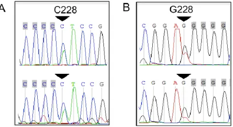
First, we analyzed for the incidence of TERT promoter mutations in 116 bone and STS tumors of 22 different subtypes (Table1). We found mutations in 11/116 patients (9.5%), namely in four cases of MLS, one solitary fibrous tumor (SFT), two malignant fibrous histiocytomas (MFH) or undifferentiated pleomorphic sarcoma (UPS), one pleomorphic sarcoma, one poorly differentiated sarcoma NOS and two chondrosarcomas (Figure1; Table2). Of note, 10 out of 11 (91%) mutations found were C228T and only in one patient with chondrosarcoma we observed a TERT promoter mutation at position C250T (Table3). In Table1, mutation frequencies are summarized in a side-by-side comparison with different studies reported in the literature.
Int. J. Mol. Sci. 2018, 19, 608 3 of 13
Int. J. Mol. Sci. 2018, 19, 608 3 of 13
Figure 1. Representative chromatograms of heterozygous TERT promoter mutations in two bone and
soft tissue sarcomas. Sequencing direction: (A) sense, (B) antisense. An arrow indicates mutations.
Table 1. Overview of the frequency of TERT promoter mutations in soft tissue and bone sarcomas
reported in the literature: Number of mutated cases/total cases (mutation rate, in percentage).
Present Study (n = 116) 22 Subtypes Saito 2016 (n = 180) 19 Subtypes Campanella 2016 (n = 68) 15 Subtypes Koelsche 2014 (n = 341) 16 Subtypes Killela 2013 (n = 95) 6 Subtypes Soft tissue sarcomas
WDLS 0/5 (0%) 0/18 (0%) 0/1 (0%) NA NA Myxoid liposarcoma 4/9 (44%) 3/13 (23%) 2/9 (22.2%) 29/39 (74%) 19/24 (79.1%) Pleomorphic liposarcoma 1/4 (25%) NA 1/1 (100%) 0/15 (0%) NA Dedifferentiated liposarcoma 0/12 (0%) NA 1/1 (100%) 0/61 (0%) NA Liposarcoma 0/7 (0%) NA NA NA NA Leiomyosarcoma 0/9 (0%) 0/24 (0%) 0/13 (0%) 0/27 (0%) NA Cytosarcoma Phyllodes 0/1 (0%) NA NA NA NA Angiosarcoma 0/3 (0%) NA NA 0/9 (0%) NA Rabdomyosarcoma 0/8 (0%) 0/5 (0%) NA NA NA Haemangiosarcoma 0/1 (0%) NA NA NA NA MPNST 0/8 (0%) 0/1 (0%) 0/7 (0%) 2/35 (6%) NA UPS (previous MFH) 2/9 (22.2%) 1/22 (4.5%) 0/12 (0%) 0/40 (0%) NA Fibrosarcoma NA NA 0/1 (0%) NA 1/3 (33.3%) Myofibroblastic sarcoma 0/3 (0%) NA 0/3 (0%) NA NA Myxofibrosarcoma 0/1 (0%) 0/6 (0%) 0/5 (0%) 0/17 (0%) 1/10 (10%) NOS 0/11 (0%) NA NA NA NA
NOS, poorly differentiated 1/8 (12.5%) NA NA NA NA
Clear cell sarcoma 0/1 (0%) 0/1 (0%) 0/1 (0%) 0/5 (0%) NA Synovial sarcoma 0/9 (0%) 0/7 (0%) 0/9 (0%) 1/25 (4%) NA Ewing sarcoma 0/1 (0%) 0/6 (0%) NA NA NA SFT 1/1 (100%) 5/40 (12.5%) 0/1 (0%) 4/31 (13%) 2/10 (20%) Bone sarcomas Chondrosarcoma 2/8 (25%) 0/10 (0%) NA 0/8 (0%) 1/2 (50%) Osteosarcoma 0/3 (0%) 0/14 (0%) NA NA 1/23 (4.3%) Overall 11/116 (9.5%) 9/167 (5.4%) 4/68 (5.9%) 36/341 (10.5%) 25/72 (34.7%) NA—not available; WDS—well-differentiated liposarcoma; UPS—undifferentiated pleomorphic sarcoma; MPNST—malignant peripheral nerve sheath tumor; SFT—solitary fibrous tumor; MFH— malignant fibrous histiocytoma; NOS—not otherwise specified; UPS—undifferentiated pleomorphic sarcoma.
Figure 1.Representative chromatograms of heterozygous TERT promoter mutations in two bone and soft tissue sarcomas. Sequencing direction: (A) sense, (B) antisense. An arrow indicates mutations.
Table 1. Overview of the frequency of TERT promoter mutations in soft tissue and bone sarcomas reported in the literature: Number of mutated cases/total cases (mutation rate, in percentage).
Present Study (n = 116) 22 Subtypes Saito 2016 (n = 180) 19 Subtypes Campanella 2016 (n = 68) 15 Subtypes Koelsche 2014 (n = 341) 16 Subtypes Killela 2013 (n = 95) 6 Subtypes Soft tissue sarcomas
WDLS 0/5 (0%) 0/18 (0%) 0/1 (0%) NA NA Myxoid liposarcoma 4/9 (44%) 3/13 (23%) 2/9 (22.2%) 29/39 (74%) 19/24 (79.1%) Pleomorphic liposarcoma 1/4 (25%) NA 1/1 (100%) 0/15 (0%) NA Dedifferentiated liposarcoma 0/12 (0%) NA 1/1 (100%) 0/61 (0%) NA Liposarcoma 0/7 (0%) NA NA NA NA Leiomyosarcoma 0/9 (0%) 0/24 (0%) 0/13 (0%) 0/27 (0%) NA Cytosarcoma Phyllodes 0/1 (0%) NA NA NA NA Angiosarcoma 0/3 (0%) NA NA 0/9 (0%) NA Rabdomyosarcoma 0/8 (0%) 0/5 (0%) NA NA NA Haemangiosarcoma 0/1 (0%) NA NA NA NA MPNST 0/8 (0%) 0/1 (0%) 0/7 (0%) 2/35 (6%) NA UPS (previous MFH) 2/9 (22.2%) 1/22 (4.5%) 0/12 (0%) 0/40 (0%) NA Fibrosarcoma NA NA 0/1 (0%) NA 1/3 (33.3%) Myofibroblastic sarcoma 0/3 (0%) NA 0/3 (0%) NA NA Myxofibrosarcoma 0/1 (0%) 0/6 (0%) 0/5 (0%) 0/17 (0%) 1/10 (10%) NOS 0/11 (0%) NA NA NA NA
NOS, poorly differentiated 1/8 (12.5%) NA NA NA NA
Clear cell sarcoma 0/1 (0%) 0/1 (0%) 0/1 (0%) 0/5 (0%) NA
Synovial sarcoma 0/9 (0%) 0/7 (0%) 0/9 (0%) 1/25 (4%) NA Ewing sarcoma 0/1 (0%) 0/6 (0%) NA NA NA SFT 1/1 (100%) 5/40 (12.5%) 0/1 (0%) 4/31 (13%) 2/10 (20%) Bone sarcomas Chondrosarcoma 2/8 (25%) 0/10 (0%) NA 0/8 (0%) 1/2 (50%) Osteosarcoma 0/3 (0%) 0/14 (0%) NA NA 1/23 (4.3%) Overall 11/116 (9.5%) 9/167 (5.4%) 4/68 (5.9%) 36/341 (10.5%) 25/72 (34.7%) NA—not available; WDS—well-differentiated liposarcoma; UPS—undifferentiated pleomorphic sarcoma; MPNST—malignant peripheral nerve sheath tumor; SFT—solitary fibrous tumor; MFH—malignant fibrous histiocytoma; NOS—not otherwise specified; UPS—undifferentiated pleomorphic sarcoma.
Int. J. Mol. Sci. 2018, 19, 608 4 of 13
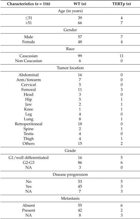
Table 2.Clinical characteristics of the selected cohort of soft tissue and bone sarcoma patients.
Characteristics (n = 116) WT (n) TERTp (n)
Age (in years)
≤51 39 4 >51 66 7 Gender Male 57 7 Female 48 4 Race Caucasian 99 11 Non Caucasian 6 0 Tumor location Abdominal 16 0 Arm/forearm 7 0 Cervical 5 0 Femoral 11 3 Head 3 0 Hip 5 1 Jaw 2 1 Knee 1 1 Leg 4 0 Lung 8 1 Retroperitoneal 18 0 Spine 2 1 Testis 4 0 Thigh 4 1 Others 15 2 Grade G1/well differentiated 16 5 G2-G3 86 6 NA 3 0 Disease progression No 53 5 Yes 45 3 NA 7 3 Metastasis Absent 55 6 Present 42 2 NA 8 3
N—number of cases. Tumor location “Others” included bladder, ear, gluteus, groin, liver, maxilla, nose, paravertebral, parotis, pleural, prostate, renal, rip/sternum, shoulder, skin, Vena cava. NA—not available. Disease progression during follow-up. Metastasis during follow-up.
2.2. Tumor Grading Correlates with the Presence of TERT Promoter Mutations
We next focused on whether the presence of TERT promoter mutations correlated with clinical features of our sarcoma cohorts. We did not observe any significant differences comparing the initial tumor localization: head/neck 10% (1/10), extremities and superficial trunk 18% (5/27), retroperitoneal 0% (0/18), abdomino-thoracic 4% (1/24) or others 13% (4/30). Since the histological grading is one of the most important factors for clinical decision-making, we compared patients according to histological grading. Patients with histopathological G1 grading/well-differentiated tumors showed significant higher percentage of TERT promoter mutations (5/21, 24%) compared to patients with G2-3 grading/poor differentiation (6/92, 7%, p = 0.030, see Table2). No further clinical characteristics
Int. J. Mol. Sci. 2018, 19, 608 5 of 13
such as age, gender, race, disease progression status or development of metastasis (Table2) differed significantly among WT and TERT promoter-mutated patients.
Table 3.Details of TERT promoter (TERTp) mutated-patients.
Diagnosis Age/Gender Location TERTp Mutation Tumor Grade Metastasis Progression
MLS 61/F thigh C228T G1 NA NA
MLS 71/M femoral C228T G1 yes yes
MLS 23/M lung C228T G1 NA NA
MLS 43/F spine C228T G2 yes yes
PS 58/F femoral C228T G2 no no
MFH 68/M skin C228T G2 no no
MFH 78/M hip C228T G3 yes no
CS 37/M femoral C228T G2 no no
CS 46/F jaw C250T G1 no no
SFT 64/F NA C228T well diff. yes yes
NOS 68/M ear C228T G2 no no
Age given in years. MLS—myxoid liposarcoma; PS—pleomorphic sarcoma; MFH—malignant fibrous histiocytoma; CS—chondrosarcoma; SFT—solitary fibrous tumor; NOS—not otherwise specified; NA—not available; well diff.—well-differentiated tumor. Metastasis and disease progression during follow-up.
2.3. Myxoid Liposarcomas (MLSs) Exhibit Longer Telomeres than Wildtype Tumors
Next, we focused on MLS based on the incidence of TERT promoter mutations—ranging in the literature from 22% to 79% (Table1)—as well as their homogenous genetic background characterized by the translocation t(12;16)(q13; p11), a genetic hallmark of MLS. Among the wildtype tumors, MLS only one (1/5 = 20%) presented distant metastasis in contrast to 2 out of 2 (100%) TERT promoter-mutated tumors from which patient clinical data was available.
Since telomere length declines with age, the comparison of the age of the patients did not reveal significant differences between MLS wildtype (61.4
±
6.0 years, n = 5) and TERT promoter-mutated tumors (49.5±
10.6 years, n = 4; p = 0.33). Interestingly, telomere length of MLS samples with TERT promoter mutations was found to be significantly less shortened (8.10±
0.50 kb; n = 4) compared to the telomere length of wildtype tumors (6.61±
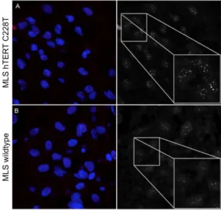
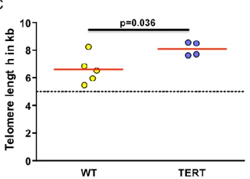
1.06 kb; n = 5; p < 0.037) (Figure2A–C). No statistically significant correlations between elongated telomere length and age, gender, tumor location, tumor grade, disease progression, and development of metastasis were observed likely due to small sample size. Assessment of ALT revealed no evidence of significant heterogeneity of telomere sizes, which would reveal the presence of ALT, in either TERT promoter MLS or wiltype tumors (Figure S1).Int. J. Mol. Sci. 2018, 19, 608 5 of 13
Table 3. Details of TERT promoter (TERTp) mutated-patients. Diagnosis. Age/Gender Location TERTp
mutation
Tumor
grade Metastasis Progression
MLS 61/F thigh C228T G1 NA NA
MLS 71/M femoral C228T G1 yes yes
MLS 23/M lung C228T G1 NA NA
MLS 43/F spine C228T G2 yes yes
PS 58/F femoral C228T G2 no no MFH 68/M skin C228T G2 no no MFH 78/M hip C228T G3 yes no CS 37/M femoral C228T G2 no no CS 46/F jaw C250T G1 no no SFT 64/F NA C228T well
diff. yes yes
NOS 68/M ear C228T G2 no no
Age given in years. MLS—myxoid liposarcoma; PS—pleomorphic sarcoma; MFH—malignant fibrous histiocytoma; CS—chondrosarcoma; SFT—solitary fibrous tumor; NOS—not otherwise specified; NA—not available; well diff.—well-differentiated tumor. Metastasis and disease progression during follow-up.
2.3. Myxoid Liposarcomas (MLSs) Exhibit Longer Telomeres than Wildtype Tumors
Next, we focused on MLS based on the incidence of TERT promoter mutations—ranging in the literature from 22% to 79% (Table 1)—as well as their homogenous genetic background characterized by the translocation t(12;16)(q13; p11), a genetic hallmark of MLS. Among the wildtype tumors, MLS only one (1/5 = 20%) presented distant metastasis in contrast to 2 out of 2 (100%) TERT promoter-mutated tumors from which patient clinical data was available.
Since telomere length declines with age, the comparison of the age of the patients did not reveal significant differences between MLS wildtype (61.4 ± 6.0 years, n = 5) and TERT promoter-mutated tumors (49.5 ± 10.6 years, n = 4; p = 0.33). Interestingly, telomere length of MLS samples with TERT promoter mutations was found to be significantly less shortened (8.10 ± 0.50 kb; n = 4) compared to the telomere length of wildtype tumors (6.61 ± 1.06 kb; n = 5; p < 0.037) (Figure 2A–C). No statistically significant correlations between elongated telomere length and age, gender, tumor location, tumor grade, disease progression, and development of metastasis were observed likely due to small sample size. Assessment of ALT revealed no evidence of significant heterogeneity of telomere sizes, which would reveal the presence of ALT, in either TERT promoter MLS or wiltype tumors (Figure S1).
Int. J. Mol. Sci. 2018, 19, 608 6 of 13
Int. J. Mol. Sci. 2018, 19, 608 6 of 13
Figure 2. Representative Q-FISH images captured by confocal laser scanning microscopy: (A) C228T
TERT promoter-mutated myxoid liposarcoma, and (B) a TERT promoter wildtype myxoid
liposarcoma patient. Magnification 630×. (C) Quantification of telomere length after Q-FISH analysis (arbitrary units, a.u.). The dashed line represents 5 kb, the threshold of critical short telomeres.
3. Discussion
Our study introduces the third largest cohort of bone and STS analyzed for TERT promoter
mutations among literature [37,38] and the first systematic analysis of telomere length in TERT
promoter-mutated MLS. We report the prevalence of TERT promoter mutations (overall incidence
near to 9.5%) to vary widely among different bone and STS subtypes (ranging from 0–44%), with the
highest mutation frequency found within MLS subgroup (44% harboring a C228T mutation at the
promoter region of the TERT gene). The two existing mutations C228T and C250T were found to be
mutually exclusive in every case. Our results add value to previous studies on smaller cohorts or
limited number of sarcoma subtypes, as those same hotspot mutations have also previously been
characterized as recurrent in MLS but rather uncommon in other subtypes (Table 1) [13,37–39].
However, the mutational frequency in the MLS subgroup appears to fluctuate substantially among
studies (22–23% vs. 40% vs. 74–79%) apparently due to differences in the size of the cohorts (Table 1)
[13,37–39]. Of note, the C228T is the predominant mutation found in our sarcoma cohort similar to
the results of previous studies [13,37–39]. Yet, we describe the first case of a C250T TERT promoter
mutation in a chondrosarcoma, after Killela et al. [13] who reported the other known published case.
TERT promoter mutations occur mainly in tumors that are derived from tissues with low rates
of self-renewal [13,14]. In line with this observation, we found significant more TERT promoter
mutations in G1 tumors than in G2–3 tumors. The high frequency of TERT promoter mutations in
just two nucleotide positions strongly suggests a possible role as drivers, i.e., primary or secondary
events in tumor pathogenesis [12]. Chiba et al. have indeed shown that TERT promoter mutations
are sufficient to overcome the proliferative barrier imposed by critical telomere shortening without
additional tumor-selected mutations [40]. In addition, some studies suggest TERT promoter
mutations are among the earliest genetic events in bladder cancer [41], thyroid carcinoma [42],
cutaneous melanoma [14,43,44], basal cell and squamous cell carcinoma [45] and oligodendroglioma
[46]. In contrast, other studies implicate that the occurrence of TERT mutations is more likely a
secondary genetic event following the activation of an oncogenic signaling pathway, such as MAPK
signaling in melanoma [14] or Wnt signaling in hepatocellular carcinoma [47].
Based on these observations, we aimed to analyze the impact of the TERT promoter mutations
on telomere lengths. Therefore, we used MLS as an ideal model to study telomere lenght in sarcoma
as this sarcoma subtype is not only characterized by a typical initial oncogenic driver translocation,
i.e., the t(12;16)(q13; p11) [26,48], but also by a substantially higher incidence of TERT promoter
mutations compared to other sarcomas subtypes [13,37–39]. Our telomere analysis on MLS
histopathological slides revealed significantly less shortened telomeres in MLS with TERT promoter
mutations compared to wildtype tumors after age matching. Of note, the telomere length of wildtype
Figure 2.Representative Q-FISH images captured by confocal laser scanning microscopy: (A) C228T TERT promoter-mutated myxoid liposarcoma, and (B) a TERT promoter wildtype myxoid liposarcoma patient. Magnification 630×. (C) Quantification of telomere length after Q-FISH analysis (arbitrary units, a.u.). The dashed line represents 5 kb, the threshold of critical short telomeres.
3. Discussion
Our study introduces the third largest cohort of bone and STS analyzed for TERT promoter mutations among literature [37,38] and the first systematic analysis of telomere length in TERT promoter-mutated MLS. We report the prevalence of TERT promoter mutations (overall incidence near to 9.5%) to vary widely among different bone and STS subtypes (ranging from 0–44%), with the highest mutation frequency found within MLS subgroup (44% harboring a C228T mutation at the promoter region of the TERT gene). The two existing mutations C228T and C250T were found to be mutually exclusive in every case. Our results add value to previous studies on smaller cohorts or limited number of sarcoma subtypes, as those same hotspot mutations have also previously been characterized as recurrent in MLS but rather uncommon in other subtypes (Table 1) [13,37–39]. However, the mutational frequency in the MLS subgroup appears to fluctuate substantially among studies (22–23% vs. 40% vs. 74–79%) apparently due to differences in the size of the cohorts (Table1) [13,37–39]. Of note, the C228T is the predominant mutation found in our sarcoma cohort similar to the results of previous studies [13,37–39]. Yet, we describe the first case of a C250T TERT promoter mutation in a chondrosarcoma, after Killela et al. [13] who reported the other known published case.
TERT promoter mutations occur mainly in tumors that are derived from tissues with low rates of self-renewal [13,14]. In line with this observation, we found significant more TERT promoter mutations in G1 tumors than in G2–3 tumors. The high frequency of TERT promoter mutations in just two nucleotide positions strongly suggests a possible role as drivers, i.e., primary or secondary events in tumor pathogenesis [12]. Chiba et al. have indeed shown that TERT promoter mutations are sufficient to overcome the proliferative barrier imposed by critical telomere shortening without additional tumor-selected mutations [40]. In addition, some studies suggest TERT promoter mutations are among the earliest genetic events in bladder cancer [41], thyroid carcinoma [42], cutaneous melanoma [14,43,44], basal cell and squamous cell carcinoma [45] and oligodendroglioma [46]. In contrast, other studies implicate that the occurrence of TERT mutations is more likely a secondary genetic event following the activation of an oncogenic signaling pathway, such as MAPK signaling in melanoma [14] or Wnt signaling in hepatocellular carcinoma [47].
Based on these observations, we aimed to analyze the impact of the TERT promoter mutations on telomere lengths. Therefore, we used MLS as an ideal model to study telomere lenght in sarcoma as this sarcoma subtype is not only characterized by a typical initial oncogenic driver translocation, i.e., the t(12;16)(q13; p11) [26,48], but also by a substantially higher incidence of TERT promoter mutations
Int. J. Mol. Sci. 2018, 19, 608 7 of 13
compared to other sarcomas subtypes [13,37–39]. Our telomere analysis on MLS histopathological slides revealed significantly less shortened telomeres in MLS with TERT promoter mutations compared to wildtype tumors after age matching. Of note, the telomere length of wildtype tumor was close to the range where telomeres become critically short (known to be near 3–5 kb [49–52]) as e.g., similar to the telomere length of patients with dyskeratosis congenita, a disease characterized by mutations within the telomerase complex [53]. In line with our telomere data, Kabjorn et al. found that MLS typically display a major population of senescent cells [54]. It is tempting to speculate that in some of the cases described critically short telomeres might contribute to cell senescene due to unresolved telomere crisis.
One explanation for our observations might be the upregulation of telomerase activity by frequently occurring TERT promoter mutations leading to telomere elongation/maintenance in TERT promoter-mutated tumors. Due to poor quality of the RNA extracted from older paraffin embedded tumor samples in our patient cohort, analysis of TERT expression and/or telomerase activity is extremely limited to further support our hypothesis. Other mechanisms for telomere elongation as e.g., ALT do not play an important role in our patient cohort, in line with the observed low frequency (approx. 5–13%) estimated by other studies [33,34]. No data is available with regard to the possibility of increased expression of C/EBP—due to FUS-DDIT3—increasing TERT expression in tumor cells. However, the high incidence of TERT promoter mutations in MLS implies that C/EBP has no major role in telomerase regulation and telomere maintenance.
Alternatively, TERT promoter mutations preferentially occur in tumor cells with longer telomeres or occur before the typical initial oncogenic driver translocation. The latter seems less likely due to substantially higher incidence of oncogenic driver as opposed to (presumably secondary) TERT promoter mutations. To clarify this issue, telomere length and TERT promoter status of the tissue from where MLS originates should be analyzed. However, the cell-of-origin in MLS sarcomas, most likely transformed mesenchymal stem cells and/or their immediate lineage progenitors [55,56], remains unknown, thus limiting definitive conclusions.
Based on our experimental data, we propose that the acquisition of TERT promoter mutations is a secondary event in disease progression of MLS required to protect MLS cells from growth arrest due to replication-induced critically short telomeres (see proposed model in Figure S2)
Although is it evident that TERT promoter mutation status and telomere regulation is variable and complex among different cancers types, particularly due to the variety related to tumor cell origin, tumor subtype [29–39,57], our proposed model is further substantiated by others studies. In follicular and atypical thyroid adenoma [42] as well as gliomas, TERT promoter mutations lead to significantly increased telomerase activity [12,13,58] explaining longer telomeres observed in our cohort of mutated MLS. Similarly, in bladder cancer it has been shown that TERT promoter mutations promote a mechanism for high-level telomerase reactivation associated with stable telomere length [59].
TERT promoter mutations alone have been linked with worse prognosis in various tumor entities such as melanoma [60,61], glioblastoma multiforme [13], medulloblastoma [62], urogenital cancer [63,64], laryngeal tumors [65], and with larger tumors and lymph node metastasis in the case of conventional papillary thyroid carcinomas [12,66–68]. Unfortunately, the low number of MLS cases in our study did not allow correlations with clinical outcome. Based on the role of TERT promoter mutations in other tumor entities and the perception that its occurrence is a secondary event in MLS, it is tempting to see clinical similarities between MLS and e.g., gliomas [69]. However, further studies are needed to unravel the role of TERT promoter mutations in MLS.
In summary, we observed that in a subset of MLS patients, TERT promoter mutations likely occur as a secondary event in a setting of critically short telomere length, most likely reflecting a compensatory mechanism that allows telomeres to elongate/maintain. Further studies are needed to further analyze the impact of TERT promoter mutations and telomere length on clinical outcome.
Int. J. Mol. Sci. 2018, 19, 608 8 of 13
4. Material and Methods
4.1. Sarcoma Samples
Formalin-fixed paraffin-embedded (FFPE) slices of 116 patients diagnosed between 2007 and 2014 with a large variety of histological subtypes of bone and STS were analyzed. Samples were obtained from diagnostic or surgical resections from the archive of the Institute of Pathology of the University Hospital RWTH Aachen. Informed consent for retention and analysis of the tissue for research purposes was obtained from all patients (local ethical review board of the Medical Faculty of the RWTH Aachen University, EK-206/09). In this study, the following sarcoma subtypes were included: undifferentiated pleomorphic sarcoma (n = 9); dedifferentiated liposarcoma (n = 12); well differentiated liposarcoma (n = 5); pleomorphic sarcoma (n = 4); liposarcoma (n = 7); MLS (n = 9); sarcoma not otherwise specified (NOS, n = 11); synovial sarcoma (n = 9); chrondroma (n = 1); chondrosarcoma (n = 9); osteosarcoma (n = 3); rhabdomyosarcoma (n = 8); leiomyosarcoma (n = 9); myofibroblastic sarcoma (n = 3); myxofibrosarcoma (n = 1); malignat peripheral nerve sheath tumor (n = 8); solitary fibrous tumor (n = 1); clear cell sarcoma (n = 1); Ewing sarcoma (n = 1); Cytosarcoma phyllodes (n = 1); epitheloid haemangiosarcoma (n = 1); and angiosarcoma (n = 3). Tumors were staged according to the TNM classification of malignant tumors (TNM) staging system of the Union for International Cancer Control (UICC). Detailed clinical patient characteristics are summarized in Table2. For TERT promoter-mutated patients additional parameters are shown in Table3.
4.2. Polymerase Chain Reaction (PCR) Amplification and Sanger Sequencing
One single 10 µm slice was prepared from each FFPE tumor block and used for extraction of genomic DNA. Extraction was done using the GeneRead DNA FFPE kit (Qiagen, Germany), according to the manufacturer’s instructions. The extracted DNA was quantified by spectrophotometry using Nanodrop spectrophotometer (Thermo Fischer, Schwerte, Germany). The protocol for polymerase chain reaction (PCR) amplification followed previously published protocols [10]. Briefly, 150 ng of DNA template in a total volume of 50 µL were used, 2.5 U/reaction Taq Biotherm DNA polymerase (Genecraft, Köln, Germany) were used and cycling conditions included initial denaturation at 94◦C for 120 s, followed by 35 cycles with denaturation at 94◦C for 60 s, annealing at 60 ◦C for 30 s and extension at 72 ◦C for 30 s. PCR primers amplifying a fragment of the hTERT promoter region containing the sites of the c.-124 C>T (C228T) and c.-146 C>T (C250T) mutations were used to screen for mutations. The primers hTERT-286-R: 50-CTCCCAGTGGATTCGCGGGC-30and hTERT-27-F: 50-CCCACGTGCGCAGCAGGAC-30, yielding a 260 bp product, were used for all samples. When amplification of the larger fragment failed, the alternative primers hTERT-200-R: 50-CACCCGTCCTGCCCCTTCACCTT-30and hTERT-26-F: 50-GGCTTCCCACGTGCGCAGCAGGA-3, yielding a 192 bp product, were used. A run 1.6% agarose gel was used to confirm the presence of amplified products. PCR products were clean with the Clean & Concentrator Kit-5 (Zymo Research, Irvine, KY, USA) before being sent to Eurofins Genomics (Ebersberg, Germany) for Sanger sequencing analysis to confirm the presence/absence of mutations. Analysis of chromatograms and sequences was performed with GENtle 2.0 software (open source).
4.3. Telomere Length Analysis by Quantitative In Situ Hybridization (Q-FISH)
For quantitative in situ hybridization (Q-FISH), 5-µm archive FFPE slices were prepared and stained as previously published [49,70–73]. Deparaffinized and dehydrated sections were incubated with a Cy3-(C3TA2)-PNA (Panagene, Daejeon, Korea) probe for 2 h before being washed and nuclei counterstained using 0.1 µg/mL DAPI (Sigma, München, Germany). Stained slides were stored at 4◦C and laser confocal microscopy images captured within 72 h after sample processing. Telomere signals were acquired with a Zeiss LSM710 (Jena, Germany) confocal laser-scanning microscope. Images were captured at optical magnification of 63
×
, with additional 1.2×
digital zoom under multi-tracking mode with 1 µm steps (maximum intensity projections of 5 steps were prepared).Int. J. Mol. Sci. 2018, 19, 608 9 of 13
Five tumorigenic areas (>100 cells total) were captured for each slice. Patients with or without TERT promoter mutation were assessed in parallel. Telomere length was quantified using Definiens Developer Software (Definiens 2.3, München, Germany). Nuclei and telomeres were detected based on the respective DAPI and Cy3 intensities. Telomere length was calculated in kb using internal controls as described previously [49,70–73].
4.4. Alternative Lengthening of Telomeres (ALT) Assessment
The presence of ALT phenotype was assessed by the identification of very large and bright intranucelar foci of telomere FISH signals after Q-FISH staining. Criteria for ALT positivity was
≥
1% tumor cells displaying ultra-bright intranuclear foci of telomere FISH signals with foci intesity being 10-fold higher than mean intesity of all telomeric signals in non-tumor stroma cells wihtin each sample [74]. A glioblastoma sample was included as positive control for the presence of ALT. 4.5. Statistical AnalysisGraphPad Prism 5.0 (GraphPad, La Jolla, CA, USA) was used for statistical analysis. Differences in telomere length between the groups were evaluated by unpaired t-testing. Chi-square test was used to compare patient’s characteristics between WT and TERT promoter-mutated groups. p < 0.05 was statistically significant.
Supplementary Materials:The following are available online atwww.mdpi.com/1422-0067/19/2/608/s1.
Acknowledgments:We thank Melanie Coeuru and Anne Abels for the excellent technical assistance. We thank the Confocal Laser Scanning Microscope Facility of the RWTH University Hospital Aachen for their pleasant cooperation. We thank the RWTH Aachen Medical Faculty for the START funding given to Monica S. Ventura Ferreira and Martina Crysandt and the Reentry Stipendium given to Martina Crysandt. We thank the Euro regional comprehensive cancer center Aachen (ECCA) for assistance in data collection.
Author Contributions: Monica S. Ventura Ferreira, Tim H. Brümmendorf and Fabian Beier conceived and designed the experiments; Monica S. Ventura Ferreira and Anne-Sophie Bouillon performed the experiments; Monica S. Ventura Ferreira, Martina Crysandt, Edgar Jost, Till Braunschweig, Barbara Voss, Ruth Knuechel, Tim H. Brümmendorf and Fabian Beier analyzed the data; Martina Crysandt, Till Braunschweig, Edgar Jost, Barbara Voss, Ruth Knuechel and Anne-Sophie Bouillon contributed clinical data and patient samples; Monica S. Ventura Ferreira, Tim H. Brümmendorf and Fabian Beier wrote the paper.
Conflicts of Interest:The authors declare no conflict of interest.
References
1. De Lange, T. Protection of mammalian telomeres. Oncogene 2002, 21, 532–540. [PubMed]
2. Hayflick, L. The limited in vitro lifetime of human diploid cell strains. Exp. Cell Res. 1965, 37, 614–636. [CrossRef]
3. Bodnar, A.G.; Ouellette, M.; Frolkis, M.; Holt, S.E.; Chiu, C.P.; Morin, G.B.; Harley, C.B.; Shay, J.W.; Lichtsteiner, S.; Wright, W.E. Extension of life-span by introduction of telomerase into normal human cells. Science 1998, 279, 349–352. [CrossRef] [PubMed]
4. Morales, C.P.; Holt, S.E.; Ouellette, M.; Kaur, K.J.; Yan, Y.; Wilson, K.S.; White, M.A.; Wright, W.E.; Shay, J.W. Absence of cancer-associated changes in human fibroblasts immortalized with telomerase. Nat. Genet. 1999, 21, 115–118. [CrossRef] [PubMed]
5. Ziegler, P.; Schrezenmeier, H.; Akkad, J.; Brassat, U.; Vankann, L.; Panse, J.; Wilop, S.; Balabanov, S.; Schwarz, K.; Martens, U.M.; et al. Telomere elongation and clinical response to androgen treatment in a patient with aplastic anemia and a heterozygous htert gene mutation. Ann. Hematol. 2012, 91, 1115–1120. [CrossRef] [PubMed]
6. Hanahan, D.; Weinberg, R.A. Hallmarks of cancer: The next generation. Cell 2011, 144, 646–674. [CrossRef] [PubMed]
7. Kim, N.W.; Piatyszek, M.A.; Prowse, K.R.; Harley, C.B.; West, M.D.; Ho, P.L.; Coviello, G.M.; Wright, W.E.; Weinrich, S.L.; Shay, J.W. Specific association of human telomerase activity with immortal cells and cancer. Science 1994, 266, 2011–2015. [CrossRef] [PubMed]
Int. J. Mol. Sci. 2018, 19, 608 10 of 13
8. Bryan, T.M.; Englezou, A.; Dalla-Pozza, L.; Dunham, M.A.; Reddel, R.R. Evidence for an alternative mechanism for maintaining telomere length in human tumors and tumor-derived cell lines. Nat. Med.
1997, 3, 1271–1274. [PubMed]
9. Ropio, J.; Merlio, J.P.; Soares, P.; Chevret, E. Telomerase activation in hematological malignancies. Genes 2016, 7, 61. [CrossRef] [PubMed]
10. Horn, S.; Figl, A.; Rachakonda, P.S.; Fischer, C.; Sucker, A.; Gast, A.; Kadel, S.; Moll, I.; Nagore, E.; Hemminki, K.; et al. Tert promoter mutations in familial and sporadic melanoma. Science 2013, 339, 959–961. [CrossRef] [PubMed]
11. Huang, F.W.; Hodis, E.; Xu, M.J.; Kryukov, G.V.; Chin, L.; Garraway, L.A. Highly recurrent tert promoter mutations in human melanoma. Science 2013, 339, 957–959. [CrossRef] [PubMed]
12. Vinagre, J.; Almeida, A.; Populo, H.; Batista, R.; Lyra, J.; Pinto, V.; Coelho, R.; Celestino, R.; Prazeres, H.; Lima, L.; et al. Frequency of tert promoter mutations in human cancers. Nat. Commun. 2013, 4, 2185. [CrossRef] [PubMed]
13. Killela, P.J.; Reitman, Z.J.; Jiao, Y.; Bettegowda, C.; Agrawal, N.; Diaz, L.A., Jr.; Friedman, A.H.; Friedman, H.; Gallia, G.L.; Giovanella, B.C.; et al. Tert promoter mutations occur frequently in gliomas and a subset of tumors derived from cells with low rates of self-renewal. Proc. Natl. Acad. Sci. USA 2013, 110, 6021–6026. [CrossRef] [PubMed]
14. Heidenreich, B.; Rachakonda, P.S.; Hemminki, K.; Kumar, R. Tert promoter mutations in cancer development. Curr. Opin. Genet. Dev. 2014, 24, 30–37. [CrossRef] [PubMed]
15. Liu, T.; Yuan, X.; Xu, D. Cancer-specific telomerase reverse transcriptase (tert) promoter mutations: Biological and clinical implications. Genes 2016, 7, 38. [CrossRef] [PubMed]
16. Bell, R.J.; Rube, H.T.; Xavier-Magalhaes, A.; Costa, B.M.; Mancini, A.; Song, J.S.; Costello, J.F. Understanding tert promoter mutations: A common path to immortality. Mol. Cancer Res. MCR 2016, 14, 315–323. [CrossRef] [PubMed]
17. Chibon, F.; Lagarde, P.; Salas, S.; Perot, G.; Brouste, V.; Tirode, F.; Lucchesi, C.; de Reynies, A.; Kauffmann, A.; Bui, B.; et al. Validated prediction of clinical outcome in sarcomas and multiple types of cancer on the basis of a gene expression signature related to genome complexity. Nat. Med. 2010, 16, 781–787. [CrossRef] [PubMed] 18. Rieker, R.J.; Weitz, J.; Lehner, B.; Egerer, G.; Mueller, A.; Kasper, B.; Schirmacher, P.; Joos, S.; Mechtersheimer, G. Genomic profiling reveals subsets of dedifferentiated liposarcoma to follow separate molecular pathways. Virchows Arch. Int. J. Pathol. 2010, 456, 277–285. [CrossRef] [PubMed]
19. Segal, N.H.; Pavlidis, P.; Antonescu, C.R.; Maki, R.G.; Noble, W.S.; DeSantis, D.; Woodruff, J.M.; Lewis, J.J.; Brennan, M.F.; Houghton, A.N.; et al. Classification and subtype prediction of adult soft tissue sarcoma by functional genomics. Am. J. Pathol. 2003, 163, 691–700. [CrossRef]
20. De Alava, E. Molecular pathology in sarcomas. Clin. Transl. Oncol. 2007, 9, 130–144. [CrossRef] [PubMed] 21. Ron, D.; Habener, J.F. Chop, a novel developmentally regulated nuclear protein that dimerizes with
transcription factors c/ebp and lap and functions as a dominant-negative inhibitor of gene transcription. Genes Dev. 1992, 6, 439–453. [CrossRef] [PubMed]
22. Fornace, A.J., Jr.; Nebert, D.W.; Hollander, M.C.; Luethy, J.D.; Papathanasiou, M.; Fargnoli, J.; Holbrook, N.J. Mammalian genes coordinately regulated by growth arrest signals and DNA-damaging agents. Mol. Cell. Biol.
1989, 9, 4196–4203. [CrossRef] [PubMed]
23. Goransson, M.; Andersson, M.K.; Forni, C.; Stahlberg, A.; Andersson, C.; Olofsson, A.; Mantovani, R.; Aman, P. The myxoid liposarcoma fus-ddit3 fusion oncoprotein deregulates nf-kappab target genes by interaction with nfkbiz. Oncogene 2009, 28, 270–278. [CrossRef] [PubMed]
24. Riggi, N.; Cironi, L.; Provero, P.; Suva, M.L.; Stehle, J.C.; Baumer, K.; Guillou, L.; Stamenkovic, I. Expression of the fus-chop fusion protein in primary mesenchymal progenitor cells gives rise to a model of myxoid liposarcoma. Cancer Res. 2006, 66, 7016–7023. [CrossRef] [PubMed]
25. Perez-Losada, J.; Pintado, B.; Gutierrez-Adan, A.; Flores, T.; Banares-Gonzalez, B.; del Campo, J.C.; Martin-Martin, J.F.; Battaner, E.; Sanchez-Garcia, I. The chimeric fus/tls-chop fusion protein specifically induces liposarcomas in transgenic mice. Oncogene 2000, 19, 2413–2422. [CrossRef] [PubMed]
26. Perez-Losada, J.; Sanchez-Martin, M.; Rodriguez-Garcia, M.A.; Perez-Mancera, P.A.; Pintado, B.; Flores, T.; Battaner, E.; Sanchez-Garcia, I. Liposarcoma initiated by fus/tls-chop: The fus/tls domain plays a critical role in the pathogenesis of liposarcoma. Oncogene 2000, 19, 6015–6022. [CrossRef] [PubMed]
Int. J. Mol. Sci. 2018, 19, 608 11 of 13
27. Avigad, S.; Naumov, I.; Ohali, A.; Jeison, M.; Berco, G.H.; Mardoukh, J.; Stark, B.; Ash, S.; Cohen, I.J.; Meller, I.; et al. Short telomeres: A novel potential predictor of relapse in ewing sarcoma. Clin. Cancer Res.
2007, 13, 5777–5783. [CrossRef] [PubMed]
28. Xie, H.; Wu, X.; Wang, S.; Chang, D.; Pollock, R.E.; Lev, D.; Gu, J. Long telomeres in peripheral blood leukocytes are associated with an increased risk of soft tissue sarcoma. Cancer 2013, 119, 1885–1891. [CrossRef] [PubMed]
29. Montgomery, E.; Argani, P.; Hicks, J.L.; DeMarzo, A.M.; Meeker, A.K. Telomere lengths of translocation-associated and nontranslocation-associated sarcomas differ dramatically. Am. J. Pathol. 2004, 164, 1523–1529. [CrossRef]
30. Jeyapalan, J.N.; Mendez-Bermudez, A.; Zaffaroni, N.; Dubrova, Y.E.; Royle, N.J. Evidence for alternative lengthening of telomeres in liposarcomas in the absence of alt-associated pml bodies. Int. J. Cancer 2008, 122, 2414–2421. [CrossRef] [PubMed]
31. Henson, J.D.; Hannay, J.A.; McCarthy, S.W.; Royds, J.A.; Yeager, T.R.; Robinson, R.A.; Wharton, S.B.; Jellinek, D.A.; Arbuckle, S.M.; Yoo, J.; et al. A robust assay for alternative lengthening of telomeres in tumors shows the significance of alternative lengthening of telomeres in sarcomas and astrocytomas. Clin. Cancer Res.
2005, 11, 217–225. [PubMed]
32. Liau, J.Y.; Tsai, J.H.; Jeng, Y.M.; Lee, J.C.; Hsu, H.H.; Yang, C.Y. Leiomyosarcoma with alternative lengthening of telomeres is associated with aggressive histologic features, loss of atrx expression, and poor clinical outcome. Am. J. Surg. Pathol. 2015, 39, 236–244. [CrossRef] [PubMed]
33. Lee, J.C.; Jeng, Y.M.; Liau, J.Y.; Tsai, J.H.; Hsu, H.H.; Yang, C.Y. Alternative lengthening of telomeres and loss of atrx are frequent events in pleomorphic and dedifferentiated liposarcomas. Mod. Pathol. 2015, 28, 1064–1073. [CrossRef] [PubMed]
34. Costa, A.; Daidone, M.G.; Daprai, L.; Villa, R.; Cantu, S.; Pilotti, S.; Mariani, L.; Gronchi, A.; Henson, J.D.; Reddel, R.R.; et al. Telomere maintenance mechanisms in liposarcomas: Association with histologic subtypes and disease progression. Cancer Res. 2006, 66, 8918–8924. [CrossRef] [PubMed]
35. Gocha, A.R.; Nuovo, G.; Iwenofu, O.H.; Groden, J. Human sarcomas are mosaic for telomerase-dependent and telomerase-independent telomere maintenance mechanisms: Implications for telomere-based therapies. Am. J. Pathol. 2013, 182, 41–48. [CrossRef] [PubMed]
36. Johnson, J.E.; Varkonyi, R.J.; Schwalm, J.; Cragle, R.; Klein-Szanto, A.; Patchefsky, A.; Cukierman, E.; von Mehren, M.; Broccoli, D. Multiple mechanisms of telomere maintenance exist in liposarcomas. Clin. Cancer Res. 2005, 11, 5347–5355. [CrossRef] [PubMed]
37. Saito, T.; Akaike, K.; Kurisaki-Arakawa, A.; Toda-Ishii, M.; Mukaihara, K.; Suehara, Y.; Takagi, T.; Kaneko, K.; Yao, T. Tert promoter mutations are rare in bone and soft tissue sarcomas of japanese patients. Mol. Clin. Oncol.
2016, 4, 61–64. [CrossRef] [PubMed]
38. Koelsche, C.; Renner, M.; Hartmann, W.; Brandt, R.; Lehner, B.; Waldburger, N.; Alldinger, I.; Schmitt, T.; Egerer, G.; Penzel, R.; et al. Tert promoter hotspot mutations are recurrent in myxoid liposarcomas but rare in other soft tissue sarcoma entities. J. Exp. Clin. Cancer Res. CR 2014, 33, 33. [CrossRef] [PubMed]
39. Campanella, N.C.; Penna, V.; Abrahao-Machado, L.F.; Cruvinel-Carloni, A.; Ribeiro, G.; Soares, P.; Scapulatempo-Neto, C.; Reis, R.M. Tert promoter mutations in soft tissue sarcomas. Int. J. Biol. Markers 2016, 31, e62–e67. [CrossRef] [PubMed]
40. Chiba, K.; Johnson, J.Z.; Vogan, J.M.; Wagner, T.; Boyle, J.M.; Hockemeyer, D. Cancer-associated tert promoter mutations abrogate telomerase silencing. eLife 2015, 4, e07918. [CrossRef] [PubMed]
41. Kinde, I.; Munari, E.; Faraj, S.F.; Hruban, R.H.; Schoenberg, M.; Bivalacqua, T.; Allaf, M.; Springer, S.; Wang, Y.; Diaz, L.A., Jr.; et al. Tert promoter mutations occur early in urothelial neoplasia and are biomarkers of early disease and disease recurrence in urine. Cancer Res. 2013, 73, 7162–7167. [CrossRef] [PubMed] 42. Wang, N.; Liu, T.; Sofiadis, A.; Juhlin, C.C.; Zedenius, J.; Hoog, A.; Larsson, C.; Xu, D. Tert promoter mutation
as an early genetic event activating telomerase in follicular thyroid adenoma (fta) and atypical fta. Cancer
2014, 120, 2965–2979. [CrossRef] [PubMed]
43. Hosler, G.A.; Davoli, T.; Mender, I.; Litzner, B.; Choi, J.; Kapur, P.; Shay, J.W.; Wang, R.C. A primary melanoma and its asynchronous metastasis highlight the role of braf, cdkn2a, and tert. J. Cutan. Pathol. 2015, 42, 108–117. [CrossRef] [PubMed]
Int. J. Mol. Sci. 2018, 19, 608 12 of 13
44. Shain, A.H.; Yeh, I.; Kovalyshyn, I.; Sriharan, A.; Talevich, E.; Gagnon, A.; Dummer, R.; North, J.; Pincus, L.; Ruben, B.; et al. The genetic evolution of melanoma from precursor lesions. N. Engl. J. Med. 2015, 373, 1926–1936. [CrossRef] [PubMed]
45. Scott, G.A.; Laughlin, T.S.; Rothberg, P.G. Mutations of the tert promoter are common in basal cell carcinoma and squamous cell carcinoma. Mod. Pathol. 2014, 27, 516–523. [CrossRef] [PubMed]
46. Arita, H.; Narita, Y.; Fukushima, S.; Tateishi, K.; Matsushita, Y.; Yoshida, A.; Miyakita, Y.; Ohno, M.; Collins, V.P.; Kawahara, N.; et al. Upregulating mutations in the tert promoter commonly occur in adult malignant gliomas and are strongly associated with total 1p19q loss. Acta Neuropathol. 2013, 126, 267–276. [CrossRef] [PubMed]
47. Nault, J.C.; Mallet, M.; Pilati, C.; Calderaro, J.; Bioulac-Sage, P.; Laurent, C.; Laurent, A.; Cherqui, D.; Balabaud, C.; Zucman-Rossi, J. High frequency of telomerase reverse-transcriptase promoter somatic mutations in hepatocellular carcinoma and preneoplastic lesions. Nat. Commun. 2013, 4, 2218. [CrossRef] [PubMed]
48. Knight, J.C.; Renwick, P.J.; Dal Cin, P.; Van den Berghe, H.; Fletcher, C.D. Translocation t(12;16)(q13;p11) in myxoid liposarcoma and round cell liposarcoma: Molecular and cytogenetic analysis. Cancer Res. 1995, 55, 24–27. [PubMed]
49. Hummel, S.; Ventura Ferreira, M.S.; Heudobler, D.; Huber, E.; Fahrenkamp, D.; Gremse, F.; Schmid, K.; Muller-Newen, G.; Ziegler, P.; Jost, E.; et al. Telomere shortening in enterocytes of patients with uncontrolled acute intestinal graft-versus-host disease. Blood 2015, 126, 2518–2521. [CrossRef] [PubMed]
50. Ventura Ferreira, M.S.; Crysandt, M.; Ziegler, P.; Hummel, S.; Wilop, S.; Kirschner, M.; Schemionek, M.; Jost, E.; Wagner, W.; Brummendorf, T.H.; et al. Evidence for a pre-existing telomere deficit in non-clonal hematopoietic stem cells in patients with acute myeloid leukemia. Ann. Hematol. 2017, 96, 1457–1461. [CrossRef] [PubMed]
51. Collado, M.; Blasco, M.A.; Serrano, M. Cellular senescence in cancer and aging. Cell 2007, 130, 223–233. [CrossRef] [PubMed]
52. Deng, Y.; Chan, S.S.; Chang, S. Telomere dysfunction and tumour suppression: The senescence connection. Nat. Rev. Cancer 2008, 8, 450–458. [CrossRef] [PubMed]
53. Mitchell, J.R.; Wood, E.; Collins, K. A telomerase component is defective in the human disease dyskeratosis congenita. Nature 1999, 402, 551–555. [CrossRef] [PubMed]
54. Kabjorn Gustafsson, C.; Stahlberg, A.; Engtrom, K.; Danielsson, A.; Turesson, I.; Aman, P. Cell senescence in myxoid/round cell liposarcoma. Sarcoma 2014, 2014, 208786. [CrossRef] [PubMed]
55. Rodriguez, R.; Rubio, R.; Menendez, P. Modeling sarcomagenesis using multipotent mesenchymal stem cells. Cell Res. 2012, 22, 62–77. [CrossRef] [PubMed]
56. Xiao, W.; Mohseny, A.B.; Hogendoorn, P.C.; Cleton-Jansen, A.M. Mesenchymal stem cell transformation and sarcoma genesis. Clin. Sarcoma Res. 2013, 3, 10. [CrossRef] [PubMed]
57. Scheel, C.; Schaefer, K.L.; Jauch, A.; Keller, M.; Wai, D.; Brinkschmidt, C.; van Valen, F.; Boecker, W.; Dockhorn-Dworniczak, B.; Poremba, C. Alternative lengthening of telomeres is associated with chromosomal instability in osteosarcomas. Oncogene 2001, 20, 3835–3844. [CrossRef] [PubMed]
58. Huang, D.S.; Wang, Z.; He, X.J.; Diplas, B.H.; Yang, R.; Killela, P.J.; Meng, Q.; Ye, Z.Y.; Wang, W.; Jiang, X.T.; et al. Recurrent tert promoter mutations identified in a large-scale study of multiple tumour types are associated with increased tert expression and telomerase activation. Eur. J. Cancer 2015, 51, 969–976. [CrossRef] [PubMed]
59. Borah, S.; Xi, L.; Zaug, A.J.; Powell, N.M.; Dancik, G.M.; Cohen, S.B.; Costello, J.C.; Theodorescu, D.; Cech, T.R. Cancer. Tert promoter mutations and telomerase reactivation in urothelial cancer. Science 2015, 347, 1006–1010. [CrossRef] [PubMed]
60. Griewank, K.G.; Murali, R.; Puig-Butille, J.A.; Schilling, B.; Livingstone, E.; Potrony, M.; Carrera, C.; Schimming, T.; Moller, I.; Schwamborn, M.; et al. Tert promoter mutation status as an independent prognostic factor in cutaneous melanoma. J. Natl. Cancer Inst. 2014, 106. [CrossRef] [PubMed]
61. Populo, H.; Boaventura, P.; Vinagre, J.; Batista, R.; Mendes, A.; Caldas, R.; Pardal, J.; Azevedo, F.; Honavar, M.; Guimaraes, I.; et al. Tert promoter mutations in skin cancer: The effects of sun exposure and x-irradiation. J. Investig. Dermatol. 2014, 134, 2251–2257. [CrossRef] [PubMed]
Int. J. Mol. Sci. 2018, 19, 608 13 of 13
62. Remke, M.; Ramaswamy, V.; Peacock, J.; Shih, D.J.; Koelsche, C.; Northcott, P.A.; Hill, N.; Cavalli, F.M.; Kool, M.; Wang, X.; et al. Tert promoter mutations are highly recurrent in shh subgroup medulloblastoma. Acta Neuropathol. 2013, 126, 917–929. [CrossRef] [PubMed]
63. Rachakonda, P.S.; Hosen, I.; de Verdier, P.J.; Fallah, M.; Heidenreich, B.; Ryk, C.; Wiklund, N.P.; Steineck, G.; Schadendorf, D.; Hemminki, K.; et al. Tert promoter mutations in bladder cancer affect patient survival and disease recurrence through modification by a common polymorphism. Proc. Natl. Acad. Sci. USA 2013, 110, 17426–17431. [CrossRef] [PubMed]
64. Wu, S.; Huang, P.; Li, C.; Huang, Y.; Li, X.; Wang, Y.; Chen, C.; Lv, Z.; Tang, A.; Sun, X.; et al. Telomerase reverse transcriptase gene promoter mutations help discern the origin of urogenital tumors: A genomic and molecular study. Eur. Urol. 2014, 65, 274–277. [CrossRef] [PubMed]
65. Qu, Y.; Dang, S.; Wu, K.; Shao, Y.; Yang, Q.; Ji, M.; Shi, B.; Hou, P. Tert promoter mutations predict worse survival in laryngeal cancer patients. Int. J. Cancer 2014, 135, 1008–1010. [CrossRef] [PubMed]
66. Melo, M.; da Rocha, A.G.; Vinagre, J.; Batista, R.; Peixoto, J.; Tavares, C.; Celestino, R.; Almeida, A.; Salgado, C.; Eloy, C.; et al. Tert promoter mutations are a major indicator of poor outcome in differentiated thyroid carcinomas. J. Clin. Endocrinol. Metab. 2014, 99, E754–E765. [CrossRef] [PubMed]
67. Liu, T.; Wang, N.; Cao, J.; Sofiadis, A.; Dinets, A.; Zedenius, J.; Larsson, C.; Xu, D. The age- and shorter telomere-dependent tert promoter mutation in follicular thyroid cell-derived carcinomas. Oncogene 2014, 33, 4978–4984. [CrossRef] [PubMed]
68. George, J.R.; Henderson, Y.C.; Williams, M.D.; Roberts, D.B.; Hei, H.; Lai, S.Y.; Clayman, G.L. Association of tert promoter mutation, but not braf mutation, with increased mortality in ptc. J. Clin. Endocrinol. Metab.
2015, 100, E1550–E1559. [CrossRef] [PubMed]
69. Gao, K.; Li, G.; Qu, Y.; Wang, M.; Cui, B.; Ji, M.; Shi, B.; Hou, P. Tert promoter mutations and long telomere length predict poor survival and radiotherapy resistance in gliomas. Oncotarget 2016, 7, 8712–8725. [CrossRef] [PubMed]
70. Schneider, R.K.; Schenone, M.; Ferreira, M.V.; Kramann, R.; Joyce, C.E.; Hartigan, C.; Beier, F.; Brummendorf, T.H.; Germing, U.; Platzbecker, U.; et al. Rps14 haploinsufficiency causes a block in erythroid differentiation mediated by s100a8 and s100a9. Nat. Med. 2016, 22, 288–297. [CrossRef] [PubMed]
71. Werner, B.; Beier, F.; Hummel, S.; Balabanov, S.; Lassay, L.; Orlikowsky, T.; Dingli, D.; Brummendorf, T.H.; Traulsen, A. Reconstructing the in vivo dynamics of hematopoietic stem cells from telomere length distributions. eLife 2015, 4, e08687. [CrossRef] [PubMed]
72. Beier, F.; Masouleh, B.K.; Buesche, G.; Ventura Ferreira, M.S.; Schneider, R.K.; Ziegler, P.; Wilop, S.; Vankann, L.; Gattermann, N.; Platzbecker, U.; et al. Telomere dynamics in patients with del (5q) mds before and under treatment with lenalidomide. Leuk. Res. 2015, 39, 1292–1298. [CrossRef] [PubMed] 73. Beier, F.; Foronda, M.; Martinez, P.; Blasco, M.A. Conditional trf1 knockout in the hematopoietic compartment
leads to bone marrow failure and recapitulates clinical features of dyskeratosis congenita. Blood 2012, 120, 2990–3000. [CrossRef] [PubMed]
74. Heaphy, C.M.; Subhawong, A.P.; Hong, S.M.; Goggins, M.G.; Montgomery, E.A.; Gabrielson, E.; Netto, G.J.; Epstein, J.I.; Lotan, T.L.; Westra, W.H.; et al. Prevalence of the alternative lengthening of telomeres telomere maintenance mechanism in human cancer subtypes. Am. J. Pathol. 2011, 179, 1608–1615. [CrossRef] [PubMed]
© 2018 by the authors. Licensee MDPI, Basel, Switzerland. This article is an open access article distributed under the terms and conditions of the Creative Commons Attribution (CC BY) license (http://creativecommons.org/licenses/by/4.0/).