In
fluence of aerobic exercise training on the neural correlates of motor
learning in Parkinson's disease individuals
C. Duchesne, MA
a,b,c, F. Gheysen, PhD
a,b,g, A. Bore, MA
a,b, G. Albouy, PhD
a,b,c, A. Nadeau, BS
a,b,c,
M.E. Robillard, MSc
a,b, F. Bobeuf, PhD
a, A.L. Lafontaine, MD, MSc, FRCPC
f, O. Lungu, PhD
a,b,d,e,
L. Bherer, PhD
a,b,h, J. Doyon, PhD
a,b,c,⁎
aCentre de Recherche de l'Institut Universitaire de Gériatrie de Montréal, Montréal, Québec, Canada bUnité de Neuroimagerie Fonctionelle, Montréal, Québec, Canada
c
Département de psychologie, Université de Montréal, Montréal, Québec, Canada
d
Département de psychiatrie, Université de Montréal, Montréal, Québec, Canada
e
Centre for Research in Aging, Donald Berman Maimonides Geriatric Centre, Montréal, Québec, Canada
f
McGill Movement Disorder Clinic, McGill University, Montréal, Québec, Canada
g
Ghent University, Ghent, Belgium
hPERFORM Centre, Concordia University, Montréal, Québec, Canada
a b s t r a c t
a r t i c l e i n f o
Article history: Received 4 April 2016
Received in revised form 1 September 2016 Accepted 10 September 2016
Available online 14 September 2016
Background: Aerobic exercise training (AET) has been shown to provide general health benefits, and to improve motor behaviours in particular, in individuals with Parkinson's disease (PD). However, the influence of AET on their motor learning capacities, as well as the change in neural substrates mediating this effect remains to be ex-plored.
Objective: In the current study, we employed functional Magnetic Resonance Imaging (fMRI) to assess the effect of a 3-month AET program on the neural correlates of implicit motor sequence learning (MSL).
Methods: 20 healthy controls (HC) and 19 early PD individuals participated in a supervised, high-intensity, sta-tionary recumbent bike training program (3 times/week for 12 weeks). Exercise prescription started at 20 min (+5 min/week up to 40 min) based on participant's maximal aerobic power. Before and after the AET program, participants' brain was scanned while performing an implicit version of the serial reaction time task.
Results: Brain data revealed pre-post MSL-related increases in functional activity in the hippocampus, striatum and cerebellum in PD patients, as well as in the striatum in HC individuals. Importantly, the functional brain changes in PD individuals correlated with changes in aerobicfitness: a positive relationship was found with in-creased activity in the hippocampus and striatum, while a negative relationship was observed with the cerebellar activity.
Conclusion: Our results reveal, for thefirst time, that exercise training produces functional changes in known motor learning related brain structures that are consistent with improved behavioural performance observed in PD patients. As such, AET can be a valuable non-pharmacological intervention to promote, not only physical fitness in early PD, but also better motor learning capacity useful in day-to-day activities through increased plas-ticity in motor related structures.
© 2016 Published by Elsevier Inc. This is an open access article under the CC BY-NC-ND license (http:// creativecommons.org/licenses/by-nc-nd/4.0/). Keywords: Parkinson's disease Exercise Motor learning fMRI 1. Introduction
Parkinson's disease (PD) is a progressive disorder of multifactorial etiology characterized by motor symptoms such as tremor, rigidity, bra-dykinesia and gait difficulties. Motor deficits in PD are due to an abnor-mal neuronal activity of the motor circuit in the basal ganglia,
predominantly involving the striatum and the putamen (Amano et al., 2013). Such brain dysfunctions lead to motor functional difficulties, in-cluding impairments in learning new motor skills (Clark, 2014), hence adding burden to day-to-day activities and leading sometimes to inac-tivity and exacerbation of disease manifestation (van Nimwegen et al., 2011). As a result, both the known striatal pathology in PD and the lack of physical activity imposed by the disease can jeopardize the pa-tients' capacity to acquire new motor skilled behaviours throughout the progression of the neurodegenerative process.
Apart from the treatments commonly used to managed PD symp-toms (e.g., dopaminergic-derived drug treatments and DBS), alternative
⁎ Corresponding author at: Functional Neuroimaging Unit, University of Montreal, Quebec Bio-Imaging Network, Bureau de coordination UNF, 4565, chemin Queen Mary, Montréal, Québec H3W 1W5, Canada.
E-mail address:julien.doyon@umontreal.ca(J. Doyon).
http://dx.doi.org/10.1016/j.nicl.2016.09.011
2213-1582/© 2016 Published by Elsevier Inc. This is an open access article under the CC BY-NC-ND license (http://creativecommons.org/licenses/by-nc-nd/4.0/).
Contents lists available atScienceDirect
NeuroImage: Clinical
treatments, such as physical activity, have recently been investigated in PD. Such a new approach is based on an increasing number of studies in animals and humans, which indicate that physical exercise may attenu-ate symptoms of the disease, and even exert a neuroprotective effect (Hirsch and Farley, 2009). In rodents for example, mice model of PD forced to exercise on a treadmill did not develop behavioural deficits and significantly preserved nigrostriatal neuronal connections as well as striatal dopamine levels compared to sedentary mice (Pothakos et al., 2009; Yoon et al., 2007).Petzinger et al. (2007)have also found that a similar exercise intervention improves efficiency of brain dopa-mine cells more in mice who exercised as compared to those that did not. Altogether, these results suggest that exercise improves dopami-nergic neurotransmission efficiency by modifying the areas of the brain (e.g., the substantia nigra and basal ganglia) where dopamine sig-nals originate (Petzinger et al., 2007). Given that movement is modulat-ed through dopamine,findings in rodents suggest a possible interaction between behaviour and neuronal cerebral viability in the striatum after cardiovascular exercise (Petzinger et al., 2010).
In humans, studies in PD individuals have demonstrated significant improvement in gait and mobility following supervised physical exer-cise (Beall et al., 2013; Herman et al., 2007; Nadeau et al., 2014; Ridgel et al., 2011, 2009; van Eijkeren et al., 2008). Among the many different types of physical training programs (e.g., resistance training,flexibility, coordination, etc.), aerobic exercise training (AET) has been the most studied so far, and the one that has shown the most unequivocal bene-fits on health across the life span (Voss et al., 2011). Although the under-lying mechanisms in PD still remain conjectural, it has been hypothesized that exercise-dependent plasticity following AET acts on the brain in a similar manner as the dopaminergic-derived treatments, using the same pathways to produce symptomatic relief (Beall et al., 2013). It is thus possible that exercise dependent change can have ben-eficial effects on motor skill learning processes. More specifically, AET may restore motor sequence learning (MSL), a capacity that is frequent-ly impaired in PD (Clark, 2014), by improving brain functioning at the level of the striatum or motor-related associated circuits.
Despite behavioural impairment observed when performing MSL tasks, studies have demonstrated that similar motor-related brain re-gions seem to be recruited in individuals with PD (Carbon et al., 2010; Schendan et al., 2013; Werheid et al., 2003) as in healthy aging controls (Albouy et al., 2015; Doyon et al., 2003). Specifically, functional brain imaging studies with healthy participants have shown that brain plas-ticity associated with MSL relies on recruitment of striatum and hippo-campus, as well as the cerebellum, motor cortical regions, and prefrontal and parietal cortex (Albouy et al., 2015; Doyon et al., 2009). Studies with PD individuals yielded similar regions, but different pat-terns of interaction between them, in the cortico-striatal and medio-temporal lobe networks (Carbon et al., 2010; Schendan et al., 2013; Werheid et al., 2003). Furthermore, thesefindings could be influenced by the disease stage and the impact of dopaminergic medication (see
Ruitenberg et al., 2015for a review). However, studies directly compar-ing the neuronal substrate involved in MSL in healthy and PD are few and those directly comparing the effects of aerobic exercise on the MSL neuronal substrate in healthy and PD are non-existent. In fact, one study demonstrated that a single bout of high-intensity aerobic ex-ercise facilitates motor sequence learning in healthy individuals (Mang et al., 2014), but the neural correlates of these effects have never been investigated at the whole brain level; and furthermore, they have never been specific to the chronic effects of exercise.
Thus, to date, there is a lack of evidence regarding the effects of aer-obic training exercise on the MSL functional neuronal substrate in both healthy and PD. In response to this knowledge gap, the principal aim of the present study was to investigate, with whole brain functional mag-netic resonance imaging (fMRI), the neural substrate mediating the ef-fects of cardiorespiratory exercise on MSL in both PD and healthy control (HC) individuals. We have already presented behavioural evi-dence that 3-month AET improved MSL capacity in both healthy and
PD individuals, as well as executive functions in PD (Duchesne et al., 2015). In the current article, we present the neuroimaging data related to MSL from the same study and we hypothesize that 1) AET will im-prove MSL capacity through augmented activity in the cortico-striatal (CS) system in PD, and 2) MSL-related cerebral activity changes ob-served after AET will be linked to improvement in aerobicfitness in both PD and HC groups.
2. Methods 2.1. Participants
Forty-two participants (21 PD patients and 23 HC) were eligible for participation after the completion of thefirst evaluation. However, be-tween the evaluation and prior to the onset of the exercise regimen, two HC participants decided to withdraw from the project for personal reasons. One PD participant and one HC were excluded after the begin-ning for health security. One PD patient was excluded from analysis be-cause of extreme results on several outcomes, even if this person respected all inclusion criteria. PD patients had to be classified as stage 1 or 2 according to Hoehn and Yahr's scale (Hoehn and Yahr, 1967) based upon evaluation of a certified neurologist. Participants who were under medication continued their treatment all throughout the study (testing and training). A total of 39 participants (18 women) aged between 40 and 80 years old participated in the present study; 19 were diagnosed with PD and 20 were healthy control (HC) subjects. They were screened for appropriateness for testing in an MR environ-ment (e.g., no metallic implants that could interfere with testing, no claustrophobia etc.). Then, the Physical Activity Readiness Question-naire (PAR-Q) (Warburton et al., 2011) was used to verify the participant's safety in participating in a physical program. To control for pre-existing experience in tasks requiring highly coordinatedfinger dexterities, PD and HC subjects had to have no previous training in playing a musical instrument, nor any training as a professional typist. Other exclusion criteria included the presence of other neurological dis-orders, comorbidities likely to affect gait, or any history of smoking or heart diseases, and high baseline physical activity.
These two groups were well matched with respect to their sex distri-bution, age, number of years of education andfitness level (as there were no significant differences on any of these variables prior to the AET intervention program, seeTable 1). Yet, and as expected, significant group differences were found on Beck's depression and anxiety scales, with PD patients showing higher scores on both scales as compared with HC participants.
Table 1
Demographics of the two groups of participants.
Variablesa HC (n = 20) PD (n = 19) p-Value Sex (male/female) 8/12 13/6 0,07b Age (years) 64 (8.19) 59 (7.11) 0.06c Education (years) 15.7 (2.36) 15.05 (2.78) 0.43c Fitnessd 2.1 (1.17) 1.84 (1.26) 0.51c Cognition (MMSE/MOCA)e 29.18/28.56 (1.25/1.51) 28.4/27.21 (1.34/1.85) 0.275/0.08c Depressionf 4.8 (4.5) 10.5 (8.3) *0.01c Anxietyf 2.1 (2.7) 8.6 (8.4) *0.002c
Hoehn and Yahr score N/A 2 (0) N/A UPDRS total score N/A 21.84 (6.16) N/A Years diagnosed N/A 8.1 (9.12) N/A
a
Values represent mean (standard deviation), except for‘sex’, where values represent counts.
b
p-Value from chi-square test.
c p-Value from ANOVA.
d Jackson's questionnaire assessing activity level at baseline. e
5 PD and 11 HC were assessed with MMSE and 14 PD and 9 HC with MOCA.
f
Beck depression inventory and Beck anxiety were used.
2.2. Clinical assessment of health and cardiovascularfitness
First, PD subjects were recruited based on a neurological assessment performed by an expert neurologist. Clinical assessments of the partici-pants' health and physical activity level were then performed by a geriatrist, neuropsychologist and physiologist using the following tests: the United Parkinson's Disease Rating Scale (UPDRS) (Goetz et al., 2008), the Montreal Cognitive Assessment (MOCA) or Mini Mental State Evaluation (MMSE) (Nazem et al., 2009), the Beck Depression In-ventory (BDI) (Beck and Beamesderfer, 1974) and Beck Anxiety Inven-tory (BAI) (Osman et al., 1997), as well as the Jackson's Questionnaire (Jackson et al., 1990).
Participants' cardiovascularfitness level was evaluated using either a submaximal aerobic test (11 HC, 5 PD) or a medically supervised maxi-mal oxygen uptake test (9 HC, 14 PD). The protocol required afive mi-nutes resting period before measuring heart rate, blood pressure, and aerobic capacity. For the evaluated with a sub-maximal test (using a re-cumbent bike), the test started with a workload stage of 35 or 50 W (for women and men, respectively), lasting one minute; the intensity in-creasing progressively by 15 W every minute until they reached 85% of their predicted maximal heart rate (HRmax = 208− 0.7 × age). When the target heart rate was reached, participants were asked to complete the current stage (if possible). The power level (in W) of the last completed stage was used to extrapolate the maximal aerobic power (MAP). Then, VO2max estimation was calculated with body
weight (kg) and MAP (W) measures, using the American College Sport Medicine's formula: VO2= (10,8 × W/weight) + 7; MAP
estima-tion was used for exercise prescripestima-tion. For the participants that were evaluated with a graded cycling (one minute workload stage) maximal test, the protocol started again at 35 W or 50 W (for women and men, respectively), and the intensity progressively increased by 15 W every minute until maximal efforts criteria were fulfilled (e.g., age predicted maximal heart rate ± 10 bpm and VO₂ plateau), or until subjects could not maintain the required workload anymore (fatigue). Aerobic capacity (VO2max) was measured directly using a breath-by-breath
system. The last completed stage power (W) was recorded and used for exercise prescription (American College of Sport Medicine, 2006). Importantly, PD individuals performed the stress test while following their usual medication regimen, which did not change during the study period. In addition, for each patient, the stress tests were administered at the same time of day and did not change pre-to-post AET.
2.3. Motor sequence learning task
Subjects' implicit MSL capacity was evaluated using a version of the serial reaction time (SRT) task (Nissen and Bullemer, 1987), in which participants were asked to press buttons on a keyboard in re-sponse to corresponding visual stimuli presented on a screen. Unbe-known to the participants, stimuli were presented in a random or in a repeating sequential order, in blocks of 40 stimuli each. These blocks were presented in triplets (sequence, random, sequence), with a total of 12 triplets during the entire training session. For the sequential blocks, two 8-item, second order conditional sequences (2-4-3-1-4-2-1-3 and 3-1-2-4-1-3-4-2) were used and counterbalanced between sessions (pre-, post-AET) and subjects, in order to avoid repetition of the learned sequence for the same in-dividual. For each trial, reaction time (i.e., the time between the on-sets of the stimulus to the completion of the response. To assess sequence learning, reaction times (RTs) and accuracy were com-pared between the sequence and random blocks, with faster RTs and better accuracy for sequence as compared to the random blocks reflecting greater motor sequence learning capacity. More informa-tion on motor task procedure can be found in previous paper (Duchesne et al., 2015).
2.4. Behavioural statistical data analysis
The main independent variables included in our analyses were the presence/absence of the disease (i.e., PD vs. HC groups, between group variable) and the effect of aerobic training, expressed as the time of test-ing (pre- vs. post- intervention, within-group variable). Dependent var-iables included the following: physicalfitness (expressed as VO2max
estimate) and the subject's performance (learning score) on the MSL task. Wefirst assessed the relationship between independent and de-pendent variables using a mixed repeated measures analysis of variance (ANOVA), with group as between-subjects factor and time of testing as within-subjects factor. In order to account for the effect of multiple com-parisons, the statistical significance was adjusted using the Bonferroni method. The two psychological variables (depression and anxiety) were used as covariates in all subsequent analyses involving behaviour-al data, to control for group differences at baseline on these variables. All analyses were performed with SPSS 20 (IBM Inc., Armonk, NY). 2.5. Experimental procedure
Participants were tested on two time points: pre and post training. Each occasion was similar and included two different days of testing that occurred at least 48 h apart. On day 1, participants werefirst in-formed in details about the study protocol before signing a consent form, which was approved by the“Regroupement Neuroimagerie Qué-bec” Ethics Review Committee. Participants were then evaluated with respect to their functional capacities and cardiorespiratoryfitness level, as well as to their medical and psychological status. This was followed by giving instructions to participants regarding the scanning procedure and the motor sequence learning task while lying in a mock scanner. During this familiarization session, participants had time to practice a randomly assigned trial to correct the positioning of the fin-gers on the response box. On Day 2, participants' brain was then scanned while completing the motor sequence task while positioned in the magnetic resonance imaging system available at the“Unité de Neuroimagerie Fonctionnelle” from the “Université de Montréal”, the latter session occurring before and after the AET program.
2.5.1. The exercise protocol procedure
The cardiorespiratoryfitness program involved three months of re-cumbent bike-training, three times a week, 1 h/session, which occurred at the same time of day. The exercise intensity prescription was based on the subject's maximal aerobic power output from the maximum vol-ume of oxygen (VO₂peak) uptake conducted on pre-test day (American College of Sport Medicine, 2006). Duration of the exercise program started at 20 min and 60% of intensity per session, and was then in-creased by steps of 5 min and 5% of intensity every week until partici-pants reached 40 min of training at 80% intensity. To reach a high-intensity level, bike speed was maintained at 60 rpm. As such, to achieve the desired bike resistance power and adjust intensity level (if needed), the work intensity was based on power output (Watt), controlling for subject's heart rate. In addition, rate of perceived exertion (Borg scale) (Borg, 1982) was assessed on each training session. The target of 75% or more in participation rate in thefitness training program was achieved. In addition, PD individuals did not change their medication regimen for the entire duration of the study.
2.6. fMRI data acquisition and analysis
Functional MRI-series were acquired using a 3.0T TIM TRIO scanner system (Siemens, Erlangen, Germany), equipped with a 12-channel head coil. Multislice T2*-weighted fMRI images were obtained with a gra-dient echo-planar sequence using axial slice orientation in an ascending order (TR = 2650 ms, TE = 30 ms, FA = 90°, 43 transverse slices, 3 mm slice thickness, 10% inter-slice gap, FoV = 220 × 220 mm2, matrix
T1-weigthed 3D MP-RAGE sequence (TR = 2300 ms, TE = 2.98 ms, TI = 900 ms, FA = 9°, 176 slices, FoV = 256 × 256 mm2, matrix size =
256 × 256 × 176, voxel size = 1 × 1 × 1 mm3) was also acquired in all
subjects. Head movements were minimized using cushions and instruc-tions prior to scanning.
Functional volumes were pre-processed and analysed using SPM8 (http://www.fil.ion.ucl.ac.uk/spm/software/spm8/;Wellcome Depart-ment of Imaging Neuroscience, London, UK) impleDepart-mented in MATLAB. Functional scans of each session were realigned using rigid body transfor-mations, iteratively optimized to minimize the residual sum of squares between thefirst and each subsequent image separately for each session, hence creating a mean realigned image. The mean functional image was then coregistered to the structural T1-image using a rigid body transfor-mation optimized to maximize the normalized mutual infortransfor-mation be-tween the two images. Coregistration parameters were then applied to the realigned BOLD time series. An average subject-based template was created using DARTEL in SPM8, and registered to MNI space (Montreal Neurological Institute,http://www.bic.mni.mcgill.ca). All functional and anatomical images were then normalized using the resulting template. Fi-nally, all functional images were spatially smoothed using an isotropic 8-mm full-width at half-maximum (FWHM) Gaussian kernel.
The analysis of fMRI data, based on a summary statistics approach, was conducted in 2 serial steps, accounting respectively forfixed and random effects. For each subject, changes in brain regional responses were estimated through a model including responses to the sequence and random conditions separately in each practice session (pre and post). These four regressors consisted of boxcars convolved with the ca-nonical hemodynamic response function. Instructions as well as move-ment parameters derived from realignmove-ment of the functional volumes were also included as covariates of no interest. High-passfiltering was implemented in the design matrix using a cut-off period of 128 s to re-move slow drifts from the time series. Serial correlations in fMRI signal were estimated using an autoregressive (order 1) plus white noise model and a restricted maximum likelihood (ReML) algorithm.
The contrast of interest explored the main motor sequence learning effect (Sequence - Random) between sessions (POST - PRE). The resulting contrast images of each subject were then further spatially smoothed (Gaussian kernel 6 mm FWHM) and entered in a second-level analysis, accounting for inter-subject variance. In the second level analyses, one sample t-tests were performed to explore main motor sequence learning effect between-sessions within each group (HC and PD) separately. Subsequently, two-sample t-tests were per-formed in order to compare this effect between groups (HC vs PD).
Furthermore, we performed an additional analysis in order to assess the relationship between these changes in brain activation and aerobicfitness across sessions. Specifically, the main sequence learning effect between sessions was regressed against changes in aerobicfitness to ascertain whether these effects were related to the AET. As before, one sample t-tests were performed to explore the regression effects within each group (HC and PD) separately, and two-sample t-tests were performed in order to compare the regression effects between groups (HC vs PD).
The set of voxel activation values resulting from each analysis described above (activation and regression analyses) were displayed in statistical maps at a threshold of pb 0.005 (uncorrected for multiple comparisons). However, all statistical inferences were performed at a threshold of pb 0.05 after family-wise error (FWE) correction for multiple comparisons over small spherical volumes (10 mm radius) located in a priori defined structures of interest reported in published work on motor learning. 3. Results
3.1. Behavioural results 3.1.1. Aerobic exercise training
The 3-months aerobic training regimen yielded a significant im-provement in aerobicfitness as indicated by the estimated VO2maxin
the two groups combined (F1,35= 17.333, pb 0.001), as well as in
both HC (F1,35= 15.188, pb 0.001) and PD (F1,35= 9.984, pb 0.003)
groups separately (see Supplemental materials for more details). Also the estimated power level increased significantly following AET in the two groups combined (F1,35= 16.461, pb 0.001) as well as in the HC
(F1,35= 14.930, pb 0.001) and PD (F1,35= 10.500, pb 0.003) groups
separately. However, there was no significant group × session interac-tion, nor any group differences for all of thesefitness measures. Alto-gether, these results indicate that the effect of exercise had similar physiological benefits in both groups of participants who significantly increased their individual level of aerobicfitness (Duchesne et al., 2015).
3.1.2. Motor sequence learning
The detailed results of the MSL task for the two conditions (sequence and random) and groups (PD and HC), both pre and post training ses-sion are reported elsewhere (Duchesne et al., 2015) and here in Supple-mental materials. Even though the interaction between groups and conditions regardless of time of testing (i.e. pre- vs. post-AET) was not significant (F1,35= 0,266, p = 0.6), across all participants, we observed
a significant difference in reaction time between conditions (F1,35=
17.712, pb 0.001), with performance during the sequence being faster than during the random condition; an indicator of motor sequence learning capacity. In addition, there was a marginally significant interac-tion between condiinterac-tion and session (F1,35= 3.650, p = 0.064) across
the two groups, hence demonstrating a trend that the participant's motor sequence learning capacity tended to improve more than their performance in the random condition following AET.
Within-group analyses revealed between-session improvement in reaction time for both sequence (F1,35= 12.525, pb 0.001) and random
conditions (F1,35= 8.422, pb 0.006); whereas PD individuals showed
AET-related improvement in RT only for the sequential condition (F1,35= 5.173, pb 0.029). Most importantly, when assessing motor
se-quence learning capacity in each group, we identified a significant se-quence learning effect (i.e., sequential - random conditions) pre-AET in HC (F1,35= 4.059, p = 0.052), but not in PD (F1,35= 1.427, p =
0.24). After AET, however, both groups showed significant sequence learning (F1,35= 7.134, pb 0.011, for HC and F1,35= 8.878, pb 0.005).
Table 2
Functional imaging results of the changes in the main learning effect following aerobic ex-ercise training in PD.
Area X mm Y mm Z mm K Z psvc
2.1. Main effect of session on sequence learning [POST– PRE] × [Sequence – Random]
PD
Right temporal lobe 30 12 −36 92 3,85 0,002 Left temporal lobe −30 12 −38 36 3,45 0,008 Left striatum ventral −18 6 −12 48 3,76 0,003 Left hippocampus −34 −16 −12 99 3,66 0,004 Right cerebellum lobules 8 and 9 16 −46 −54 38 3,1 0,022 Left cerebellum lobules 8 and 9 −20 −42 −52 38 3,14 0,02 Right cerebellum crus 1 28 −72 −38 51 3,1 0,022 2.2. Main effect of session on sequence learning [POST– PRE] × [Sequence –
Random] regressed against changes in VO2max
Positive
Left hippocampus −18 −34 −2 182 3,19 0,017 Left hippocampus −18 −24 −10 23 3,12 0,021 Right hippocampus 36 −32 −8 170 3,4 0,01 Left putamen dorsal −28 0 18 45 3,04 0,025 Negative
Cerebellum lobule 7 −2 −76 −24 167 3,98 0,001 Left cerebellum lobules 8 and 9 −4 −62 −36 65 3,17 0,018 −16 −60 −46 110 3,11 0,021 Right cerebellum lobules 8 and 9 26 −64 −46 58 3,23 0,015 Statistical inferences were performed at a threshold of pb 0.05 after correction for multi-ple comparisons over small spherical volumes (SVC). K represents the number of voxels in each cluster reported (determined at a threshold of p = 0.005).
3.2. Brain imaging results
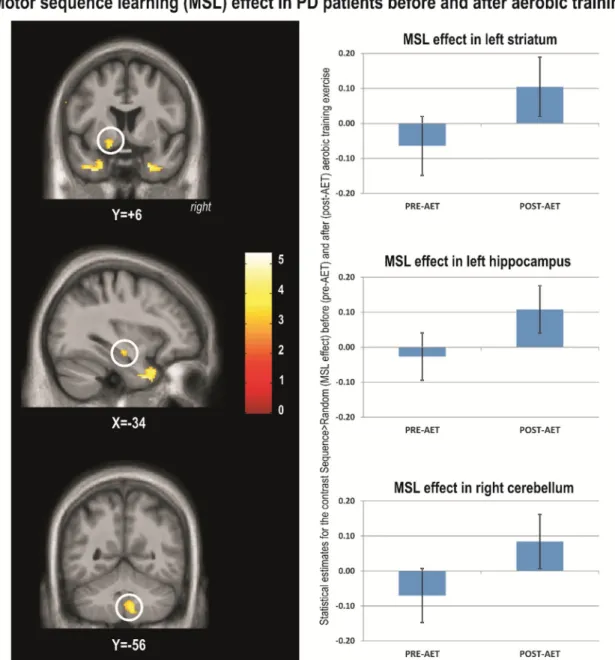
3.2.1. Functional brain changes related to motor learning capacity following AET in PD
To investigate the effects of AET on the neural correlates specific to motor sequence learning, we contrasted the main sequence learning ef-fect [Sequence– Random] by session [POST – PRE] within PD group. The results of this analysis revealed significant training-related changes for PD's individuals. Specifically, brain responses reflecting motor sequence learning capacity increased significantly post-AET in the temporal lobes, left ventral striatum, left hippocampus, cerebellar lobules 8 and 9 bilat-erally and right crus (Table 2,Fig. 1).
To test whether pre-post functional brain changes expressing motor learning capacity were indeed related to improvement in aerobic fit-ness, we conducted a regression analysis between changes in estimated VO2max and changes in MSL brain responses ([Sequence –
Random] × session [POST– PRE]). In PD patients, increases in VO2max
(indicating improved aerobicfitness as a result of training) correlated positively and significantly with increases activity in the hippocampus
bilaterally (Table 2.Fig. 2) and the left dorsal striatum. This indicated that as the aerobic capacity improved in PD patients, the motor learning specific activation also increased in these brain regions. In contrast, a significant negative correlation was observed in bilateral cerebellum lobe 7, 8 and 9 (Table 2,Fig. 2).
While the regions corresponding to the main sequence learning ef-fect and those resulting from the regression analysis were found in sim-ilar macro-structures (i.e. hippocampus, striatum), they did not spatially overlap.
3.2.2. Functional brain changes related to motor learning capacity following AET in HC
The main learning effect [Sequence– Random] by session [POST – PRE] assessed within the HC group revealed no significant change in ac-tivity between sessions. The regression analysis [Sequence – Random] × session [POST– PRE] also revealed no significant correlation between AET-related changes in aerobic capacity and brain changes re-lated to motor learning capacity for HC group alone.
3.2.3. Between group differences in functional brain activity related to mo-tor learning capacity following AET
When comparing the two groups (PD vs. HC) on the main learning effect [Sequence– Random] by session [POST – PRE], PD patients showed significantly higher levels of motor learning related activation
than HC, post-AET as compared to pre-AET, in the left cerebellum (lob-ules 8 and 9), right globus pallidus, and left ventral striatum (Table 3,
Fig. 3). The left hippocampus was marginally significant (0.06) in this contrast. However, no significant differences were found in the opposite direction (i.e., HC greater than PD) for the same contrast.
3.2.4. Functional brain changes following AET in PD and HC regressed against changes in aerobicfitness
Between-group differences in regression between VO2maxchanges
and MSL-related brain activity changes were observed in the right hip-pocampus (Table 3,Fig. 4) and in left cerebellum lobules 8, 9 and 7 (neg-ative correlation). These between-group effects were both driven by the findings in the PD group. Again, there was no spatial overlap between the regions obtained from the regression analysis (Section 3.2.4) and those obtained from the group contrast (Section 3.2.3), albeit they were found in the same macro-structures (i.e. hippocampus, cerebel-lum lobule 8 and 9). Also, no significant differences were found in the opposite direction (i.e., HC greater than PD).
4. Discussion
We recently reported that 12 weeks of progressive intense AET re-sulted in significant improvement in aerobic fitness as well as motor skill learning in both PD and HC groups (Duchesne et al., 2015). AET-re-lated MSL improvements were global in HC individuals as they were ob-served in both conditions (sequence and random), but were specific to the sequence condition in PD patients. In addition, PD patients im-proved their sequence-specific motor learning capacity after aerobic training as indicated by the significant difference between sequence and random conditions after the training period only. Interestingly, in the present study, the neuroimaging results corroborate those behav-iouralfindings and suggest that AET-related changes in motor sequence learning capacity (i.e., difference between sequence and random condi-tions) are mainly seen in PD individuals. Specifically, increases in MSL-specific brain activity due to training were found in the temporal lobe,
Table 3
Functional imaging results of the main sequence learning effect following aerobic exercise training between PD and HC.
Area X mm Y mm Z mm K Z psvc
3.1. Main effect of session on sequence learning [POST– PRE] × [Sequence – Random]
PD-HC
Left cerebellum lobules 8 and 9 −2 −62 −40 95 3,5 0,007 −20 −48 −54 181 3,41 0,009 −24 −48 −44 169 3,3 0,013 Right globus pallidus 14 2 −10 11 3,15 0,019 Left striatum ventral −18 6 −12 15 3,1 0,022 Left hippocampus −36 −16 −14 5 2,68 0,06a
HC-PD
No significant responses
3.2. Main effect of session on sequence learning [POST– PRE] × [Sequence – Random] regressed against changes in VO2max
PD-HC Positive
Right hippocampus 20 −28 −6 38 3,15 0,019 Negative
Cerebellum lobule 7 −2 −76 −24 95 3,48 0,007 Left cerebellum lobules 8 and 9 0 −64 −32 2,85 0,041 HC-PD
Positive
No significant responses Negative
No significant responses
Statistical inferences were performed at a threshold of pb 0.05 after correction for multi-ple comparisons over small spherical volumes (SVC). K represents the number of voxels in each cluster reported (determined at a threshold of p = 0.005).
a
Marginally significant.
hippocampus, striatum and cerebellum in the PD group. Importantly, there was also a positive relation between the patients' change in aero-bicfitness and MSL-related changes in the hippocampal and striatal ac-tivity, while we found a negative relation between the change infitness level and activity in the cerebellum in PD patients. Most importantly, the effects at the cerebral level are larger in the PD than in the HC group. 4.1.1.1. MSL exercise-dependent plasticity related to the striatum and hip-pocampus in PD individuals
The current study is thefirst to examine the changes in neural sub-strate supporting MSL following aerobic exercise training in both healthy controls and PD patients. Ourfindings thus extend the neuroim-aging literature on implicit MSL in PD individuals and HC participants (Gamble et al., 2014; Ruitenberg et al., 2015; Siegert et al., 2006), by spe-cifically highlighting the positive effects of AET on the MSL-related ac-tivity in the striatum, cerebellum and; all structures that are typically involved in motor learning (Albouy et al., 2013; Doyon et al., 2009, 2011). In fact, our hypothesis regarding changes in MSL specific to striatal changes following AET was confirmed in PD individuals. This finding is consistent with previous studies, which have revealed AET-dependent neuroplasticity in the central nervous system due to neuro-chemical changes in the striatum, the latter having been proposed as one possible mechanism of action to explain the gains in performance on motor tasks in PD (Petzinger et al., 2010, 2011, 2013, 2015). Indeed,
research in animal models of PD using dopaminergic neurotoxins has shown that behavioural motor ameliorations following physical exer-cise are associated with increased efficiency in dopaminergic and gluta-matergic neurotransmission (Fisher et al., 2013; Paillard et al., 2015; Petzinger et al., 2013, 2010, 2015), which together are thought to reduce the cortically-driven hyper-excitability observed in PD patients. These authors have reported an increase in dopamine availability coupled with a greater expression of dopamine D2 receptors, hence producing a better dopaminergic signaling in the striatum overall. Furthermore, physical exercise has been found to diminish the amount of synaptic glutamate release, which is known to decrease the level of cortical excit-ability (Fisher et al., 2004). Although conjectural, it is thus probable that the AET-specific changes in striatal activity observed in the current study may reflect this type of neurochemical mechanism, which would in turn explain the improvement in motor learning.
Interestingly, our results revealed that AET-related and MSL-specific functional brain changes are not only observed in the striatum, but within the hippocampus and cerebellum as well. Thesefindings could suggest that such brain structures are part of the functional network compensating for the typical dysfunction within the cortico-striatal cir-cuits seen in PD. In fact, the reported positive relationship between changes in aerobicfitness and AET-related MSL brain changes in the same macro-structures (hippocampus and striatum) is consistent with emerging data suggesting that links between the hippocampus and the dopaminergic systems in PD are important in memory and learning
(Calabresi et al., 2013). In their review,Calabresi et al. (2013)suggested that, while dopamine-dependent impairment (involving a complex mo-lecular dysfunction at glutamatergic synapses) of hippocampal long-term potentiation (LTP) might contribute to cognitive impairments in PD, such interactions could also be a potential source of symptoms re-duction in PD patients. Clinical interventions such as AET, for example, could potentially stimulate neurogenesis in the dysfunctional hippo-campus of PD's individuals, as it has been shown in rodent's studies (Voss, Vivar, Kramer, and van Praag, 2013). This type of interventions could also be responsible for the associated involvement of neurotroph-ic factors, as a greater expression in brain-derived neurotrophneurotroph-ic factor (BDNF) and LTP mechanisms following exercise has also been proposed as a putative mechanism responsible for the neuroprotective effects and functional improvements in this clinical population (Audiffren, André, and Albinet, 2011; Dishman et al., 2006; Fabel and Kempermann, 2008; Gomez-Pinilla, Zhuang, Feng, Ying, and Fan, 2011; Mustroph et al., 2012; Voss, Vivar, Kramer, and van Praag, 2013). Based on evidence that exercise-dependent brain changes within the hippocampus have been associated with functional changes in memory (Erickson et al., 2011; Szabo et al., 2011), we can thus conjecture that the improvement observed during MSL in PD following AET could be explained by LTP-like processes, due to greater expression of BDNF in the hippocampus.
In light of these recentfindings (Calabresi et al., 2013), it can be hy-pothesized that the beneficial effect that AET has on MSL-related activ-ity in both of these regions, may be based on the interaction or functional connectivity between them. It is possible that plastic AET-and MSL-related changes mayfirst occur within the hippocampus, and then propagate in basal ganglia via interactions between the MTL and dopaminergic system. This assumption is based on the fact that while MSL-related changes were observed in both hippocampus and striatum in PD individuals, only in the hippocampus we found regions in which MSL-related changes in activity remained positively correlated with im-provements in aerobicfitness when PD and HC participants were com-pared. Yet further functional connectivity investigations are still necessary to elucidate such AET-related brain reorganisation.
4.1.1.2. MSL exercise-dependent plasticity related to the cerebellum in PD individuals
In the current study, we found that AET-related and MSL specific changes in the cerebellum correlated negatively with changes in aerobic capacity. Although apparently contradictory, this result can be interpreted in light of several lines of evidence from previous studies: First, changes in the cerebellum have been interpreted as a compensato-ry functional system in animal models of PD model following exercise (Holschneider, Yang, Guo, and Maarek, 2007; Wang, Guo, Myers, Heintz and Holschneider, 2015a; Wang, Guo, Myers, Heintz, Peng, Maarek and Holschneider, 2015b), and a similar assumption has been proposed following striatal dysfunction in PD (Doyon, 2008). Second, human studies have indicated that both the cortico-striatal (CS) and cortico-cerebellar (CC) systems play distinctive roles in MSL (Doyon, 2008; Doyon and Benali, 2005; Doyon et al., 2003; Leggio and Molinari, 2015), despite the fact that cerebellum and basal ganglia are known to be interconnected (Bostan and Strick, 2010; Hoshi, Tremblay, Feger, Carras, and Strick, 2005). Given the interaction be-tween the CC and CS systems during MSL (Doyon, 2008; Doyon et al., 2009; Doyon and Benali, 2005) and the fact that previous functional im-aging data revealed that cerebellar hemispheres are hyperactivated, while the striatum is hypoactivated in PD patients as compared to healthy controls (Yu, Sternad, Corcos, and Vaillancourt, 2007), it is thus possible that the cerebellum is capable of compensating for the de-ficient basal ganglia activity observed in PD. Hence, in the current study, the PD participants who improved less their aerobic capacity used pre-dominantly the cerebellum during MSL, whereas those who had higher changes in aerobicfitness used more the hippocampus and striatum,
indicating a‘restoration’ of functionality in the network typically seen in MSL.
According to the motor sequence learning literature, both the cere-bellum and striatum are known to be involved in the early MSL phase, while only the striatum has been shown to maintain its activity in later learning stages when the memory trace has been consolidated (Doyon et al., 2009, 2011, 2002). Therefore, while modest improve-ments in aerobic capacity linked with increases in cerebellar activity may reflect learning in the early stage tested here, the fact that large im-provements in aerobicfitness were associated with an enhanced hippo-campal and striatal functioning may reflect a transition towards a more durable stage of MSL. These MSLfindings are thus in line with previous structural and physiological investigations, which parallel the functional changes that aerobic exercise can exert in general (e.g., (Hillman, Erickson, and Kramer, 2008) for a review), and in PD, in particular (Ahlskog, 2011; Alberts, Linder, Penko, Lowe and Phillips, 2011; Goodwin, Richards, Taylor, Taylor, and Campbell, 2008; Hirsch and Farley, 2009; Speelman et al., 2011) for reviews).
4.1.1.3. MSL and exercise in HC individuals
Compared to PD individuals, AET did not have a statistically signi fi-cant effect on changes in the neural correlates of MSL in the HC group. Such divergence between the HC and PD groups could be explained by a potential ceiling effect observed at baseline for the HC group in regards to their MSL capacity at both the behavioural and cerebral level. Given that HC individuals already showed a good MSL capacity prior to AET, they obviously had less room for sequence-specific behav-ioural improvement compared to PD patients. This is supported by the fact that analysis of the behavioural results revealed that, while HC indi-viduals improved their motor performance in both random and se-quence condition after AET, they also maintained a similar magnitude in RT difference between these two conditions (Duchesne et al., 2015). This pattern of results can be interpreted as HC individuals having a more efficient medial temporal lobe, CS, CC functioning networks prior to AET, as compared to PD patients, which leaves also less room for improvements to be seen in functional activity in these brain areas. 5. Limitations and conclusions
One of the limitations in our study was the heterogeneity of the pa-tient population. Similar to many other studies with PD papa-tients, disease characteristics were diverse (e.g., motor, cognitive, neuropsychiatric, etc.). In fact, it was often a challenge to control for all symptoms and ob-tain a homogeneous sample. However, we used several strategies to ad-dress this limitation. First, we matched the PD group with the HC group with respect to sex distribution, age, years of education, cognitive and fitness level. In addition, given that the two groups differed in their de-pression and anxiety symptoms, we accounted for these variables in our statistical analyses by considering them as covariates in the analyses of the behavioural data. Another limitation of our study was the lack of a PD control group for the type of exercise. Despite this constraint, how-ever, it is worth noting the fact that we conducted a regression analysis highlighting that there were functional brain changes that were related to changes in aerobicfitness level, which indicate that there were brain changes specific to the AET program in PD patients.
To conclude, this study is thefirst to assess the neural correlates of MSL following AET in PD. Functional reorganisation of brain activity in early PD following aerobic training was observed within the hippocam-pus, the striatum and the cerebellum, resulting in improvement of their learning capacity. Most importantly, in the same brain structures, albeit in different sub-regions, we have also showed that MSL specific brain plasticity correlated with changes in aerobicfitness, showing a positive relationship within the hippocampus and the striatum and a negative relationship within the cerebellum. Altogether our study makes two im-portant contributions to thefield. First, we show that a 12-week
progressive aerobic training regimen has beneficial effects on motor skill learning in sedentary PD and healthy individuals. Second, we high-light the neurophysiological changes underlying exercise-dependent plasticity in PD, with particular emphasis on the neuronal substrate of MSL. Our results thus pave the way for other studies to further explore the structural and functional organization within brain regions and how exercise-dependent plasticity manifests itself within the medial tempo-ral lobe, CS and CC systems following chronic or acute bouts of aerobic activities in PD. As a result, thesefindings have important clinical impli-cations for the rehabilitation of PD individuals, as the design and the use of non-pharmacological interventions based on physical exercise can be implemented to improve motor learning capacity and restore motor functions in patients afflicted with this debilitating disease.
Acknowledgments
1. This work was supported by the Fonds de Recherche du Québec -Santé under Grant 26409.
2. The authors wish to thank Dr. Juan Manuel Villalpando and Dr. Thien Tuong Minh Vu who kindly accepted to supervise physical assess-ment during testing.
Appendix A. Supplementary data
Supplementary data to this article can be found online athttp://dx. doi.org/10.1016/j.nicl.2016.09.011.
References
Ahlskog, J.E., 2011. Does vigorous exercise have a neuroprotective effect in Parkinson disease? Neurology 77 (3), 288–294.http://dx.doi.org/10.1212/ WNL.0b013e318225ab66.
Alberts, J.L., Linder, S.M., Penko, A.L., Lowe, M.J., Phillips, M., 2011. It is not about the bike, it is about the pedaling: forced exercise and Parkinson's disease. Exerc. Sport Sci. Rev. 39 (4), 177–186.http://dx.doi.org/10.1097/JES.0b013e31822cc71a.
Albouy, G., King, B.R., Maquet, P., Doyon, J., 2013. Hippocampus and striatum: dynamics and interaction during acquisition and sleep-related motor sequence memory con-solidation. Hippocampus 23 (11), 985–1004.http://dx.doi.org/10.1002/hipo.22183. Albouy, G., Fogel, S., King, B.R., Laventure, S., Benali, H., Karni, A., ... Doyon, J., 2015.
Main-taining vs. enhancing motor sequence memories: respective roles of striatal and hip-pocampal systems. NeuroImage 108, 423–434. http://dx.doi.org/10.1016/j. neuroimage.2014.12.049.
Amano, S., Roemmich, R.T., Skinner, J.W., Hass, C.J., 2013. Ambulation and Parkinson dis-ease. Phys. Med. Rehabil. Clin. N. Am. 24 (2), 371–392.http://dx.doi.org/10.1016/j. pmr.2012.11.003.
American College of Sport Medicine, 2006.ACSM's Guidelines for Exercise Testing and Prescription. 6th ed. Lippincott Williams & Wilkins, Lea & Febiger, USA, Philadelphia.
Audiffren, M., André, N., Albinet, C., 2011. Effets positifs de l'exercice physique chronique sur les fonctions cognitives des seniors: bilan et perspectives. Revue de neuropsychologie neurosciences cognitives et cliniques 3 (4), 207–225.http://dx. doi.org/10.3917/rne.034.0207.
Beall, E.B., Lowe, M.J., Alberts, J.L., Frankemolle, A.M.M., Thota, A.K., Shah, C., Phillips, M.D., 2013. The effect of forced-exercise therapy for Parkinson's disease on motor cortex functional connectivity. Brain Connectivity 3 (2), 190–198.http://dx.doi.org/10. 1089/brain.2012.0104.
Beck, A.T., Beamesderfer, A., 1974. Assessment of depression: the depression inventory. Mod. Probl. Pharmacopsychiatry 7 (0), 151–169http://www.ncbi.nlm.nih.gov/ pubmed/4412100.
Borg, G., 1982. Psychophysical bases of perceived exertion. Med. Sci. Sports Exerc. 14, 377–381http://www.ncbi.nlm.nih.gov/pubmed/7154893.
Bostan, A.C., Strick, P.L., 2010.The cerebellum and basal ganglia are interconnected. Neuropsychol. Rev. 20 (3), 261–270.
Calabresi, P., Castrioto, A., Di Filippo, M., Picconi, B., 2013. New experimental and clinical links between the hippocampus and the dopaminergic system in Parkinson's disease. Lancet Neurol. 12 (8), 811–821.http://dx.doi.org/10.1016/S1474-4422(13)70118-2. Carbon, M., Reetz, K., Ghilardi, M.F., Dhawan, V., Eidelberg, D., 2010.Early Parkinson's dis-ease: longitudinal changes in brain activity during sequence learning. Neurobiol. Dis. 37 (2), 455–460.
Clark, G.M., Lum, J.A.G., Ullman, M.T., 2014.A meta-analysis and meta-regression of serial reaction time tast performance in Parkinson's disease. Neuropsychology.
Dishman, R.K., Berthoud, H.R., Booth, F.W., Cotman, C.W., Edgerton, V.R., Fleshner, M.R., ... Zigmond, M.J., 2006.Neurobiology of exercise. Obesity (Silver Spring) 14 (3), 345–356.
Doyon, J., 2008.Motor sequence learning and movement disorders. Curr. Opin. Neurol. 21 (4), 478–483.
Doyon, J., Benali, H., 2005. Reorganization and plasticity in the adult brain during learning of motor skills. Curr. Opin. Neurobiol. 15 (2), 161–167.http://dx.doi.org/10.1016/j. conb.2005.03.004.
Doyon, J., Song, A.W., Karni, A., Lalonde, F., Adams, M.M., Ungerleider, L.G., 2002. Experi-ence-dependent changes in cerebellar contributions to motor sequence learning. Proc. Natl. Acad. Sci. U. S. A. 99 (2), 1017–1022.http://dx.doi.org/10.1073/pnas. 022615199.
Doyon, J., Penhune, V., Ungerleider, L.G., 2003.Distinct contribution of the cortico-striatal and cortico-cerebellar systems to motor skill learning. Neuropsychologia 41 (3), 252–262.
Doyon, J., Bellec, P., Amsel, R., Penhune, V., Monchi, O., Carrier, J., ... Benali, H., 2009. Contribu-tions of the basal ganglia and functionally related brain structures to motor learning. Behav. Brain Res. 199 (1), 61–75.http://dx.doi.org/10.1016/j.bbr.2008.11.012. Doyon, J., Orban, P., Barakat, M., Debas, K., Lungu, O., Albouy, G., ... Benali, H., 2011.
Func-tional brain plasticity associated with motor learning. Med. Sci. (Paris) 27 (4), 413–420.
Duchesne, C., Lungu, O., Nadeau, A., Robillard, M.E., Bore, A., Bobeuf, F., ... Doyon, J., 2015. Enhancing both motor and cognitive functioning in Parkinson's disease: aerobic exer-cise as a rehabilitative intervention. Brain Cogn. 99, 68–77.http://dx.doi.org/10.1016/ j.bandc.2015.07.005.
Erickson, K.I., Voss, M.W., Prakash, R.S., Basak, C., Szabo, A., Chaddock, L., ... Kramer, A.F., 2011. Exercise training increases size of hippocampus and improves memory. Proc. Natl. Acad. Sci. U. S. A. 108 (7), 3017–3022.http://dx.doi.org/10.1073/pnas. 1015950108.
Fabel, K., Kempermann, G., 2008.Physical activity and the regulation of neurogenesis in the adult and aging brain. Neruomol. Med. 10 (2), 59–66.
Fisher, B.E., Petzinger, G.M., Nixon, K., Hogg, E., Bremmer, S., Meshul, C.K., Jakowec, M.W., 2004.Exercise-induced behavioral recovery and neuroplasticity in the 1-methyl-4-phenyl-1,2,3,6-tetrahydropyridine-lesioned mouse basal ganglia. J. Neurosci. Res. 77 (3), 378–390.
Fisher, B.E., Li, Q., Nacca, A., Salem, G.J., Song, J., Yip, J., ... Petzinger, G.M., 2013. Treadmill exercise elevates striatal dopamine D2 receptor binding potential in patients with early Parkinson's disease. NeuroReport 24 (10), 509–514.http://dx.doi.org/10.1097/ WNR.0b013e328361dc13.
Gamble, K.R., Cummings Jr., T.J., Lo, S.E., Ghosh, P.T., Howard Jr., J.H., Howard, D.V., 2014. Implicit sequence learning in people with Parkinson's disease. Front. Hum. Neurosci. 8, 563.http://dx.doi.org/10.3389/fnhum.2014.00563.
Goetz, C.G., Tilley, B.C., Shaftman, S.R., Stebbins, G.T., Fahn, S., Martinez-Martin, P., ... LaPelle, N., 2008. Movement Disorder Society-sponsored revision of the Unified Parkinson's Disease Rating Scale (MDS-UPDRS): scale presentation and clinimetric testing results. Mov. Disord. 23 (15), 2129–2170.http://dx.doi.org/10.1002/mds. 22340.
Gomez-Pinilla, F., Zhuang, Y., Feng, J., Ying, Z., Fan, G., 2011.Exercise impacts brain-de-rived neurotrophic factor plasticity by engaging mechanisms of epigenetic regulation. Eur. J. Neurosci. 33 (3), 383–390.
Goodwin, V.A., Richards, S.H., Taylor, R.S., Taylor, A.H., Campbell, J.L., 2008. The effective-ness of exercise interventions for people with Parkinson's disease: a systematic re-view and meta-analysis. Mov. Disord. 23 (5), 631–640.http://dx.doi.org/10.1002/ mds.21922.
Herman, T., Giladi, N., Gruendlinger, L., Hausdorff, J.M., 2007. Six weeks of intensive tread-mill training improves gait and quality of life in patients with Parkinson's disease: a pilot study. Arch. Phys. Med. Rehabil. 88 (9), 1154–1158.http://dx.doi.org/10.1016/ j.apmr.2007.05.015.
Hillman, C.H., Erickson, K.I., Kramer, A.F., 2008.Be smart, exercise your heart: exercise ef-fects on brain and cognition. Nat. Rev. Neurosci. 9 (1), 58–65.
Hirsch, M.A., Farley, B.G., 2009.Exercise and neuroplasticity in persons living with Parkinson's disease. Eur. J. Phys. Rehabil. Med. 45 (2), 215–229.
Hoehn, M.M., Yahr, M.D., 1967.Parkinsonism: onset, progression and mortality. Neurolo-gy 17 (5), 427–442.
Holschneider, D.P., Yang, J., Guo, Y., Maarek, J.M., 2007. Reorganization of functional brain maps after exercise training: importance of cerebellar-thalamic-cortical pathway. Brain Res. 1184, 96–107.http://dx.doi.org/10.1016/j.brainres.2007.09.081. Hoshi, E., Tremblay, L., Feger, J., Carras, P.L., Strick, P.L., 2005. The cerebellum
communi-cates with the basal ganglia. Nat. Neurosci. 8 (11), 1491–1493http://www.nature. com/neuro/journal/v8/n11/suppinfo/nn1544_S1.html.
Jackson, A.S., Blair, S.N., Mahar, M.T., Wier, L.T., Ross, R.M., Stuteville, J.E., 1990. Prediction of functional aerobic capacity without exercise testing. Med. Sci. Sports Exerc. 22 (6), 863–870http://www.ncbi.nlm.nih.gov/pubmed/2287267.
Leggio, M., Molinari, M., 2015. Cerebellar sequencing: a trick for predicting the future. Cer-ebellum 14 (1), 35–38.http://dx.doi.org/10.1007/s12311-014-0616-x.
Mang, C.S., Snow, N.J., Campbell, K.L., Ross, C.J., Boyd, L.A., 2014. A single bout of high-in-tensity aerobic exercise facilitates response to paired associative stimulation and pro-motes sequence-specific implicit motor learning. J. Appl. Physiol. (1985) 117 (11), 1325–1336.http://dx.doi.org/10.1152/japplphysiol.00498.2014.
Mustroph, M.L., Chen, S., Desai, S.C., Cay, E.B., DeYoung, E.K., Rhodes, J.S., 2012. Aerobic ex-ercise is the critical variable in an enriched environment that increases hippocampal neurogenesis and water maze learning in male C57BL/6J mice. Neuroscience 219, 62–71.http://dx.doi.org/10.1016/j.neuroscience.2012.06.007.
Nadeau, A., Pourcher, E., Corbeil, P., 2014. Effects of 24 wk of treadmill training on gait performance in Parkinson's disease. Med. Sci. Sports Exerc. 46 (4), 645–655.http:// dx.doi.org/10.1249/MSS.0000000000000144.
Nazem, S., Siderowf, A.D., Duda, J.E., Ten Have, T., Colcher, A., Horn, S.S., ... Weintraub, D., 2009. Montreal cognitive assessment performance in patients with Parkinson's dis-ease with“normal” global cognition according to mini-mental state examination score. J. Am. Geriatr. Soc. 57 (2), 304–308.http://dx.doi.org/10.1111/j.1532-5415. 2008.02096.x.
Nissen, Bullemer, 1987.Attentional requirements of learning: evidence from performance measures. Cogn. Psychol. 19 (1), 32.
Osman, A., Kopper, B.A., Barrios, F.X., Osman, J.R., Wade, T., 1997. The Beck Anxiety Inven-tory: reexamination of factor structure and psychometric properties. J. Clin. Psychol. 53 (1), 7–14. http://dx.doi.org/10.1002/(SICI)1097-4679(199701)53:1b7::AID-JCLP2N3.0.CO;2-S.
Paillard, T., Rolland, Y., de Souto Barreto, P., 2015.Protective effects of physical exercise in Alzheimer's disease and Parkinson's disease: a narrative review. J. Clin. Neurophysiol. 11 (3), 212–219.
Petzinger, G.M., Walsh, J.P., Akopian, G., Hogg, E., Abernathy, A., Arevalo, P., ... Jakowec, M.W., 2007.Effects of treadmill exercise on dopaminergic transmission in the 1-methyl-4-phenyl-1,2,3,6-tetrahydropyridine-lesioned mouse model of basal ganglia injury. J. Neurosci. 27 (20), 5291–5300.
Petzinger, G.M., Fisher, B.E., Van Leeuwen, J.E., Vukovic, M., Akopian, G., Meshul, C.K., ... Jakowec, M.W., 2010. Enhancing neuroplasticity in the basal ganglia: the role of exer-cise in Parkinson's disease. Mov. Disord. 25 (Suppl. 1), S141–S145.http://dx.doi.org/ 10.1002/mds.22782.
Petzinger, G.M., Fisher, B.E., Akopian, G., Holschneider, D.P., Wood, R., Walsh, J.P., ... Jakowec, M.W., 2011.The role of exercise in facilitating basal ganglia function in Parkinson's disease. Neurodegener. Dis. Manag. 1 (2), 157–170.
Petzinger, G.M., Fisher, B.E., McEwen, S., Beeler, J.A., Walsh, J.P., Jakowec, M.W., 2013. Ex-ercise-enhanced neuroplasticity targeting motor and cognitive circuitry in Parkinson's disease. Lancet Neurol. 12 (7), 716–726.http://dx.doi.org/10.1016/ S1474-4422(13)70123-6(S1474-4422(13)70123-6 [pii] ).
Petzinger, G.M., Holschneider, D.P., Fisher, B.E., McEwen, S., Kintz, N., Halliday, M., ... Jakowec, M.W., 2015.The effects of exercise on dopamine neurotransmission in Parkinson's disease: targeting neuroplasticity to modulate basal ganglia circuitry. Brain Plast. 1 (1), 29–39.
Pothakos, K., Kurz, M.J., Lau, Y.S., 2009.Restorative effect of endurance exercise on behav-ioral deficits in the chronic mouse model of Parkinson's disease with severe neurode-generation. BMC Neurosci. 10, 6.
Ridgel, A.L., Vitek, J.L., Alberts, J.L., 2009. Forced, not voluntary, exercise improves motor function in Parkinson's disease patients. Neurorehabil. Neural Repair 23 (6), 600–608.http://dx.doi.org/10.1177/1545968308328726.
Ridgel, A.L., Kim, C.H., Fickes, E.J., Muller, M.D., Alberts, J.L., 2011. Changes in executive function after acute bouts of passive cycling in Parkinson's disease. J. Aging Phys. Act. 19 (2), 87–98http://www.ncbi.nlm.nih.gov/pubmed/21558565.
Ruitenberg, M.F., Duthoo, W., Santens, P., Notebaert, W., Abrahamse, E.L., 2015. Sequential movement skill in Parkinson's disease: a state-of-the-art. Cortex 65, 102–112.http:// dx.doi.org/10.1016/j.cortex.2015.01.005.
Schendan, H.E., Tinaz, S., Maher, S.M., Stern, C.E., 2013. Frontostriatal and mediotemporal lobe contributions to implicit higher-order spatial sequence learning declines in aging and Parkinson's disease. Behav. Neurosci. 127 (2), 204–221.http://dx.doi.org/ 10.1037/a0032012.
Siegert, R.J., Taylor, K.D., Weatherall, M., Abernethy, D.A., 2006.Is implicit sequence learn-ing impaired in Parkinson's disease? A meta-analysis. Neuropsychology 20 (4), 490–495.
Speelman, A.D., van de Warrenburg, B.P., van Nimwegen, M., Petzinger, G.M., Munneke, M., Bloem, B.R., 2011. How might physical activity benefit patients with Parkinson disease? Nat. Rev. Neurol. 7 (9), 528–534.http://dx.doi.org/10.1038/nrneurol.2011. 107.
Szabo, A.N., McAuley, E., Erickson, K.I., Voss, M., Prakash, R.S., Mailey, E.L., ... Kramer, A.F., 2011. Cardiorespiratoryfitness, hippocampal volume, and frequency of forgetting in older adults. Neuropsychology 25 (5), 545–553.http://dx.doi.org/10.1037/a0022733. van Eijkeren, F.J.M., Reijmers, R.S.J., Kleinveld, M.J., Minten, A., Bruggen, J.P., Bloem, B.R., 2008.Nordic walking improves mobility in Parkinson's disease. Mov. Disord. 23 (15), 2239–2243.
van Nimwegen, M., Speelman, A., Hofman-van Rossum, E., Overeem, S., Deeg, D., Borm, G., ... Munneke, M., 2011.Physical inactivity in Parkinson's disease. J. Neurol. 1–8.
Voss, M.W., Nagamatsu, L.S., Liu-Ambrose, T., Kramer, A.F., 2011. Exercise, brain, and cog-nition across the life span. J. Appl. Physiol. 111 (5), 1505–1513.http://dx.doi.org/10. 1152/japplphysiol.00210.2011.
Voss, M.W., Vivar, C., Kramer, A.F., van Praag, H., 2013. Bridging animal and human models of exercise-induced brain plasticity. Trends Cogn. Sci. 17 (10), 525–544.
http://dx.doi.org/10.1016/j.tics.2013.08.001.
Wang, Z., Guo, Y., Myers, K.G., Heintz, R., Holschneider, D.P., 2015a. Recruitment of the prefrontal cortex and cerebellum in Parkinsonian rats following skilled aerobic exer-cise. Neurobiol. Dis. 77 (0), 71–87.http://dx.doi.org/10.1016/j.nbd.2015.02.020. Wang, Z., Guo, Y., Myers, K.G., Heintz, R., Peng, Y.-H., Maarek, J.-M.I., Holschneider, D.P.,
2015b. Exercise alters resting-state functional connectivity of motor circuits in par-kinsonian rats. Neurobiol. Aging 36 (1), 536–544.http://dx.doi.org/10.1016/j. neurobiolaging.2014.08.016.
Warburton, D.E., Jamnik, V.K., Bredin, S.D., Gledhill, N., 2011.The Physical Activity Readi-ness Questionnaire for Everyone (PAR-Q+): French North America Version (Ques-tionnaire sur l'aptitude à l'activité physique pour tous (Q-AAP+)). Health Fit. J. Canada 4 (2), 1–4.
Werheid, K., Zysset, S., Müller, A., Reuter, M., von Cramon, D.Y., 2003. Rule learning in a serial reaction time task: an fMRI study on patients with early Parkinson's disease. Cogn. Brain Res. 16 (2), 273–284.http://dx.doi.org/10.1016/s0926-6410(02)00283-5. Yoon, M.C., Shin, M.S., Kim, T.S., Kim, B.K., Ko, I.G., Sung, Y.H., ... Kim, C.J., 2007.Treadmill exercise suppresses nigrostriatal dopaminergic neuronal loss in 6-hydroxydopamine-induced Parkinson's rats. Neurosci. Lett. 423 (1), 12–17.
Yu, H., Sternad, D., Corcos, D.M., Vaillancourt, D.E., 2007. Role of hyperactive cerebellum and motor cortex in Parkinson's disease. NeuroImage 35 (1), 222–233.http://dx. doi.org/10.1016/j.neuroimage.2006.11.047(S1053-8119(06)01175-X [pii]).