HAL Id: hal-01274236
https://hal-univ-rennes1.archives-ouvertes.fr/hal-01274236
Submitted on 22 Mar 2016HAL is a multi-disciplinary open access archive for the deposit and dissemination of sci-entific research documents, whether they are pub-lished or not. The documents may come from teaching and research institutions in France or abroad, or from public or private research centers.
L’archive ouverte pluridisciplinaire HAL, est destinée au dépôt et à la diffusion de documents scientifiques de niveau recherche, publiés ou non, émanant des établissements d’enseignement et de recherche français ou étrangers, des laboratoires publics ou privés.
New in vitro and in vivo models to evaluate antibiotic
efficacy in Staphylococcus aureus prosthetic vascular
graft infection
Matthieu Revest, C. Jacqueline, R. Boudjemaa, J. Caillon, V. Le Mabecque,
A. Breteche, K. Steenkeste, Pierre Tattevin, G. Potel, Christian Michelet, et
al.
To cite this version:
Matthieu Revest, C. Jacqueline, R. Boudjemaa, J. Caillon, V. Le Mabecque, et al.. New in vitro and in vivo models to evaluate antibiotic efficacy in Staphylococcus aureus prosthetic vascular graft infection. Journal of Antimicrobial Chemotherapy, Oxford University Press (OUP), 2016, 71 (5), pp.1291-1299. �10.1093/jac/dkv496�. �hal-01274236�
Title page
New in vitro and in vivo Models to evaluate Antibiotic Efficacy in Staphylococcus aureus Prosthetic Vascular Graft Infection
M. Revest1,2,3, C. Jacqueline1, R. Boudjemaa4, J. Caillon1, V. Le Mabecque1, A. Breteche1, K.
Steenkeste4, P. Tattevin2,3, G. Potel1, C. Michelet2,3, MP. Fontaine-Aupart4, D. Boutoille1,5
1 Université Nantes, Faculté Médecine EA3826 Nantes, France
2 CHU Rennes Infectious Diseases and Intensive Care Unit, Pontchaillou Hospital, 35033
Rennes Cedex, France
3 CIC Inserm 1414, Rennes 1 University, Pontchaillou Hospital, 35033 Rennes Cedex, France 4 Institut des Sciences Moléculaires Orsay, CNRS, Université Paris-Sud, 91405 Orsay, France 5 CHU Nantes, Infectious Diseases Unit, Hôtel Dieu, Nantes, France
Corresponding author: Dr Matthieu Revest,
CHU Rennes Infectious Diseases and Intensive Care Unit, Pontchaillou Hospital, 35033 Rennes Cedex, France
Phone: +33 2 99 28 37 98 Fax: +33 2 99 28 94 64
matthieu.revest@chu-rennes.fr
Running title: evaluation of different antibiotics for vascular prosthesis infections treatment Key words: Staphylococcus aureus, animal models, biofilm, surgical infection, vascular prosthesis infections
Abstract:
Objective. Prosthetic vascular graft infection (PVGI) is an emerging disease, mostly due to
staphylococci, with limited data regarding efficacy of current antistaphylococcal agents. We aimed to assess the efficacy of different antibiotic regimens. Methods. Six different strains of methicillin-susceptible (MSSA) and methicillin-resistant S. aureus (MRSA) were used. We compared results of minimal biofilm inhibitory and eradicating concentrations (MBICs and MBECs) obtained with a Calgary Biofilm Pin lid Device (CBPD) to those yielded by an original Dacron®-related minimal inhibitory and eradicating concentrations measure model. We then used an original murine model of Staphylococcus aureus vascular material infection to evaluate efficacy of different antibiotic regimens: vancomycin and daptomycin combined or not with rifampicin for MRSA and same groups with cloxacillin and cloxacillin associated with rifampicin for MSSA. Results. We demonstrated that classical measures of MBICs and MBECs with CPBD could overestimate the decrease of antibiotic susceptibility in material-related infections and that the nature of the support used might influence the measure of biofilm susceptibility since results yielded by our Dacron®-related minimal eradicating assay were lower than those found on a plastic device. In our in vivo model, we showed that daptomycin was significantly more bactericidal than comparators for some strains of MRSA or MSSA but not for all. For the majority of strains, it was as efficient as comparators. The addition of rifampicin to daptomycin did not enhance daptomycin efficacy. Conclusions. Despite the heterogeneity of results according to bacterial strains, these innovative models represent an option to better evaluate in vitro efficacy of antibiotics on Dacron®-related biofilm S. aureus infections, and to screen different antibiotic regimens in a mouse-model of PVGIs.
New in vitro and in vivo Models to evaluate Antibiotic Efficacy in Staphylococcus aureus Prosthetic Vascular Graft Infection
M. Revest1,2, C. Jacqueline1, R. Boudjemaa3, J. Caillon1, V. Le Mabecque1, A. Breteche1, K.
Steenkeste3, P. Tattevin2, G. Potel1, C. Michelet2, MP. Fontaine-Aupart3, D. Boutoille1
1Université Nantes, Faculté Médecine EA3826 Nantes, 2CHU Rennes Infectious Diseases
Unit, 3 Institut des Sciences Moléculaires Orsay, Université Paris-Sud France
Introduction
More than 400 000 vascular grafts are inserted annually in the United States1 and approximately 50 000 in France.2 Prosthetic vascular graft infections (PVGIs) are among the most serious complications associated with these procedures,1 with 30 day- and one year-mortality rates of, respectively, 10–25%, and 50%.3 Staphylococcus aureus is the main
pathogen, and methicillin resistant S. aureus (MRSA) accounts for almost 50% of S. aureus PVGIs in North America.4 Clinical data regarding the optimal antibiotic therapy for these infections are scarce, in the absence of any comparative clinical trial dealing with the treatment of PVGIs.5 Hence, experimental data are needed to better identify the optimal antibiotic regimens for PVGIs.
Biofilm developed onto the vascular prosthesis plays a significant role in the difficulties encountered for treating PVGIs. Firstly, biofilm acts as a mechanical barrier against antibiotic penetration. Secondly, bacteria embedded in a mature biofilm enter into an altered metabolic state associated with a dramatic decrease of susceptibility to most antibiotics.6, 7 Many studies showed that minimal biofilm inhibitory concentrations (MBIC) and minimal biofilm eradicating concentrations (MBEC) of antibiotics are much higher than their respective minimum inhibitory concentration (MIC), and minimum bactericidal concentration (MBC), as measured on planktonic bacteria.8-10 However, most studies evaluated antibiotic efficacy on artificial materials not used in clinical practice, although the nature of the material may influence these results. In addition, although helpful, in vitro data may fail to capture what
happens in vivo. Animal models of PVGIs have mainly been used to evaluate techniques to prevent PVGIs,11-14 but no animal model assessing the efficacy of antibiotics on infection of the Dacron® vascular prosthesis used in clinical daily practice has been described so far. In this study, we investigated the activities of cloxacillin, vancomycin, daptomycin and rifampicin against different strains of methicillin-susceptible S. aureus (MSSA) and MRSA in vitro. Those activities were determined against planktonic bacteria (MIC and MBC), and against adherent bacteria (MBIC and MBEC). To evaluate whether the nature of the support influences results for MBIC and MBEC, two different techniques were used: a largely used modified version of the Calgary Biofilm Pin Lid Device (CBPD) and an original model of Dacron®-related biofilm. Then, cloxacillin, vancomycin, daptomycin whether or not combined with rifampicin, were evaluated in a new mouse vascular-material infection model, using the same strains of S. aureus.
Materials and methods
Bacterial strains
Six different strains of S. aureus were used for all experiments. For MSSA: two different clinical strains, thereafter named 171 and 176, isolated from patients with S. aureus bloodstream infections, and one strain from the American Type Culture Collection: ATCC 27217. For MRSA: two different strains, thereafter named BCB8 and 117, also isolated from blood cultures, and one ATCC strain: ATCC 33591. Bacteria were stored in a cryovial bead preservation system at -80°C.
Antimicrobial agents
Clinical forms of the following antibiotics were used: cloxacillin (Astellas Pharma, Levallois-Perret, France) stored in a 100 mg/mL stock-solution, vancomycin (Sandoz, Levallois-Levallois-Perret,
France) stored in a 50 mg/mL stock-solution, daptomycin (Novartis Pharma SAS, Rueil-Malmaison, France) stored in a 50 mg/mL stock-solution and rifampicin (Sanofi-Aventis, Paris, France) stored in a 60 mg/mL stock-solution. All the stock-solutions were prepared in sterile and pyrogen-free 0.9% saline except for rifampicin, which was prepared in sterile water, and stored at -80°C before utilization.
In vitro experiments
All the following experiments were performed at least in duplicate, with all the staphylococcal strains evaluated. Biofilm formation was compared between polystyrene (CPBD) and Dacron® using confocal microscopy.
MICs and MBCs
The MICs and the MBCs values for cloxacillin, vancomycin, daptomycin and rifampicin were determined by the broth macrodilution method in cation-adjusted Mueller-Hinton broth (CAMHB), according to the European Committee on Antimicrobial Susceptibility Testing (EUCAST). Media were supplemented with 50 mg/L Ca2+ for daptomycin.
MBICs and MBECs
A modified Calgary device was used as previously described.8, 15 Briefly, biofilm was formed
by immersing pegs of a modified microtiter lid into wells of a flat-bottom 96-well microtiter plate. Each well was filled with 150 μL of a 3 McFarland S. aureus broth medium solution. After a 24-h incubation at 37°C, pegs lids were rinsed 3 times in sterile water, placed onto flat-bottom microtiter plates containing antibiotic twofold dilutions in 150 μL of CAMHB per well, and incubated for 24 h at 37°C. The MBIC were defined as the minimal concentration of antibiotic inhibiting bacterial growth, as determined by reading turbidity of media at 650 nm. Pegs with no bacterial growth were again rinsed 3 times with sterile water and sonicated (Aquasonic sonicator, 35 kHz for 5 minutes) followed by vortexing for 30 seconds to remove
biofilm from the support. A new cover plate was added and this new device was cultured for 24 h at 37°C in CAMHB. The MBEC was defined as the minimal concentration of antibiotic where no bacterial growth was documented.
Dacron®-related minimal biofilm inhibitory concentrations (dMBICs) and the Dacron®
-related minimal biofilm eradicating concentrations (dMBECs)
Commercially available woven Dacron® grafts (CardialTM, Bard, Saint-Etienne, France) were cut into 1 cm x 1 cm squares and sterilized. To cover the biomaterial with proteins and thus to facilitate the bacterial graft, these Dacron® sheets were incubated at 37°C under sterile conditions with horse serum for 24 h. They were rinsed 3 times with sterile water to remove the horse serum and then incubated for 24 h at 37°C with 1 mL per patch of Mueller-Hinton broth (MHB) containing 108 colony forming units (CFU)/mL of S. aureus. Again, they were rinsed 3 times in sterile water to eliminate the planktonic bacteria, dried with a sterile gauze compress, and plunged into tubes filled with CAMHB containing none (control groups) or serial antibiotic dilutions, and incubated for 24 h at 37°C. dMBIC for each antibiotic and strain couple was defined as the antibiotic concentration of the first tube with no visible bacterial growth. Then, Dacron® patches with no bacterial growth were removed from their tubes, rinsed 3 times in sterile water, dried with a sterile gauze compress and plunged again in MHB. A sonication (35kHz for 5 minutes) was performed followed by vortexing for 30 seconds. These MHB containing the Dacron® sheets were incubated for 24 h at 37°C.
dMBECs were defined as the antibiotic concentration of the first tube with no bacterial growth.
Confocal microscopy evaluating biofilm formation depending on the support
Twenty-four h-biofilms prepared as previously described were observed using a Leica TCS SP5 confocal laser-scanning microscope (Leica Microsystems, France). Bacteria were stained with 2.5 µM Syto9® (Invitrogen), which is able to penetrate all bacteria, and 5 µL of
1-mg/mL propidium iodide (PI, Invitrogen), which can only penetrate dead cells. Syto9® and PI were excited at 488 nm and 543 nm, respectively, and their fluorescence emissions were collected between 500 and 600 nm for Syto9® and between 640 and 750 nm for PI. Images were acquired using a 63x with a 1.4 numerical aperture oil immersion objective. The size of the confocal images was 512 x 512 pixels (82 x 82 µm²), recorded with a z-step of 1 µm and a 3x zoom. For each biofilm, at least four different regions were analysed.
In vivo experiments
All experiments were approved by the French ministry of research and the regional committee of animal ethics and animals were cared for in line with national guidelines.
Animals
Four-weeks old female Swiss mice (RjOrl/SWISS, Janvier laboratory St Berthevin, France) weighing approximately 20 g were maintained on a 12-h light/dark cycle with free access to food and water. Eight to 15 animals per group were used for analysis.
Biomaterial
Sterile 1 cm2 squares of commercially used Dacron® were incubated with sterile serum of healthy female Swiss mice during 24 h at 37°C. Then, they were implanted into mice.
Surgical procedures
Mice were anesthetized with ketamine (70 mg/kg) and Xylazine (10 mg/kg) through intraperitoneal (IP) injection. Under sterile conditions, a 10-mm horizontal incision in the center of the back was made to create a subcutaneous pocket. A sterile Dacron® patch was implanted into this pocket. Skin was closed with sutures (Vicryl 5/0). Two days after Dacron®
implantation, a saline solution (0.2 mL) containing 107 CFU of S. aureus was transcutaneously inoculated onto the graft surface. During inoculation, mice were anesthetized with isoflurane (0.8 L/min, 3%).
Antimicrobial treatment regimens
All the antibiotics used were administered at dose regimens resulting in serum concentrations similar to those obtained in humans. Mice were randomized into 14 groups. For MRSA: no treatment (controls); vancomycin group (subcutaneous injection (SC), 110 mg/kg/12 h);16
daptomycin group (50 mg/kg/24 h, SC);17 Rifampicin group (30 mg/kg/12 h, IP);18 vancomycin-rifampicin group; daptomycin-rifampicin group; for MSSA: same groups + cloxacillin group (200 mg/kg/12 h, SC)19 and cloxacillin-rifampicin group. Vancomycin and cloxacillin solutions were prepared in sterile 0.9% saline, daptomycin in sterile Ringer-Lactate and rifampicin in sterile 5% glucose serum. Mice were treated for 48 h and then euthanized following international guidelines.
Bacterial counts
Dacron® patches were removed under aseptic conditions, homogenized in 0.5 mL of saline buffer and vortexed during 30 seconds. Fifty microliters of this solution were used for quantitative bacterial cultures. The Dacron® patches were then sonicated (35kHz for 5
minutes) and 50 µL of the supernatant was inoculated for cultures on Tryptic Soy (TS) and Chapman agar plates, incubated at 37°C. The bacterial count was performed after 48 h of incubation. Spleens were also homogenized in 1 mL of saline buffer for bacterial cultures. Animals for which the spleen bacterial cultures were positive were considered to be bacteriemic. In case of a positive bacterial culture, in vitro drug susceptibility testings were performed using the Vitek 2 automated identification and susceptibility testing system with the Advanced Expert System (bioMerieux, Lyon, France), and interpreted according to the EUCAST criteria.
Statistical analysis
GraphPad prism software (version 6.0; GraphPad Software, San Diego, CA, USA) was used. Normally distributed data were analyzed using analysis of variance to compare the effects
between the different groups, followed by a Bonferroni’s test to compare the groups two by two. P < 0.05 was considered to be statistically significant.
Results
In vitro efficacy of antibiotics in biofilms are influenced by the nature of the support
Confocal microscopy confirmed a biofilm formation for all strains on polystyrene (Figure 1a), or Dacron® (Figure 1b). However, biofilms formed on Dacron® sheets were less dense and
thick as compared to those found with the modified CPBD.
MBIC and MBEC measures demonstrated a dramatic reduction of bacterial susceptibilities to all antibiotics tested, for all strains. dMBICs and dMBECs were also higher than MICs and MBCs for all strains tested. However, decreases of susceptibilities were less pronounced on Dacron® than on polystyrene, with the modified Calgary device (Table 1): for all the
conditions evaluated, MBECs were much higher than dMBECs.
Daptomycin efficacy on MRSA vascular material infection is superior to comparators
for one strain but not for others
There was no difference between bacterial cultures before and after Dacron® sonication. For
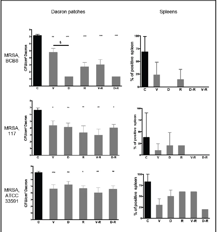
MRSA BCB8, daptomycin was significantly more bactericidal than vancomycin with a dramatic reduction of bacterial load after a 48 h-treatment (- 5.85 log10 CFU/cm2 as compared
to the control group, P < 0.001, and - 3.47 log10 CFU/cm2 as compared to vancomycin, P <
0.001), whether or not combined with rifampicin. The bactericidal activity of vancomycin was significantly improved when combined with rifampicin, although this combination remained less bactericidal than daptomycin alone on this isolate (P < 0.001). Rifampicin monotherapy demonstrated good efficacy (- 4.5 log10 CFU/cm2 vs control group, P < 0.001)
(Figure 2). No emergence of antibiotic resistance was observed on bacteria recovered from positive cultures.
For MRSA 117 and 33591, results were strikingly different. For both strains, there were no significant differences between daptomycin, vancomycin and rifampicin monotherapies. Combined therapies demonstrated no significant benefit as compared to monotherapies (Figure 2). Although not statistically significant, there was a trend towards higher efficacy of vancomycin-rifampicin as compared to daptomycin-rifampicin. Once again, no antibiotic resistance was documented after treatment for both strains.
Spleen cultures were more frequently positive for controls than for therapeutic groups. There was no significant difference between therapeutic groups.
Daptomycin efficacy on MSSA vascular material infection depends also on bacterial
strains
As for MRSA, bacterial cultures after sonication did not differ from those without sonication. Daptomycin was significantly more bactericidal than cloxacillin for MSSA 176 (– 1.27 log10
CFU/cm2 (CI 95%: 0.32 to 2.22), P < 0.05), but not for MSSA 27217 and MSSA 171 (Figure 3). A trend towards better efficacy of daptomycin vs vancomycin on MSSA 176 (- 1.1 log10
CFU/cm2; CI 95%: -0.16 to 2.37) was observed. Daptomycin and cloxacillin demonstrated higher efficacy than vancomycin for MSSA 171 (respectively – 2.72 log10 CFU/cm2 (CI 95%:
0.75 to 3.79) and – 2.1 log10 CFU/cm2 (CI 95%: 0.55 to 3.65); P < 0.001 for both).
Combination of cloxacillin, vancomycin, or daptomycin, with rifampicin was more bactericidal than monotherapies for the 27217 strain. This effect was less pronounced for 171 and 176. Surprisingly, the association of daptomycin and rifampin was less efficient than cloxacillin-rifampicin (- 1.76 log10 CFU/cm2 (CI 95%: 0.61 to 2.9); P < 0.01) or
vancomycin-rifampicin (- 2.17 log10 CFU/cm2 (CI 95%: 1.1 to 3.25); P < 0.001), for MSSA 27217. No
emergence of antibiotic resistance was documented for any of the conditions tested.
Spleen cultures were less often positive for MSSA than for MRSA, and no significant difference between the different conditions were noticed.
Discussion
The main findings of these experimental studies are: i) the results of the in vitro biofilm antibiotic susceptibility assays may vary according to the nature of the support used; ii) our mouse model of S. aureus vascular graft infection allowed us to test a large number of bacterial strains and antibiotic regimens and could pave the way toward a better understanding of antibiotic efficacy in PVGIs; iii) although more efficient than comparators for some bacterial strains, daptomycin was most of the time not superior to vancomycin or cloxacillin; and iv) combination of rifampicin and daptomycin did not enhance the bactericidal effect of daptomycin in this model.
Decrease of antibiotic efficacy in material-related infections has been highlighted by many studies using biofilm susceptibility tests, including the Calgary device.8, 15, 20, 21 Although not
recommended in routine,22 those techniques illustrate that MBICs and MBECs are much higher than MIC and MBC, with limited perspective of clinical cure since these concentrations are not achievable in humans.9, 21 However, those techniques use polystyrene devices, significantly different from biomaterials used for vascular prosthesis. Therefore, results yielded with those procedures may not be relevant for PVGIs. We developed a model to evaluate specific MBICs and MBECs on Dacron®, referred as dMBICs and dMBECs, to better assess the decrease of antibiotics efficacy on bacteria embedded inside the biofilm on vascular prostheses. In this model, while dMIBCs and MBICs were comparable, dMBECs
were lower than MBECs, although higher than MBCs. For instance, dMBICs and dMBECs of rifampicin were in the range of concentrations achievable in humans for most bacterial strains tested. Biofilm developed onto plastic was more dense, and thicker, than the one found on Dacron® (figure 1), and adhesion of bacteria onto the Dacron® appeared weaker, explaining
why antibiotics are less efficient in the polystyrene model. Our results demonstrated that classical techniques used to measure MBICs and MBECs could overestimate the decrease of antibiotic efficacy in the particular context of PVGIs, and that our model, specific to PVGIs, could yield more relevant findings. They do not question the recommendations of removing all infected material whenever possible for PVGI.23, 24 However, our data suggest that
antibiotics alone may be a reasonable therapeutic option in selected cases when surgery would be associated with a high probability of severe adverse outcomes.25-27
Animal models constitute a critical step for the evaluation of antibiotics for PVGIs. A rat model of PVGI using a Dacron® patch implanted in a dorsal subcutaneous pouch evaluated different prophylactic procedures.13, 28-30 Other models mimicked prosthetic-joint infections
using a Teflon®-cage implanted in a dorsal subcutaneous pouch in Guinea-pigs,31-35 or rats.10 To our knowledge, no specific model evaluated the curative treatment of PVGI so far. We combined two approaches to evaluate different antibiotic regimens in a PVGI mouse model. For technical reasons, it was impossible to implant our Dacron® along the vascular system, and this represents one weakness of our work. Some authors described rabbit,36 pig14 or dog11
models of aortic graft infections, but those models do not allow the use of a large number of animals, and consequently are not appropriate for the screening of antibiotic strategies on different bacterial strains. Moreover, in a clinical setting, most PVGIs occur from the wound or from an adjacent infectious focus, and not through hematogenous route.37 Therefore, the infection process usually starts along the external part of the vascular prosthesis, not the endoluminal layer. Our model reproduces this pathway.
Although all antibiotics tested in this model demonstrated efficacy when compared to controls, antibiotic efficacy varied according to bacterial strains. For instance, daptomycin was more bactericidal than vancomycin for MRSA BCB8 and MSSA 171, and more bactericidal than cloxacillin for MSSA 176, but this was not superior to other antibiotics for other strains. For MRSA BCB8, dMBIC and dMBEC were lower for daptomycin than for vancomycin and this could partially explain the differences noticed. One other explanation could be the different capabilities of antibiotics to penetrate the biofilm in vivo., although this would not explain the striking differences observed between strains.38 Daptomycin has already demonstrated better efficacy against MRSA than vancomycin in vitro39, 40 and in
animal models.40, 41 This higher efficacy is thought to be linked to a better biofilm penetration for daptomycin than for vancomycin,42 and a better bactericidal activity against bacteria in stationary phase.40 However, some authors did not found any difference between daptomycin and vancomycin efficacy.43, 44 Our results highlight a possible differential activity of daptomycin according to the bacterial strain, that may explain the discrepancies between previous studies.
Rifampicin has demonstrated its efficacy for the treatment of material-related staphylococcal infection45 and its use in combination is largely recommended. In the present study, we did not found any improvement of therapeutic efficacy when rifampicin was added to daptomycin for MRSA, but rifampicin enhanced the activity of vancomycin. The combination of daptomycin and rifampicin increased only moderately the bactericidal activity of daptomycin for MSSA 27217, while the addition of rifampicin to vancomycin or cloxacillin strongly enhanced their bactericidal effect for this strain. These results are in contrast with those of Sakoulas et al.,46 who demonstrated in a rat model of MRSA endocarditis a synergistic effect of daptomycin plus rifampicin. Saleh-Mghir et al. found similar results in a rabbit model of prosthetic joint infection.47 However, an in vitro pharmacodynamic model evaluating
daptomycin and rifampicin against different MRSA strains found variable activity of this association (i.e. increased bactericidal activity with combinations in some, but not all strains tested.48 Likewise, the addition of rifampicin to daptomycin did not enhance the bactericidal activity of daptomycin in a rabbit model of MRSA endocarditis.49 Some studies suggested
that daptomycin-fosfomycin or daptomycin-cloxacillin may be more synergistic.10, 50
Our model could not capture the utility of combinations to prevent the emergence of resistance. Indeed, even when bacterial load was high after treatment, we did not document any bacterial resistance. This was unexpected, since emergence of resistance under treatment is one of the main caveats with rifampicin,41 or daptomycin monotherapies.41, 47 An
experimental study evaluating the in vivo fitness of rifampicin-resistant S. aureus mutants in a mouse biofilm infection model found that rifampicin-resistant strains appeared after 3 to 9 days of treatment.18 This delay may explain why our 48 h-treatment regimens were not associated with emergence of resistance. Thus, although no resistance was documented in our model, clinical data indicate that rifampicin must be used in combination for PVGI, as for any other infection.
In conclusion, we found that biofilm formation and bacterial adhesion are weaker onto the Dacron® than onto polystyrene devices, resulting in a less pronounced biofilm-related decrease of antibiotic efficacy in the particular setting of PVGIs. We implemented an innovative mouse model of PVGI allowing the evaluation of a large number of antibiotic regimens. In this model, we demonstrated that daptomycin was more efficient than comparators for some strains but not all, and that the addition of rifampicin did not enhance daptomycin efficacy. However, due to the variability of findings according to bacterial strains, we were not able to determine the best antibiotic regimen for PVGIs.
Acknowledgements: The authors want to acknowledge the Collège des enseignants des Maladies Infectieuses et Tropicales (CMIT) and the Société Française de Pathologie Infectieuse de Langue Française (SPILF) for their support. They also want to acknowledge Prof Erwan Flecher who kindly provided the Dacron®.
Funding: This work was supported by a grant from Novartis and a grant from the Collège des enseignants des Maladies Infectieuses et Tropicales (CMIT). Sponsors had no role in the design and the realization of this work, and had no access to results until this work was submitted.
Transparency Declarations: Matthieu Revest has been supported by MSD and Pfizer laboratories to attend international conferences. The other authors have nothing to declare.
a
b
MSSA 27217 MRSA BCB8
Figure 1. Visualization of MSSA and MRSA 24h-biofilms on polystyrene (a) and on Dacron (b) using section views to observe biofilm thickness. All bacteria were stained green with Syto9®. The acquisition was performed on the whole biofilm thickness with an axial displacement of 1 µm. Images dimension is 82 x 82 µm². The scale bars correspond to 20 µm. Only biofilm for MSSA 27217 and MRSA BCB8 are represented since they were representative of all the biofilms visualized for other strains.
Figure 2. Results of bacterial count on Dacron patches and spleens after 48 h of treatment for
MRSA infection.
C: controls; V: vancomycin; D: daptomcyin; R: rifampicin; V-R: vancomycin-rifampicin; D-R: daptomycin-rifampicin. CFU: Colony Forming Unit
Stars represent results of comparison between control groups and each therapeutic group. * P < 0.05; ** P < 0.01; *** P < 0.001. § P < 0.001, comparison between vancomycin and daptomycin for MRSA BCB8.
Number of mice per condition:
BCB8: n= 12 per antibiotic regimen
117: n= 10 per antibiotic regimen
Figure 3. Results of bacterial count on Dacron sheets and spleen after 48 h of treatment for
MRSA infection.
C: controls; V: vancomycin; D: daptomcyin; R: rifampicin; V-R: vancomycin-rifampicin; D-R: daptomycin-rifampicin. CFU: Colony Forming Unit
Stars represent results of comparison between control groups and each therapeutic group. * P < 0.05; *** P < 0.001. P < 0.05, comparison between controls and daptomycin for MSSA 176.
¤
P < 0.01, comparison between cloxacillin-rifampicin and daptomycin-rifampicin forMSSA 27217. # P < 0.001, comparison between vancomycin-rifampicin and daptomycin-rifampicin for MSSA 27217
Number of mice per condition:
171: n= 8 to 12 per antibiotic regimen
176: n= 10 to 15 per antibiotic regimen
Table 1. In vitro drug-susceptibility testing for the bacterial strains used in this study
Bacterial
strains Antibiotic
Classical methods Biofilm on Dacron Biofilm on polystyrene
MIC MBC dMBIC dMBEC MBIC MBEC
MRSA BCB8 Vancomycin 1 1 1 > 32 1 > 256 Daptomycin 0.125 0.125 0.5 8 1 > 256 Rifampicin < 0.06 < 0.06 < 0.125 > 1 0,015 > 8 117 Vancomycin 1 4 4 16 1 > 256 Daptomycin 1 2 2 64 1 > 256 Rifampicin 0.015 0.03 0.015 > 0.5 0,015 > 8 33591 Vancomycin 1 2 4 16 2 > 256 Daptomycin 0.25 0.5 2 > 16 1 > 256 Rifampicin 0.0075 0.015 0.015 0.5 0,0075 > 8 MSSA 171 Cloxacillin 0.25 0.5 2 > 32 0,25 > 256 Vancomycin 1 1 8 32 4 > 256 Daptomycin 0.5 0.5 4 > 32 4 > 256 Rifampicin 0.015 0.03 0.125 > 0.25 0,015 > 8 176 Cloxacillin 0.5 0.5 2 > 32 0,5 > 256 Vancomycin 1 1 2 16 1 > 256 Daptomycin 0.5 0.5 2 > 64 0,5 > 256 Rifampicin 0.015 0.03 0.125 > 0.5 0,015 > 8 27217 Cloxacillin 0.5 0.5 1 16 0,25 > 256 Vancomycin 1 1 8 > 32 8 > 256 Daptomycin 0.25 0.25 1 > 8 2 > 256 Rifampicin < 0.06 0.125 2 2 0,03 > 8
MIC: Minimum inhibitory concentration; MBC: minimum bactericidal concentration; dMBIC: Dacron-related minimal biofilm inhibitory concentration; dMBEC: Dacron-related minimal biofilm eradicating concentration; MBIC: minimal biofilm inhibitory concentration; MBEC: minimal biofilm eradicating concentration
References
1 Darouiche RO. Treatment of infections associated with surgical implants. N Engl J Med 2004; 350: 1422-9.
2 HAS. Implants vasculaires. Révision de catégories homogènes de dispositifs médicaux. . Saint Denis, La Plaine; 2013.
3 FitzGerald SF, Kelly C and Humphreys H. Diagnosis and treatment of prosthetic aortic graft infections: confusion and inconsistency in the absence of evidence or consensus. J Antimicrob Chemother 2005; 56: 996-9.
4 Jansen KU, Girgenti DQ, Scully IL et al. Vaccine review: "Staphyloccocus aureus vaccines: problems and prospects". Vaccine 2013; 31: 2723-30.
5 Revest M, Camou F, Senneville E et al. Medical treatment of prosthetic vascular graft infections: Review of the literature and proposals of a Working Group. Int J Antimicrob Agents 2015.
6 Stewart PS and Franklin MJ. Physiological heterogeneity in biofilms. Nat Rev Microbiol 2008; 6: 199-210.
7 Richards JJ and Melander C. Controlling bacterial biofilms. Chembiochem 2009;
10: 2287-94.
8 Ceri H, Olson ME, Stremick C et al. The Calgary Biofilm Device: new technology for rapid determination of antibiotic susceptibilities of bacterial biofilms. J Clin Microbiol 1999; 37: 1771-6.
9 LaPlante KL and Mermel LA. In vitro activities of telavancin and vancomycin against biofilm-producing Staphylococcus aureus, S. epidermidis, and Enterococcus faecalis strains. Antimicrob Agents Chemother 2009; 53: 3166-9.
10 Garrigos C, Murillo O, Lora-Tamayo J et al. Fosfomycin-daptomycin and other fosfomycin combinations as alternative therapies in experimental foreign-body infection by methicillin-resistant Staphylococcus aureus. Antimicrob Agents Chemother 2013; 57: 606-10.
11 Javerliat I, Goeau-Brissonniere O, Sivadon-Tardy V et al. Prevention of Staphylococcus aureus graft infection by a new gelatin-sealed vascular graft prebonded with antibiotics. J Vasc Surg 2007; 46: 1026-31.
12 Hirose K, Marui A, Arai Y et al. Sustained-release vancomycin sheet may help to prevent prosthetic graft methicillin-resistant Staphylococcus aureus infection. J Vasc Surg 2006; 44: 377-82.
13 Cirioni O, Mocchegiani F, Ghiselli R et al. Daptomycin and rifampin alone and in combination prevent vascular graft biofilm formation and emergence of antibiotic resistance in a subcutaneous rat pouch model of staphylococcal infection. Eur J Vasc Endovasc Surg 2010; 40: 817-22.
14 Gao H, Sandermann J, Prag J et al. Rifampicin-soaked silver polyester versus expanded polytetrafluoro-ethylene grafts for in situ replacement of infected grafts in a porcine randomised controlled trial. Eur J Vasc Endovasc Surg 2012; 43: 582-7.
15 Moskowitz SM, Foster JM, Emerson J et al. Clinically feasible biofilm susceptibility assay for isolates of Pseudomonas aeruginosa from patients with cystic fibrosis. J Clin Microbiol 2004; 42: 1915-22.
16 Crandon JL, Kuti JL and Nicolau DP. Comparative efficacies of human simulated exposures of telavancin and vancomycin against methicillin-resistant Staphylococcus aureus with a range of vancomycin MICs in a murine pneumonia model. Antimicrob Agents Chemother 2010; 54: 5115-9.
17 Dandekar PK, Tessier PR, Williams P et al. Pharmacodynamic profile of daptomycin against Enterococcus species and methicillin-resistant Staphylococcus aureus in a murine thigh infection model. J Antimicrob Chemother 2003; 52: 405-11.
18 Yu J, Wu J, Francis KP et al. Monitoring in vivo fitness of rifampicin-resistant Staphylococcus aureus mutants in a mouse biofilm infection model. J Antimicrob Chemother 2005; 55: 528-34.
19 Domenech A, Ribes S, Cabellos C et al. Experimental study on the efficacy of combinations of glycopeptides and beta-lactams against Staphylococcus aureus with reduced susceptibility to glycopeptides. J Antimicrob Chemother 2005; 56: 709-16.
20 Hengzhuang W, Wu H, Ciofu O et al. In vivo pharmacokinetics/pharmacodynamics of colistin and imipenem in Pseudomonas aeruginosa biofilm infection. Antimicrob Agents Chemother 2012; 56: 2683-90.
21 Abbanat D, Shang W, Amsler K et al. Evaluation of the in vitro activities of ceftobiprole and comparators in staphylococcal colony or microtitre plate biofilm assays. Int J Antimicrob Agents 2014; 43: 32-9.
22 Hoiby N, Bjarnsholt T, Moser C et al. ESCMID guideline for the diagnosis and treatment of biofilm infections 2014. Clin Microbiol Infect 2015; 21 Suppl 1: S1-S25.
23 Lyons OT, Patel AS, Saha P et al. A 14-year experience with aortic endograft infection: management and results. Eur J Vasc Endovasc Surg 2013; 46: 306-13.
24 Tong SY, Davis JS, Eichenberger E et al. Staphylococcus aureus infections: epidemiology, pathophysiology, clinical manifestations, and management. Clin Microbiol Rev 2015; 28: 603-61.
25 Maze MJ, Laws P, Buckenham T et al. Outcomes of infected abdominal aortic grafts managed with antimicrobial therapy and graft retention in an unselected cohort. Eur J Vasc Endovasc Surg 2013; 45: 373-80.
26 Erb S, Sidler JA, Elzi L et al. Surgical and antimicrobial treatment of prosthetic vascular graft infections at different surgical sites: a retrospective study of treatment outcomes. PLoS One 2014; 9: e112947.
27 Legout L, Sarraz-Bournet B, D'Elia PV et al. Characteristics and prognosis in patients with prosthetic vascular graft infection: a prospective observational cohort study. Clin Microbiol Infect 2012; 18: 352-8.
28 Giacometti A, Cirioni O, Ghiselli R et al. Temporin A soaking in combination with intraperitoneal linezolid prevents vascular graft infection in a subcutaneous rat pouch model of infection with Staphylococcus epidermidis with intermediate resistance to glycopeptides. Antimicrob Agents Chemother 2004; 48: 3162-4.
29 Turgut H, Sacar S, Kaleli I et al. Systemic and local antibiotic prophylaxis in the prevention of Staphylococcus epidermidis graft infection. BMC Infect Dis 2005; 5: 91.
30 Atahan E, Gul M, Ergun Y et al. Vascular graft infection by Staphylococcus aureus: efficacy of cefazolin, teicoplanin and vancomycin prophylaxis protocols in a rat model. Eur J Vasc Endovasc Surg 2007; 34: 182-7.
,31 Oliva A, Furustrand Tafin U, Maiolo EM et al. Activities of fosfomycin and rifampin on planktonic and adherent Enterococcus faecalis strains in an experimental foreign-body infection model. Antimicrob Agents Chemother 2014; 58: 1284-93.
32 Furustrand Tafin U, Majic I, Zalila Belkhodja C et al. Gentamicin improves the activities of daptomycin and vancomycin against Enterococcus faecalis in vitro and in an experimental foreign-body infection model. Antimicrob Agents Chemother 2011; 55: 4821-7.
33 Furustrand Tafin U, Corvec S, Betrisey B et al. Role of rifampin against Propionibacterium acnes biofilm in vitro and in an experimental foreign-body infection model. Antimicrob Agents Chemother 2012; 56: 1885-91.
34 Zimmerli W, Waldvogel FA, Vaudaux P et al. Pathogenesis of foreign body infection: description and characteristics of an animal model. J Infect Dis 1982; 146: 487-97.
35 Schwank S, Rajacic Z, Zimmerli W et al. Impact of bacterial biofilm formation on in vitro and in vivo activities of antibiotics. Antimicrob Agents Chemother 1998; 42: 895-8.
36 Shimabukuro K, Hirose H, Mori Y et al. Local treatment of Dacron patch graft infected with biofilm-producing Staphylococcus epidermidis using antibiotic-releasing porous apatite ceramic: an experimental study in the rabbit. J Vasc Surg 2004; 39: 1361.
37 Jones L, Braithwaite BD, Davies B et al. Mechanism of late prosthetic vascular graft infection. Cardiovasc Surg 1997; 5: 486-9.
38 Boudjemaa R, Briandet R, Fontaine-Aupart M et al. Diffusion, Bioavailability and Reactivity of Antibiotics against Staphylococcus aureus Biofilms: a New Approach by Dynamic Fluorescence Imaging. International Conference on Antimicrobial Agents and Chemotherapy. Washington, DC; 2014. p. Abstract C-1415.
39 Edmiston CE, Jr., Goheen MP, Seabrook GR et al. Impact of selective antimicrobial agents on staphylococcal adherence to biomedical devices. Am J Surg 2006; 192: 344-54.
40 Murillo O, Garrigos C, Pachon ME et al. Efficacy of high doses of daptomycin versus alternative therapies against experimental foreign-body infection by methicillin-resistant Staphylococcus aureus. Antimicrob Agents Chemother 2009; 53: 4252-7.
41 Garrigos C, Murillo O, Euba G et al. Efficacy of usual and high doses of daptomycin in combination with rifampin versus alternative therapies in experimental foreign-body infection by methicillin-resistant Staphylococcus aureus. Antimicrob Agents Chemother 2010; 54: 5251-6.
,42 Stewart PS, Davison WM and Steenbergen JN. Daptomycin rapidly penetrates a Staphylococcus epidermidis biofilm. Antimicrob Agents Chemother 2009; 53: 3505-7.
43 Smith K, Perez A, Ramage G et al. Comparison of biofilm-associated cell survival following in vitro exposure of meticillin-resistant Staphylococcus aureus biofilms to the antibiotics clindamycin, daptomycin, linezolid, tigecycline and vancomycin. Int J Antimicrob Agents 2009; 33: 374-8.
44 Lefebvre M, Jacqueline C, Amador G et al. Efficacy of daptomycin combined with rifampicin for the treatment of experimental meticillin-resistant Staphylococcus aureus (MRSA) acute osteomyelitis. Int J Antimicrob Agents 2010; 36: 542-4.
45 Senneville E, Joulie D, Legout L et al. Outcome and predictors of treatment failure in total hip/knee prosthetic joint infections due to Staphylococcus aureus. Clin Infect Dis 2011; 53: 334-40.
46 Sakoulas G, Eliopoulos GM, Alder J et al. Efficacy of daptomycin in experimental endocarditis due to methicillin-resistant Staphylococcus aureus. Antimicrob Agents Chemother 2003; 47: 1714-8.
47 Saleh-Mghir A, Muller-Serieys C, Dinh A et al. Adjunctive rifampin is crucial to optimizing daptomycin efficacy against rabbit prosthetic joint infection due to methicillin-resistant Staphylococcus aureus. Antimicrob Agents Chemother 2011; 55: 4589-93.
48 Rose WE, Leonard SN and Rybak MJ. Evaluation of daptomycin pharmacodynamics and resistance at various dosage regimens against Staphylococcus aureus isolates with reduced susceptibilities to daptomycin in an in vitro
pharmacodynamic model with simulated endocardial vegetations. Antimicrob Agents Chemother 2008; 52: 3061-7.
49 Miro JM, Garcia-de-la-Maria C, Armero Y et al. Addition of gentamicin or rifampin does not enhance the effectiveness of daptomycin in treatment of experimental endocarditis due to methicillin-resistant Staphylococcus aureus. Antimicrob Agents Chemother 2009; 53: 4172-7.
50 Garrigos C, Murillo O, Lora-Tamayo J et al. Efficacy of daptomycin-cloxacillin combination in experimental foreign-body infection due to methicillin-resistant Staphylococcus aureus. Antimicrob Agents Chemother 2012; 56: 3806-11.