Monoacylglycerol as a metabolic coupling factor in
glucose-stimulated insulin secretion
par Shangang Zhao
Département de Biochimie Faculté de Médecine
Mémoire présentée à la Faculté des Etudes Supérieures en vue de l’obtention du grade de maître ès sciences
en Biochimie
Décembre 2010
Ce mémoire intitulée :
Monoacylglycerol as a metabolic coupling factor in glucose-stimulated insulin secretion
Présenté par : Shangang Zhao
a été évaluée par un jury composé des personnes suivantes:
Dr Tony Antakly, président-rapporteur Dr Marc Prentki, directeur de recherche Dr Ashok K. Srivastava, membre du jury
Résumé
Les cellules beta pancréatiques sécrètent l’insuline lors d’une augmentation post-prandiale du glucose dans le sang. Ce processus essentiel est contrôlé par des facteurs physiologiques, nutritionnels et pathologiques. D’autres sources d’énergie, comme les acides aminés (leucine et glutamine) ou les acides gras potentialisent la sécrétion d’insuline. Une sécrétion d’insuline insuffisante au besoin du corps déclanche le diabète. Le rôle que joue l’augmentation du calcium intracellulaire et les canaux K+/ATP dans la sécrétion d’insuline est bien connu. Bien que le mécanisme exact de la potentialisation de la sécrétion d’insuline par les lipides est inconnu, le cycle Glycérolipides/Acides gras (GL/FFA) et son segment lipolytique ont été reconnu comme un composant essentiel de la potentialisation lipidique de la sécrétion d’insuline. Le diacylglycérol, provenant de la lipolyse, a été proposé comme un signal lipidique important d’amplification. Cependant, l’hydrolyse des triglycérides et des diacylglycérides a été démontrée essentielle pour la sécrétion d’insuline stimulée par le glucose, en suggérant un rôle du monoacylglycérol (MAG) dans ce processus.
Dans cette étude, on démontre que la réduction de la sécrétion d’insuline stimulée par le glucose, lors d’une inhibition de la lipolyse, est restaurée par l’addition de MAG. Dans les cellules beta pancréatiques, le niveau de MAG augmente en présence des concentrations élevées du glucose, et également lorsqu’on inhibe l’enzyme MAG hydrolase abhydrolase-6 (ABHD6) avec l’inhibiteur spécifique WWL70.
L’analyse lipidomique a démontré qu’après la stimulation des cellules beta pancréatiques avec le glucose et aussi avec le WWL70, l’espèce la plus accumulée de MAG était le 1-stearoylglycérol (1-SG). L’addition de 1-SG, de 1-palmitoylglycérol (1-PG) ou de WWL70 augmente la sécrétion d’insuline stimulée par le glucose, et cette augmentation est indépendante de la génération de acides gras à partir de MAG. Cela suggère que le MAG est un signal lipidique pour la potentialisation de la sécrétion d’insuline stimulée par le glucose. De plus, la surexpression du gène d’ABHD6 dans les cellules INS832/13 cause une réduction de la sécrétion d’insuline, due probablement à la diminution des niveaux intracellulaire de MAG.
Avec le but de comprendre le mécanisme moléculaire impliqué dans la potentialisation de la sécrétion d’insuline par le MAG, on a bloqué l’action du récepteur vanilloid-1 (TRPV1) liant le MAG par l’agent pharmacologiste, AMG9810. Le traitement des cellules beta pancréatique par AMG9810 entraîne une diminution de la potentialisation de la sécrétion de l’insuline induite par le MAG. Il est a noter que le MAG pourrait activer TRPV1 par une liaison physique dans la membrane cellulaire interne; ce qui entraînerai l’entrée du calcium dans la cellule, et ensuite la stimulation de l’exocytose des granules à insuline. En soutien de cette hypothèse, on a trouvé une diminution du calcium intracellulaire lorsqu’on traite au AMG9810 des cellules beta pancréatique de rat (provenant des îlots dispersés) stimulées au glucose et au WWL70.
L’ensemble des résultats suggère que le MAG est un médiateur de la potentialisation lipidique de la sécrétion d’insuline stimulée par le glucose. Vu que l’inhibition pharmacologique d’ABHD6 augmente la sécrétion d’insuline, on pourra conclure que cette enzyme représente une cible thérapeutique potentielle dans le développement des médicaments anti-diabétiques, visant une augmentation de la sécrétion d’insuline.
Abstract
Insulin secretion by the pancreatic -cell in response to post-prandial increase in blood glucose levels is an essential physiological process that is governed by cellular, nutritional and pathological factors. Other fuels including amino acids like leucine and glutamine and also fatty acids contribute to further augment insulin secretion. Failure to secrete adequate amount of insulin according to the changing demands of the body by -cell is a key determinant of diabetes. The role played by the elevated Ca2+influx and K+-ATP channels in insulin secretion is well known. Even though the precise mechanism of the lipid amplification of insulin secretion and the involved molecular signals are not clear, Glycerolipid/Free fatty acid (GL/FFA) cycle and its lipolytic segment have been recognized as essential components in the lipid amplification pathway of insulin secretion. Diacylglycerol produced by lipolysis was proposed as an important lipid amplification signal. However, hydrolysis of triglycerides and also of diacylglycerols is shown to be essential for glucose stimulated insulin secretion (GSIS), indicating a possible role for monoacylglycerol (MAG) in this process.
In the present study we demonstrate that the obliterated GSIS due to lipolysis inhibition in -cells can be restored by providing exogenous MAG. In the -cells MAG levels increase significantly in the presence of high glucose concentration and specific inhibition of the major MAG hydrolase, abhydrolase-6 (ABHD6), in -cells and islets with WWL70 leads to accumulation of MAG with concomitant increase in insulin secretion. Lipidomics analysis
indicated that the major MAG species that is elevated by high glucose as well as WWL70 addition is 1-stearoylglycerol (1-SG). Exogenously added 1-SG and also 1-palmitoylglycerol (1-PG) strongly enhanced GSIS and this augmentation is not dependent on the generation of FFA by these MAGs. This indicates that MAG is a potential candidate for being the lipid signal for GSIS amplification. Further evidence for this was provided by the observation that overexpression of the MAG hydrolase ABHD6 in INS832/13 cells, resulted in decreased insulin secretion, probably owing to the lowered MAG level inside the -cells.
Pharmacological studies using AMG9810, a specific antagonist of transient receptor potential vanilloid-1 (TRPV1) receptor that binds MAG, revealed that a blockade of TRPV1 strongly attenuated the MAG-augmented insulin secretion. Since MAG is a potential activator of TRPV1, it is likely that MAG binds on the inner surface of the cell membrane to TRPV1, which in turn triggers rapid influx of Ca2+ thereby promoting insulin granule exocytosis. Thus, AMG9810 was found to lower Ca2+influx into dispersed rat islet cells that was induced by high glucose and also WWL70.
These results collectively suggest that MAG is the potential mediator of the lipid amplification of glucose-stimulated insulin secretion. Our results also indicate that pharmacological intervening at the ABHD6 hydrolysis step enhances insulin secretion; this enzyme protein can be a promising thrapeutic target for the development of anti-diabetic drugs that promote insulin secretion.
Keywords: Diabetes, glucose-stimulated insulin secretion, monoacylglycerol, ABHD6, TRPV1
Table des matières
RÉSUMÉ………...i
ABSTRACT………...iv
Table des matières………...vii
Liste des figures………...x
Liste des abréviations………xii Remerciements……… xv
1 Background……….……….1
1.1 Obesity and diabetes………...2
1.1.1 Current situation of obesity and diabetes in the world………...2
1.1.2 Type 1 diabetes………....3
1.1.3 Gestational diabetes………...…5
1.1.4 Type 2 diabetes………...………6
1.1.4.1 Insulin resistance………...10
1.1.4.2 Beta cell failure………...…...13
1.1.4.2.1 Glucotoxicity………...….14 1.1.4.2.2 Lipotoxicity………...………15 1.1.4.2.3 Glucolipotoxicity………...………...16 1.1.4.2.4 ER stress………...………18 1.1.4.2.5 Inflammation………...………..20 1.2 Insulin secretion………...…………...20
1.2.1 Insulin biosynthesis and exocytosis……….………21
1.2.2 Inhibitors and stimulators of insulin secretion………25
1.2.2.1 Glucose………27
1.2.2.2 Fatty acids……….28
1.2.2.3 Amino acids………..30
1.2.2.4 GLP-1 and GIP………...31 1.2.2.5 Acet yl ch olin e……… ……… …… …………...3 1
1.2.2.6 Somatostatin……….…………...32
1.2.2.7 Galanin……….…...32
1.2.2.8 (Nor)epinephrine………33
1.2.3 Metabolic signals promoting insulin secretion……….33
1.2.3.1 Mitochondrial signals: anaplerosis, cataplerosis and pyruvate cycling………..……34
1.2.3.2 NADPH, Adenine and Guanine Nucleotides, and Glutamate…… …36
1.2.3.3 Lipid metabolism dependent amplification of beta cell metabolic signaling………...………37
1.3 Glycerolipid/free fatty acid cycle (GL/FFA cycle)……….39
1.3.1 The process of GL/FFA cycle and related enzymes………..………39
1.3.2 MAG lipases………43
1.3.3 Functions of GL/FFA cycle……….45
1.3.4 Regulation of insulin secretion by GL/FFA cycle in beta cells….…………49
1.4 TRP channels and insulin secretion………51
1.4.1 TRP channels identified in beta cells………53
1.4.1.1 TRPC subfamily……….…………..53
1.4.1.2 TRPM channels……….………...54
1.4.1.3 TRPV subfamily………..……….55
1.4.2 TRPV1 and beta cell function……….……….56
1.5 Hypothesis………...……….57
2 MATERIALS AND METHODS………...………..59
2.1 Materials………...………..59
2.2 Cell culture………...………..59
2.3 Islet isolation………...………59
2.4 Insulin secretion measurement………...……….60
2.5 Effect of Lipase inhibitors on [U-14C]-glucose incorporation into lipids…….…61
2.6 LC/MS/MS measurements……….……62
2.8 Quantitative real-time RT-PCR………63
2.9 Immunoblot analysis………...………64
2.10 Cytosolic Ca2+ measurement in rat islets……….64
2.11 Statistical analysis………...………65
3 Results………...………...66
3.1 Effect of monoacylglycerols (MAGs) in restoring normal GSIS in INS832/13 beta cells treated with the panlipase inhibitor orlistat ……….……66
3.2 Expression of MAGL and ABHD6 in rat tissues, islets and beta cell lines……68
3.3 The effect of inhibition MAG lipase and ABHD6 on GSIS……….70
3.4 14C-Glucose incorporation into neutral lipids: Effect of glucose concentration, orlistat and WWL70………...………..72
3.5 Profile of different MAG species, measured by LC-MS/MS analysis……….…74
3.6 The effect of exogenous MAG on insulin secretion………….…………75
3.7 The effect of overexpression of ABHD6 on GSIS……….………..76
3.8 TRPV1 is possibly responsible for WWL70-enhanced insulin secretion………78
4 Discussion…...83
5 Conclusion and perspective... ...95
Liste des figures
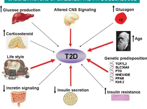
Figure 1 The possible causal factors of T2D………..7
Figure 2 Islet beta cell failure and natural history of T2D………..………9
Figure 3 Stimulators and inhitors of insulin secretion……….26
Figure 4 Triggering and K+-ATP independent/ amplification pathways in beta cell meabolic signaling………….………28
Figure 5 Tricycling and FFA induced insulin secretion………29
Figure 6 Candidate coupling factors in fuel-induced insulin secretion……….34
Figure 7 Pyruvate cycling and insulin secretion……….36
Figure 8 Interactions between glucose and fatty acid metabolism in nutrient-secretion coupling………38
Figure 9 Enzymes and intermediates of the triacylglycerol/free fatty acid cycle. ………40
Figure 10 Biological processes regulated by GL/FFA cycling………45
Figure 11 GL/FFA cycling and nutrient glucolipodetoxification……….…………....47
Figure 12 Model illustrating the role of the free fatty acids (FFA) and the glycerolipid/fatty acid (GL/FA) cycle in the regulation of insulin secretion as a function of the nutritional state………50
Figure 13 Effect of MAGs in restoring normal GSIS in INS832/13 beta cells treated with the panlipase inhibitor orlistat…………..……….………67
Figure 14 Expression of MAGL and ABHD6 in rat tissues, islets and beta cells……...….69
Figure 15 Effect of MAGL and ABHD6 inhibitors on insulin secretion in beta cells……..71
Figure 17 Effect of exogenous MAG addition on insulin secretion……….76
Figure 18 Effect of over-expression of ABHD6 on insulin secretion in INS832/13 cells....78
Figure 19 TRPV1 is possibly responsible for WWL70-enhanced insulin secretion….…...81
Figure 20 Lipolysis-derived MAG as a MCF for insulin secretion, targeting TRPV1 channels, PKD and Munc13-1………..………97
Liste des abréviations
1-OG 1-oleoylglycerol
1-PG 1-pamitoylglycerol
1-SG 1-stearoylglycerol
2-AG 2-arachidonoyl glycerol
2-OG 2-oleoylglycerol
ABHD6 alpha/beta-hydrolase domain 6
ABHD12 alpha/beta-hydrolase domain 12
ADA American Diabetes Association
ADPR ADP ribose
ATF6 activating transcription factor 6 ATGL adipose tissue triglyceride lipase
CHOP C/EBP homologous protein
DAG diacylglycerol
GAD65 glutamic acid decarboxylase
GDM gestational diabetes mellitus
GIP glucose-dependent insulinotropic peptide GL/FFA cycle glycerolipid/ free fatty acid cycle
GLP-1 glucagon-like peptide-1
GLUT2 glucose transporter 2
GSIS glucose-stimulated insulin secretion
HSL hormone sensitive lipase
IRE1 inositol requiring 1
IRSs insulin receptor substrates
JNK c-JUN NH2-terminal kinase
MAG monoacylglycerol
MAGL monoacylglycerol lipase
MCF metabolic coupling factor
MDP morphologically docked pool
NSF soluble N-ethylmaleimide-sensitive factor
PDX-1 pancreas-duodenum homobox-1
PERK PKR-like kinase
PI3K phosphatidylinositol 3-kinase
PKC protein kinase C
ROS reactive oxygen speices
SNAP-25 synaptosomal-associated protein 25 SNARE soluble NSF attachment protein receptor
RRP readily released granules SRP signal recognition particle
T1D type 1 diabetes
T2D type 2 diabetes
TG triacylglycerol
TRP channel transient receptor potential channels
UPR unfolded protein response
VAMP-2 vesicle-associated membrane protein 2
ZDF Zucker Diabetic Fatty
ZnT7 zinc transporter 7
Remerciements
I thank my director Dr Marc Prentki who gave me the chance to study in his famous laboratory in Canada as well as for all the support for my experiments.
I thank Dr S. R. Murthy Madiraju, who always guided and encouraged me with my experiments and always gave me good suggestions about my daily life and some suggestion related to taking care of my baby.
Thanks a lot to Drs Erik Joly and Marie-line Peyot for their help and discussions about my experiments.
Thanks a lot to the students Julien Lamontagne, Annie Barbeau, Marco Pineda, Emile Pepin, and also the technicians Roxane, Johane Morin, Melanie for their help in the experiments and help improve my English and French.
Particular thanks to Jose and Yves for their help and daily discussions of my results. Thanks a lot to my wife Xiaomeng Xu and our baby Chloe Zhao, who gave me big support in my daily life and much happiness.
1.1.1 The current situation in the world
Economic development and industrialization in many countries offer a wide variety of choice of foods to people. This has led more and more people to choose foods with elevated levels of saturated fatty acids, sugar-sweetened beverages and starchy food and reduced consumption of fibre. In conjunction with more sedentary lifestyle of people the energy from food intake has often considerably surpassed the daily energy expenditure. The surplus energy is predominantly stored as fat in the adipocytes and other tissues, therby contributing to obesity among these people and this is particularly marked for individuals who are genetically predisposed to obesity (Alappat and Awad, 2010).
Obesity has become a major socio-economic problem all over the world, especially in the developed countries. According to the American Obesity Association, nealy one third of the total US population is affected by obesity with a similar situation in Canada. A report from the National Diabetes Surveillance System indicated that approximately 25% of all Canadian adults are obese as are 10% of the children (Anis et al., 2010).
The global obesity epidemic is a primary cause for the increasing world-wide incidence of diabetes in the recent years. According to the World Health Organization and the International Diabetes Federation, the number of diabetes patients has increased from 100– 135 million in 1994–1995 to approximately 246 million in 2007, worldwide. With a further
7 million people developing diabetes each year, this number is expected to hit 438 million by 2030. In Canada, approximately 1.9 million men and women had been diagnosed with diabetes in 2005-2006, which represents about 1 in 17 Canadians - 5.5 % of all women and 6.2 % of all men. And now in 2010, more than 9 million Canadians live with undiagnosed or diagnosed diabetes, or prediabetes.
Diabetes is defined in simple terms, as the pathological situation where inadequate amount of insulin is secreted to meet the needs of body, with the resultant hyperglycemia, the hallmark of diabetes. However, diabetes is a highly complex metabolic disorder with a multitude of mechanisms for its causes, including both genetic and environmental factors depending on different types of diabetes. Diabetes, on the basis of its causes, could be divided into three major types- type 1 diabetes (T1D), type 2 diabetes (T2D) and gestational diabetes as well as other types, such as permanent neonatal diabetes (Gloyn et al., 2004).
1.1.2 Type 1 Diabetes (T1D)
T1D that is also named Juvenile diabetes, accounts for about 5 -10% of all cases of diabetes. In the United-States, 1 million people have been diagnosed with T1D in 2000. There are potentially another million patients, mostly adults, who have been possibly misdiagnosed as having type 2 diabetes (Prevalence of Diabetes in the United States based on US Population:
275 million in 2000). Additional studies predict that the incidence of T1D may be increasing at a rate of 3 - 4% a year (Dahlquist, 1998; Gloyn et al., 2004).
Type 1 diabetes is in the vast majority of its cases a chronic autoimmune disease, in which T cells and macrophages invade pancreatic islets and specifically target and kill beta cells in genetically predisposed individuals causing near complete loss of insulin secretion with the resultant hyperglycemia (Sera et al., 1999; Winter, 1996); At present, the exact mechanisms underlying the causes whereby the body's immune system is triggered to attack the beta cells is still not well understood. Some studies indicated that autoimmune, genetic, and environmental factors, possibly viruses, are involved in this process. Generally, T1D most often affects children and young adults, but can also appear at any age. Symptoms include increased thirst and urination, constant hunger, weight loss, blurred vision, and extreme fatigue. If not treated with insulin, a person with type 1 diabetes can lapse into a life-threatening diabetic coma, also known as diabetic ketoacidosis. Although the symptoms of T1D generally develop within a short period, the beta cell destruction maybe occurring over the span of several years prior the development of overt T1D. Thus, this allows for the possible prevention of T1D transition to overt diabetes. More than 30 years ago, it was observed that certain antibodies (named as islet cell antibodies) in TID patients could specifically bind to pancreatic islets. Presently, three major autoantigens: glutamic acid decarboxylase (GAD65) (Baekkeskov et al., 1989), a protein tyrosine phosphatase-like molecule (IA-2) (Notkins et al., 1996) and insulin (Palmer, 1987) have been identified. Using assays that employ islet cell autoantibodies, it is possible to
accurately identify people at higher risk for T1D. Once the risk is identified, these people can have time to use different strategies to delay or prevent the incidence of T1D, such as taking oral insulin capsules (Skyler et al., 2005). For people who are already suffering with T1D, the secure and relatively convenient way is to treat the disease by daily administration of insulin, because presently there is no “cure” for T1D. Even though islet transplantation has shown to be a promising therapy for T1D for many years (Gruessner et al., 2004; Rickels et al., 2006), it is not widely used due to its high price, graft immuno-suppression associated problems, and limitation of islet donors. Another promising strategy for T1D is the regeneration of beta cells from stem cells, precursor cells or the remaining beta cells (Meier et al., 2005). Thus, murine adult pancreatic precursor cell likely exists that could differentiate into cells with the characteristics of ¦Âcells (Seaberg et al., 2004). Another study showed that the prinicipal source of new beta cells is from the pre-existing beta cells, rather than pluripotent stem cells (Dor et al., 2004).
1.1.3 Gestational diabetes
Gestational diabetes mellitus (GDM) is defined as any degree of glucose intolerance that is first detected during pregnancy (Jovanovic et al., 1998). According to the American Diabetes Association (ADA), about 7% of all the pregnancies in USA are diagnosed with having GDM (Simmons et al., 2010). Another report indicates that the total incidence of the GDM can be as high as 17.8% (Metzger et al., 2010). Once diagnosed with GDM, the mother as well as its offspring, who has been exposed to hyperglycemic intrauterine
environment, has increased risk of developing type 2 diabetes mellitus in the future (Bellamy et al., 2009; Clausen et al., 2008). Because of the lack of uniform diagnostic criteria (Karagiannis et al., 2010), the global incidence of GDM is difficult to estimate. But it is well-accepted that GDM is a common disorder in pregnancy (Yogev et al., 2004). The mechanism related to incidence of GDM has not been fully elucidated, but at least three different factors have been shown to be implicated in this process: 1) auto-immune destruction of beta cell; 2) genetic abnormalities leading to defective insulin secretion; 3) beta cell dysfunction related to insulin resistance. As a consequence, the incidence of GDM is not only influenced by environmental factors, but also by genetic factors, and it shares some similarities to both T1D and T2D. So treatment of GDM may be different based on the individuals. The importance of nutrition in the management of GDM is well established (Amann-Gassner and Hauner, 2008). Since glucose is the primary fuel for the fetus and the characteristic of GDM is hyperglycemia, a healthy pregnancy outcome has to be met in the context of aberrant regulation of glucose metabolism. The main objective of nutritional recommendation for women with GDM is to maintain maternal normoglycemia and to reduce inadvertent accelerated fetal growth. The dietary advises are expected to have a lifelong positive impact for women with GDM so that maternal health is maintained and the risk for developing type 2 diabetes mellitus in the future is alleviated.
T2D is characterized with inadequate insulin secretion by pancreatic beta cells leading to hyperglycemia in individuals with a long history of insulin resistance and is predominantly due to the dysfunction of beta cells. T2D is thought to be a complex syndrome of polygenic nature (Fig 1) and many genes, such as CAPN10, ENPP1, HNF4A, ACDC, SLC308A, IDE-KIF11, EXT2-ALX4 that contribute to the development of T2D have been identified (Perry and Frayling, 2008; Sladek et al., 2007) with the help of the new high-density array technology that permits the simultaneous genotyping of thousands of polymorphisms. Some of the identified susceptible genes, such as zinc transporter SLC308A, are exclusively expressed in beta cells (Potapov et al., 2010). Many other genes that are also related to T2D still need to be identified. Discovery of new T2D risk genes will help in identifying novel players in T2DM pathogenesis, and will unravel novel mechanisms and lead to more efficient therapeutics.
Figure 1 The possible causal factors of T2D. The onset of T2D is complex, not only related to the environmental factors, such as change of life style, but also the genetic background. Several risk factors have been linked with onset of T2D and four factors, including life style, decreased insulin secretion, altered CNS signaling and genetic predisposition shown in red arrow play crucial roles in T2D.
T2D is the more common type of diabetes and accounts for approximately 90 to 95 percent of all cases of diabetes. Approximately 150 million people worldwide were affected by T2D in the year 2000 but this number has climbed to ~250 million in 2010. According to the Canadian Diabetes Association, it is estimated that nearly 3 million Canadians suffer from T2D in 2010. Because of the global obesity epidemic and increasing number of
people with insulin resistance, it is predicted that this number could increase to 370 million by 2030. Incidence of T2D is strongly associated with older age, obesity, family history of diabetes, previous history of gestational diabetes, physical inactivity and ethnicity (Al-Adsani et al., 2009). There has been an alarming increase in childhood obesity associated with T2D (Amed et al., 2010). However, nationally representative data on prevalence of type 2 diabetes in youth are not available.
Unlike T1D, the symptoms of type 2 diabetes develop gradually. T2D is actually considered as a syndrome rather than a disease such as anemia and hypothyroidism. Even though there are no overt symptoms for T2D such as fever, pain etc., if the hyperglycemia of these patients is not corrected soon with pharmacological, nutritional or physical intervention, gradually the function of several organs, such as kidney, foot as well as eyes and brains, can be compromised. Thus untreated T2D can become a serious health and social problem due to secondary complications (Renard, 2009), affecting the quality of life and even threatening life. Even though our knowledge related to the onset of T2D is still poor, some crucial factors have been identified and proven to be implicated. Numerous studies show that most individuals with T2D have insulin resistance (Lillioja et al., 1993; Prentki and Nolan, 2006) and more importantly, insulin resistance appears before the development of hyperglycemia in subjects that eventually develop T2D (Martin et al., 1992; Prentki and Nolan, 2006). Furthermore, it is now recognized that T2D develops in insulin-resistant subjects with the onset of beta cell dysfunction (Prentki and Nolan, 2006). So
insulin resistance and beta cell dysfunction are two crucial factors that play significant role in the onset of T2D as shown in Fig 2.
Figure 2 Islet beta cell failure and natural history of T2D. T2D develops in response to overnutriton and lack of physical activity in subjects that have underlying genetic and acquired predispositions to both insulin resistance (and/or hyperinsulinemia) and beta cell dysfunction. Over time, islet beta cell compensation for the insulin resistance fails, resulting in a progressive decline in -cell function. As a consequence, subjects progress from normal glucose tolerance (NGT) to IGT and finally to established T2D. Even after diagnosis of T2D, -cell function continues to worsen such that subjects progress from needing changes in diet/exercise only to requiring oral hypoglycemic agents and eventually insulin for achievement of adequate glycemic control. Future therapies will be directed not only to achievement of euglycemia, but also changing the course of the disease by reversing the processes of -cell failure (Nolan et al., 2006b).
Insulin sensitivity is commonly described as the ability of insulin to lower plasma glucose levels by suppressing hepatic glucose production and stimulating glucose utilization in skeletal muscle and adipose tissue (Fujimoto, 2000). Reduced insulin sensitivity makes the insulin-target tissues to respond poorly to insulin, leading to insulin resistance, which is an important link between obesity and T2D. Development of insulin resistance depends both on inherited and acquired factors. The inherited factors are most evident in the off-springs of T2D, who have more chance to be affected by insulin resistance (Warram et al., 1990). Some inherited defects have been identified, such as mutations in insulin receptors, glucose transporters and signaling proteins, but these mutations are relatively rare, and cannot explain the most common forms of insulin resistance, which is usually closely related to T2D. More work needs to be done to identify these inherited defects. With regard to the acquired causes of insulin resistance, some of the responsible factors include less physical activity, fuel surfeit, aging, hyperglycemia, hyperlipidemia and high level of plasma free fatty acids (FFA) as well as some medications. The mechanisms whereby these factors lead to insulin resistance are largely unknown. Some evidences indicate that impaired insulin signal transduction may be responsible. Insulin signaling involves a cascade of events initiated by insulin binding to its cell surface receptor, followed by receptor autophosphorylation, and activation of receptor tyrosine kinases, which result in tyrosine phosphorylation of insulin receptor substrates (IRSs)(Choi and Kim, 2010). IRSs further activate phosphatidylinositol 3-kinase (PI3K) and as a consequence, Akt will be activated and then phosphorylate its downstream targets, such as AS 160, which is implicated in the
process of glucose transport. IRS-1/PI3K/Akt signaling pathway is crucial in the regulation of glucose transport in response to insulin in skeletal muscle, the major site of glucose disposal (Koopmans et al., 1997). Loss of insulin signaling leads to severe insulin resistance in hepatocytes and progressive liver dysfunction (Pessin and Saltiel, 2000). Restoration of IRS-1/PI3K/Akt signaling pathway using insulin-sensitizer agents, such as metfomin, troglitazone and other thiazolidinediones could reverse insulin resistance and delay, or even prevent the onset of T2D. Interestingly, a polymorphism associated with T2D has been found within the IRS1 gene locus (Rung et al., 2009).
Insulin resistance is closely related to lipid metabolism. FFA derived from lipid metabolism (both do novo synthesis and lipolysis) is an important inducer of insulin resistance. From one side, high levels of FFA could directly interrupt the insulin signaling and decreased plasma membrane translocation of Glut4 in muscle and therefore lead to lower glucose disposal (Boden et al., 1991). Decreasing the FFA level in obese nondiabetic individuals is helpful to ameliorate insulin sensitivity in skeletal muscle (Santomauro et al., 1999). Blood levels of FFA are influenced by dietary intake and also by contributions from lipolysis of depot fats in adipose and other tissues. Adipose tissue triglyceride lipase (ATGL) is an important enzyme in lipolysis and its deficiency leads to TG accumulation and decreased FFA release, as a result, increasing insulin sensitivity and glucose tolerance (Hoy et al., 2010). On the other hand, increased level of FFA causes DAG and ceramide accumulation and DAG has been shown to disrupt normal insulin signaling by serine phosphorylation of
IRS1, through the activation of some atypical protein kinase C isoforms (Itani et al., 2002). Ceramides could activate protein phosphatase 2A, leading to dephosphorylation of Akt, thereby blocking insulin signaling and inhibiting Glut4 translocation to the cell membrane, thus disrupting insulin-mediated glucose uptake into skeletal muscle (Bonen et al., 2004). Besides the level of circulating FFA, intracellular content of FFA is also an important determinant of insulin resistance that acts independently of cellular energy balance and stress (Matsuzaka et al., 2007). Additional metabolic signaling pathways that could be central to insulin resistance are the AMP-kinase/malonyl-CoA signaling network and the glycerolipid/FFA cycle. Thus, elevated glucose and FFA will lead to AMPK inhibition, acetyl-CoA carboxylase activation and enhanced malonyl-CoA accumulation leading to reduced FFA oxidation and a rise in DAG that can activate C-kinase enzyme and thus cause reduced insulin signaling (Prentki and Corkey, 1996; Ruderman and Prentki, 2004). The evidence has been reviewed recently that altered GL/FFA cycling, with resulting accumulation of particular GL, such as DAG, MAG and phosphatidate, is implicated in insulin resistance and tissue malfunction during fuel surfeit (Prentki and Madiraju, 2008). Besides FFA, Sphingolipids metabolism, such as glucosylceramides are also implicated in the pathogenesis of insulin resistance. It has been shown that pharmacological inhibition or genetic ablation of enzymes controlling sphingolipid synthesis in rodents ameliorates insulin resistance (Holland and Summers, 2008; Summers and Nelson, 2005).
It is widely accepted that insulin resistance plays crucial role in pathogenesis of T2D and other diseases associated with the metabolic syndrome. Amelioration of insulin resistance is necessary to delay or prevent the onset of T2D. Some evidence showed that angiotensin-converting-enzyme inhibitors could prevent the onset of T2D (Solski and Longyhore, 2008). And also physical activity could improve insulin sensitivity and glucose tolerance in the skeletal muscle, and therefore it is also a good measure to prevent T2D. Certainly, our current knowledge related to insulin resistance is not enough to prevent T2D. More efforts are needed to completely understand the molecular mechanisms, and to find better treatments to prevent and cure insulin resistance and T2D.
1.1.4.2 Beta cell dysfunction
Beta cell is the only cell that synthesizes insulin and is responsible for insulin release, which plays key role in glucose homeostasis. Multiple evidences indicate that beta cell dysfunction is present in individuals with T2D and acts as an integral part in the pathogenesis of T2D. The identified mechanisms responsible for beta cell dysfunction include glucotoxicity, lipotoxicity, glucolipotoxicity, inflammation and ER stress. We will separately introduce these different aspects.
1.1.4.2.1 Glucotoxicity
Glucose toxicity for beta cells is defined as dysfunction induced by the exposure of these cells to chronically high concentrations of glucose, which causes impaired
glucose-stimulated insulin secretion and insulin gene expression as well as apoptosis under some conditions (Gleason et al., 2000). After chronic high glucose treatment, beta cell lines, such as HIT-T15 and INS-1 have reduced glucose-stimulated insulin secretion and also reduced insulin content (Olson et al., 1993). Studies showed that the insulin promoter activity and pancreas-duodenum homobox-1 (PDX-1) and MafA binding activity to the insulin promoter also decreased (Olson et al., 1993; Olson et al., 1995). Similar results have been obtained in rat islets cultured ex vivo for up to six weeks (Briaud et al., 1999).
The mechanism regarding glucose toxicity is closely related to the remodeling of glucose metabolism in the beta cell. Instead of glucose being metabolized via glycolysis to produce pyruvate that enters the Krebs’ cycle, additional pathways are activated that include glyceraldehyde autooxidation, lipogenesis with DAG formation and PKC activation, sorbitol metabolism leading to the accumulation of reactive oxygen speices (ROS) (Poitout and Robertson, 2002). Due to poor defense mechanisms against ROS in the beta cells (low expression of superoxide dismutases and virtually no expression of catalase or glutathione peroxidase) ROS eventually cause beta cell dysfunction. Interestingly, beta-cell specific overexpression of glutathione peroxidase preserves intranuclear MafA and reverses diabetes in db/db mice (Harmon et al., 2009).
By virtue of glucotoxicity leading to oxidative stress, some antioxidants may be beneficial in protecting beta cell from this insult. This hypothesis has been supported in studies using
the anti-oxidants N-acetylcysteine and aminoguanidine. In the Zucker Diabetic Fatty (ZDF) rats, treatment with these antioxidants has been shown to decrease markers of oxidative stress and improve glycemia in this T2D model (Briaud et al., 1999). In db/db mice, treatment with N-acetylcysteine enhanced insulin secretion, ameliorated glycemia, reduced apoptosis and increased beta cell mass (Kaneto et al., 1999).
1.1.4.2.2 Lipotoxicity
The concept of lipid toxicity derived from the phenomenon that T2D is commonly linked with elevated levels of triacylglycerol (TG) and FFA in the plasma. Similar to glucotoxicity, lipotoxocity refers to chronically elevated level of FFA eventually causing beta cell dysfunction (Gremlich et al., 1997). Different fatty acid species have differential effect in inducing beta cell apoptosis. Saturated fatty acids, such as palmitate and stearate have strong effect in inducing cell apoptosis. By contrast, un-saturated fatty acids, such as oleate, have been shown to protect beta-cells from apoptosis (Cnop et al., 2001; El-Assaad et al., 2003; Maedler et al., 2003; Morgan et al., 2008). And this protective effect may be due to the strong ability of unsaturated fatty acid in inducing TG synthesis (Cnop et al., 2001). But this conclusion has been under debate, as other results indicate the protective effect is due to reasons other than the metabolism of FFA (Morgan et al., 2008).
Even though the concept of lipotoxicity has been there for many years, it is still short of strong experimental support. Our and other people’s results indicate that FFA can induce
beta cell dysfunction only in combination with elevated levels of glucose. In the presence of low glucose, high FFA (at reasonable concentrations) does not change both in vitro and in vivo the total insulin content of the ß-cell and only slightly affects glucose-stimulated insulin secretion. So from this standpoint, the concept of glucolipotoxicity (Prentki et al., 1998), combination of chronic effect of both high glucose and FFA, is more reasonable to explain the toxic effect of FFA in beta cells and therefore gained more focus in recent research.
1.1.4.2.3 Glucolipotoxicity
Glucolipotoxicity is a combination of glucotoxicity and lipotoxicity, but importantly incorporates the concept of synergy of the toxicity of these fuels when present in excess simultaneously. This concept has been initially advanced advanced by our lab together with Dr B Corkey (Prentki and Corkey, 1996) and has been widely accepted. Glucolipotoxicity indicates the synergistic effects in chronic effect of glucose and fatty acid in inducing beta cell dysfunction, and therefore glucolipotoxicity shows some similar features as glucotoxicity, such as decreased glucose-stimulated insulin secretion and decreased total insulin content of beta cells. Glucolipotoxicity also induces beta cell dysfunction in a unique fashion. When INS832/13 cells, derived from rat beta cells were incubated in different conditions with high glucose (20mM glucose, no FFA), or high FFA (0.4mM palmitate, 5mM glucose), or high glucose (20mM) plus high FFA (0.4mM), high FFA alone has no effect in comparison to high glucose, indicating only high FFA per se has no
toxic effect. Of special importance, the combination of high glucose and high FFA showed highest apoptosis, compared to the group with low glucose (El-Assaad et al., 2003). Further study indicates that this strong apoptosis inducing effect was closely related to lipid esterification processes, TG accumulation and ceramide deposition as well as activation cascade-3 pathway in the cells (El-Assaad et al., 2010). Similar results were also reported in dispersed rat and human islets (Buteau et al., 2004). Some in vivo results also favor the concept of glucolipotoxicity. A 72 h infusion of glucose and intravenous fat emulsion in 6-month-old rats leads to insulin resistance and reduced insulin secretion in vivo. This was associated with diminished glucose-stimulated second-phase insulin secretion and proinsulin biosynthesis and lower insulin content as well as reduced expression of typical beta cell genes in isolated islets (Fontes et al., 2010).
The mechanisms related to glucolipotoxicity are strongly associated with glucose and FFA metabolism. As proposed by our lab, glucose functions as the main determinant of fatty acid partitioning inside the beta cells. When glucose concentration is in the low to normal range, fatty acids are transported into mitochrondria through CPT-1, for beta oxidation without causing any toxic effect. When glucose and fatty acids are both in elevated concentration, glucose is converted to citrate through TCA cycle and then leads to the synthesis of malonyl-CoA, which inhibits CPT-1 activity. Inhibition of beta oxidation, at the CPT-1 step causes fatty acid partitioning from beta oxidation to esterification. High glucose also leads to enhanced lipolysis, and if the rates of esterfication and lipolysis are
the same, there is little toxic effect even in the presence of high glucose and high fatty acid. But if these rates of esterfication and lipolysis are not balanced, many lipid derivatives may accumulate and induce beta cell dysfunction (Poitout and Robertson, 2008; Prentki et al., 2002; Prentki and Madiraju, 2008).
1.1.4.2.4 Endoplasmic reticulum (ER) stress
The main function of pancreatic beta cell is insulin synthesis according to the body’s demand. As estimated, beta cell can produce 18-75 pg insulin per minutes per islets under basal conditions. This large amount underscores the special “insulin factory” characteristic of beta cells with a highly developed ER, which is the major site responsible for post-translational modification, folding and assembly of newly synthesized secretory proteins, and a cellular calcium store. ER is also an organelle that controls cell survival. A myriad of pathological and physiological factors, such as impairment of protein transport from the ER to the Golgi, and calcium depletion from the ER lumen, can compromise the function of the ER, termed as ER stress (Laybutt et al., 2007). Beta cells have to employ certain cytoprotective mechanisms to mitigate ER stress, referred to as the unfolded protein response (UPR), also named as ER stress signaling, that is elicited by ER stress (Eizirik and Cnop, 2010). The UPR can reduce ER stress and maintain ER function to produce and process proper amounts of proteins. In the event that the UPR cannot maintain ER homeostasis, cells activate at least three apoptosis pathways to induce cell apoptosis (Kim et al., 2006): the transcriptional induction of the genes for CHOP (C/EBP homologous
protein)/ GADD153 pathway (Oyadomari and Mori, 2004), the c-JUN NH2-terminal kinase (JNK) pathway (Kaneto et al., 2005), and the ER-localized cysteine protease caspase-12 pathway (Shiraishi et al., 2006),. Multiple studies have shown that ER stress is implicated in beta cell apoptosis and may be responsible for the reduction of beta cell mass in individuals with T2D.
Three ER membrane-associated proteins, inositol requiring 1 (IRE1), PKR-like kinase (PERK), and activating transcription factor 6 (ATF6) (Yoshida, 2007), have been identified as master regulators in ER stress signaling and shown to regulate glucose homeostasis. Pancreatic beta cells deficient in PERK are more susceptible to ER stress-induced apoptosis. And PERK-deficient mice develop severe hyperglycemia soon after birth in virtue of defects in islet proliferation and increased apoptosis (Harding et al., 2001).
However, our understanding of the UPR in beta-cells is far from complete. The complexity of UPR pathways as well as its master regulators has not been completely investigated. We need to identify the cross talk between UPR pathway and other signaling pathways, such as mTOR, which have been shown to decrease ER stress (Qin et al., 2010), and also focus on identifying endogenous molecules and chemical compounds that could modulate ER stress and protecting beta cells from metabolic stress-induced apoptosis.
It has been recognized that cytokines are one of the main “players” in inducing T1D (Mandrup-Poulsen et al., 1996). The main types of cytokines are interleukin-1beta (IL-1beta), tissue necrosis factor 1alpha and interferon-¦Ã. But the role of cytokines in the pathogenesis of T2D is still under debate. It has been reported that high glucose can induce human islets to produce IL-1beta, which causes beta cell dysfunction (Donath et al., 2003). But these results could not be reproduced by others. Most recently, it has been shown in a clinical trial that a drug blocking binding of IL-1beta to its receptor improved beta cell function and ameliorated the hyperglycemia in human T2D, indicating the important roles of IL-1beta exerting negative effects on beta cells (Mandrup-Poulsen et al., 2010). However, large-scale clinical studies in human T2D should be conducted to confirm these results.
1.2 Insulin secretion
As indicated in the first chapter, despite the huge variations in beta cell dysfunction in each individual affected with diabetes, the common hallmark for all types of diabetes is hyperglycemia, due to insufficient insulin secretion after stimulation. Insulin was discovered by Banting and Best in 1922 and its primary structure was deduced in 1951 by Fred Sanger, who was awarded the Nobel Prize (Sanger, 2001). Insulin is composed of two polypeptide chains referred to as the A chain and B chain, which are linked together by two disulfide bonds, and an additional disulfide is formed within the A chain. In most species, the A chain consists of 21 amino acids and the B chain of 30 amino acids. Although the amino acid sequence of insulin varies among species, certain segments of the molecule are
highly conserved, including the positions of the three disulfide bonds, both ends of the A chain and the C-terminal residues of the B chain. These similarities in the amino acid sequence of insulin lead to a three dimensional conformation of insulin that is very similar among different species, and thus insulin from one animal is very likely biologically active in other species.
1.2.1 Insulin biosynthesis and insulin exocytosis
Insulin biosynthesis is a complex process and involves the formation of several intermediates, including preproinsulin, proinsulin and other intermediate cleavage products (Halban, 1991). The initial precursor of insulin from translation of insulin mRNA is preproinsulin, which is synthesized as a single peptide in the cytoplasm and contains a characteristic signal peptide with hydrophobic N-terminal 24 amino acid residues. This signal peptide can be recognized by the signal recognition particle (SRP) (Egea et al., 2005) and then cotranslationally translocated into the lumen of the ER. Then the signal peptide of preproinsulin is cleaved by a specialized signal peptidase located on the inner surface of the rough ER membrane (Patzelt et al., 1978) and produces proinsulin with three disulfide bonds to stabilize the structure. The ER is an important site for insulin biosynthesis and the folding of newly synthesized proinsulin. IRE1a, an ER-resident protein kinase, has a crucial function in insulin biosynthesis. IRE1a phosphorylation is coupled to insulin biosynthesis in response to transient exposure to high glucose; inactivation of IRE1a signaling by siRNA
or inhibition of IRE1a phosphorylation inhibits insulin biosynthesis (Lipson et al., 2006). After the formation of properly folded proinsulin, it is then transported into the Golgi apparatus for packaging into secretory granules, where the mature insulin is produced. Convertases are responsible for conversion of proinsulin to insulin in the secretory granules. At least two convertases, named PC2 and PC1/3 have been identified in the beta cells. Using immunostaining, PC2 and PC1/3 have been shown to colocalize with proinsulin, indicating its role in cleavage of proinsulin (Ugleholdt et al., 2006). The difference between PC2 and PC1/3 is the cleavage site in the proinsulin. PC2 specifically cleaves the proinsulin at the A-chain junction, and PC1/3 is shown to cleave B-chain junction. After cleavage, the final products are equal amount of mature insulin and C-peptide, and therefore, C-peptide is a good indicator of insulin secretion and insulin stability.
Calcium and Zinc play important roles in regulating insulin biosynthesis. By pulse-chase radiolabelling of isolated rat islets of Langerhans, it has been shown that the Ca2+ ion is required for prohormone processing in the granules. Depletion of calcium from ER inhibits the conversion of proinsulin to insulin (Guest et al., 1997). Zinc co-crystallizes with insulin in dense core secretory granules. Cellular zinc homeostasis is largely maintained by zinc transporters and intracellular zinc binding proteins. Over-expression of zinc transporter 7 (ZnT7) in beta cells leads to an increase of insulin mRNA expression and subsequent insulin protein synthesis in the cells by modulating Mtf1 transcriptional activity (Huang et al., 2010). Using Zinc transporter-8 (ZnT8) deficient mice, ZnT8 was shown to be essential
for the formation of insulin crystals and the efficient packaging of insulin in beta cells (Lemaire et al., 2009).
The mature insulin is stored in secretory granules, and awaits the signal to release the hormone into the blood. The release process is referred as insulin exocytosis. Insulin vesicle exocytosis is a complex process involving many steps, including vesicle movement, docking, priming, and finally fusion with the plasma membrane and each step requires careful regulation in order to release appropriate amounts of insulin in response to diffevarious stimuli. In order to easily explain the vesicle exocytosis, the vesicles have been divided into three different groups depending on their release competence following beta cell stimulation: the readily released granules (RRP), the morphologically docked pool (MDP) and the reserve pool (Bratanova-Tochkova et al., 2002). As its name implies, RRP is the pool of granules that are immediately available to be released after stimulation. MDP means that the granules have already docked on the plasma membrane and some of the granules are already primed and others not. With regard to the reserve pools, they are larger and more complex than RRP. Several reports indicated that some newly synthesized insulin is stored in the reserve pools and is released preferentially. Capacitance studies showed that in mouse beta cells, the total number of granules is estimated as13000, of which 0.3-0.7% belong to the RRP group, 1-7% to the MDP, and the rest are comprised in the reserve pool. That is, the total number of RRP granules ranges from 40 to 100, while the total number of granules in the MDP and reserve pools is approximately 1,000 and 11,600, respectively
(Bratanova-Tochkova et al., 2002). Insulin secretion is a biphasic process (Rorsman et al., 2000). As RRP is responsible for the first phase of glucose stimulated insulin secretion, it is obvious that the sustained second phase of glucose-stimulated release must involve translocation of granules from reserve pools to the readily releasable pool or transformation of morphologically docked granules to release competency before exocytosis.
Several hypotheses have been provided to explain the process of insulin vesicle exocytosis. The soluble N-ethylmaleimide-sensitive factor (NSF) attachment protein receptor (SNARE) hypothesis has attracted more attention (Lang, 1999). This hypothesis proposes the formation of complex between the proteins located in the plasma membrane, syntaxin and synaptosomal-associated protein 25 (SNAP-25) and the protein from the vesicle-associated membrane protein 2 (VAMP-2)/synaptobrevin-2 (Lang and Jahn, 2008). At least 5 protein super-families have been identified to be involved in this process, such as SNAREs, Sec1/Munc18 (SM) proteins, Synaptotagmins, Rab proteins and Endocytic proteins. Different proteins participate at different steps and play crucial roles. Loss of the fusion between the membrane protein and vesicle completely blocked vesicle exocytosis. Knocking down the protein “Munc18-1” decreased the second phase of insulin secretion (Tomas et al., 2008). Rab plays a critical role in the formation, trafficking, and tethering of vesicles to the target compartment in endocrine and nonendocrine cells. Rab27a deficient mice showed decreased number of docked granules and insulin secretion (Kasai et al., 2005). Besides these core proteins, the remodeling of F-actin also plays important roles in
insulin exocytosis. F-actin negatively regulates exocytosis via binding and blocking Syntaxin 4 accessibility (Jewell et al., 2008). But further studies indicate that metabolic amplifying pathway increases both phases of insulin secretion and is independent on actin microfilaments (Mourad et al., 2010).
The mechanism that underlies the biphasic nature of the insulin secretion process are still poorly understood, but two well-established pathways have been characterized: the KATP-dependent/triggering pathway and the KATP-independent/amplification pathway(Gilon et al., 2002)
1.2.2 Inhibitors and stimulators of insulin secretion
It is well-accepted that insulin vesicle exocytosis needs some signal to switch from “quiescence state” to “release state”. The signals that are implicated in stimulating insulin secretion, named as stimulators (Fig 3), include mainly glucose, amino acids, FFA, Glucagon, GLP-1, GIP, and acetylcholine. At the same time, the beta cells also employ some signals to inhibit/ slow down insulin secretion, referred to as “inhibitors” (Fig 3). Many inhibitors have been identified, in particular somatostatin, epinephrine and galanin. In the following part, we will separately discuss the effect of each stimulator and inhibitor on insulin secretion.
Figure 3 Stimulators and inhitors of insulin secretion. Insulin vesicle exocytosis needs some signal to switch between “quiescence state” and “release state”. As its name implied, stimulators promote insulin secretion and inhibitors shut down insulin secretion. Several stimulators, such as glucose, amino acids, fatty acids, glucagons, GLP-1, GIP and acetylcholine have been identified and shown to enhance insulin secretion, while some inhibitors, such as somatostatin, galanin, adrenalin and norepinephrine have been shown to inhibit insulin secretion through different mechanisms as indicated in the text.
1.2.2.1 Glucose
Glucose is the most potent nutrient signal for the beta cell. The mechanism concerning glucose as the potent signal is closely related to its metabolism inside the cells (Fig 4). Following the increase in blood glucose concentration, the glucose sensing mechanisms located in the beta cell drive glucose transporter 2 (GLUT2) to transport glucose into the
cells, where it is oxidized via glycolysis and the TCA cycle to produce ATP. An increase in the ratio ATP-to-ADP closes the ATP-sensitive K+ (K+ATP) channels, causing depolarization of the plasma membrane, which in turn opens L-type voltage dependent Ca2+ channels. Along with Ca2+ efflux from ER, it would take part in priming the secretory vesicles and also exocytosis. Additional mechanisms implicated in glucose signaling to insulin secretion are discussed below.
Figure 4 Triggering and K+-ATP independent/ amplification pathways in beta cell metabolic signaling. Multiple signals take part in inducing insulin secretion. After glucose enters the cytosol through the Glut-2 transporter, it is converted into pyruvate. Pyruvate enters mitochondrial and is oxidized in the tricarboxylate (TCA) cycle generating NADH through electron transport chain that eventually results in ATP production. A rise in cytosolic ATP causes the K+ channels of the plasma membrane to close with resultant influx of Ca2+. Increased Ca2+ in the cytosol triggers the release of insulin from secretory
granules into the extracellular space. Metabolites derived from TCA cycle also act as signaling molecules in the amplification pathways of insulin secretion.
1.2.2.2 FFA
At basal glucose concentration, FFA barely induces insulin secretion. But at high glucose concentration, FFA potentiates glucose-stimulated insulin secretion. The mechanism related to the effect of FFA can be explained by the "trident model" (Fig 5) (Nolan et al., 2006b). The first two arms of this model related to the intracellular metabolism of FFA, and the third arm implicates the FFA receptor (also known as GPR40) located in the plasma membrane. The AMP-activated protein kinase/malonyl-CoA/long-chain acyl-CoA (LC-CoA) signaling network is the first arm. At high glucose the level of malonyl-CoA increases causing the switch in FFA metabolism from beta oxidation to esterification, resulting in the availablitity of LC-CoA and acylglycerol (DAG, lysophosphatidic acid, phosphatidic acid) intermediates for signaling purpose. The second arm is related to glycerolipid/ free fatty acid (GL/FFA) cycling, which will be introduced in detail in the next chapter. The third arm is related to signaling via the activation of the highly expressed receptor GPR40, by medium to long chain FFA. GPR40 deficient mice have decreased FFA potentiation of glucose-stimulated insulin secretion (Kebede et al., 2008).
Figure 5 The trident model of beta cell lipid signaling. Three interdependent arms of lipid signaling are proposed by which FFAs augment insulin secretion. First, glucose and other nutrient secretagogues such as glutamine (Gln), Leucine (Leu), andα-ketoisocaproate (KIC) contribute to anaplerosis, which allows cataplerotic efflux of citrate from mitochondria. This results in malonyl-CoA (MALCoA) formation, inhibition of CPT-1 activity, and FA oxidation, and accumulation of LC-CoAs that stimulate insulin secretion directly or by the formation of complex lipids such as DAG and various phospholipids (PL). The second arm involves glucose-responsive TG/FFA cycling, due to the effects of glucose to concomitantly promote FA esterification (provision of glycerol-3-phosphate [G-3-P] and Mal-CoA inhibition of FA oxidation) and lipolysis processes. This allows, particularly in the presence of exogenous FFAs, for the accumulation of cycle intermediates (LC-CoA, DAG, PL, and FFA) that have signaling roles. Third, exogenous FFAs activate the cell surface FA receptor, FFAR1/GPR40, which causes an increase in intracellular Ca2+. FFAs formed from the hydrolysis of TG can cross the cell membrane and, together with exogenous FFAs, activate FFAR1. Thus, the “trident pathways” of lipid amplification intercommunicate and synergize to promote insulin secretion.
.
1.2.2.3 Amino acids
Individual amino acids alone do not induce insulin secretion at their physiological concentrations. However, combination of some amino acids at high concentrations, such as
L-alanine and glutamine and leucine (Newsholme et al., 2007), show strong stimulation of insulin secretion. The mechanisms vary differently dependent on the type of amino acids. L-Arginine, a cationically charged amino acid, can directly depolarize the plasma membrane and causes insulin secretion; but this effect is dependent on the presence of glucose. L-alanine has a more complex mechanism to stimulate insulin secretion. On one hand, L-alanine can be co-transported with Na+, and thus can depolarize the plasma membrane; on the other hand, L-alanine can be metabolized, to produce ATP, leading to KATP channel closure. Both patways appear to promote Ca2+ influx and insulin secretion. Leucine is known to activate glutamate dehydrogenase and thus strongly potentiates the effect of L-glutamine in inducing insulin secretion via anaplerotic input (see below) into the Krebs cycle (Yang et al., 2010b).
1.2.2.4 GLP-1 and GIP
Glucose-dependent insulinotropic peptide (GIP) and glucagon-like peptide-1 (GLP-1) are two important gluco-incretins, and play crucial roles in inducing insulin secretion. GLP-1 and GIP contribute to more than 50% of the total post-prandial insulin secretion (Vilsboll and Holst, 2004). GLP-1 is produced from enteroendocrine L- cells of the intestinal mucosa and is released into the portal circulation in response to meal ingestion (Dalla Man et al., 2010). GIP is produced from endocrine K-cells in the duodenum after ingestion of carbohydrate and fat. The effect of GLP-1 in inducing insulin is not dependent on intracellular fuel metabolism in the beta cell (Peyot et al., 2009a). Both GLP-1 and GIP
exert their effects on insulin secretion through binding to their respective cell surface receptors in beta cells and elevating the production of cAMP and Ca2+, which activate downstream signaling patways that trigger insulin exocytosis.
1.2.2.5 Acetylcholine
Acetylcholine (Ach) is synthesized using the substrates acetyl-CoA and choline by choline acetyltransferase in the cytoplasm. Then ACh is transported from the cytoplasm into the vesicles by an antiporter (Su et al., 2007). Multiple evidences have shown that Ach released by cholinergic nerve terminals that innevate the pancreas plays important roles in stimulating insulin secretion, particularly during the “cephalic phase” of insulin secretion that occurs prior to food intake when one sees a meal.
1.2.2.6 Somatostatin
Somatostatin, a growth-hormone-release inhibiting factor initially isolated from the hypothalamus, inhibits insulin secretion. Somatostatin directly acts on beta cells, and inhibit the initial and the late sustained phase of insulin secretion (Alberti et al., 1973). This inhibitory effect is mediated by a G-protein receptor, which can decrease Ca2+ entry through voltage-dependent Ca2+ channels. (Hsu et al., 1991). The somatostatin receptor participates in the regulation of insulin secretion and glucose homeostasis. Somatostatin receptor subtype 5 deficient mice show decreased blood glucose and plasma insulin and increased leptin and glucagon concentrations (Strowski et al., 2003).
1.2.2.7 Galanin
Galanin is a 29-amino-acid peptide, and is widely expressed in the peripheral and central nervous systems. Galanin strongly inhibit insulin secretion in vitro and its in vivo administration results in sustained hyperglycemia (McDonald et al., 1985). Further studies have indicated that galanin inhibits insulin release, through a pathway involving a pertussis-toxin-sensitive, guanine-nucleotide-binding regulatory protein, negatively coupled to adenylate cyclase (Amiranoff et al., 1988). Galanin also inhibits exocytosis at a site distal to and independent of the gating of K+ channels and the inhibition of adenylate cyclase activity (Ullrich and Wollheim, 1989).
1.2.2.8 (Nor) epinephrine
Epinephrine in the blood and norepinephrine released by adrenergic nerve terminals play a role in regulating insulin secretion and action during stress and exercise. Epinephrine, acting primarily through a ¦Â-adrenergic receptor, markedly reduce tissue insulin sensitivity and this effect results from both peripheral and hepatic resistance to the action of insulin (Deibert and DeFronzo, 1980) These catecholamines via α-adrenergic receptors reduce insulin secretion and are particularly important to prevent hypoglycemia when muscle contraction promotes glucose uptake by tissues, and when enhanced whole body glucose oxidation is required (Gerich et al., 1991).
1.2.3 Metabolic signals promoting insulin secretion
Nutrient secretagogues induce activation of beta cell metabolism and ATP production, which precedes the rise in Ca2+ and secretion. The availability of substrates and their metabolism causes the production of metabolic coupling factors (MCF) leading to the insulin release (Fig 6). The initial metabolism of fuels in the beta cells appears to be governed by a “push” (substrate availability) than a “pull” (Ca2+ influx driving mitochondrial metabolism), unlike other tissues (Peyot et al., 2009a).
Figure 6 Candidate coupling factors in fuel-induced insulin secretion. Nutrient secretagogues induce activation of beta cell metabolism and ATP production. The availability of substrates and their metabolism causes the production of metabolic coupling factors (MCF) leading to the release of insulin. Several MCF, including ATP, ADP, GTP, Malonyl-CoA, DAG, citrate, ROS, NADPH have been proposed to play key role in promoting insulin secretion.
Cataplerosis and anaplerosis are processes that represent the efflux of Krebs’ cycle intermediates from mitochondria into the cytosol, and their replenishment, respectively (Farfari et al., 2000; Schuit et al., 1997). Both processes are quantitatively similar because the Krebs’ cycle is not a sink for glucose carbons (Brun et al., 1996; Schuit et al., 1997). In the beta cells, anaplerosis and cataplerosis pathways are thought to provide the carbon precursors (eg citrate) of MCF for insulin secretion (Fig 7). Cytosolic citrate increases at high glucose concentration in the beta cell, and it has been shown that pyruvate cycling allows for high glycolytic flux via NADH reoxidation and the production of two MCF, malonyl-CoA (MalCoA) and NADPH (Guay et al., 2007). Altered flux through anaplerosis, in response to glucose correlates with MCF production and insulin secretion (Jensen et al., 2008). The expression of pyruvate caboxylase (PC), malic enzyme (ME) and ATP-citrate lyase (ACL), enzymes of the pyruvate/citrate cycle is defective in islets from humans with T2D (Jitrapakdee et al., 2010). PC expression is reduced in islets of the diabetic GK and ZDF rats (MacDonald et al., 1996). RNAi-knockdown of PC (Hasan et al., 2008), ME (Guay et al., 2007; Ronnebaum et al., 2008) and ACL (Guay et al., 2007) curtailed GSIS. Although some studies have questioned the role of ACL and ME in beta cell signaling (Ronnebaum et al., 2008), there is a general consensus that pyruvete cycling processes are implicated in the regulation of beta cell fuel signaling. Thus, inhibition of one cycling process may in some studies be compensated by enhanced flux through another pyruvate cycling pathway.
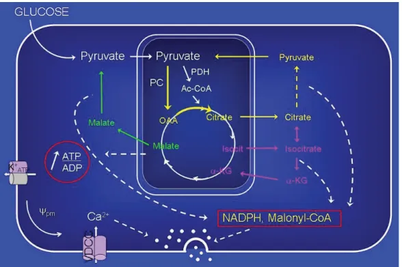
Figure 7 pyruvate cycling and insulin secretion. Pyruvate-derived from glucose metabolism enters in the tricarboxylic acid cycle via its carboxylation by PC, producing oxaloacetate (OAA). This anaplerotic process leads to the formation and accumulation of citrate within the mitochondria. At increasing levels, mitochondrial citrate is exported into the cytoplasm in exchange with malate. Once in the cytosol, citrate is cleaved by ACL, producing pyruvate, leading to the production of NADPH. Pyruvate re-enters the mitochondria via the pyruvate transporter. The pyruvate/citrate cycling together with glucose-derived pyruvate leads to the net synthesis of malonyl-CoA, NAD+, NADPH, and indirectly ATP, which increased insulin secretion.
1.2.3.2 NADPH, Adenine and Guanine Nucleotides and Glutamate
Ample evidence implicates cytosolic NADPH as a MCF. GSIS positively correlates with the NADPH/NADP ratio and NADPH directly induces exocytosis in single beta cells. Recent studies (Reinbothe et al., 2009) indicate that NADPH acts via glutaredoxin-1 that may control exocytotic effectors and/ or voltage K+ channels Kv 2.1 to promote the rise of
Ca2+. Several studies demonstrated that ATP and ADP act as MCF in the beta cell. It has also been suggested that mitochondrial GTP positively regulates GSIS via mitochondrial Ca2+ handling (Kibbey et al., 2007). Glucose-derived glutamate, via the glutamate dehydrogenase reaction, was proposed to act as MCF. Thus, activating mutations in GDH are associated with hyperinsulinemia (Kranendijk et al., 2010). However, the view that glutamate as a MCF has been challenged (Malaisse et al., 1979), and the evidence indicates that anaplerotic oxidative deamination of glutamate via GDH, producing alpha-ketoglutarate in the Krebs cycle (Agarwal et al., 2006), is implicated in this type of hypersulinemia.
1.2.3.3 Lipid metabolism dependent amplification of beta cell metabolic signaling Glucose metabolism leads to a rise in MalCoA, which inhibits carnitine palmitoyltransferase-1 (CPT-1) and blocks beta-oxidation and thus causes a fuel shift in beta cells from fatty acid to glucose utilization (figure 8). Overwhelming support for the MalCoA/CPT-1 interaction and fatty acyl-CoA, or their derivations such as diacylglycerol (DAG), in beta cell fuel stimulation was provided by our lab and others. Thus, overexpression of MalCoA decarboxylase (MCD) in the cytosol or a MalCoA-insenstive CPT-1 mutant in the beta cell curtailed GSIS (Roduit et al., 2004). Nutrient inhibition of fatty acid oxidation is accompanied by elevated glycerolipid (GL) formation and significant increases in the total mass of DAG, triacylglycerol (TG) and phosphatidic acid (PA) were shown to occur in glucose-stimulated beta cells. Inhibition of beta oxidation by MalCoA
leads to a rise in cytosolic FACoA, and, as far as signaling is concerned, FACoA, FFA themselves and complex lipids (DAG, lysophophatidic acid and PA) are potential MCF, as these compounds directly influence signaling pathways and secretion. MalCoA/lipid signaling is important not only in beta cells as we initially proposed, but also in the control of insulin action, appetite and body weight.
Figure 8 Interactions between glucose and fatty acid metabolism in nutrient-secretion coupling. This model illustrates the synergistic interaction between glucose and FA metabolism and the generation of lipid signaling molecules that augment GSIS. Glucose gives rise to pyruvate, which, when channeled through pyruvate dehydrogenase (PDH), contributes to induction of insulin secretion via ATP production and the KATP-dependent triggering pathway. Pyruvate alternatively can be channeled via pyruvate carboxylase (PC) into the anaplerosis/cataplerosis pathway, which contributes to increases in cytosolic oxaloacetate (OAA) and citrate. Glucose increases malonyl-CoA (Mal-CoA), which blocks FA oxidation by inhibiting CPT-1. Inhibition of FA oxidation allows LC-CoA esters to accumulate in the cytosol. LC-CoAs are formed from FFAs supplied externally or produced by the lipolysis of endogenous TG. LC-CoAs can be esterified with glycerol-3-phosphate (Glyc-3-P) to form complex lipids such as TG, DAG, and phospholipids (PL). Glucose also