Université de Montréal
LOX and LOX-Like Proteins as Potential Therapeutic
Target for Atrial Fibrillation
par
Doa’a Ghazi Al-u’datt
Département de Pharmacologie et Physiologie, Université de Montréal Faculté de Médecine
Thèse présentée à la Faculté des études supérieures en vue de l’obtention du grade de PhD
en Physiologie
Janvier, 2019
Université de Montréal
Facult
éde
MédecineCette thèse intitulée:
LOX and LOX-Like Proteins as Potential Therapeutic
Target for Atrial Fibrillation
Présentée par:
Doa’a Ghazi Al-u’datt
A été évaluée par un jury composé des personnes suivantes:
Dr. Yahye Merhi
Président-rapporteur
Dr. Stanley Nattel
Directeur de recherche
Dr. Bruce Allen
Co-directeur de recherche
Dr. Guy Rousseau
Membre du jury
Dr. Zamaneh Kassiri
Examinateur externe
Dr.
René Cardinal
Représentant du doyen
Résumé
Les lysyl-oxydases (LOX) et LOX-like (LOXL-1, 2, 3 et 4) influencent le remodelage de la matrice extracellulaire (MEC) lors d’anomalies cardiaques comme l’insuffisance cardiaque (IC) ou la fibrose. L’objectif principal était d’étudier les fonctions matricedépendantes et -indépendantes des LOX et LOXL dans la transduction des signaux favorisant la fibrose et la fibrillation atriales (FA). Dans l’oreillette gauche (OG) de chiens IC, nous avons observé une augmentation de la régulation de : LOX et LOXL-1 dans le tissue atrial, LOX et LOXL-2 dans les fibroblastes ainsi que LOX, LOXL-1, LOXL-3 et LOXL-4 dans les myocytes. Nous avons évalué le rôle des isoformes des LOX dans la signalisation de la fibrose et de la FA dans l’OG de rats avec infarctus du myocarde (IM). Chez le chien et les cellules cardiaques de rats néonataux, nous avons exploré le rôle des LOX et LOXL sur la fonction matrice-dépendante et -indépendante des fibroblastes et myocytes cardiaques, en utilisant un traitement à l’angiotensine-II (Ang II) et un knockdown par si-ARN des isoformes de LOX.
L’augmentation de l’expression des ARNm de LOX et LOXL était associée à une augmentation de l’expression des ARNm de COL 1A1, FN 1, TGF-β1, CTGF, periostin, α-SMA et MMP-2 dans la zone infarcie du ventricule gauche (VG). L’expression des ARNm de LOXL-1, LOXL-3, COL 1A1, TGF-β1 et periostin était significativement augmentée dans l’OG. L’administration de β-aminopropionitrile (BAPN) post-IM a diminué significativement l’expression des ARNm de LOXL-1,2,3. Le BAPN a aussi diminué l’expression d’ARNm de marqueurs pro-fibrotiques dans l’OG. Ces changements n’étaient pas significatifs dans le VG. Le BAPN a diminué la fibrose dans l’OG ainsi que le ratio de cross-linking du collagène, mais pas significativement dans le VG. Le BAPN a réduit les remodelages structuraux et fonctionnels
de l’OG sans influencer significativement ceux-ci dans le VG. L’IM était associé à une augmentation de la durée de l’onde P ainsi que la durée et l’inductibilité des FA que le BAPN a significativement réduit. Chez des rats néonataux, des fibroblastes et des myocytes de ventricule ont été mis en culture et traités à l’Ang II.
Les LOX et LOXL-2 sécrétées et le ratio de cross-linking du collagène ont été augmentés; et contribueraient au remodelage de la MEC. Chez le chien, la contractilité des myocytes de l’OG a été augmentée suivant le knockdown de LOX et LOXL-1 associé à des changements de concentrations calciques. Dans les fibroblastes, le knockdown de LOXL-3 a réduit l’expression des ARNm de LOXL-2,3,4, associé à une réduction de la prolifération cellulaire et de l’expression des ARNm de COL 1A1, COL 3A1 et CCNE 2. Ces résultats suggèrent que LOXL-2,3,4 influenceraient la prolifération des fibroblastes et la synthèse du collagène. Le knockdown de LOXL-4 a augmenté ratio de l’expression en ARNm de BAX/BLC-2, ainsi LOXL-4 pourrait avoir un effet protecteur contre l’apoptose.
En conclusion, les LOX et LOXL sont des médiateurs potentiels de la fibrose et la FA dans l’OG chez le rat infarci. Les LOX et LOXL seraient alors impliqués dans la régulation des fonctions des fibroblastes et myocytes cardiaques.
Mot-clefs: Insuffisance cardiaque, Fibrillation atriale, fibrose, Lysyl-oxydase (LOX), LOX-like
Abstract
Lysyl oxidase (LOX) and LOX-like (LOXL-1, 2, 3 and 4) proteins have a crucial role in extracellular matrix (ECM) remodeling in several types of heart disease, such as heart failure (HF) and fibrosis. The main objective of this thesis was to address the matrix-dependent and matrix-independent functions of LOX and LOXL proteins in signal transduction, leading to atrial fibrosis and atrial fibrillation (AF). We noted upregulation of LOX and LOXL-1 in tissues, LOX and LOXL-2 in fibroblasts and LOX, LOXL-1, LOXL-3 and LOXL-4 in myocytes of the canine left atrium (LA) in congestive heart failure (CHF). Based on these findings, we studied the roles of LOX isoforms as upstream targets in the signaling pathways of LA fibrosis and AF in a rat model following myocardial infarction (MI). Additionally, we explored the physiological roles of LOX and LOXL proteins in matrix-dependent and matrix-independent functions of cardiac fibroblasts and myocytes through angiotensin II (Ang II) treatment and siRNA-mediated knockdown of LOX isoforms in canine and neonatal rat cells.
Upregulation of the mRNA expression of LOX and LOXL was accompanied by an increase in mRNA expression of COL 1A1, FN 1, TGF-β1, CTGF, periostin, α-SMA and MMP-2 in the infarcted area of the left ventricle (LV). mRNA expression of LOXL-1, LOXL-3, COL 1A1, TGF-β1 and periostin were significantly increased in the LA post-MI. Administration of β-aminopropionitrile (BAPN) post-MI significantly reduced the mRNA expression of LOXL-1, LOXL-2 and LOXL-3 along with a decrease in the mRNA expression of several profibrotic markers in the LA without significant changes to those in the LV. Moreover, the administration of BAPN post-MI reduced LA fibrosis and the collagen cross-linking ratio without significantly changing those in the LV. BAPN administration post-MI reduced the adverse structural and
functional remodeling of LA without significantly changing those in the LV. Furthermore, MI caused an increase in the P-wave duration, AF duration and AF inducibility, while the values of those parameters were significantly reduced upon BAPN administration post-MI.
The protein expression of secreted LOX and LOXL-2 were increased in cultured neonatal rat ventricular fibroblasts and myocytes along with an increase in the collagen cross-linking ratio in fibroblasts upon treatment with Ang II. The secretion of LOX and LOXL-2 from cardiac fibroblasts and myocytes may contribute to ECM remodeling. Moreover, the contractility of canine LA myocytes was enhanced upon knockdown of LOX or LOXL-1 along with slight changes in Ca2+ transients. Upon knockdown of LOXL-3 in fibroblasts, LOXL-2, LOXL-3 and
LOXL-4 mRNA expression levels were reduced along with reduced proliferation and mRNA expression of COL 1A1, COL 3A1 and CCNE 2. The results showed that LOXL-2, LOXL-3 and LOXL-4 may promote fibroblast proliferation and collagen synthesis. LOXL-4 knockdown increased the mRNA expression of the BAX/BCL-2 ratio, suggesting that LOXL-4 may protect against apoptosis in cardiac fibroblasts and myocytes.
In conclusion, LOX and LOXL proteins are prominent mediators of LA fibrosis and AF post-MI. Additionally, these findings suggested that LOX and LOXL proteins are implicated in the regulation of various aspects of cardiac fibroblast and myocyte functions.
Keywords: Heart failure, Atrial fibrillation, Fibrosis, Lysyl oxidase (LOX), LOX-like (LOXL)
Table of contents
Résumé ... iii
Abstract ... v
Table of contents ... vii
List of tables ... xii
List of figures ... xiii
List of abbreviations ... xxiii
Acknowledgements ... xxx
Chapter 1: Introduction ... 1
Part I– Cardiac pathophysiology and management ... 2
1. Overview ... 2
2. Cardiac anatomy and physiology ... 3
2.1 Cardiac structure and function ... 3
2.2 Cellular components ... 6
2.2.1 Cardiomyocytes ... 6
2.2.2 Fibroblasts ... 7
2.2.3 Other cardiac cell types ... 8
2.3 Cardiomyocyte‒fibroblast communication ... 8
2.4 Cardiac ECM components ... 9
2.4.1 Collagen ... 10
2.4.2 Other ECM proteins ... 11
3. Cardiac remodeling ... 12
4. Atrial fibrillation (AF) ... 13
4.1 Risk factors ... 16
4.2 Mechanisms ... 17
4.2.1 Ca2+ handling abnormalities ... 18
4.2.2 Electrical remodeling ... 19
4.2.3 Autonomic nerve remodeling ... 20
4.2.4 Structural remodeling... 20
4.3 Complications ... 21
5.1 Pathogenesis ... 21
5.2 Differentiation of fibroblasts into myofibroblasts ... 23
5.3 Molecular pathways ... 25 5.3.1 TGF-β ... 25 5.3.2 Ang II ... 26 5.3.3 ET-1 ... 27 5.3.4 PDGF ... 27 5.3.5 miRNAs ... 28 5.3.6 CTGF ... 28 5.3.7 MMPs and TIMPs ... 29 5.3.8 Oxidative stress ... 29
5.4 Types of cardiac fibrosis ... 30
5.4.1 Replacement fibrosis ... 30
5.4.2 Ventricular interstitial fibrosis ... 30
5.4.3 Atrial interstitial fibrosis ... 31
6. Myocardial infarction (MI) ... 35
6.1 Stages of post-MI healing ... 36
6.1.1 Early inflammatory stage ... 36
6.1.2 Mid-proliferative stage... 36
6.1.3 Late maturation stage ... 37
6.2 MI-associated complications ... 38
7. Pharmacological therapies targeting cardiac fibrosis and AF ... 38
Part II– Role of the lysyl oxidase enzyme family in cardiac function and disease ... 41
Article 1 ... 41 Contributions of authors ... 41 Abstract ... 43 Abbreviations ... 44 1. Introduction ... 45 2. Historical overview ... 47
3. LOX-family structure and biochemical function ... 48
3.2 LOX-dependent enzymatic reactions in heart... 49
3.3 Biosynthesis, secretion and activation of LOX... 50
4. Overview of LOX expression and function in different systems ... 51
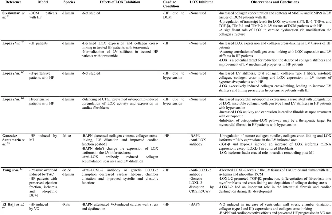
5. Role of LOX-family enzymes in heart disease ... 52
5.1 Role of LOX-family enzymes in ventricular dysfunction ... 52
5.2 LOX-family enzymes in atrial disorders... 54
6. Therapeutic modulation of LOX-family protein function in cardiac diseases ... 55
7. Conclusions ... 58
References ... 58
Part III– Hypothesis and objectives ... 83
1. Thesis rationale ... 83
2. Thesis hypotheses ... 84
3. Objectives ... 84
Chapter 2: Targeting the lysyl oxidase protein signaling pathway in atrial fibrosis and fibrillation ... 85 Contributions of authors ... 86 Abstract ... 88 1. Introduction ... 89 2. Methods... 91 2.1 Animal Models... 91 2.1.1 Dog model ... 91 2.1.2 Rat model ... 92 2.2 MI surgery ... 92
2.3 Assessment of transthoracic echocardiography ... 93
2.4 In vivo electrophysiological study via trans-jugular stimulation ... 95
2.5 Fibrosis quantification by Masson’s trichrome staining ... 96
2.6 RNA extraction, reverse transcription (RT) and quantitative real-time polymerase chain reaction (qPCR) ... 97
2.7 Protein quantification using immunoblot analysis ... 98
2.8 Soluble, insoluble and cross-linking collagen analysis... 99
3. Results ... 101
3.1 Increased LOX family expression in LA tissues of CHF dogs ... 101
3.2 Administration of BAPN post-MI in rats decreased the P-wave duration, AF duration and AF inducibility ... 101
3.3 Administration of BAPN post-MI in rats attenuated LA fibrosis without changing LV fibrosis ... 102
3.4 BAPN administration decreased MI-induced LA remodeling in rats ... 103
3.5 BAPN administration had no effect on MI-induced LV remodeling in rats... 104
3.6 Administration of BAPN post-MI in rats attenuated LA collagen cross-linking without a change in LV collagen cross-linking ... 106
3.7 Administration of BAPN post-MI in rats decreased the mRNA expression of some profibrotic markers in the LA tissues ... 106
3.8 Administration of BAPN post-MI in rats had no effect on the mRNA expression of profibrotic markers in LV tissues ... 107
3.9 Administration of BAPN post-MI in rats had no effect on the protein expression of LOX and LOXL proteins in LA and LV tissues... 108
4. Discussion ... 108
5. Conclusions ... 114
References ... 114
Chapter 3: Characterization of matrix-dependent and matrix-independent roles for lysyl oxidase (LOX) and LOX-like (LOXL) proteins in regulating cardiac fibroblast and myocyte functions ... 146 Contributions of authors ... 147 Abstract ... 149 1. Introduction ... 150 2. Methods... 152 2.1 Animal Model ... 152
2.2 Cardiac fibroblast and myocyte isolation ... 152
2.3 Cell culture and treatments ... 154
2.4 Cell transfection ... 154
2.6 Protein quantification by immunoblotting ... 156
2.7 Determination of mRNA by qPCR ... 157
2.8 Soluble and insoluble collagen analysis ... 158
2.9 Measurement of Ca2+ fluorescence and cell shortening ... 158
2.10 Statistical analysis ... 159
3. Results ... 159
3.1 Increased LOX and LOXL protein expression in LA fibroblasts and myocytes of CHF dogs ... 159
3.2 Ang II increased LOX and LOXL-2 secretion from neonatal rat ventricular fibroblasts and myocytes ... 160
3.3 Ang II stimulated collagen cross-linking and BAPN attenuated collagen cross-linking in neonatal rat ventricular fibroblasts ... 161
3.4 LOX and LOXL immunoreactivity was reduced by specific siRNA approaches in neonatal rat ventricular fibroblasts and myocytes ... 162
3.5 Knockdown of individual LOX isoforms altered the expression of profibrotic markers in neonatal rat ventricular fibroblasts ... 163
3.6 Knockdown of individual LOX isoforms altered proliferation and the expression of proliferation and apoptotic markers in neonatal rat ventricular cells ... 163
3.7 Knockdown or overexpression of individual LOX isoforms altered cell shortening with little change in Ca2+ transients in canine LA myocytes ... 164
4. Discussion ... 165
5. Conclusions ... 169
References ... 170
Chapter 4: General discussion and conclusions ... 199
1. Major findings and original contribution to the literature ... 200
2. Discussion and relationship to prior work area... 201
3. Potential limitations ... 206
4. Future research directions ... 207
5. Conclusions ... 209
List of tables
Chapter 1
Part II
Table 1. Summary of investigations of LOX-family isoforms in heart failure (HF) ... 78 Table 2. Summary of investigations of LOX-family isoforms in hypertrophic cardiac conditions
... 80
Table 3. Summary of investigations of LOX-family isoforms in atrial disease and atrial
fibrillation (AF) ... 82
Chapter 2
Table 1. Effect of BAPN administration on echocardiographic parameters (ventricular and atrial
structural and functional remodeling) post-MI in rats ... 124
Table S1. Sequences of custom-made SYBR Green primers used in this study ... 136 Table S2. Statistical linear regression comparison (slopes and Y-intercepts) between WMSI and
echocardiographic parameters in MI and MI+BAPN rats ... 137
Chapter 3
Table S1. Sequences of siRNA for LOX isoforms used in this study ... 190 Table S2. Sequences of custom-made SYBR Green primers used in this study ... 191
List of figures
Chapter 1
Part I
Figure 1. Anterior view of the human heart. This figure shows the cardiac chambers, valves
and major vessels ... 5
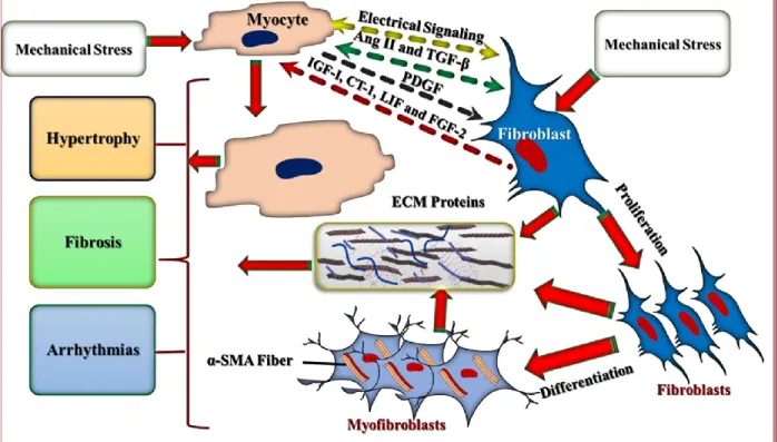
Figure 2. Fibroblast-cardiomyocyte interactions. Fibroblasts and cardiomyocytes respond to
mechanical stress and communicate via electrical and chemical coupling. Several cytokines and growth factors act in the paracrine and/or autocrine manner and induce fibrosis, arrhythmias and cardiomyocyte hypertrophy ... 14
Figure 3. Illustration of heart with normal sinus rhythm and atrial fibrillation (AF).
Cardiac rhythm is initiated by the sinoatrial (SA) node causing atrial contraction, followed by atrioventricular conduction through the atrioventricular (AV) node and His-Purkinje system, leading to ventricular contraction. AF causes highly irregular and rapid atrial contraction ... 15
Figure 4. Schematic diagram for the pathophysiology of atrial fibrillation, including clinical risk factors, mechanisms (Ca2+ handling abnormalities, structural, autonomic nerve (sympathetic activation) and electrical remodeling) and complications. Focal ectopic firing usually results from DADs that produce a spontaneous
action potential. The susceptible re-entry substrate needs shortening of refractoriness and disturbances in conduction ... 22
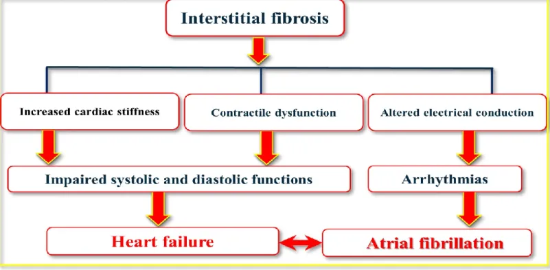
Figure 5. Schematic showing the proposed signaling pathways of cardiac fibrosis ... 33 Figure 6. Overview of cardiac interstitial fibrosis complications contributing to heart failure and atrial fibrillation... 34
Part II
Figure 1. Schematic representation of collagen biosynthesis and cross-linking. Following
translation, procollagen α-chains are imported into the endoplasmic reticulum (ER) and Golgi apparatus to form triple-helical procollagen (two α1-chains and one α2-chain).
These immature collagen helices are secreted into the extracellular space and then converted to mature collagen through cleavage by procollagen N-proteinase (PNPase) and C-proteinase (PCPase). Mature collagen fibrils are self-assembled and then cross-linking is initiated by enzymatic (lysyl oxidase (LOX) family) and non-enzymatic (advanced glycation end products (AGEs)) processes ... 73
Figure 2. Structures of lysyl oxidase (LOX) and LOX-like proteins (LOXL-1, LOXL-2, LOXL-3 and LOXL-4) ... 74 Figure 3. Mechanism of collagen cross-linking as catalyzed by lysyl oxidase (LOX) enzymes. (A) Sequences of peptidyl lysine and hydroxylysine in collagen. (B) Oxidation
of peptidyl lysine and hydroxylysine to peptidyl aldehyde (allysyl and hydroxyallysyl) in collagen. (C) Condensation of peptidyl aldehyde (allysyl and hydroxyallysyl) and lysine to dehydrolysinonorleucine and aldol. (D) Maturation of dehydrolysinonorleucine and aldol condensation products to pyridinoline and pyrrole ... 75
Figure 4. Schematic representation of biosynthesis, secretion and activation of lysyl oxidase (LOX) enzyme in heart tissues. LOX gene is transcribed in the nucleus, LOX
mRNA is translated, pre-protein (pre-pro-LOX) enters the endoplasmic reticulum (ER), and is transported as a prolysyl oxidase (pro-enzyme) from the ER to the Golgi apparatus. In the Golgi apparatus, glycosylation occurs, followed by association with cellular copper and formation of a lysine tyrosylquinone (LTQ) cofactor. Prolysyl oxidase is cleaved between Gly168 and Asp169 at the surface of cardiac cells by procollagen C-proteinase (PCPase; bone morphogenetic protein 1 (BMP-1)) to form the active (mature) LOX (30 kDa) and LOX propeptide (18 kDa), which are then secreted into the extracellular matrix (ECM). Transforming growth factor beta (TGF-β) and prostaglandin E2 modulate LOX mRNA transcription ... 76
Figure 5. Schematic overview of the principal role of LOX-family enzymes in heart disease
... 77
Chapter 2
Figure 1. In vivo experimental timeline. β-aminopropionitrile (BAPN) was administered to
and day 27 post-MI. All animals were subjected to electrophysiological study prior to sacrifice... 125
Figure 2. Increased protein expressions of lysyl oxidase (LOX) and LOX-like protein 1 (LOXL-1) in the left atrial (LA) tissues of congestive heart failure (CHF) dogs. Basal
relative protein expressions of LOX isoforms in the LA tissues (n = 5) of a CHF (2 weeks of ventricular tachypacing (VTP)) model were quantified by Western blot analysis, including (A) band intensities of Western blot images were normalized to glyceraldehyde 3-phosphate dehydrogenase (GAPDH), (B) LOX, (C) 1, (D) 2, (E) LOXL-3 and (F) LOX-4 ... 126
Figure 3. Administration of β-aminopropionitrile (BAPN) post-myocardial infarction (MI) in rats decreased P-wave duration, atrial fibrillation (AF) duration and AF inducibility. Electrophysiological (A-C) and electrocardiogram (ECG; D-H)
measurements for four rat groups (Sham; n = 16-18, Sham+BAPN; n = 16-18, MI; n = 16-22; MI+BAPN; n = 16-18) were assessed at day 28 post-surgery, including (A) AF inducibility, (B) AF duration, (C) effective refractory period (ERP), (D) P-wave duration, (E) P-P interval, (F) P-R interval, (G) QRS duration and (H) QT duration ... 127
Figure 4. Administration of β-aminopropionitrile (BAPN) post-myocardial infarction (MI) in rats attenuated left atrial (LA) fibrosis without changing the left ventricular (LV) fibrosis. LA and LV fibrosis in four rat groups (Sham; n = 6, Sham+BAPN; n = 6, MI
from remote (Rem) and infarct (Inf) areas (MI, MI-Rem and MI-Inf; n = 5) and MI+BAPN (MI+BAPN, MI+BAPN-Rem and MI+BAPN-Inf; n = 6)) was evaluated on day 28 post-surgery, including (A) representative Masson’s trichrome staining images of LA and LV tissue sections for fibrosis quantification, (B) LA fibrous tissue quantification, (C) LV fibrous tissue quantification and (D) LV scar area ... 128
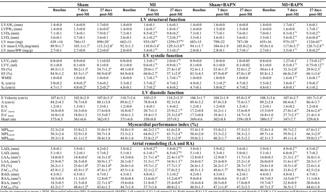
Figure 5. β-aminopropionitrile (BAPN) administration decreased myocardial infarction (MI)-induced left atrial (LA) remodeling in rats. LA structural and functional
remodeling in four rat groups was assessed by echocardiography, including Sham (n = 21), Sham+BAPN (n = 18-20), MI (n = 23-24) and MI+BAPN (n = 22), on day 27 post-MI. (A) LA diameter at end systole (LADs), (B) LA diameter at end diastole (LADd),
and (C) fractional shortening (FS). Comparison between echocardiographic assessment of the wall motion score index (WMSI) and LA structural and functional parameters in MI rats treated with vehicle or BAPN on day 27 post-surgery. Linear correlations of WMSI with (D) LADs, (E) LADd, and (F) FS ... 129 Figure 6. β-aminopropionitrile (BAPN) administration had no effect on myocardial infarction (MI)-induced left ventricular (LV) remodeling in rats. LV structural and
functional remodeling in four rat groups was assessed by echocardiography, including Sham (n = 17-21), Sham+BAPN (n = 18-20), MI (n = 18-24) and MI+BAPN (n = 14-22), on day 27 post-surgery. (A) LV diameter at end systole (LVDs), (B) LV diameter at
end diastole (LVDd), (C) LV mass, (G) ratio of early diastolic transmitral filling velocity
(E) to atrial transmitral filling velocity (A), (H) ejection fraction (EF) and (I) wall motion score index (WMSI). Comparison between echocardiographic assessment of the WMSI and LV structural and functional parameters in MI rats treated with vehicle or BAPN on day 27 post-MI. Linear correlations of WMSI with (D) LVDs, (E) LVDd, (F) LV mass, (J)
ratio of E/A and (K) EF ... 130
Figure 7. Administration of β-aminopropionitrile (BAPN) post-myocardial infarction (MI) in rats attenuated collagen cross-linking in the left atrium (LA) without changing that in the left ventricle (LV). Collagen content in the LA and LV from four rat groups
(Sham; n = 6, Sham+BAPN; n = 6, MI from remote (Rem) and infarct (Inf) areas (MI, MI-Rem and MI-Inf; n = 6) and MI+BAPN (MI+BAPN, MI+BAPN-Rem and MI+BAPN-Inf; n = 6)) was measured by the QuickZyme assay on day 28 post-surgery, including (A) soluble collagen in LA tissues, (B) insoluble collagen in LA tissues, (C) collagen cross-linking ratio in LA tissues, (D) soluble collagen in LV tissues, (E) insoluble collagen in LV tissues and (F) collagen cross-linking ratio in LV tissues ... 131
Figure 8. Administration of β-aminopropionitrile (BAPN) post-myocardial infarction (MI) in rats decreased the mRNA expression of several profibrotic markers in left atrial (LA) tissues. mRNA expression levels of profibrotic markers in the LA tissues from four
rat groups (Sham; n = 6, Sham+BAPN; n = 6, MI; n = 6 and MI+BAPN; n = 6) were quantified by qPCR on day 28 post-surgery, including (A) lysyl oxidase (LOX), (B) LOX-like protein-1 (LOXL-1), (C) LOXL-2, (D) LOXL-3, (E) LOXL-4, (F) collagen 1A1
(COL 1A1), (G) collagen 3A1 (COL 3A1), (H) transforming growth factor β1 (TGF-β1), (I) connective tissue growth factor (CTGF), (J) periostin, and (K) α-smooth muscle actin (α-SMA) ... 132
Figure S1. Increased mRNA expressions of lysyl oxidase (LOX) isoforms in the left atrial (LA) tissues of congestive heart failure (CHF) dogs. Basal relative mRNA (A-E)
expressions of LOX isoforms in the LA tissues (n = 5) of CHF (2 weeks of ventricular tachypacing (VTP)) model were quantified by qPCR, including (A) LOX, (B) LOX-like protein 1 (LOXL-1), (C) LOXL-2, (D) LOXL-3 and (E) LOX-4 ... 138
Figure S2. Effect of β-aminopropionitrile (BAPN) administration post-myocardial infarction (MI) on the surface electrocardiogram (ECG) and atrial fibrillation (AF) induction. (A) representative surface ECG recording and (B) representative recording of
burst pacing-induced AF in four rat groups (Sham, Sham+BAPN, MI and MI+BAPN) at day 28 post-surgery ... 139
Figure S3. β-aminopropionitrile (BAPN) administration decreased myocardial infarction (MI)-induced left atrial (LA) remodeling in rats. LA structural and functional
remodeling in four rat groups was assessed by echocardiography, including Sham (n = 21), Sham+BAPN (n = 18-20), MI (n = 23-24) and MI+BAPN (n = 22) on day 27 post-surgery. (A) LA area at end systole (LAAs), (B) LA area at end diastole (LAAd) and (C)
fractional area changing of LA (FACLA). Comparison between echocardiographic
assessment of the wall motion score index (WMSI) and LA structural and functional parameters in MI rats treated with vehicle or BAPN on day 27 post-MI. Linear correlations of WMSI with (D) LAAs, (E) LAAd, and (F) FACLA ... 140 Figure S4. β-aminopropionitrile (BAPN) had no effect on myocardial infarction
(MI)-induced left ventricular (LV) remodeling in rats. LV structural and functional
remodeling in four rat groups was assessed by echocardiography, including Sham (n = 17-21), Sham+BAPN (n = 18-20), MI (n = 18-24) and MI+BAPN (n = 14-22) on day 27 post-surgery. (A) LV anterior wall thickness at end diastole (LVAWd), (B) LV posterior
wall thickness at end diastole (LVPWd), (C) ratio of LV mass to LV diameter at end
annulus moving velocity during early filling at septal wall (e'Septal,), (E) ratio of E to mitral
annulus moving velocity during early filling at lateral wall (e'Lateral), (F) E wave
deceleration time (EDT), (N) LV volume at end diastole (LVVd), (O) LV volume at end
systole (LVVs), (P) fractional shortening (FS), (Q) septal systolic contractility (SS), (R)
lateral wall systolic contractility (SL) and (S) myocardial performance index (MPIGlobal).
Comparison between echocardiographic assessment of the wall motion score index (WMSI) and LV structural and functional parameters in MI rats treated with vehicle or BAPN on day 27 post-MI. Linear correlations of WMSI with (G) LVAWd, (H) LVPWd,
(I) ratio of LV mass to LVDd, (K) ratio of E to e'Septal (L) ratio of E to e'Lateral, (M) EDT,
(T) LVVd, (U) LVVs, (V) FS, (W) SS, (X) SL and (Y) MPIGlobal ... 141 Figure S5. Administration of β-aminopropionitrile (BAPN) post-myocardial infarction (MI) in rats decreased the mRNA expression profibrotic markers in the left atrial (LA) tissues. Transcript levels of profibrotic markers in the LA tissues from four rat
groups, including Sham (n = 6), Sham+BAPN (n = 6), MI (n = 6) and MI+BAPN (n = 6) were quantified by qPCR on day 28 post-surgery. (A) fibronectin 1 (FN 1), (B) connexin 43 (Cx 43), (C) matrix metalloproteinase 2 (MMP-2) and (D) MMP-9 ... 142
Figure S6. Administration of β-aminopropionitrile (BAPN) post-myocardial infarction (MI) in rats had no effect on the abundance of transcripts for profibrotic markers in left ventricular (LV) tissues. mRNA levels of profibrotic markers in the LV tissues from
four rat groups (Sham; n = 6, Sham+BAPN; n = 6, MI from remote (Rem) and infarct (Inf) areas (MI-Rem and MI-Inf; n = 6) and MI+BAPN (MI+BAPN-Rem and MI+BAPN-Inf; n = 6)) were quantified by qPCR on day 28 post-surgery, including (A) lysyl oxidase (LOX), (B) LOX-like protein-1 (LOXL-1), (C) LOXL-2, (D) LOXL-3, (E) LOXL-4, (F) collagen 1A1 (COL 1A1), (G) collagen 3A1 (COL 3A1), (H) fibronectin 1 (FN 1), (I) transforming growth factor β1 (TGF-β1), (J) connective tissue growth factor (CTGF), (K) periostin, (L) connexin 43 (Cx 43), (M) α-smooth muscle actin (α-SMA), (N) matrix metalloproteinase-2 (MMP-2) and (O) MMP-9 ... 143
Figure S7. Administration of β-aminopropionitrile (BAPN) post-myocardial infarction (MI) in rats had no effect on the protein expression of lysyl oxidase (LOX) isoforms in left atrial (LA) tissues. Western blot analysis was used to evaluate the protein
expression of LOX isoforms in the LA tissues from four rat groups, including Sham (n = 6), Sham+BAPN (n = 6), MI (n = 6) and MI+BAPN (n = 6) on day 28 after surgery. (A) LOX, (B) LOX-like protein-1 (LOXL-1), (C) LOXL-2, (D) LOXL-3, (E) LOXL-4 and (F) representative immunoblot images of protein quantification ... 144
Figure S8. Administration of β-aminopropionitrile (BAPN) post-myocardial infarction (MI) in rats had no effect on the amount of lysyl oxidase (LOX) isoform immunoreactivity in left ventricular (LV) tissues. Western blot analysis was used to
evaluate the LOX isoform immunoreactivity in the LV tissues from four rat groups, including Sham (n = 4), Sham+BAPN (n = 4), MI from remote (Rem) and infarct (Inf) areas (MI-Rem and MI-Inf; n = 4) and MI+BAPN (MI+BAPN-Rem and MI+BAPN-Inf; n = 4) on day 28 after surgery. (A) LOX, (B) LOX-like protein-1 (1), (C) LOXL-2, (D) LOXL-3, (E) LOXL-4 and (F) representative immunoblot images of protein quantification ... 145
Chapter 3
Figure 1. Lysyl oxidase (LOX) and LOX-like (LOXL) immunoreactivity was increased in the left atrial (LA) fibroblasts and myocytes from congestive heart failure (CHF) dogs. Basal immunoreactivity of LOX isoforms in cardiac fibroblasts (Fbs; n = 7; A-E);
(A) LOX, (B) LOXL-1, (C) LOXL-2, (D) LOXL-3 and (E) LOXL-4 and myocytes (CM; n = 7; F-J); (F) LOX, (G) LOXL-1, (H) LOXL-2, (I) LOXL-3 and (J) LOXL-4. (K) Representative Western blots of LOX isoforms from Fbs. (L) Representative Western blots of LOX isoforms from CM ... 178
Figure 2. The abundance of lysyl oxidase (LOX) isoform mRNA expressions were increased in the left atrial (LA) fibroblasts and myocytes from congestive heart failure (CHF) dogs. Basal abundance of LOX isoform transcripts in cardiac fibroblasts
(Fbs; n = 8; A-E); (A) LOX, (B) LOX-like protein 1 (1), (C) 2, (D) LOXL-3 and (E) LOXL-4 and myocytes (CM; n = 9; F-J); (F) LOX, (G) LOXL-1, (H) LOXL-2, (I) LOXL-3 and (J) LOXL-4 ... 179
Figure 3. Angiotensin II (Ang II) increased lysyl oxidase (LOX) and LOX-like protein 2 (LOXL-2) secretion from neonatal rat ventricular fibroblasts and myocytes. Effect
of treatment with different concentrations of Ang II (0.1, 1.0 and 10.0 µM) on the abundance of LOX and LOXL-2 immunoreactivity in conditioned media from cardiac fibroblast cultures (Fbs; n = 4; (A) LOX, (B) LOXL-2, (C) representative Western blots of LOX and (D) representative Western blots of LOXL-2) and myocytes (CM; n = 4; (E) LOX, (F) LOXL-2, (G) representative Western blots of LOX and (H) representative Western blots of LOXL-2) ... 180
Figure 4. Angiotensin II (Ang II) stimulated collagen cross-linking and β-aminopropionitrile (BAPN) attenuated collagen cross-linking in neonatal rat ventricular fibroblasts. Effect of treatment with different concentrations of Ang II (0.1,
1.0 and 10.0 µM; A-C) and BAPN (0.1, 1.0, 10.0 and 100.0 µM; D-F) in fibroblasts on the collagen content, including (A) Ang II doses vs. soluble collagen, (B) Ang II doses vs. insoluble collagen, (C) Ang II doses vs. collagen cross-linking ratio, (D) BAPN doses vs. soluble collagen, (E) BAPN doses vs. insoluble collagen and (F) BAPN doses vs. collagen cross-linking ratio ... 181
Figure 5. Lysyl oxidase (LOX) isoform expression was efficiently suppressed in neonatal rat ventricular fibroblasts using siRNA. The efficiency and specificity of LOX isoform
knockdown was estimated by qPCR (A-E) and validated by Western blot (F). Effect of knocking down individual LOX isoforms in fibroblasts (n = 5) using siRNA on the mRNA of (A) LOX, (B) LOX-like protein 1 (LOXL-1), (C) LOXL-2, (D) LOXL-3, (E) LOXL-4 and (F) representative Western blots of LOX isoforms ... 182
Figure 6. Knockdown of individual lysyl oxidase (LOX) isoforms altered the expression of profibrotic markers in neonatal rat ventricular fibroblasts. Effect of siRNA-mediated
knockdown of individual LOX isoforms in fibroblasts (n = 5) on the abundance of (A) collagen 1A1 (COL 1A1), (B) COL 3A1, (C) fibronectin 1 (FN 1), (D) transforming growth factor β 1 (TGF-β1), (E) connective tissue growth factor (CTGF), (F) periostin, (G) α-smooth muscle actin (α-SMA), (H) matrix metalloproteinase-2 (MMP-2) and (I) MMP-9 mRNA ... 183
Figure 7. Knockdown of individual lysyl oxidase (LOX) isoforms in neonatal rat ventricular fibroblasts altered proliferation and the expression of proliferation and
apoptotic markers. Effect of siRNA-mediated knockdown of individual LOX isoforms
in fibroblasts (n = 5) on (A) cell proliferation and (B) cyclin E2 (CCNE 2), (C) cyclin D1 (CCND 1), (D) B-cell lymphoma 2 (BCL-2)-associate X protein (BAX), and (E) BCL-2 mRNA and (F) the ratio of BAX/BCL-2 mRNA ... 184
Figure 8. Knockdown of individual lysyl oxidase (LOX) isoforms in neonatal rat ventricular myocytes altered the expression of apoptosis markers. Effect of
siRNA-mediated knockdown of individual LOX isoforms in cardiomyocytes (n = 4) on the abundance of (A) B-cell lymphoma 2 (2)-associate X protein (BAX), and (B) BCL-2 mRNA and (C) the ratio of BAX/BCL-BCL-2 mRNA ... 185
Figure 9. Knocking down lysyl oxidase-like protein 1 (LOXL-1) in canine left atrial (LA) myocytes increased cell shortening without altering Ca2+ transients. Effect of
siRNA-mediated knockdown of individual LOX isoforms in cardiomyocytes (n = 5-15 cells) on (A) cell shortening, (B) diastolic Ca2+ level, (C) Ca2+ amplitude and (D) decay time
constant ... 186
Figure S1. Angiotensin II (Ang II) failed to change the abundance of lysyl oxidase (LOX) isoform mRNA or immunoreactivity in neonatal rat ventricular fibroblasts (Fbs).
Effect of treatment with different concentrations of Ang II (0.1, 1.0 and 10.0 µM) in Fbs on the relative quantity of (A) LOX, (B) LOX-like protein 1 (LOXL-1), (C) LOXL-2, (D) 3 and (E) LOX-4 mRNA and the intracellular (cell lysate) of (F) LOX, (G) LOXL-1, (H) LOXL-2, (I) LOXL-3 and (J) LOXL-4 immunoreactivity. (K) Representative Western blots of LOX family proteins ... 192
Figure S2. Angiotensin II (Ang II) increased lysyl oxidase like protein-1 (LOXL-1) immunoreactivity in neonatal rat ventricular myocytes (CM). Effect of treatment with
different concentrations of Ang II (0.1, 1.0 and 10.0 µM) in CM on (A) LOX, (B) LOXL-1, (C) LOXL-2, (D) LOXL-3 and (E) LOXL-4 mRNA and the intracellular (cell lysate) of (F) LOX, (G) LOXL-1, (H) LOXL-2, (I) LOXL-3 and (J) LOXL-4 immunoreactivity. (K) Representative Western blots of LOX family proteins ... 193
Figure S3. β-aminopropionitrile (BAPN) doesn’t change the abundance of lysyl oxidase (LOX) isoform mRNA in neonatal rat ventricular fibroblasts (Fbs). Effect of
treatment with different concentrations of BAPN (0.1, 1.0, 10.0 and 100.0 µM) in Fbs on the abundance of (A) LOX, (B) LOX-like protein 1 (1), (C) 2, (D) LOXL-3 and (E) LOXL-4 mRNA ... 194
Figure S4. Lysyl oxidase (LOX) isoform expression was efficiently suppressed using siRNA in neonatal rat ventricular myocytes. The efficiency and specificity of siRNA-mediated
knockdown of individual LOX isoforms were estimated by qPCR (A-E) and validated by Western blot (F). Effect of siRNA knockdown of LOX isoforms in cardiomyocytes on the abundance of (A) LOX, (B) LOX-like protein 1 (LOXL-1), (C) LOXL-2, (D) LOXL-3 and (E) LOXL-4 mRNA. (F) Representative Western blots of LOX isoforms. mRNA was normalized to that of glyceraldehyde 3-phosphate dehydrogenase (GAPDH) ... 195
Figure S5. The expression of lysyl oxidase (LOX) isoforms was efficiently suppressed by specific siRNAs in canine left atrial (LA) myocytes. The knockdown efficiency and
specificity of LOX isoforms were estimated by qPCR. The effect of siRNA-mediated knockdown of individual LOX isoforms in cardiomyocytes (n = 4) on the abundance of (A) LOX, (B) LOX-like protein 1 (1), (C) 2, (D) 3 and (E) LOXL-4 mRNA... 196
Figure S6. Adenovirally-mediated lysyl oxidase (LOX) overexpression increased LOX mRNA in canine left atrial (LA) myocytes. Effect of overexpression of LOX in
cardiomyocytes (n = 4) using adenovirus (Aden-LOX) on the abundance of (A) LOX, (B) LOX-like protein 1 (LOXL-1), (C) LOXL-2, (D) LOXL-3 and (E) LOXL-4 mRNA ... 197
Figure S7. Overexpression of lysyl oxidase (LOX) in canine left atrial (LA) myocytes reduced cell shortening without altering Ca2+ transients. Effect of LOX
overexpression (n = 5-15 cells) using adenovirus (Aden-LOX) in cardiomyocytes on (A) cell shortening, (B) diastolic Ca2+ level, (C) Ca2+ amplitude and (D) decay time constant
List of abbreviations
2-ΔΔCt: Comparative threshold cycle.
αβ: Integrin receptor α and β subunits.
α-SMA: α-smooth muscle actin.
e': Mitral annulus moving velocity during early filling.
A: Atrial transmitral filling velocity.
ACE: Angiotensin-converting enzyme.
Ac-SDKP: N-acetyl-seryl-aspartyl-lysyl-proline.
Aden-LOX: Adenovirus encoding lysyl oxidase.
Aden-GFP: Adenovirus encoding green fluorescent protein.
AF: Atrial fibrillation.
AGEs: Advanced glycation end products.
ALK: Activin receptor-like kinase.
Ang I: Angiotensin I.
Ang II: Angiotensin II.
Anget: Angiotensinogen.
ANOVA: Analysis of variance.
AP-1: Activator protein 1.
APD: Action potential duration.
ARBs: Angiotensin receptor blockers.
AS: Aortic stenosis.
ATP: Adenosine triphosphate.
AT1R: Ang II type I receptor.
AT2R: Ang II type II receptor.
AV node: Atrioventricular node.
B2m: β2 microglobulin.
BAPN: β-aminopropionitrile.
BAX: BCL-2-associated X protein.
BCL-2: B-cell lymphoma 2.
BMP-1: Bone morphogenetic protein 1.
BW: Body weight.
CCND 1: Cyclin D1.
CCNE 2: Cyclin E2.
CHF: Congestive heart failure.
CM: Cardiomyocyte.
COL 1: Collagen 1.
COL 1A1: Collagen 1A1.
COL 3A1: Collagen 3A1.
CRL: Cytokine receptor-like.
CT-1: Cardiotrophin-1.
CTGF: Connective tissue growth factor.
CVD: Cardiovascular disease.
Cx: Connexins.
DADs: Delayed afterdepolarization.
DCM: Dilated cardiomyopathy.
DDR: Discoidin domain receptor.
E: Early diastolic transmitral filling velocity.
EADs: Early afterdepolarizations.
ECG: Electrocardiogram.
ECM: Extracellular matrix.
EDA: Extra domain A.
EDB: Extra domain B.
EF: Ejection fraction.
EGF: Epidermal growth factor.
EMT: Epithelial-mesenchymal transition.
EndMT: Endothelial-mesenchymal transition.
ER: Endoplasmic reticulum.
ERK: Extracellular signal-regulated kinases.
ERP: Effective refractory period.
ET: Endothelin.
FACLA: Fractional area changing of LA.
FACRA: Fractional area changing of RA.
FBS: Fetal bovine serum.
FGF: Fibroblast growth factor.
FN: Fibronectin.
FS: Fractional shortening.
FSP1: Fibroblast-specific protein 1.
G6PD: Glucose 6-phosphate dehydrogenase.
GAGs: Glycosaminoglycans.
GAPDH: Glyceraldehyde 3-phosphate dehydrogenase.
GJA1 Cx 43: Gap junction protein connexin43.
Grb2: Growth-factor receptor-binding protein 2.
HF: Heart failure.
HFD: High-fat diet.
HIF-1α: Hypoxia-inducible factor 1-α.
HRP: Horseradish peroxidase.
HW/BW: Heart weight/body weight.
ICaL: L-type Ca2+ current.
IFN: Interferon.
IGF-1: Insulin-like growth factor-1.
IK1: Background K+ current.
IKACh: Constitutive acetylcholine-regulated K+ current.
IL: Interleukin.
INa: Na+ current.
Inf: Infarct.
IP3R: Inositol trisphosphate receptor.
Ito: Transient outward K+ current.
JAK: Janus kinase.
JNK: c-jun N-terminal kinases.
Kir: Inwardly rectifying K+ channels.
Kv4.3: Voltage-dependent K+ channel 4.3.
LA: Left atrium.
LAAd: LA area at end diastole.
LADd: LA diameter at end diastole.
LADs: LA diameter at end systole.
LIF: Leukemia inhibitory factor.
LOX: Lysyl oxidase.
LOXL: Lysyl oxidase like proteins.
LTQ: Lysine tyrosylquinone.
LV: Left ventricle.
LVAWd: LV anterior wall thickness at end diastole.
LVDd: LV diameter at end diastole.
LVDs: LV diameter at end systole.
LVPWd: LV posterior wall thickness at end diastole.
LVVd: LV volume at end diastole.
LVVs: LV volume at end systole.
MAPK: Mitogen-activated protein kinase.
MI: Myocardial infarction.
miRNAs: MicroRNAs.
MMPs: Matrix metalloproteinases.
mTORC: Mammalian target of rapamycin complex 1.
MPI: Myocardial performance index.
MRTF: Myocardin-related transcription factor.
MVco: Mitral valve closing to opening.
NADPH: Nicotinamide adenine dinucleotide phosphate.
NCX: Na+-Ca2+ exchanger.
NF-κB: Nuclear factor-kappa β.
NFAT: Nuclear factor of activated T-cells.
NFDM: Non-fat dry milk.
OPN: Osteopontin.
P: Phosphorus group.
PCPase: Procollagen C-proteinase.
PDGF: Platelet derived growth factor.
PGs: Proteoglycans.
PKC: Protein kinase C.
PLC: Phospholipase C.
PMSF: Phenylmethanesulfonyl fluoride.
PNPase: Procollagen N-proteinase.
P/S: Penicillin/streptomycin.
PUFAs: Polyunsaturated fatty acids.
PVDF: Polyvinylidene difluoride.
PW: Pulsed wave.
qPCR: Quantitative real-time polymerase chain reaction.
R: Receptor.
R2: Correlation coefficient.
RA: Right atrium.
RAAd: RA area at end diastole.
RAAs: RA area at end systole.
RAAS: Renin-angiotensin-aldosterone system.
Rac 1 GTPase: Rac family small GTPase 1.
RADs: RA diameter at end systole.
RacET: Transgenic mice with Rac1 overexpression.
Rem: Remote.
ROS: Reactive oxygen species.
RT: Reverse transcription.
RV: Right ventricle.
RyRs: Ryanodine receptors.
S6K1: Ribosomal protein S6 kinase β-1.
SA node: Sinoatrial node.
ScRNA: Scrambled control.
SDS-PAGE: Sodium dodecyl sulfate polyacrylamide gel electrophoresis.
SEM: Standard error mean.
SERCA: SR Ca2+ ATPase.
Shc: Src homologous and collagen protein.
siRNA: Small interfering RNA.
SM22α: Smooth muscle protein 22α.
SMAD: Mothers against decapentaplegic homolog transcription factor.
SOS: Son of sevenless protein.
SPARC: Secreted protein acidic and rich in cysteine
SR: Sarcoplasmic reticulum.
Src: Sarcoma proto-oncogene tyrosine kinase.
SRCR: Scavenger receptor cysteine rich.
SRF: Serum-response transcription factor.
Ss: Septum systolic contractility.
STAT: Signal transducers and activators of transcription.
TAC: Transverse aortic constriction.
TBS: Tris-buffered saline.
TCF: Transcription factor.
TGF-β: Transforming growth factor β.
TgLOX: Transgenic mice with overexpression of LOX.
Thy: Thymus cell antigen.
TIMPs: Tissue inhibitors of metalloproteinases.
TKs: Tyrosine kinases.
TMF: Transmitral flow.
TNF-α: Tumor necrosis factor α.
TSPs: Thrombospondins.
VEGF: Vascular endothelial growth factor.
VO: Volume overload.
VSMCs: Vascular smooth muscles cells.
VTP: Ventricular tachypacing.
Dedication
To my husband.
To my mother and father.
Acknowledgements
In the Name of God, the Most Gracious and the Most Merciful
First and foremost, I would like to express my deepest and sincere appreciation to my supervisor and mentor, Dr. Stanley Nattel, for his flexible attitude, enthusiasm, patience, guidance, encouragement and tremendous support during my PhD study. You always encouraged and taught me to explore an innovative research idea through critical thinking and rigorous questioning. Dr. Nattel offered me a great chance to be one of his research team and I feel lucky to be one of Dr. Nattel’s research team. I would also like to gratefully thank my co-supervisor and mentor, Dr. Bruce Allen, for his support, constructive suggestions, patience, guidance and encouragement throughout this project. I greatly appreciate his support when I was going through some difficulties in biochemistry part and making his laboratory facilities available for me.
I am very grateful to acknowledge my deep thanks for the members of the jury for evaluating this thesis; Dr. Yahye Merhi, Dr. Guy Rousseau, Dr. René Cardinal and Dr. Zamaneh Kassiri. I am also thankful to Dr. Eric Thorin for valuable suggestions during the accomplishment of my project. I would like to thank Dr. Martin Sirois and Cynthia Torok for their support and supervision the histology part. I greatly appreciate the assistance of Dr. Yanfen Shiand Dr. Jean-Claude Tardif in the echocardiographic analysis.
I would gratefully like to express my deep thanks to Dr. Roddy Hiram, an exceptional postdoc in Dr. Nattel’s lab, for his help, time, suggestion, generosity, and great advises. I am also extremely grateful to Dr. Patrice Naud for his patience and help in technically challenging
experiments, I extremely appreciate your feedbacks and advises in writing protocols, manuscripts and presentations as well as your time towards my project. I sincerely thank Dr. Jiening Xiao and Dr. Xiaoyan Qi for their friendship, help, time, patience and support. I am also thankful to Jennifer Anne Bacchi, Dr. Nattel’s administrative assistance, for her support in processing and organizing my documents. My special deep thanks to Chantal St-Cyr, the most honest, organized and helpful person I met in my life. I greatly appreciate the assistance from Nathalie LHeureux in organizing the in vivo studies, lab meeting and animal protocols. I would also like to thank my colleagues in Dr. Nattel’s lab; Dr. Abhijit Takawale, Dr. Donghai Liu, Dr. Hua, Dr. Anna Garcia, Dr. Sirirat Surinkaew, Dr. Martin Aguilar, Dr. Yu Chen, Mozhdeh, Faeze, Xixiao, Dr. Feng Xiong, Dr. Tao Liu, Dr. Jonathan Melka, Eric Duong, Dr. Patrick Vigneault, Dr. Jean-Baptiste Guichard and Dr. Wang Zq as well as from Dr. Allen’s lab, Dr. Sherin Nawaito, Dr. Fatiha Sahmi and Dr. Pramod Sahadevan. I would also like to extend my thanks to Natacha Daquette for performing the MI surgery.
I merely do not have appropriate words to express my deep thanks and appreciation to my grateful husband (Muhammad) and my little kids (Sarah and Amro) whose numerous sacrifices to make my education possible. My truthful and warm appreciation goes to my father (Ghazi), my mother (Abeer), my father-in-law (Hussein), my mother-in-law (Seteh), my late grandmother (Sheika), my aunt (Samieha), my brothers (Bilal, Mohammad and Ahmad), my sisters (Walaa, Razan and Leena), my brothers-in-law (Hashem, Waleed, Ahmad, Rezeq, Anas and Mutasem) and my sisters-in-law (Suha and Fatima) for their understanding, support and encouragement. Financial support, in the form of a scholarship from Jordan University of Science and Technology, is gratefully acknowledged. Finally, I gift this work to all residing in my heart, to whom concerned with my matter, to who’s tried to help even with a smile.
Part I– Cardiac pathophysiology and management
1. Overview
Chronic fibrosis, a common pathological disorder, is characterized by deposition of extracellular matrix (ECM) proteins, which affects many organ systems, such as the skin, lungs, kidneys, heart and liver 1. Furthermore, fibrosis leads to severe stiffness and impaired organ
function 2. Worldwide, 45 % of the total deaths per year are caused by fibrotic diseases 3. The
biological key point of fibrosis is the deposition of cross-linked collagen and elastin within the ECM, which is initiated by enzymatic (lysyl oxidase (LOX) and LOX-like (LOXL) proteins) and non-enzymatic (advanced glycation end-products; AGEs) pathways 4-6. Cardiac fibrosis causes a
substantial proportion of deaths among other types of fibrosis 3. Cardiac fibrosis has been involved
in various forms of cardiovascular disease (CVD). CVD is the main cause of death in the world, representing 31 % of total deaths annually 7. Cardiac fibrosis is initially an adaptive and protective
mechanism. Nevertheless, prolonged fibrosis leads to adverse remodeling and distinct impairment of organ function 1, 8. The incidence of and death from cardiac fibrosis have increased in aged
individuals 8. In the heart, increased ECM protein deposition is caused by several pathological
conditions, such as pressure overload, myocardial infarction (MI), diabetes and cardiomyopathy 9.
Chen and Frangogiannis 10 reported that activated inflammatory and fibrogenic signaling pathways
induce heart failure (HF). HF is associated with significant structural remodeling (fibrosis) of the atria that can lead to an increase in atrial fibrillation (AF) susceptibility 11. Recently, the AF
incidence has reached 13 % among the world population 12. AF is the most common type of
sustained cardiac arrhythmia and is associated with high mortality and morbidity rates 13. AF is
To date, the role of LOX and LOXL proteins in cardiac fibrosis underlying the development of AF are not well understood. The main objective of this thesis was to address the roles of LOX and LOXL proteins in the signal transduction, leading to atrial fibrosis, AF and atrial remodeling. The literature review in this thesis is divided into two parts: part I provides information and discusses topics related to the pathophysiology of AF, cardiac fibrosis and MI. Following this part is a review paper that covers a substantial number of studies related to the role of the LOX family in cardiac function and diseases, which was accepted in Cardiovascular Research Journal (Chapter 1- Part II).
2. Cardiac anatomy and physiology
2.1 Cardiac structure and functionThe heart, a muscular pump, directs oxygenated blood throughout the body through the circulatory system 17-19. The normal human heart beats 100,000 times/day and pumps more than
16,000 liters of blood to all organs 17, 19. The heart consists of four chambers, including two inferior
chambers (right and left ventricles; RV and LV, respectively) and two superior chambers (right and left atria; RA and LA, respectively) 17, 18. The RV and LV are separated by the interventricular
septum, whereas the RA and LA are separated by the interatrial septum 17-19. The heart is encircled
by coronary arteries that carry all of the blood supply to the heart 17, 18. The main coronary arteries
are the left anterior descending, left coronary, right coronary and circumflex coronary 17, 18.
Furthermore, the heart is enclosed by a fibroelastic sac called the pericardium, which is important for cardiac protection from surrounding infection and prevention of heart overfilling 17, 18. The LV
and LA are responsible for maintaining the systemic circulation, while the RV and RA are essential for sustaining the pulmonary circulation 17, 19. The systemic circulation passes the oxygenated
blood to all body parts and returns the deoxygenated blood to the lungs 19-21. The ventricles have
thicker and stronger muscular walls compared with those of the atria, enabling the ventricles to pump the blood to distant areas of the body. Usually, the atria and ventricles exist in either relaxation (diastole) or contraction (systole) status 20, 22. Cardiac output is the volume of ejected
blood by the heart in one minute, with a value of 5-5.5 liters/min during rest 17, 18, 22. Figure 1
illustrates the anatomy of the heart.
The sinoatrial node (SA node) is considered as the pacemaker of the heart and is located in the superior wall of the RA 18. Normally, the SA node spontaneously fires to start stimulating the
atria, followed by the atrioventricular node (AV node) 18. Following AV node stimulation, signals
slowly move to the ventricles to initiate filling of the ventricles 18. Finally, the stimulation proceeds
to the His Purkinje system, leading to a more robust and coordinated contraction of the ventricles
18. Normal cardiac rhythm, sinus rhythm, mediates the accurate stimulation of the whole heart in
the proper sequence 18. However, an arrhythmia is defined as any deviation from sinus rhythm 23.
An electrocardiogram (ECG) records the electrical activity of the heart and serves as an important indicator of several heart diseases 23, 24. A normal ECG shows a single synchronized electrical
wave during beating of the heart 24. In an ECG, a P-wave is produced by atrial depolarization, a
QRS wave is generated by ventricular depolarization and a T wave is produced by ventricular repolarization 24. Differences in the shape of the waves and the distance between the ECG peaks
Figure 1. Anterior view of the human heart. This figure shows the cardiac chambers, valves and
major vessels. The black arrows indicate the normal blood flow direction due to the contraction of the heart chambers.
2.2 Cellular components
The cardiac wall is composed of three layers, including the endocardium, myocardium and epicardium 25, 26. The heart interstitium consists of different cell types, such as cardiomyocytes,
fibroblasts, endothelial cells, smooth muscle cells, macrophages, pericytes and mast cells 27, 28.
Cardiac fibroblasts and myocytes account for 27 % and 56 % of the total cell number in the adult murine heart, respectively 29. Cardiac cells communicate with each other in several ways, including
through direct cell-cell interactions as well as through the paracrine and autocrine effects of released factors 25.
2.2.1 Cardiomyocytes
Cardiomyocytes, the most important cardiac cells, constitute approximately 75 % of the heart tissue volume and are essential for maintaining the automaticity and contractility of the heart 30.
During the fetal life, cardiomyocytes proliferate at a rapid rate and become unable to proliferate immediately after birth 31. Thus, upon neurohormonal stimulation or mechanical stress,
cardiomyocytes are elongated and hypertrophied to maintain the stroke volume 31. Cardiomyocytes
are firmly bound and linked by gap junctions to pass the ions 32. Derived action potentials from
the pacemaker cells stimulate the cardiomyocytes and then proceed to neighboring cardiomyocytes to produce an organized contraction of the ventricles and atria 32. Several factors are implicated in
the hypertrophy of cardiomyocytes during cardiac remodeling, such as transforming growth factor β (TGF-β), angiotensin II (Ang II) and tumor necrosis factor α (TNF-α) 33, 34.
2.2.2 Fibroblasts
Fibroblasts are flat spindle-shaped cells in perivascular and interstitial matrices that originate from circulating fibrocytes, cardiac resident fibroblasts, perivascular cells, epithelial cells via epithelial-mesenchymal transition (EMT), bone marrow-derived progenitor cells or endothelial cells via endothelial-mesenchymal transition (EndMT) 35-39. Fibroblasts are well known as the
most predominant cells in the adult heart 29. Recently, endothelial cells and cardiomyocytes have
been recognized as the most predominant cells among all cardiac cell types 40. Fibroblasts are
found between cardiomyocytes as sheets and strands and have a crucial role in preserving cardiac structure and function 40. During the heart development, fibroblasts activate the proliferation of
cardiomyocytes through the secretion of fibroblast growth factor (FGF) and periostin 1, 28, 41, 42.
Fibroblasts are characterized by a high membrane resistance that makes them an excellent passive follower for electrical signals 1, 39, 43. Cardiomyocyte generates action potential that drives
fibroblast membrane polarization due their limited capacity and high resistance 39, 43. Fibroblasts
don’t generate an action potential in response to electrical stimuli, but they respond to mechanical stimuli with changes in their membrane potential 44-46. Upon exposure of fibroblasts to mechanical
stretch, Ca2+, K+ and Na+ channels are stimulated and depolarize the fibroblast membrane 1, 44-46.
Fibroblasts play numerous roles in the heart, including tissue repair, synthesis and degradation of ECM proteins, inflammation, proliferation, angiogenesis, scar formation and fibrosis 39, 47, 48. Fibroblasts produce ECM components, such as structural proteins (elastin and
collagen), adhesive (fibronectin (FN) and laminin) proteins, glycosaminoglycans (GAGs) and proteoglycans (PGs) 47. Furthermore, fibroblasts regulate ECM degradation through the production
of matrix metalloproteinases (MMPs) and tissue inhibitors of metalloproteinases (TIMPs) 47.
paracrine and autocrine signaling pathways 1, 49, 50. Moreover, fibroblasts activate the synthesis of
vascular endothelial growth factor (VEGF), FGF, connective tissue growth factor (CTGF) and platelet-derived growth factor (PDGF), which have important roles in maintaining proper vascularization of the heart 1, 51. Different stimuli can alter fibroblast functions, such as mechanical,
electrical and biochemical stimuli 52, 53. There are several stimulators of fibroblast proliferation,
differentiation and collagen secretion, such as TGF-β1, vasopressin, PDGF, cardiotrophin-1 (CT-1), FGF 2, endothelin (ET), mast cell-specific proteases (chymase and tryptase), MMPs, TIMPs and Ang II 48, 54-56.
2.2.3 Other cardiac cell types
The coronary artery consists of endothelial cells that participate in tissue healing via stimulation of myofibroblasts and new blood vessel formation 35, 57. Communication of endothelial
cells with fibroblasts modulates the formation of new blood vessels via released VEGF and FGF from fibroblasts during wound healing 39, 58. Travers et al. 59 reported that fibroblasts derived from
circulating progenitor and endothelial cells have a vital role in the fibrotic response. During an injury, macrophages spread and secrete MMPs and cytokines within the myocardium to induce cardiac remodeling 60. Pericytes are located in the perivascular area and are important for
preserving cardiac vessel integrity 61. Additionally, pericytes release several cytokines that can
modify fibroblast status 62.
2.3 Cardiomyocyte‒fibroblast communication
Cardiac fibroblasts are imbedded in the matrix and distributed around the myocytes 39.
contraction and transduction of mechanical and electrical signals 63. Cardiac myocytes and
fibroblasts communicate either directly through gap junctions or indirectly through paracrine factors 63. Moreover, Ongstad and Kohl 43 reported that cardiac fibroblasts have direct
communication with cardiomyocytes via gap junctional proteins called connexins, such as Cx 45, Cx 40 and Cx 43. Cardiac fibroblasts cannot be excited, but they have the ability to transfer currents between myocytes via connexins 64. Cardiac myocytes release several factors that can
alter the functions of fibroblasts and other heart cell types, leading to cardiac fibrosis and hypertrophy 65. Furthermore, cardiac fibroblasts stimulate myocyte proliferation via the ECM,
heparin-binding epidermal growth factor (EGF), FGF and the FN-integrin β1 pathway 28, 42. Ang
II stimulates cardiomyocyte hypertrophy through the secretion of ET and TGF-β1 from fibroblasts
66. However, cardiomyocytes secrete several molecules, such as TGF-β, Ang II and ET that
activate fibroblast differentiation, proliferation and secretion of ECM proteins 66, 67.
Cardiomyocyte-specific Ang II type I receptor (AT1R) knockout leads to decreased fibroblast proliferation around myocytes 67.
2.4 Cardiac ECM components
The ECM is a highly organized acellular network that surrounds fibroblasts, myocytes, and vascular and immune cells 68. It is primarily composed of matricellular proteins, PGs, structural
fibrous proteins, GAGs and adhesive glycoproteins 68. The ECM has various functions in the heart,
such as cell proliferation and motility, structural framework formation, mechanical signal distribution and blood flow modulation 1, 68-70. Additionally, the ECM contains cytokines, MMPs,
TIMPs and growth factors that affect ECM homeostasis by regulating the synthesis and degradation of ECM components 68. In response to cardiac injury, MMPs, TGF-β, VEGF and FGF
are activated and released into the ECM to participate in cardiac remodeling 71. Disruption of ECM
homeostasis following a cardiac injury is the main cause of cardiac fibrosis and dysfunction 72.
2.4.1 Collagen
Collagens, major fibrous ECM proteins, are classified into four groups, namely membrane-associated collagens with interrupted triple helices, fibril-membrane-associated collagens with interrupted triple helices, multiple triple-helix domains and fibrillar collagens (such as types I, II, III, V, and XI) 73. Collagen types I and III are the most predominant structural fibrous ECM proteins and are
synthesized by fibroblasts and myofibroblasts 68. Collagen type I represents approximately 85 %
of the total collagen and is characterized by thick fibers with high tensile strength, while collagen type III constitutes approximately 11 % of the total collagen and is described as thin fibers with elastic properties 74, 75. Collagen type I, a triple helix, is composed of three polypeptide α chains,
including the two α1 chains and one α2 chain 76. The triple helix of collagen type III consists of
three identical α1 chains, and each α chain contains three repeating amino acids (glycine, proline and hydroxyproline) 76.
Collagens are produced by fibroblasts and myofibroblasts as large inactive molecules called preprocollagens and then secreted into the ECM for processing, assembly and cross-linking 77.
Preprocollagens are subjected to cleavage of their signal peptides in the endoplasmic reticulum to form procollagens, followed by packing of these procollagens into vesicles to be released in the extracellular area for further modifications, including the elimination of C- and N- terminal propeptides by procollagen C-proteinase (PCPase) and procollagen N-proteinase (PNPase),
respectively 78. The resulting mature collagen self-assembles to form collagen microfibrils and is
step during collagen processing is essential for increasing the tensile strength and stability of collagen fibers 40, 80-83. The mature collagen fibers are very stable, with a half-life of 80 to 120 days 84. Excessive deposition of collagen in the interstitial space leads to an increase in ventricular
stiffness and impairs diastolic function 84. Kong et al. 85 reported that the changes in ECM
composition stimulate fibroblast migration and proliferation. In the healthy heart, there is a tight balance between the formation of new collagen and degradation of old collagen 84, 86, 87, while the
disruption of this balance leads to great changes in cardiac structure and function 84.
2.4.2 Other ECM proteins
The cardiac ECM also contains less abundant proteins, such as laminin, fibrillin, elastin, other collagen types (IV, V, and VI) and FN 88. Laminin, a glycoprotein, consists of different
binding domains for ECM components, such as cell membrane receptors and collagens 89. Laminin
is primarily produced by cardiomyocytes, endothelial cells and vascular smooth muscle cells (VSMCs) 89, 90. Elastin proteins are important as they provide tissues with elastic properties,
allowing them to be stretched without breakage 47. FN, a glycoprotein, is found as cellular
(insoluble) and plasma (soluble) forms in tissues and controls cell migration and adhesion 91. FN
is composed of homologous domains that are spliced into a longer protein with two extra domains: A and B (EDA and EDB, respectively) 91. Various cardiac cells, such as endothelial cells,
fibroblasts and macrophages, secrete FN 92, 93, which is involved in different cardiac functions,
including ECM stability, mechanotransduction, cell adhesion, collagen deposition and cell migration 94, 95. Moreover, FN plays a crucial role in fibrillin-1 assembly into structural networks 96. Arslan et al. 97 demonstrated that EDA domain deletion attenuates the adverse cardiac