Université de Montréal
Implication des biofilms dans la rhinosinusite chronique et l’évaluation des traitements avec un modèle in vitro
Par Zohra Bendouah
Département de sciences biomédicales Faculté de médecine
Thèse présentée à la Faculté des études supérieures
en vue de l’obtention du grade de maîtrise en sciences biomédicales option recherche clinique
Août, 2008
Université de Montréal Faculté des études supérieures
Cette thèse intitulée :
Implication des biofilms dans la rhinosinusite chronique et l’évaluation des traitements avec un modèle in vitro
Présentée par : Zohra Bendouah
a été évaluée par un jury composé des personnes suivantes :
Président-rapporteur : John S. D. Chan
Directeur de recherche : Martin Desrosiers, MD, FRCSC
Codirecteur : Jean Barbeau Ph D
Résumé
Introduction : La chronicité de la rhinosinusite, sa résistance aux antibiotiques, et ses exacerbations aiguës laissent croire que les biofilms sont impliqués dans la rhinosinusite chronique. Objectifs : Nous avons évalué la capacité des bactéries Pseudomonas
aeruginosa, staphylocoques à coagulase négative et Staphylococcus aureus à former des
biofilms par un essai in vitro, et si cette capacité de formation a un lien avec l’évolution de la maladie. Nous avons évalué in vitro l’effet de la moxifloxacine, un antibiotique utilisé dans le traitement de la rhinosinusite chronique sur des biofilms matures de Staphylococcus
aureus. Méthodes : Trent et une souches bactériennes ont été isolées de 19 patients atteints de rhinosinusite chronique et qui ont subit au moins une chirurgie endoscopique des sinus. L’évolution de la maladie a été notée comme "bonne" ou "mauvaise" selon l’évaluation du clinicien. La production de biofilm a été évaluée grâce à la coloration au crystal violet. Nous avons évalué la viabilité du biofilm après traitement avec la moxifloxacine. Ces résultats ont été confirmés en microscopie confocale à balayage laser et par la coloration au LIVE/DEAD BacLight. Résultat et Conclusion : Vingt deux des 31 souches ont produit un biofilm. La production d’un biofilm plus importante chez Pseudomonas aeruginosa et
Staphylococcus aureus était associée à une mauvaise évolution. Ceci suggère un rôle du
biofilm dans la pathogenèse de la rhinosinusite chronique. Le traitement avec la moxifloxacine, à une concentration de 1000X la concentration minimale inhibitrice réduit le nombre des bactéries viables de 2 à 2.5 log. Ces concentrations (100 µg/ml - 200 µg/ml) sont faciles à atteindre dans des solutions topiques. Les résultats de notre étude suggèrent que l’utilisation de concentrations supérieure à la concentration minimale inhibitrice sous forme topique peut ouvrir des voies de recherche sur de nouveaux traitements qui peuvent être bénéfiques pour les patients atteints de forme sévère de rhinosinusite chronique surtout après une chirurgie endoscopique des sinus.
Mots-clés : Biofilm, rhinosinusite chronique, Pseudomonas aeruginosa, Staphyloccocus
aureus, chirurgie endoscopique des sinus, Microscope confocal à balayage laser,
Abstract
Introduction: The role of biofilms in chronic diseases is increasingly recognized. Chronic rhinosinusitis, with its chronic indolent course, resistance to antibiotics, and acute exacerbations, has an evolution that parallels that of other biofilm-related diseases. Objectives: 1-To develop an in vitro method to assess the biofilm formation capacity. 2- To determine whether biofilm-forming capacity of bacteria demonstrated in chronic rhinosinusitis has an impact on persistence of the disease following endoscopic sinus surgery. 3- To determine the in vitro activity of moxifloxacin against Staphyylococcus
aureus in biofilm form. Method: Thirty-one bacterial strains recovered from 19 patients with chronic rhinosinusitis at least one year post-endoscopic sinus surgery. Evolution of disease was assessed by questionnaire and endoscopy as favorable or unfavorable. The bacteria were cultured on a 96-well culture plaque and a semi-quantitative method using crystal violet to quantify biofilm production was used. Confirmation of the effect of the antimicrobial agents on viability was performed with confocal laser microscopy, using a LIVE/DEAD BacLight staining. Results: Twenty-two of 31 samples produced a biofilm thicker or equal to the positive control. Biofilm formation was associated with a poor evolution for Pseudomonas aeruginosa and Staphylococcus aureus, but not for coagulase-negative staphylococci. Biofilm treated with moxifloxacin at 1000X (0.1mg/ml – 0.2 mg/ml) gave a 2 to 2.5 log reduction in number of viable bacteria. Conclusion: We have shown that Crystal violet method is able to detect biofilm formation. There is a correlation between in vitro biofilm production by Pseudomonas aeruginosa and Staphylococcus
aureus and unfavorable evolution after endoscopic sinus surgery, suggesting a role for
biofilm in chronic rhinosinusitis. Increased concentrations of moxifloxacin, easily attainable in topical solutions have a potential role in the management of biofilm infections. Key words: Biofilm, chronic rhinosinusitis, Pseudomonas aeruginosa, Staphylococcus
aureus, endoscopic sinus surgery, confocal laser scanning microscopy, LIVE/DEAD
Table des matières
Résumé ……….……….…….i
Abstract ……….………..…..ii
Liste des abréviations ………...v
Remerciements ………...vi
Introduction……….…...1-14 Objectifs..……….……….….…..15
Résultats sous forme d’articles
1. Bendouah Z, Barbeau J, Hamad W, Desrosiers M. Use of an in vitro assay for determination of biofilm-forming capacity of bacteria in chronic rhinosinusitis. Am J Rhinol. 2006 Sep-Oct;20(5):434-8………...16-35 2. Bendouah Z, Barbeau J, Hamad WA, Desrosiers M. Biofilm formation by
Staphylococcus aureus and Pseudomonas aeruginosa is associated with an unfavorable
evolution after surgery for chronic sinusitis and nasal polyposis. Otolaryngol Head Neck Surg. 2006 Jun;134(6):991-6…...36-57 3. Desrosiers M, Bendouah Z, Barbeau J. Effectiveness of topical antibiotics on
Staphylococcus aureus biofilm in vitro. Am J Rhinol. 2007 Mar-Apr ;
21(2):149-53………...………..58-75 Résultats non publiés
4. Bendouah Z, Desrosiers M, Barbeau J. Bacterial killing in Staphylococcus aureus biofilm doesn't improve with increasing concentration of moxifloxacin beyond 1000XCMI. (soumis)...…………...76-92
Discussion générale……….……...93-101
Conclusion ……….102-103
La liste des abréviations
CES : chirurgie endoscopique des sinus CMI : concentration minimal inhibitrice CV : crystal violet
DO : densité optique
EPS : substances polymériques extracellulaires FISH : Fluorescence in situ hybridization MCBL : microscope confocal à balayage laser MEB : microscope électronique à balayage. MOXI : moxifloxacine
P. aeruginosa : Pseudomonas aeruginosa RSC : rhinosinusite chronique
S. aureus (Sa) : Staphylococcus aureus SCN : Staphylocoques à coagulase négative UFC : unités formant des colonies
Les remerciements
Je voudrais exprimer ma gratitude aux personnes suivantes :
Un grand merci à mon directeur de recherche monsieur Martin Desrosiers ainsi que mon codirecteur monsieur Jean Barbeau pour leur soutien tout au long de ma maîtrise et pour tout ce qu’ils m’ont appris.
Annie Leduc et Jacynthe Séguin pour leur précieuse assistance au laboratoire.
Je voudrais remercier également monsieur Onder Agbaba pour avoir isolé et identifié les souches bactériennes qui ont servies à notre étude.
Je remercie madame Sylvista Archana de m’avoir bien formée pour l’utilisation de la microscopie confocale.
Introduction
Les biofilms
Depuis l’époque de Koch, les bactériologistes et les cliniciens ont focalisé leurs travaux sur les bactéries planctoniques (sous forme libre), relativement faciles à manipuler au laboratoire car détectables et prédominantes dans les infections aiguës. Par contre, comme on le sait maintenant, seulement une très petite fraction de bactéries se présente sous la forme planctonique ; la majorité des bactéries existent dans la nature sous forme attachée à une surface (biofilm) 1, 2.
Les fossiles retrouvés démontrent que tôt dans l’évolution, les microorganismes ont acquis une capacité à s’organiser en biofilm. La morphologie des biofilms a été identifiée dans des roches sédimentaires qui dataient de 3.5 milliards d’années 3 et dans des dépôts volcaniques de sulfure 4.
La capacité de formation de biofilm chez les microorganismes représente une adaptation acquise durant l’évolution. Une protection était nécessaire pour survivre contre les conditions extrêmes qui régnaient sur terre il y a des milliards d’années (températures extrêmes, pH, rayons UV) 5.
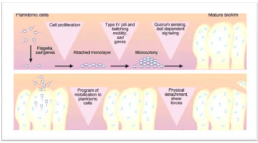
En 1978, John William Costerton a proposé le terme de biofilm en suggérant que ce serait le mode de vie naturel de la plupart des microorganismes. Il l’a définit comme une communauté organisée de microorganismes collaborant entre eux, contenus dans une substance hydratée polymérique extracellulaire (EPS) ou matrice autoproduite, adhéra à une surface inerte ou vivante. Cette matrice qui représente 85% du volume total du biofilm
est constituée essentiellement de polysaccharides, d’acides nucléiques et de protéines 6,7 (Figure 1).
Figure 1. Représentation schématique d’un biofilm 8.
La définition des biofilms a beaucoup évoluée depuis. Elle inclut maintenant l’ensemble des phénomènes où les bactéries adhérent à une surface. La surface d’adhésion des organismes sessiles peut être abiotique (les matériaux inertes : des implants médicaux, des tubes en acier, le sol... etc) ou biotique (les tissus ou cellules vivantes, des cellules épithéliales par exemple).
Canaux liquide
Microcolonies bactériennes
La définition de biofilms qui a généralement été appliquée aux interfaces solide-liquide a évolué pour inclure des interfaces air-liquide, ou pas d’interface définie comme dans le cas des agrégats de bactéries en suspension.
Il n’est cependant pas très clair si le type de surface sur laquelle se développe un biofilm joue un rôle au niveau de son métabolisme et sa physiologie. Par exemple; est ce qu’un biofilm de Pseudomonas aeruginosa attaché à une lame de verre a les mêmes propriétés qu’un biofilm attaché à des cellules épithéliales ou résidant dans un poumon d’un patient atteint d’une fibrose kystique?
La physiologie du biofilm est complexe, on peut le considérer comme un microcosme dans lequel les bactéries se multiplient et s’organisent en microcolonies (Figure 2).
Le microenvironnement joue un rôle important dans le développement ainsi que la structure du biofilm. En effet, le groupe de Stoodley a pu démontrer que des changements hydrodynamiques ainsi que la composition du milieu de culture peuvent influencer la biomasse du biofilm ainsi que plusieurs processus impliqués dans le développement de ce dernier9.
Un des défis concernant la recherche sur les biofilms consiste à comprendre pourquoi les bactéries capables de former un biofilm adhèrent spécifiquement à un certain type de surface, et comment ces bactéries résistent aux différents agents antimicrobiens.
À l’état naturel, les biofilms sont des communautés au sein desquelles on peut trouver plusieurs espèces bactériennes différentes qui sont capables d’échanger leur matériel génétique.
La communication entre les microorganismes dans un biofilm se fait grâce à la détection du quorum, appelée quorum sensing en anglais. C’est un mécanisme de régulation qui contrôle l’expression de certains gènes bactériens. Les bactéries qui utilisent la détection du quorum produisent des signaux moléculaires dits auto-inducteurs, qui contribuent a l’expression (ou la répression) de ces gènes au sein d'une population bactérienne en fonction de la densité de celle-ci.
Les différents mécanismes de résistance du biofilm
La forme planctonique des bactéries peut permettre leur dissémination dans le sang et les tissus. Ce phénomène est plutôt associé aux infections aiguës1.
Cependant, sous forme de biofilm les bactéries agissent en communauté organisée, ce qui augmente leur résistance et parfois leur virulence 5.
Les biofilms sont très résistants aux défenses immunitaires, ils ne sont pas susceptibles aux macrophages et aux anticorps, et sont très résistants aux antibiotiques11, 12. Cette résistance est dûe à plusieurs facteurs.
La matrice d’exopolysaccharide
La matrice d’EPS représente une barrière physique et confère une résistance au biofilm. Le groupe de Stewart suggère qu’elle peut retarder la diffusion d’antibiotiques 13 et diminuer leur diffusion due au gradient osmotique. Elle empêche aussi les neutrophiles ainsi que les anticorps d’accéder aux bactéries 14 et les protège contre les radicaux libres comme les dérivés toxiques de l’oxygène 15.
Plusieurs souches de Staphylococcus aureus produisent le PIA (polysaccharide intercellular adhesin) 16. Ce polysaccharide est constitué de longues chaînes de beta-1,6-glucosamines. Sa synthèse semble être sous le contrôle d’un locus nommé ica (intercellular adhesin). En plus de son rôle dans l’adhésion intercellulaire et l’augmentation de l’adhésion à des surfaces hydrophiles, le PIA est responsable de l’hémagglutination des globules rouges 17, 18.
De nombreuses souches de Pseudomonas aeruginosa produisent une substance muqueuse appelée alginate, c’est un exopolysaccharide produit par le gène algc. L’expression de ce dernier est quatre fois plus élevée en biofilms 19. L’alginate a un rôle protecteur et confère une résistance très élevée au biofilm.
Les bactéries vivant dans un biofilm ont une résistance sensiblement différentes de celles des bactéries planctoniques de la même espèce (Figure 3).
Figure 3. Diagramme d’un biofilm médical.
(a) Les bactéries planctoniques peuvent être éliminées par les anticorps, les phagocytes et sont sensibles aux antibiotiques. (b) Les bactéries adhérentes forment une communauté de biofilm résistante aux anticorps, phagocytes et antibiotiques. (c) Les phagocytes attaquent le biofilm, mais leurs tentatives échouent et les enzymes phagocytaires sont libérées. (d) Les enzymes phagocytaires endommagent les tissus entourant le biofilm et des bactéries peuvent être libérées du biofilm, menant potentiellement à une infection aiguë dans les tissus avoisinants 10.
Des études de phénotype ont démontré une différence de métabolisme entre les bactéries inclues dans le biofilm et les bactéries planctoniques. Les bactéries sous un phénotype de biofilm peuvent exprimer plusieurs gènes qui ne sont pas exprimés par les bactéries de forme planctonique20.
À l’intérieur d’un biofilm toutes les bactéries n’ont pas la même activité métabolique. Les bactéries situées dans les couches les plus profondes du biofilm ont une activité métabolique réduite; certaines bactéries peuvent désactiver leur métabolisme et adopter un état de survie. La division cellulaire est 5 à 15 fois plus lente à l’intérieur d’un biofilm que dans les conditions planctoniques 21.
L’accès limité à ces bactéries au sein du biofilm, ainsi que les modifications de leur activité métabolique, augmentent leur niveau de résistance aux attaques des défenses naturelles et aux antibiotiques.
Les persisteurs
Les antibiotiques qui en général sont efficaces contre les bactéries planctoniques montrent une efficacité réduite envers les infections chroniques5.
L’expérience clinique a montré que lorsque les défenses de l’organisme, ainsi que les traitements antibiotiques, sont insuffisants pour éliminer un biofilm mature 11, 22, la chirurgie est parfois inévitable pour éradiquer l’infection.
Les bactéries d’un biofilm peuvent survivre à des concentrations très élevées d’agents antimicrobiens 53. Ce phénomène est dû à la présence de persisteurs. Ces bactéries sont
génétiquement identiques à la population du biofilm, et sont capables de survivre même après une période prolongée de traitements avec des fortes concentrations d’antibiotiques. Les persisteurs représentent généralement 1% ou moins du total de la population bactérienne 49.
L’hypothèse des persisteurs est la plus récente explication de la résistance des biofilms aux agents antimicrobiens. Il est cependant très difficile de les étudier, à cause de leur nombre très petit d’une part, ainsi que le manque de compréhension au niveau de leur physiologie. Le rôle des biofilms dans les maladies chroniques est maintenant très reconnu. De récentes publications estiment qu’au moins 65% des infections bactériennes humaines impliquent les biofilms 23.
Les biofilms s’avèrent responsable de plusieurs maladies chroniques respiratoire telles que : la fibrose kystique, l’otite séreuse, le choléstéatome et l’adénoïdite chronique 1, 24. Les biofilms sont aussi impliqués dans les infections reliées aux implants médicaux 10.
La rhinosinusite chronique
La rhinosinusite chronique (RSC) est une maladie inflammatoire qui persiste plus de trois mois dans la région des muqueuses nasales et les cavités aérées de la face appelées sinus paranasaux 25. Elle se caractérise aussi par une colonisation bactérienne et une infection. On distingue la rhinosinusite chronique de la rhinosinusite aiguë par la durée des symptômes (Figure 4).
Figure 4. Classification de la rhinosinusite26.
La RSC est parmi les maladies chroniques les plus fréquentes, causant un inconfort chez les patients ainsi que la morbidité 27.
Quatorze pour cent des américains disent souffrir de RSC. Il n’y a pas de statistique indiquant la prévalence de cette maladie au Canada.
Un élément clef dans le développement de la RSC est la dysperméabilité du complexe ostioméatal. C’est une région anatomique située sous le cornet moyen où sont localisés les ostia de drainage des sinus maxillaires, des cellules ethmoïdales antérieures et des sinus frontaux. L’obstruction du complexe ostioméatal entraîne une diminution de la ventilation et du drainage des sinus paranasaux avec comme conséquence une stase de mucus, un risque infectieux et une inflammation de toutes les cavités rhino-sinusiennes.
Intensité des symptômes Exacerbation aiguë de la rhinosinusite chronique Rhinosinusite aiguë Rhinosinusite chronique
Figure 5. Schéma d'une coupe tomodensitométrique frontale du massif facial passant par les ostia des sinus maxillaires. 1. Cavité nasale, 2. Méat inférieur, 3. Sinus maxillaire, 4. Processus unciforme, 5. Région du méat moyen, 6. Cellule de la bulle, 7. Racine d'attache verticale du cornet moyen, 8. Lame criblée, 9. Processus crista galli, 10. Cellule supra-bullaire, 11. Os planum, 12. Canal sous-orbitaire, 13. Ostium du sinus maxillaire et gouttière uncibullaire, 14. Cornet moyen, 15. Cornet inférieur, 16. Septum nasal 28.
Les individus avec des sinusites réfractaires souffrent de symptômes persistants de rhinosinusite chronique même après une chirurgie endoscopique des sinus. La qualité de vie de ces patients est compromise, ils souffrent fréquemment de douleurs faciales et d’écoulement nasal avec des exacerbations qui nécessitent généralement un traitement antibiotique ou même une chirurgie. Le traitement chez ces patients est très difficile dû au manque de compréhension au niveau de la pathophysiologie entourant cette maladie 25.
L’étiologie de la RSC est très probablement multifactorielle. Elle inclut l’asthme, des dysfonctions ciliaires, des déficiences immunitaires, l’obstruction du complexe ostioméatal, des infections virales, bactériennes ou fongiques, la présence de superantigènes (exotoxines) et des facteurs environnementaux 26, 29.
La présence de biofilms dans les sinus pourrait expliquer l’échec des thérapies conventionnelles et de la chirurgie25. En effet, la présence du biofilm au niveau de la muqueuse sinusale chez les patients atteints de RSC fournit une présence continuelle des antigènes (surface des bactéries, éléments fongiques, exotoxines ect.), pouvant résulter en une inflammation chronique au niveau du sinus.
L’inflammation chronique de la muqueuse sinusale génère des dommages tissulaires qui pourraient favoriser la création de surfaces où le biofilm peut adhérer 10. Ce profil inflammatoire de la RSC concorde avec celui d’une infection reliée à la présence de biofilms.
Traitements de la rhinosinusite chronique :
Le traitement de la RSC comprend l’utilisation d’antibiotiques, de décongestionnants, de mucolytiques et de corticostéroïdes topiques 30. Lorsque la RSC ne répond pas aux traitements courants, on a recours à la chirurgie endoscopique des sinus pour enlever les tissus infectés et rétablir l'ouverture et le drainage au niveau des sinus (Figure 6).
Les bactéries sous forme planctonique montrent généralement une sensibilité aux antibiotiques in vitro, ce qui explique l’efficacité de l’antibiothérapie par voie orale dans le traitement de la rhinosinusite maxillaire bactérienne aigue. Cependant, ce même traitement
est particulièrement inefficace chez des patients atteints de rhinosinusite chronique qui ont subi une chirurgie endoscopique des sinus.
L’échec de l’antibiothérapie chez ces patients, l’inflammation chronique des muqueuses sinusales et le taux élevé des cultures stériles laissent croire que les biofilms sont impliqués dans la pathophysiologie de la RSC.
Figure 6. Les changements effectués après la chirurgie endoscopique des sinus facilitent la pénétration des solutions à l’intérieur des cavités sinusales.
Les bactéries impliquées dans la RSC
Pour mieux comprendre cette maladie il est crucial d’évaluer la flore bactérienne au niveau des sinus des patients atteints de rhinosinusite chronique.
Les cultures provenant de patients atteints de RSC et qui ne répondent pas à la chirurgie endoscopique des sinus ont démontré une incidence élevée de Staphylococcus aureus,
Pseudomonas aeruginosa, staphylocoques à coagulase négative, Streptococcus pneumoniae
et Haemophilus influenzae 31, 32.
Pseudomonas aeruginosa est un agent pathogène opportuniste, responsable d’infections
nosocomiales et d’infections irréversibles et mortelles chez les malades souffrant de mucoviscidose. Les staphylocoques sont des coques à Gram positif, et ils sont responsables d’infections nosocomiales. L’espèce Staphylococcus aureus se distingue généralement des autres staphylocoques appelés staphylocoques à coagulase négative (SCN) par la présence d’une coagulase. Les SCN sont responsables de l’infection des implants médicaux.
Évidences de l’implication des biofilms dans la rhinosinusite chronique
La présence des biofilms au niveau de la muqueuse sinusale des patients atteints de RSC a été suggérée par plusieurs chercheurs qui ont utilisé différentes techniques.
Le groupe de Palmer a analysé des biopsies de muqueuses sinusales provenant de 16 patients atteints de RSC. Grâce au microscope électronique à balayage (MEB), ils ont remarqué la présence de biofilm de P. aeruginosa dans 4 des spécimens 33.
Dans une autre étude menée par le groupe de Ferguson, quatre biopsies de muqueuses ont été analysées par un MEB. Des évidences morphologiques de la présence de biofilms ont été remarquées dans 2 des spécimens analysées 34.
Ramadan et al 35 ont aussi démontré grâce à la MEB la présence de biofilms sur des biopsies de muqueuses sinusales chez 5 patients atteints de RSC.
Le groupe de Hunsaker a utilisé le microscope confocale à balayage laser (MCBL) et l'hybridation fluorescente in situ (FISH) 36. L’avantage du FISH est qu’il permet l’identification des bactéries tout en gardant la structure du biofilm. Dans cette étude 18 spécimens ont été prélevés au moment de la chirurgie endoscopique des sinus de patients atteints de RSC. La présence de biofilms a été démontrée dans 14 des 18 spécimens. Les espèces prédominantes dans ces biofilms étaient : Haemophilus influenzae, Streptococcus
pneumoniae et Staphylococcus aureus. Parmi les 5 spécimens contrôles utilisés, 2 ont
démontré la présence d’un biofilm d’ Haemophilus influenzae.
Dans un modèle animal, le groupe de Perloff a analysé 22 lapins avec une sinusite maxillaire, infectés par Pseudomonas aeruginosa. En utilisant le microscope électronique à balayage, des évidences morphologiques de présence de biofilms ont été remarquées chez tous les lapins. Aucun biofilm n’a été remarqué dans les 22 contrôles dans cette étude 37.
Objectifs
Notre projet d’étude sur l’implication des biofilms dans la RSC s’est étalé sur 3 étapes : • Tout d’abord nous avons testé la capacité de Pseudomonas aeruginosa, des
staphylocoques à coagulase négative et Staphylococcus aureus, des bactéries isolées de patients atteints de RSC, à former un biofilm in vitro.
• La prochaine étape était d’évaluer si la capacité de formation de biofilm par ces bactéries a un lien avec l’évolution clinique de la maladie.
• Finalement nous avons évalué in vitro l’effet de la moxifloxacine, un antibiotique utilisé dans le traitement de la RSC sur des biofilms matures de souches cliniques de
Résultats déjà publiés
1. Bendouah Z, Barbeau J, Hamad W, Desrosiers M. Use of an in vitro
assay for determination of biofilm-forming capacity of bacteria in chronic
rhinosinusitis. Am J Rhinol 20(5):434-4388, Sep-Oct 2006.
Abstract
Introduction: Bacterial biofilms are increasingly implicated in the pathogenesis of chronic disease and have been demonstrated in several chronic ear, nose and throat (ENT) conditions, including chronic sinusitis. However, this relies upon specialised imaging methods not widely available. Objectives: We wished to assess the capacity of an easily performed, inexpensive in vitro test to assess biofilm production by bacteria recovered from individuals with chronic rhinosinusitis with or without nasal polyposis. Setting: Academic tertiary rhinology practice. Method: Biofilm formation was determined with an in vitro staining method using crystal violet. Ten isolates of Pseudomonas aeruginosa,
Staphylococcus aureus and 11 of coagulase negative staphylococcus from patients with
chronic rhinosinusitis having previously undergone endoscopic sinus surgery greater than one year previously were assessed. Samples were cultured 24 hours at 37ºC on 96 well plates in TSB 0.5% glucose medium. After staining with crystal violet, optical density at 570 nm was measured to quantify biofilm production. Biofilm-forming capacity was
compared with positive and negative controls for each species obtained from commercial sources.
Results: Positive controls all grew biofilms, with a tendency to lesser biofilm formation at higher dilutions. Twenty-two of 31 clinical samples produced a biofilm greater or equal to the positive control. Biofilm was recovered consistently for all 3 species studied. Conclusion: This in vitro assessment method is capable of detecting biofilm-forming capacity in bacteria isolated from individuals with chronic rhinosinusitis. This simple assay may be useful complement to existing techniques for clinical research.
Keywords: Chronic rhinosinusitis, bacterial biofilms, endoscopic sinus surgery Funding: Internal funding. Conflict of interest statement: None
Introduction
Biofilms are defined as an organized community of bacteria collaborating between themselves, adherent to a surface and contained in an extracellularpolymeric substances made of exopolysaccharides, nucleic acids and proteins 1.
This matrix represents 98% of thevolume of the biofilm 2, 3. Biofilms represent an ancient prokaryotic survival strategy, and constitute the most defensive life strategy that can be adopted by prokaryotic cells 4. Biofilms are highly resistant to host defence mechanisms, both innate and specific immunity. Because of their exopolysaccharide matrice and reduced metabolic rate, they are less susceptible to phagocytic macrophages and are resistant to antibiotics that attack only dividing cells 5, 6, 7. These local conditions encourage
persistence of bacteria for periods of months to years, with intermittent occurrence of acute exacerbations.
Bacterial biofilms are increasingly implicated in the pathogenesis of human chronic disease 8.While initially understood to be responsible for dental plaque leading to the development of caries and later to the development of catheter-borne infections, their presence has been documented in both and their presence has been demonstrated in several ENT conditions including chronic otitis media, cholesteatomas and chronic adenoiditis 9.
Chronic rhinosinusitis, with its chronic indolent course, acute exacerbations and resistance to antibiotics, has an evolution that parallels that of other biofilm diseases and thus seems an ideal candidate for a biofilm disease. In support of this, the presence of biofilms on the mucosa of subjects with chronic sinusitis has been demonstrated by at least two previous authors. Using scanning electron microscopy, Cryer et al, showed presence of biofilm on the mucosa of a limited number of subjects with persistent chronic rhinosinusitis 10. In a study of biopsies of the mucosa of the ethmoid sinus taken at the time of endoscopic sinus surgery (ESS), Sanclement demonstrated presence of biofilms in 24/30 affected individuals and 0/4 controls 11.
Current methods for biofilm detection include scanning electron microscopy (SEM), transmission electron microscopy (TEM), confocal laser microscopy and detection of
presence of genes identified with biofilm forming capacity 12. However, biofilm detection by confocal or electron microscopy or genetic methods is expensive and not routinely available. Specialised skills are required for performance and interpretation. As well, SEM and TEM require tissue sample for use, obviating its utility in clinical practice.
As well, while these methods can help identify the presence of a biofilm, they are unable to identify the agent(s) involved. While this can be addressed by fluorescent in situ hybridisation probes identified via laser confocal microscopy, this expertise is currently unavailable outside a limited number of specialised centers.
Methods for assessment of biofilm-forming capacity of bacteria by means of an in vitro analysis exist 13, 14. While they may have a higher false-negative rate that genetic tests 15, these techniques may nevertheless offer simple and inexpensive means for assessment of biofilm forming capacity of different species of bacteria, allowing in vitro study and facilitating research.
Objective
We wished to determine the capacity of an easily performed, low cost, in vitro detection method of biofilm-forming capacity in bacteria commonly identified in chronic rhinosinusitis.
Materials
Bacterial isolates were recovered from patients consulting in a tertiary academic-based rhinology practice for routine follow up care. All had previously undergone endoscopic sinus surgery for a diagnosis of chronic sinusitis and/or nasal polyposis according to 2003 AAO-HNS guidelines 16, and are refractory to maximal medical therapy over 12 months previously. Patients were not required to be symptomatic at the time of study.
Patients with cystic fibrosis or underlying immunosuppressive disorders were not included. Treatment with topical intranasal corticosteroids with or without nasal irrigations was allowed, however patients having taken antibiotics or oral prednisone within a 1-month period previous were not allowed.
All cultures were performed by the senior author (M.D.) under endoscopic guidance as described by Nadel et al 17. Briefly, after topical anaesthesia, the nasal ala was retracted and the endoscope was used to visualise the middle meatus and sinus cavities. A thin, flexible calcium alginate swab (Starswab Microorganism Collection and Transport system (Starplex Scientific, Etobicoke, Ontario). was then inserted under direct endoscopic control, and directed to the site of maximal purulence. Where no purulence was seen, the surface of the maxillary sinus was swabbed for a fifteen second period. Care was taken at all times to avoid contact with the lateral nasal wall or the nasal vestibule. These were rapidly transported to the hospital laboratory for culture and identification of the pathogens according to standard hospital protocol, and then frozen until the time of study.
Bacterial species selected for study were pathogens previously identified as those most frequently recovered from individuals with chronic sinusitis refractory to medical and surgical therapy: Staphylococcus aureus, coagulase-negative staphylococci and
Pseudomonas aeruginosa.
This study was approved by the Ethical review board for Human Subejcts of the Centre Hospitalier de l’Université de Montréal.
Methods
Crystal violet staining was adapted from the method previously described by Stepanovic et al 14.
Growth
Previously frozen strains were initially inoculated on blood Agar (TSA 0.5% of sheep blood). After culture for 24 hours, one to four colonies per strain were recultured on TSA (Typtic Soy Agar). These were incubated at 37ºC for 24 h in order to condition them to the TSB/TSA medium and to ensure non-contamination. The colonies grown on TSA solid medium were amplified in 5 ml of TSB medium (Typtic soy broth) with 0.5% glucose 18 and incubated at 37ºC for 24 h.
After the incubation, the optical density of the cultures was standardized in the following manner:
The amplified culture was centrifuged at 3000 RPM during 10 minutes, the supernatant removed, and the aliquot washed with 10 ml of TSB. The centrifugation was repeated and the aliquot again suspended in 5 ml of TSB with 0.5 % glucose.
Serial dilutions of the bacterial suspension were distributed in 96 well plates. Optical density (O.D) at 630 nm was measured using a spectrophotometer (Dynatech MR5000). The dilution corresponding to the D.O of 0.1 at 0.15 was determined and served as a starting point for the dilutions.
Using the bacterial suspension producing an O.D of 0.1 to 0.15, the suspensions were then distributed in the microplaques. Serial dilutions with TSB 0.5 % glucose were performed in order to obtain concentrations varying from 100 to 10-4 (1:1 to 1: 10 000). 4 replicates were used for each sample. The plates were incubated at 37ºC without agitation for 24 h. O.D at 630 nm was measured at times 0 and 24 hours in order to assess growth by evaluation of the cellular density. The supernatant was then withdrawn and the plate rinsed under running tap water. After drying, staining for adherent biofilm was performed using crystal violet.
Staining for biofilm
Crystal violet (Fisher Scientific Co) was applied during 10 minutes. The dye fixes itself to the attached biofilm giving it a characteristic purple colour. Following the 10 min of staining, the plate was again rinsed with running water and left to dry. After drying, semi-quantitative assessment of biofilm formation was obtained by extracting the crystal violet
with 100 µl per well of the following bleaching solution: 200 ml methanol, 50 ml glacial acetic acid, 250 ml H2O. This dissolved the bound crystal violet and produced a violet coloured solution in each well. The intensity of coloration was determined by measuring the absorbance at 570nm. The average value of O.D. was determined by calculating the average O.D for the four replicates.
Controls:
Commercially obtained slime producing and non-slime producing strains for each of the tested species are used as reference strains to confirm biofilm production (Table I). For
Staphylococcus aureus, no slime – negative control could be obtained.
Determination of biofilm-production was established in reference to positive controls for each of the species. An example of biofilm formation across the serial dilutions in the 96 well plate is shown in figure I.
Results
Thirty-one strains isolates were recovered from 19 patients: 10 Staphylococcus aureus (Sa), 10 Pseudomonas aeruginosa (Pa) and 11 coagulase-negative staphylococci (CNS). Patient population was 9 women and 10 men, with an average age of 52 years (range 29 to 68 years).
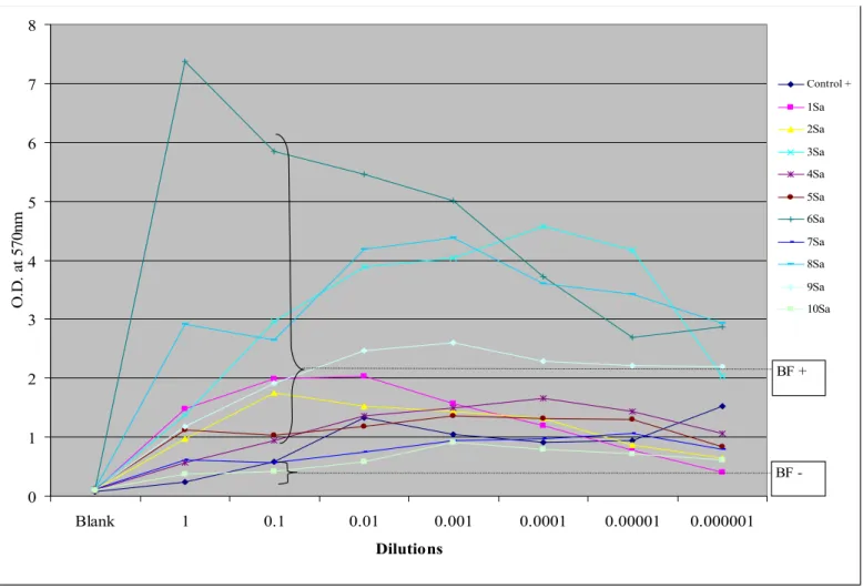
Biofilm formation was recorded for positive controls and several of the isolates from each of the three species studied. Results for each of the separate organisms studied are presented in graphical form in Figures II, III, and IV. While growth was usually maximal for the undiluted culture, for several of the S. aureus species it was maximal at the 10-1 dilution. Thus, for comparison purposes of biofilm-forming capacity with control, the O.D. at the 10-1 dilution was used.
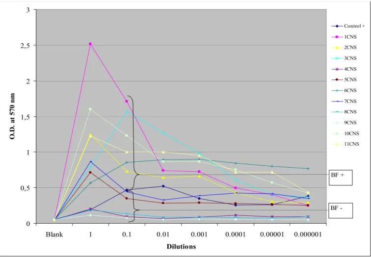
Biofilm-forming capacity was detected in 22 of the 31 isolates studied. For P. aeruginosa, biofilm formation was noted in 6/10, with some samples having quite marked biofilm production. For S. aureus, 8/10 samples produced a biofilm definitely higher than positive controls. For coagulase negative staphylococci 6/11 samples produced a biofilm greater than positive control. Of note is that the negative control for biofilm formation for CNS produced a biofilm with our method. This may however be due to the methodology used in that study (production of exopolysaccharides on Congo red agar), which differs from ours19.
It can be seen that staining intensity varies between species and according to dilution. For intensity, Staphylococcus sp. stains more intensely, despite the strong amount of slime production by the Pseudomonas sp. This is related to the site of slime production in the 96 well plate. For the Staphylococcus sp, slime production occurs over the entire surface of the floor of the well, however, for P. aeruginosa, slime tends to form mainly at the air-liquid interface on the side of the well, limiting the amount of biofilm available for binding the crystal violet after washing of the plate.
For all three species studied growth is maximal at the 10-1 dilution, it persists over a number of subsequent dilutions. For comparison purposes of biofilm-forming capacity between different isolates, the O.D at this single dilution could also be used.
Legends for Illustrations
Figure I. Example of biofilm formation the 96 well plate. A biofilm producing strain of
Staphylococcus aureus is shown. Note that intensity of staining is actually maximal in the
Figure II. Biofilm-forming capacity for Staphylococcus aureus. The average O.D. is plotted for the different serial dilutions.
BF+: Biofilm production. BF-: No biofilm production. 0 1 2 3 4 5 6 7 8 Blank 1 0.1 0.01 0.001 0.0001 0.00001 0.000001 Dilutions O. D. a t 5 70 nm Control + 1Sa 2Sa 3Sa 4Sa 5Sa 6Sa 7Sa 8Sa 9Sa 10Sa BF -BF +
Figure III. Biofilm-forming capacity for Coagulase negative staphylococci. The average O.D. is plotted for the different serial dilutions.
BF+: Biofilm production. BF-: No biofilm production. 0 0,5 1 1,5 2 2,5 3 Blank 1 0.1 0.01 0.001 0.0001 0.00001 0.000001 Dilutions O .D . at 57 0 n m Control + 1CNS 2CNS 3CNS 4CNS 5CNS 6CNS 7CNS 8CNS 9CNS 10CNS 11CNS BF -BF +
Figure IV. Biofilm-forming capacity for Pseudomonas aeruginosa. The average O.D is plotted for the different serial dilutions. Sample 1, a strong biofilm producer, has been removed to improve clarity. BF+: BF+: Biofilm production. BF-: No biofilm production. 0 0,5 1 1,5 2 2,5 3 Blank 1 0,1 0,01 0,001 0,0001 0,00001 0,000001 Dilution DO 5 70 nm control+ control-2Pa 3Pa 4Pa 5Pa 6Pa 7Pa 8Pa 9Pa 10Pa BF+
BF-Table I. Control strains for biofilm formation (ATCC: American Type Culture Collection, LSPQ: Laboratoire de Santé Publique du Québec)
Strain Biofilm formation
Staphylococcus aureus LSPQ 2520 Positive Staphylococcus epidermidis ATCC 12228 Negative Pseudomonas aeruginosa LSPQ 3332 Positive Pseudomonas aeruginosa ATCC 10145 Negative Staphylococcus epidermidis LSPQ 3027 Positive
Discussion
We evaluated the capacity of a simple, in vitro method to detect biofilm-forming capacity in commonly identified species of bacteria (S. aureus, CNS, and P. aeruginosa) recovered from individuals with chronic rhinosinusitis with or without nasal polyposis (CRS +/- NP) having previously undergone endoscopic sinus surgery for disease unresponsive to maximal medical management.
Using this method, biofilm-forming capacity could be established in 22 of 31 of the specimens, a rate comparable with the results reported by Ramadan et al using scanning electron microscopy 22. This high recovery rate in our study suggests that this technique is capable of detecting biofilm-forming capacity in chronic rhinosinusitis, and that it may offer a complement to existing methods. It is obvious that biofilm-forming capacity in vitro does not necessarily correlate to in vivo biofilm production. Certainly favourable local conditions are required to favor the expression of the biofilm forming phenotype. Establishment of the biofilm with subsequent chronic bacterial infection may help maintain the chronic course experienced by these individuals20,21.
It is interesting to note that biofilm-forming capacity is noted for all three bacterial species assessed, even CNS. What contribution this makes to disease in our patient population remains to be determined. The questions of whether all bacterial species have the same pathogenic potential remain unanswered.
Conclusion
This in vitro assessment method demonstrates biofilm-forming capacity in a majority of bacterial samples recovered from individuals with chronic rhinosinusitis with or without nasal polyposis. This suggests that this easily performed; inexpensive technique may offer a complement to other modalities currently used to study biofilms in chronic rhinosinusitis. Further experience with its use in relation to impact of biofilm-forming capacity on clinical disease and in vivo studies of biofilm modulation will offer additional information on its potential as a clinical and research tool.
Acknowledgements
References
1. Costerton JW, Stewart PS, and Greenberg EP. Bacterial biofilms: A common cause of persistent infections. Science 284: 1318–1322, 1999.
2. Donlan RM. Biofilms: Microbial life on surfaces. Emerg Infect Dis 8:881–890, 2002. 3. Fleming H-C, Wingender J, Griebe T, and Mayer C. Physicochemical properties of
biofilms. In Biofilms: Recent Advances in Their Study and Control. Evans LV (Ed.). Amsterdam: Harwood Academic Publishers, 19–34, 2000.
4. Stoodley P, Sauer K, Davies DG, and Costerton JW. Biofilms as complex ifferentiated communities. Annu Rev Microbiol 56:187–209, 2002.
5. Potera C. Forging a link between biofilms and disease. Science 283:1837–1839, 1999. 6. Amorena B, Gracia E, Monzon M, et al. Antibiotic susceptibility assay for
Staphylococcus aureus in biofilms developed in vitro. J Antimicrob Chemother 44:43–
55, 1999.
7. Monzon M, Oteiza C, Leiva J, et al. Biofilm testing of Staphylococcus pidermidis clinical isolates: Low performance of vancomycin in relation to other antibiotics. Diag Microbiol Infect Dis 44(4):319–324, 2002.
8. Costerton W, Veeh R, Shirliff M, et al. The application of biofilm sciences to the study and control of chronic bacterial infections. J Clin Invest 112:1466–1477, 2003.
9. Post JC, Stoodley P, Hall-Stoodley L, and Ehrlich GD. The role of biofilms in otolaryngologic infections. Curr Opin Otolaryngol Head Neck Surg 12:185–190, 2004. 10. Cryer J, Schipor I, Perloff J, and Palmer JN. Evidence of bacterial biofilms in human
11. Sanclement J, Webster P, Thomas J, and Ramadan H. Bacterial biofilms in surgical patients with chronic rhinosinusitis. Laryngoscope 115: 578–582, 2005.
12. Cramton SE, Gerke C, Schnell NF, et al. The intercellular adhesion (ica) locus is present in Staphylococcus aureus and is required for biofilm formation. Infect Immun 67:5427–5433, 1999.
13. Arciola CR, Campoccia D, Gamberini S, et al. Search for the insertion element IS256 within the ica locus of Staphylococcus epidermidis clinical isolates collected from biomaterial-associated infections. Biomaterials 25:4117–4125, 2004.
14. Stepanovic S, Vukovic D, Dakic I, et al. A modified microtiter-plate test for quantification of staphylococcal biofilm formation. J Microbiol Methods 40:175–179, 2000.
15. Cramton SE, Gerke C, and Gotz F. In vitro methods to study staphylococcal biofilm formation. Methods Enzymol 336:239–255, 2001.
16. Benninger MS, Ferguson BJ, Hadley JA, et al. Adult chronic rhinosinusitis: Definitions, diagnosis, epidemiology, and pathophysiology. Otolaryngol Head Neck Surg 129(suppl): S1–S32, 2003.
17. Nadel DM, Lanza DC, and Kennedy DW. Endoscopically guided cultures in chronic sinusitis. Am J Rhinol 12:233–241, 1998.
18. Gotz F. Staphylococcus and biofilms. Mol Microbiol 43:1367–1378, 2002.
19. Arciola CR, Baldassarri L, and Montanaro L. Presence of icaA and icaD genes and slime production in a collection of staphylococcal strains from catheter-associated
infections. J Clin Microbiol 39:2151–2156, 2001. 20. Bachert C, Gevaert P, and van Cauwenberge P. Staphylococcus aureus superantigens and airway disease. Curr Allergy 20. Bachert C, Gevaert P, and van Cauwenberge P. Staphylococcus aureus superantigens
and airway disease. Curr Allergy Asthma Rep 2:252–258, 2002.
21. Tripathi A, Conley DB, Grammer LC, et al. Immunoglobulin E to staphylococcal and streptococcal toxins in patients with chronic sinusitis/nasal polyposis. Laryngoscope 114:1822–1826, 2004.
2. Bendouah Z, Barbeau J, Hamad WA, Desrosiers M. Biofilm formation
by Staphylococcus aureus and Pseudomonas aeruginosa is associated with
an unfavorable evolution after surgery for chronic sinusitis and nasal
polyposis. Otolaryngol Head Neck Surg 2006 June ;134(6):991-6.
Abstract
Objectives: To determine whether biofilm-forming capacity of bacteria demonstrated in chronic rhinosinusitis (CRS) has an impact on persistence of the disease following endoscopic sinus surgery (ESS).
Method: Thirty-one bacterial strains recovered from 19 patients with CRS at least one year post-ESS. Evolution of disease was assessed by questionnaire and endoscopy as favorable of unfavorable. The bacteria were cultured on a 96-well culture plaque and a semi-quantitative method using crystal violet to quantify biofilm production was used.
Results: Twenty-two of 31 samples produced a biofilm thicker or equal to the positive control. Biofilm production was noted in 6/10 Pseudomonas aeruginosa isolates, 8/10
Staphylococcus aureus and 8/11 coagulase-negative staphylococci. Biofilm formation was
associated with a poor evolution for Pseudomonas aeruginosa and Staphylococcus aureus, but not coagulase-negative staphylococcus.
Conclusion: There is a correlation between in vitro biofilm-producing capacity by
Pseudomonas aeruginosa and Staphylococcus aureus and unfavorable evolution after ESS,
Keywords: Chronic rhinosinusitis, bacterial biofilms, endoscopic sinus surgery,
Staphylococcus aureus, Pseudomonas aeruginosa.
Running head: Biofilm formation is associated with a poor evolution following ESS Funding: Internal funding. Conflict of interest statement: None.
Presented at the 2005 AAO-HNS annual meeting, Los Angeles, CA
Introduction
Biofilms are defined as an organized community of bacteria, adherent to a surface and contained in an extracellular polymeric substances made of exopolysaccharides, nucleic acids and proteins 1. Biofilms are highly resistant to both innate and specific host defense mechanisms. In part because of their exopolysaccharide matrix and reduced metabolic rate, they are less susceptible to phagocytic macrophages and are resistant to antibiotics that attack only dividing cells2, 3. These local conditions encourage persistence of bacteria for periods of months to years, with intermittent occurrence of acute exacerbations.
The role of the biofilms in the chronic diseases is increasingly recognized 4. The frequency is such that infectious disease experts at the Centers for Disease Control and Prevention (CDC) estimate that 65% of human bacterial infections involve biofilms5. In the upper respiratory tract, the presence of biofilms has been demonstrated in several chronic diseases including chronic otitis media, cholesteatoma and chronic adenoiditis 5.
Chronic rhinosinusitis (CRS), with its chronic indolent course, resistance to antibiotics and acute exacerbations has an evolution that parallels that of other biofilm-related diseases. In support of this, the presence of biofilms on the mucosa of subjects with chronic sinusitis
has been demonstrated by at least two previous authors. Using scanning electron microscopy (SEM), Cryer et al, showed presence of biofilm on the mucosa of a limited number of subjects with persistent chronic rhinosinusitis 6. Using SEM to study biopsies of the mucosa of the ethmoid sinus taken at the time of ESS, Sanclement et al demonstrated the presence of biofilms in 24/30 affected individuals and 0/4 controls 7. However, the authors did not identify the organisms present within these biofilms nor did they establish whether the presence of biofilms plays a role in the pathophysiology of CRS.
Objectives
We wished to determine whether the biofilm-forming capacity in bacteria commonly recovered from individuals with chronic sinusitis/sino-nasal polyposis has an impact on persistence of the disease following endoscopic sinus surgery.
Materials and methods
This study was approved by the Ethical Review Board for Human Subjects of the Centre Hospitalier de l’Université de Montréal.
Bacterial species identified for study were pathogens previously identified as those most frequently recovered from individuals with chronic sinusitis refractory to medical and surgical therapy: Staphylococcus aureus, coagulase-negative staphylococci and
Pseudomonas aeruginosa 8.
Bacterial isolates were recovered from a consecutive series of patients seen for sinus disorders in the tertiary academic-based rhinology practice of the senior author (M.D.). Consecutive patients were sampled until a representative number of organisms for each species were obtained, for a total of ten each samples of Staphylococcus aureus,
Pseudomonas aeruginosa, and coagulase-negative staphylococci. Both good and poor
outcome patients were included.
All patients had previously undergone technically successful endoscopic sinus surgery (ESS) (absence of synechiae, frontal sinus obstruction or technical problems such as retained uncinate process) for a diagnosis of chronic sinusitis and/or nasal polyposis according to 2003 AAO-HNS guidelines 9, and refractory to maximal medical therapy over 12 months previously. Evolution of disease was assessed by evaluation of symptoms of CRS and rigid sinonasal endoscopy.
A favorable evolution was defined as absence of symptoms or no more than one mild symptom (defined as a score of no more than 1 on a scale 0-3) of symptoms of facial pain/ pressure, nasal discharge or nasal obstruction on an ongoing basis. This had to be accompanied by an adequate technical result documented by endoscopy defined as presence of patent cavities and absence of purulent secretions. All others were deemed to have an unfavorable evolution.
Patients with cystic fibrosis or underlying immunosuppressive disorders (HIV, insulin-dependent diabetes mellitus, and renal disease) were not included. Treatment with topical intranasal corticosteroids with or without nasal irrigations was allowed, however patients having taken antibiotics or oral prednisone within a 1-month period previous were excluded.
All cultures were performed by the senior author (M.D.) under endoscopic guidance as described by Nadel et al 10. Briefly, after topical anesthesia, the nasal ala was retracted and the endoscope used to visualize the middle meatus and sinus cavities. A thin, flexible calcium alginate swab (Starswab Microorganism Collection and Transport system (Starplex Scientific, Etobicoke, Ontario). was then inserted under direct endoscopic control, and directed to the site of maximal purulence. Where no purulence was seen, the surface of the maxillary sinus was swabbed for a fifteen second period. Care was taken at all times to avoid contact with the lateral nasal wall or the nasal vestibule.
Samples were rapidly transported to the hospital microbiology laboratory for Gram staining, culture, and identification. Clinical specimens were identified after plating and incubation according to standard procedures. Correct speciation of microorganisms was done using the Vitek2 system (Biomerieux, Marcy l’Étoile, France) for gram-positive and gram-negative strains.
Antibiotic susceptibilities were tested according to standard procedures using an automated microbroth dilution system (Vitek 2, Biomerieux, France). All procedures were conformed to NCCLS standards.
Staining for biofilm:
Crystal violet staining was adapted from the method previously described by Stepanovic et al 11.
The person performing the assay of biofilm forming capacity (ZB) was blinded to the patient outcome.
Growth
Previously frozen strains were initially inoculated on blood Agar (TSA 0.5% of sheep blood). After culture for 24 hours, one to four colonies per strain were cultured on TSA (Typtic Soy Agar). These were incubated at 37ºC for 24 h in order to condition them to the TSB/TSA medium and to ensure non-contamination. The colonies grown on TSA solid
medium were amplified in 5 ml of TSB medium (Typtic soy broth) with 0.5% glucose 12 and incubated at 37ºC for 24 h.
After the incubation, the optical density of the cultures was standardized in the following manner:
The amplified culture was centrifuged at 3000 RPM during 10 minutes, the supernatant removed, and the aliquot washed with 10 ml of TSB. The centrifugation was repeated and the aliquot again suspended in 5 ml of TSB with 0.5 % glucose.
Serial dilutions of the bacterial suspension were distributed in 96 well plates, Optical density (O.D) at 630 nm was measured using a spectrophotometer (Dynatech MR5000). The dilution corresponding to the D.O of 0.1 to 0.15 was determined and served as a starting point for the dilutions.
Using the bacterial suspension producing an O.D of 0.1 to 0.15, the suspensions were then distributed in the microplaques. Serial dilutions with TSB 0.5 % glucose were performed in order to obtain concentrations varying from 100 to 10-4 (1:1 to 1: 10 000). Four replicates were used for each sample. The plates were incubated at 37ºC without agitation for 24 h. O.D. at 630 nm was measured at times 0 and 24 hours in order to assess growth by evaluation of the cellular density. The supernatant was then withdrawn and the plate rinsed under running tap water. After drying, staining for adherent biofilm was performed using crystal violet.
Staining for biofilm
Crystal violet (Fisher Scientific Co) was applied during 10 minutes. The dye fixes itself to the attached biofilm giving it a characteristic purple color. Following the 10 min of
staining, the plate was again rinsed with running water and left to dry. After drying, semi-quantitative assessment of biofilm formation was obtained by extracting the crystal violet with 100 µl per well of the following bleaching solution: 200 ml methanol, 50 ml glacial acetic acid, 250 ml H2O. This dissolved the bound crystal violet and produced a violet colored solution in each well. The intensity of coloration was determined by measuring the absorbance at 570nm. The average value of O.D was determined by calculating the average O.D. for the four replicates.
Net O.D was calculated by subtracting the O.D of the blank (no specimen) from the one under study. For comparison purposes between isolates, the net O.D. at the 10-1 dilution were used as it best represented maximal biofilm formation for all species studied (Data not shown). Determination of biofilm-production was established in reference to positive controls for each of the species.
Controls:
Control biofilm-producing commercially obtained slime-producing and non-slime producing strains for each of the tested species were obtained from the American Type culture collection (ATCC) and the Laboratoire de Santé Publique du Québec (LSPQ) used as reference strains to confirm biofilm production.
As no well-categorized, standardized biofilm-producing agents are available (catalog listings of organisms available from central supply sources use the term ‘slime-producing’ rather than ‘biofilm-producing’ in their description), slime-production was used as a surrogate for biofilm-producing capacity when noted.
Slime producing organisms used were: LSPQ 3332 (Pseudomonas aeruginosa), LSPQ 3027 (Staphylococcus epidermidis) and LSPQ 2520 (Staphylococcus aureus). Slime negative organisms used were: ATCC 10145 (Pseudomonas aeruginosa) and ATCC 12228 (Staphylococcus epidermidis). For Staphylococcus aureus, no commercial source for slime negative control could be obtained.
Results
Patient population was 9 women and 10 men, with an average age of 52 years (range 29 to 68 years). Five were deemed to have a favorable evolution while 14 to have an unfavorable evolution. Thirty-one isolates were recovered from 19 patients: 10 Staphylococcus aureus (Sa), 10 Pseudomonas aeruginosa (Pa) and 11 coagulase-negative staphylococci (CNS).
Biofilm-forming capacity greater than control was detected in 16/19 patients or in 22 of the 31 isolates (Table I). There were marked differences between the three species studied. For
P. aeruginosa, biofilm formation was noted in 6/10, with some samples having quite
marked biofilm production. For S. aureus, 8/10 samples produced a biofilm definitely higher than positive controls. For coagulase-negative staphylococci 6/11 samples produced a biofilm greater than positive control.
The quantitative results of biofilm formation by Pseudomonas aeruginosa seem lower than those produced by Staphylococcus aureus or coagulase-negative staphylococci. This is
explained by the fact that Pseudomonas aeruginosa produces a biofilm limited to the air-liquid interface, thus after washing, only the biofilm formed around the wall remains available for staining. For staphylococci, biofilm production is distributed evenly over the well surface.
Of note is that the negative control for biofilm formation for CNS produced a biofilm with our method. This may however be due to the methodology used in that study (production of exopolysaccharides on Congo red agar), which differs from ours 13.
Isolates from each species were grouped according to clinical evolution. A poor evolution was associated with biofilm formation in-vitro for both S. aureus and P. aeruginosa, but not for CNS (Figures III, IV, and V).
Legends for Illustrations:
Figure I. Examples of biofilms in the 96 well plate for Staphylococcus aureus and coagulase-negative staphylococci after crystal violet staining. Light, medium and heavy growth are illustrated. .
Figure II. Biofilm-forming capacity for Staphylococcus aureus. Biofilm formation is reported in relation to control strain at 10-1 dilution.
0 1 2 3 4 5 6 7 Con trol +
1Sa 2Sa 3Sa 4Sa 5Sa 6Sa 8Sa 9Sa 7Sa 10Sa Isolates
D.
O a
t 570 nm
Figure III. Biofilm-forming capacity for Coagulase negative staphylococci. Biofilm formation is reported in relation to control strain at 10-1 dilution.
0 0.2 0.4 0.6 0.8 1 1.2 1.4 1.6 1.8 Control + 1CNS 2CNS 3CNS 4CNS 5CNS 8CNS 9CNS 6CNS 7CNS 10CNS 11CNS Isolates N et O .D . a t 570nm
Good outcome
Poor outcome
Figure IV. Biofilm-forming capacity for Pseudomonas aeruginosa. Sample 1Pa, a strong biofilm producer, has been removed to improve clarity. Biofilm formation is reported in relation to control strain at 10-1 dilution.
0 0.5 1 1.5 2 2.5 Control + Control
-2Pa 3Pa 4Pa 5Pa 10Pa 6Pa 7Pa 8Pa 9Pa Isolates
Ne t O . D. a t 5 70 n m
Table I. Clinical evolution, bacteriology and biofilm-forming capacity in subjects studied.
ID Sex Age Diagnosis
Biofilm-forming capacity P. aeruginosa S. aureus Coagulase-negative staphylococci Evolution 1 M 68 CRS w/NP BF- Good 2 F 52 CRS w/ o NP BF+ Poor 3 M 54 CRS w/ o NP BF+ , BF+ * Good 4 F 52 CRS w/ NP BF+ Poor 5 M 68 CRS w/ NP BF- BF- BF+ Good 6 F 66 CRS w/ o NP BF- BF- Good 7 M 40 CRS w/ NP BF- BF+ Good 8 M 57 CRS w/ NP BF+ BF+ Poor 9 F 44 CRS w/ NP BF+ Poor 10 M 75 CRS w/ NP BF+ Poor 11 M 42 CRS w/ o NP BF+ BF- , BF+ Poor 12 F 43 CRS w/ NP BF+ Poor 13 M 44 CRS w/ NP BF+ Poor 14 M 68 CRS w/ o NP BF+ Poor 15 M 36 CRS w/ NP BF- , BF- Poor 16 F 47 CRS w/ NP BF+ , BF+ Poor 17 F 54 CRS w/ o NP BF+ Poor 18 F 29 CRS w/ NP BF+ BF+ Poor 19 F 68 CRS w/ o NP BF+ BF+ Poor
* More than one isolate may be recovered from the same subject CRS: chronic rhinosinusitis
Discussion
In this preliminary study, we have shown that biofilm-forming capacity by S. aureus and
P. aeruginosa but not CNS is associated with a poor clinical evolution in individuals with
chronic rhinosinusitis with or without nasal polyposis (CRS +/- NP) having previously undergone endoscopic sinus surgery. This suggests that biofilm-producing capacity by S.
aureus and P. aeruginosa influences the clinical evolution and may help explain the
persistence of disease in CRS.
S. aureus and P. aeruginosa have both been implicated as pathogens in respiratory disease
and in CRS. S. aureus is believed to exert a role in CRS by toxin production with superantigenic stimulation of specific immunity, and possible sensitization to the toxin 14, 15. P. aeruginosa is a Gram-negative, frequently associated with long-term respiratory tract disease. Inflammation and tissue destruction from natural enzymes secreted by P.
aeruginosa is enhanced by products of the systemic defence mechanisms, such as products
of neutrophil degradation 16. However, there is a paucity of information explaining why these bacterial species remain difficult to eradicate with conventional antibiotic therapy. The presence of biofilms may help explain this and open the door to novel means of therapeutic intervention.
It is interesting to note that there is no relation between the production of biofilm by coagulase-negative staphylococci and the evolution of the patients. Coagulase negative staphylococci are frequently regarded as mere contaminants of the nasal flora and are the
agent the most frequently recovered in healthy, asymptomatic post-ESS cavities. However, in other areas CNS can be potent pathogens, and are frequently responsible for infection and failure of a variety of medical implants 17. In this study, no conclusion can be drawn on their role as a potential pathogen as results for CNS may reflect inherent difficulties in culture of bacteria from biofilms.
In this study, we have used a simple, in vitro method to detect biofilm-forming capacity instead of other methods for in situ biofilm detection such as scanning electron microscopy (SEM), transmission electron microscopy (TEM), confocal laser microscopy or detection of presence of genes identified with biofilm-forming capacity 18. However, these methods are expensive, may require tissue sample for use, and are not routinely available. These factors limit their utility in clinical practice. In addition, while these methods can help identify the presence of a biofilm, they do not permit to identify the agent(s) involved, limiting the study to specific bacterial species. Though this limitation may be addressed by use of fluorescent in situ hybridization (FISH) probes combined with laser confocal microscopy, this expertise is currently unavailable outside of a limited number of specialized centers. One of the principal limitations of in vitro staining techniques is that they have a higher false-negative rate for specimens with weak biofilm formation 19. The organisms we studied are mostly strong producers and this technique appears to perform well in our model.
It is obvious that biofilm-forming capacity in vitro does not necessarily correlate to in vivo biofilm production. Certainly favorable local conditions are required to favor expression of the biofilm-forming phenotype. It may be that the microenvironment of the inflamed sinuses in patients with CRS favors this, even in the absence of a foreign body. Changes brought about at the level of the sinus mucosa by the inflammatory process create areas of damaged epithelium with impaired local defenses 20, favoring the attachment of certain types of bacteria 21 with possible biofilm formation. Establishment of the biofilm with subsequent chronic bacterial infection may help maintain the chronic course experienced by these individuals. Once biofilm is installed, persistent infection and host response to biofilm-bound and free forms of the bacteria may contribute to continue tissue damage.
The presence of bacterial biofilms in the cavities of symptomatic post-ESS patients may help explain previously noted contradictory findings in studies of topical antibiotic therapy for post-ESS sinusitis. While topical antibiotic therapy was reported beneficial in individuals with acute exacerbations of symptomatology in post-ESS sinusitis 22, in another trial it was of no benefit in patients with chronic disease with stable symptomatology 23. In these situations, bacteria organized in biofilms may have been responsible for chronicity, with exacerbations caused by increased presence of planktonic (free) forms responsive to antibiotic therapy.
Despite the potential interest of biofilms in CRS, it is probable that biofilms do not represent the entire explanation for CRS. The initial events leading to inflammation remain to be explained, and host factors predisposing to CRS need to be better characterized. The occurrence of poor outcome in patients where cultures revealed lack of biofilm forming capacity may be due to other phenomenon responsible for clinical disease.
Conclusion
This preliminary work suggests that biofilm-forming capacity by S. aureus and P.
aeruginosa is associated with a poor evolution in individuals having previously undergone
ESS for CRS and that biofilm formation by these two organisms may play a role in the chronicity of this disorder. Before drawing definite conclusions, confirmation of these preliminary results using both larger, better powered series and more sophisticated detection methods are warranted. If biofilms are found to be implicated in CRS, therapies modulating biofilm formation and detachment may be incorporated into strategies for management of this disorder.
Acknowledgements
We thank Onder Agbaba for his excellent technical assistance with isolation and
identification of the pathogens. The financial support of the Fondation Antoine Turmel is greatly acknowledged.