Cloning
and
amplified expression
in
Streptomyces
lividans of
the
gene
encoding
the extracellular
f-lactamase
of Actinomadura
R39
Claudine PIRON-FRAIPONT, Colette DUEZ, Andre MATAGNE, Chantal MOLITOR,* Jean DUSART,t
Jean-Marie FRERE and Jean-Marie GHUYSEN
Departement de Microbiologie, Universite de Liege, InstitutdeChimie, B6, B-4000 Sart Tilman (Liege 1), Belgium
Byusing the promoter-probe plasmid pIJ424, genomic DNA fragments of Actinomadura R39were shown
to have promoter activity in Streptomyces lividans. The same 100-200-copy-number
plasmid
was used toclone in S. lividans TK24, the gene that encodes the Actinomadura R39
,-lactamase.
Genecloning
resultedin anamplified expression of the
,-lactamase
whencomparedwith theamountsof enzymeproducedbytheoriginal strain (1 mg versus 0.008 mg*litre of
culture-').
INTRODUCTION
Actinomadura R39, isolated from an African soil and
initially designated 'Streptomyces R39', has a wall
peptidoglycan of the meso-diaminopimelic acid chemo-type I(Ghuysenetal., 1973). Itsecretesduring growth a
hypersensitive DD-peptidase/penicillin-binding protein (Leyh-Bouille et al., 1972; Ghuysen et al., 1974; Frere et al., 1974, 1980; Duez et al., 1981a,b) and very low
amounts ofa f,-lactamase (Johnson et al., 1973; Duez
et al., 1982). Actinomadura spp. belong to the Order
Actinomycetales and possess a DNA G+C content of 60-72mol% (McClung, 1974), which is similar to that of thegenus Streptomyces. Given thissimilarity and the fact that several genes encoding Streptomyces enzymes
have been cloned in a Streptomyces high-copy-number
vector-host system with amplification of the expressed
proteins(Dehottayetal., 1986;Duezetal., 1987;Lenzini et al., 1987), thesame strategywas used to overproduce the Actinomadura R39
fl-lactamase.
MATERIALS AND METHODS
Bacterial strains andplasmids
Actinomadura R39 was from the local collection.
S. tividans TK24 (Hopwood et al., 1983)and S. tividans
M336,i.e. S. tividans TK24bearing thepromoter-probe plasmid pIJ424 (Ward et al., 1986), were from the John
Innes Institute, Norwich, U.K. Media and growth conditions
Cultures were grown at 28°C, with vigorous orbital
shaking, infivedifferent media: Difcobrain-heartbroth,
Merckpeptonebroth[asdescribedbyLeyh-Bouilleetal. (1971), except that peptone Merck 7213 was used],
Lennox broth (Lennox, 1955), L broth (Miller, 1972)
and E9 broth (Dehottay et al., 1986). R2YE agar
(Hopwoodetal., 1985)wasused forplating. Kanamycin
gradient plates were made with MMT medium (Katz
et al., 1983) as described (Ward et al., 1986). Spore suspensions were prepared as described by Hopwood
etal. (1985).
Recombinant DNA techniques
Chromosomal and plasmid DNAswereprepared and
Streptomyces protoplastsweretransformed asdescribed
by Hopwood etal. (1985). DNA minipreparations were
made using the alkaline-lysis procedure ofBirnboim &
Doly (1979) as modified by Kieser (1984). Treatments
with restrictionendonucleases (fromvarious commercial
sources), bacterial alkaline phosphatase (The
Radio-chemical Centre,
Amersham;
Bucks., U.K.)andphage-T4 DNA ligase (Boehringer, Mannheim, Germany),
separation of digested DNAs by agarose-gel
electro-phoresis and elution of DNA fragments from the gels
wereperformedessentiallyasdescribedin Maniatisetal.
(1982).
fl-Lactamcompounds and other antibiotics
Nitrocefin was purchased from Oxoid Ltd.,
Basing-stoke, Hants., U.K., and kanamycin from Sigma, St.
Louis, MO, U.S.A. The followingcompoundsweregifts:
thiostrepton (from S. J.Lucania,E. R. Squibb andSons, New Brunswick, NJ, U.S.A.); phenoxymethylpenicillin
(from Dr. J. Vanderhaeghe, Rega Institute, Leuven, Belgium); N-formimidoylthienamycin and cefoxitin
(from the Merck Institute for Therapeutic Research,
Rahway, NJ, U.S.A.); and cephaloridine (from E. Lilly
and Co., Indianapolis, IN, U.S.A.). fI-Lactamase activity and enzyme units
Unlessotherwisestated, nitrocefin(O'Callaghanetal.,
1972)wasusedassubstrate and thetestswereperformed
in 50mM-sodium phosphate, pH 7.0, containing 0.1mg
of bovine serum albumin-ml-'. Routinely, 5-10
Isl
samples of enzyme were added to 450,u of 100
suM-nitrocefin in buffer and incubated at 30'C. One unit is defined as the amount of enzyme hydrolysing 1 mol
ofsubstrate/min at maximalvelocity.
Kinetic parameters
Enzyme samples and various concentrations of
/-lactam substrates were incubated at 30'C in 50
mm-sodium phosphate, pH7.0, and the absorbances ofthe
Vol. 262
* Presentaddress: Smith Kline RIT, Service deGenetique,ruedel'Institut89, B-1330Rixensart, Belgium.
C.Piron-Fraipont and others
solutions were monitored until stabilization, at 482nm for nitrocefin, 260 nm forcephaloridine and 235 nm for phenoxymethylpenicillin. The reading frequency was 2 s-5 and the kinetic parameters were estimated as described in De Meester et al. (1987). The enzyme concentrations were such thatcompletion of thereactions was achieved in less than 5 min. Inactivation ofthe /,-lactamases by cefoxitin and N-formimidoylthienamycin was analysed on the basisof the simple model:
K k
E+I-Ir-E-I E-I*
where E is theenzyme, I is the ,I-lactam inactivator, Kis the dissociation constant,
k+2
is the first-order rate constant and E-I* is the acyl-enzyme.Twomethods were used. With cefoxitin, the enzyme and the inactivator were incubated at 30°C in buffer, and the residual enzyme activity was measured, by using nitrocefin as substrate, in samples removedafterincreasingincubation times. With N-formimidoylthienamycin, the reporter-substrate method (DeMeester etal.,1987)wasused.The progressivedecrease ofthe rate of hydrolysis of 100 uM-nitrocefinwas measured onternary mixtures containing theenzyme, theinactivator and nitrocefin.Enzyme purification
Actinomadura R39 was grown for 65h at 28°C and with strong aeration inatank containing 200 litresofthe Merck peptone medium, and S. lividans CM3wasgrown for 96 hat28°C,in60 1-litre flaskscontaining 500 mlof the same peptone medium. After elimination of the mycelium by centrifugation, the supernatantfluids were
stirred withDEAE-cellulose (10 g/litre) previously equi-libratedagainst 50 mM-sodium phosphate, pH 7.0, until disappearance of the enzyme activity. The
DEAE-cellu-lose was washed exhaustively with 100mM-Tris/HCl, pH 7.2, containing 0.2M-NaCl, until the absorbance of the buffer at 280nm was less than 0.2, and packed in
5cmx30cm columns (for 350 g samples of ion-exchanger). The enzymes wereelutedwith an NaCl gra-dient made in 100 mM-Tris/HCl, pH 7.2, by dropwise addition, at constant volume, of 1350 ml of 0.5M-NaCl in buffer to 600ml of 0.2 M-NaCl in buffer. The active
fractions (total volume 800 ml) were concentrated
to 32 ml byultrafiltration onaDiaflo UM10membrane (Amicon Corp.,Danvers, MA, U.S.A.). The
fl-lactamase
of S.
lividans
CM3 wasfurther purified by threesuccessive runs on a 2.2 cmx25 cm column of phenylboronic acid-agarose type B(Cartwright & Waley, 1984). Elution was carried out with 100mM-Tris/HCI
(pH7.2)/0.5
M-NaCl at a fow rate of 50 ml
h-1
(Fig. 1). With the ActinomaduraR39,3-lactamase
thethird run wasomitted and replaced by anf.p.l.c.
step on MonoQ HR5/5 (Pharmacia, Uppsala, Sweden).Proteincontent and amino acid composition
Theproteins were estimated by measuring the amounts of amino groups available for dinitrophenylation after hydrolysis by6
M-HCl,
at110
°C(Duez et al., 1978). The amino acidcomposition was establishedas described by Joris et al. (1983).Polyacrylamide-gelelectrophoresis
Separation gelelectrophoresis [7
0,
(w/v)acrylamide, pH8.4] was run using 0.7cmx9.5 cm cylindrical gels and acurrent of5mA/gel. After an overnight pre-run, thesampleswereloaded and allowedtomigrate until the Bromophenol Blue marker reached the bottom of the tube. Gel electrophoresis in the presence of SDS wascarried out as described by Laemmli & Favre (1973).
RESULTS AND DISCUSSION
Transcriptional
efficacy
ofActinomadura R39 promotersinS.
lividans
A portion (2 ,tg) ofgenomic DNA of Actinomadura R39 was cleaved with restriction endonucleases
BamHI,
BclJ
andBglIlI
respectively,and eachdigestwasseparately ligated to 1,sg
ofpromoter-probe plasmid pIJ424 (Fig. 2)previouslycleaved withBgllI
andtreated with bacterial alkaline phosphatase. The resulting ligation mixtures were used to transform S.lividans
TK24 protoplasts, and the transformants were selected on R2YE agar plates containing 50,tg of thiostreptonml-'
(Hopwood
0 20 40 O. 0. 0. 0 20 40 (c) 3 II I II I I .1-0 20 40 Fraction (5 ml) no.
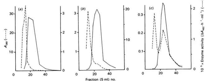
Fig. 1.Purificationof the S.Iividans CM3fl-lactamase onaphenylboronic acid-agarose affinity column
Threesuccessive runs,(a), (b)and(c),wereperformed. Fordetails, seetheMaterials andmethodssection. Enzyme activityis
expressed inarbitraryunitscorrespondingtoan absorbance increase of1.0 at482nm after 1h ofincubation. Such arbitrary
unit isequivalentto 0.71x10⁻⁶ enzyme unitasdefinedinthe Materials and methods section, nitrocefin being the substrate.
1989 -2 E C w x co 1 >-.4_ 4) E c x O o 8:50 co 004
Bg/l I
Fig. 2. Promoter-probe plasmid vector pIJ424 (Ward et al., 1986)
pIJ424 uses the promoterless aminoglycoside phospho-transferasegene(aphII) oftransposonTn5asthe indicator for promoteractivity.There is auniqueBglIIsite located downstreamof themajor terminator (ter) of the Escherichia coliphage fd(so that readthrough from upstream vector
promoters is prevented) and upstream of a ribosome-binding site, anin-framestop codon and the start codonof theaphll gene. Insertion of apromoter-containing DNA fragment in the BglII site results in the expression of kanamycin resistance in asuitablehost such as S.lividans
TK24.pIJ424 is a derivative of thepIJIO1replicon (Kieser et al., 1982); it occurs in a 100-200 copy number per chromosome (Ward et al., 1986) and also contains the thiostrepton-resistance marker(tsr).
1 4
T
...
et al., 1985). Transformation rates (numbers of trans-formants
-c/g
ofplasmid'1) ranged from 1.4to 3.0x I04. Spore suspensions of thiostrepton-resistanttrans-formants were streaked on kanamycin gradient (from 0
to 250,ug *ml-')plates. Growthwasfrequently observed uptohalf wayonthegradient (Fig. 3). S. lividans M336, which contained the original pIJ424, served as control; few coloniesgrewonlyatthe origin of the gradient. The plasmid DNAs of 30 kanamycin-resistant clones were analysed byagarose-gel electrophoresis. They all had an increased molecular mass when compared with that of pIJ424. In all likelihood these plasmids had acquired Actinomadura R39 genomic DNA fragments that con-tained promoter regions, thus permitting expression of the promoterless aphll gene ofpIJ424.
fl-Lactamase-producingS. lividans CM3
Since Actinomadura R39 promoters were recognized and effectively utilized by S. lividans, thiostrepton-resistant transformants havingthecapacityof producing an extracellular
fl-lactamase
were selected by nitrocefintest onR2YEagarplates(Dehottay et al., 1986). Nine of them produced the desired activity; seven originated from the Bcll library, two from the BamHI library and nonefromthe BglIllibrary. Their
,-lactamase-producing
capacitywas tested in fivedifferentgrowth media(see the Materials and methods section). S. lividans CM3, from the BamHI library, was the best ,-lactamase producer. Its 8.1 kb plasmid pDML150 had no Bglll site. The occurrence ofhybrid BglII/BamHI sites prevented the 1.8kb insert from being isolated.
Maximal level of 8-lactamase secretion by S. lividans CM3, i.e. - 1 mg of enzyme- litre ofculture-1, occurred
in the Merck peptone broth after 96 h ofgrowth. This
1 T
0
f.
250 Fig. 3. Growth ofS. lividans transformants onkanamycin gradient(0-250
pg.
ml-l) platesT, S. lividans M336 (TK24+pIJ424). 1-14, S. lividans strainstransformed with theligation mixtures described in thetext.
C. Piron-Fraipont and others
Table 1. Purification ofthe extracellular 8-lactamaseproduced by Actinomadura R39 and S.lividansCM3
Purification Strain step R39 CM3 Culture supernatant (200 1) DEAE-cellulose Phenylboronic
acid-agarose (two steps) F.p.l.c. MonoQ Culture supernatant (30 1) DEAE-cellulose Phenylboronic acid-agarose (three steps) Enzyme uni,ts (,umol'-min-') 1875 2280 980 980 31300 29400 12400 Total protein (mg) 96400 750 3.2 0.80 182350 1660 10 Specific activity (units mg of protein-') 0.019 3 307 1245 0.17 17.7 1230 Recovery Enrichment (0) (fold) (100) 121 52 52 (100) 94 40 158 16150 65500 104 7230
Table 2. Aminoacidcomposition of the Actinomadura R39 and S.lividans CM3 p-lactamases
Composition (mol/100mol) Residue R39f8-lactamase* CM3
,l-lactamase
Lys His Arg Trp Asx Thr Ser Glx Pro Gly Ala 2-Cys Val Met Ile Leu Tyr Phe 1.6 1.6 5.6 3.2 11.2 6.4 4.0 14.4 4.8 9.6 10.4 1.6 8.0 1.6 2.4 8.8 1.6 3.2 1.6 1.4 3.0 2.0 13.8 7.8 4.6 15.8 5.6 11.0 7.8 0.6 10.0 0.8 2.4 8.4 1.0 2.6* Data from Duez etal. (1982).
level was 100-fold higher than that observed with the
original Actinomadura R39 strain (~ 0.008mg of
en-zyme litre ofculture-') grown under identical and op-timalconditions. Asimilaramplified expressionof the
,-lactamases of Streptomyces albus G (Dehottay et al.,
1986) and Streptomyces cacaoi(Lenzinietal., 1987)had been observedaftergenecloninginS. lividansviapIJ702 (30and40mgofenzyme-litre of culture-1 asagainst0.5
and 1.5mg ofenzyme litre ofculture-1 for the original strains).
Actinomadura R39 and S. lividans CM3
fl-lactamases
Actinomadura R39 and S. lividans CM3 were grown,
and the secreted ,-lactamases were purified (see the
Materials and methods section). The last purification
stepyieldedfractions which, irrespective of the producing strain, had aconstant specific enzyme activity of about 1200units * mg ofprotein-',using nitrocefin assubstrate. Table 1 gives the enzyme recoveries and enrichments.
The Actinomadura R39 and S. lividans CM3 ,-lactamases were co-eluted byf.p.l.c. onMonoQ HR5/5. Elution occurred at 0.51M-NaCl. Upon polyacrylamide-gel electrophoresis under non-denaturing conditions, they migrated 7.8 cm towards the anode as a single protein band. Upon SDS/polyacrylamide-gel electro-phoresis, they also migrated as a single protein band with
an apparent relative molecular mass of 55-57 kDa. Upon filtration on Sephadex G-100 in 50mM-sodium phosphate (pH7.2)/0.5M-NaCl, they exhibited an elutioncoefficient value which, by reference to standard proteins, indicated an apparent molecular mass of 38.5 kDa. Great variations in the estimated molecular
mass of the Actinomadura R39
,-lactamase
prepared as described byDuezetal. (1982) had been observed. That former preparation contained about100%
(w/w) of apolydeoxyribonucleotidematerial, apparently in the form of a stable complex. Though the enzyme preparations obtained in the course of the present study lacked this contaminating material, SDS/polyacrylamide-gel electrophoresisand molecular sievingfailedtoyield
con-sistent molecular-mass values. The reason for these discrepancies is not understood. Nucleotide sequencing (S.Houba, S.Willem, C.Duez, C. Molitor &J.Dusart, unpublished work) revealed that the cloned S. lividans CM3
,J-lactamase
gene codes for a 304-amino-acid polypeptide, i.e. a 276-amino-acid mature protein of 29.27 kDa. Within the limits ofexperimentalerrors, theS. lividans CM3 ,-lactamase and the original
Actino-madura R39
,3-lactamase
(Duez et al., 1982) have thesame amino acid composition (Table 2).
TheActinomadura
R39,8-lactamase
isawide-spectrum
enzyme,penicillins
andA3-cephalosporins
being
eithergood or poor substrates. The kinetic parameters of
hydrolysis of three different
fl-lactam
antibioticsby
theActinomadura R39 and S. lividans CM3
fl-lactamases
prepared and
purified
as described above werere-determined
(Table 3).
Both enzymes behavedsimilarly.
The two
fl-lactamase
preparations
also reactedsimilarly
with two different,-lactamase
inactivators(Table 4).
1989Table3.Kinetic parameters ofActinomaduraR39and S.fividansCM3II-lactamases Each value represents the average(±S.D.) of atleast three measurements.
Enzyme Km(/,M)
VMax.*
Concentration quantity Substrate (4M) (ng) R39 CM3 R39 CM3 Nitrocefin Cephaloridine Phenoxymethylpenicillin 50-100 50-100 250-500 50-100 70-120 70-350 72+2 38+2 84+13 69+7 38+3 85+9 100+ 5 74+4 100+7 100+18 73+9 139+6*
V.ax.
values are expressed as a percentage of that with nitrocefin (= 1200 ,mol min-' mg ofprotein-1).
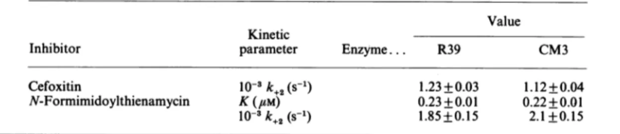
Table 4. Kinetic parameters of the inactivation of the Actinomadura R39 and S. liuvidansCM3
8-lactamases
byII-lactamcompounds Eachvalue represents the average (±S.D.) of at least three measurements.Value Kinetic
Inhibitor parameter Enzyme... R39 CM3
Cefoxitin N-Formimidoylthienamycin 10-3k+2(S-1) K(#M) 10-3k+2(S-1) 1.23+0.03 0.23 +0.01 1.85+0.15 1.12+0.04 0.22+0.01 2.1 +0.15
With cefoxitin, the rate of inactivation did not vary between 20 and 60 mm, indicating a Kvalue of much less than 20 mm. With N-formimidoylthienamycin, the values ofk1, where
k k+21C]
= [C]+K
were measured at concentrations ranging from 0.3 to 1.5 uM. The individual values of
k+2
and K were derived from plots 1/k,
versus 1/[C], where [C] is the con-centration offormimidoylthienamycin.Wethank Dr. D. A.Hopwood, Dr.T.KieserandDr. M.J. Bibb from the John Innes Institute (Norwich, U.K.) for generously providing strains and plasmids. This work was supported in part by the Fonds de la Recherche Scientifique
Medicale (contract n° 3.4507.83), an Action concertee with the Belgian Government (convention 86/91-90, the Fonds de Recherchede laFacultedeMedicineULg andacontractwith the European Economic Community (BAP-0197-B). J.D. is Research Associate at the National Fund for Scientific Research (Belgium). A.M. is indebted to the Institut pour l'Encouragementde laRechercheScientifiquedans l'Industrie
et l'Agriculturefora predoctoral fellowship.
REFERENCES
Birnboim, H.C. & Doly, J. (1979) Nucleic Acids Res. 7,
1513-1523
Cartwright, S. J. & Waley, S.G. (1984) Biochem. J. 221,
505-512
Dehottay, P., Dusart, J., Duez, C., Lenzini, M.V., Martial, J.A, Frere,J. M.,Ghuysen,J.M. &Kieser,T.(1986)Gene 42, 31-36
De Meester, F., Joris, B., Reckinger, G.,
Bellefroid-Bourguignon, C., Frere, J. M. & Waley, S. G. (1987) Biochem. Pharmacol. 36, 2393-2403
Duez, C.,Frere, J. M., Geurts, F., Ghuysen, J. M., Dierickx, L. &Delcambe, L. (1978) Biochem. J. 175, 793-800
Duez, C., Joris, B., Frere, J. M., Ghuysen, J. M. & Van Beeumen, J. (1981a) Biochem. J. 193, 83-86
Duez, C., Frere, J. M., Ghuysen, J. M., Van Beeumen, J. & Vandekerckhove, J. (1981b) Biochim. Biophys. Acta 671, 109-116
Duez, C., Frere, J. M., Ghuysen, J.M., Van Beeumen, J,. Delcambe, L.&Dierickx, L.(1982) Biochim. Biophys.Acta 700, 24-32
Duez, C., Piron-Fraipont, C., Joris, B., Dusart, J., Urdea,
M. S., Martial, J.A., Frere, J. M. &Ghuysen, J. M. (1987) Eur. J. Biochem. 162, 509-518
Frere, J. M., Ghuysen, J. M., Reynolds,P.E., Moreno, R. & Perkins, H. R. (1974) Biochem. J. 143, 241-249
Frere,J. M., Klein, D. &Ghuysen, J. M. (1980) Antimicrob. Agents Chemother. 18,506-510
Ghuysen, J. M., Leyh-Bouille, M., Campbell, J.N., Moreno, R.,Frere,J. M., Duez,C., Nieto,M.&Perkins,H.R.(1973) Biochemistry 12, 1243-1251
Ghuysen, J.M., Reynolds, P.E., Perkins, H. R., Frere, J. M.
& Moreno, R. (1974) Biochemistry 13, 2539-2547
Hopwood, D.A., Kieser, T., Wright, H.M. & Bibb, M. J. (1983) J. Gen. Microbiol. 129, 2257-2269
Hopwood,D.A.,Bibb,M.J.,Chater,K. F.,Kieser,T., Bruton, C.J., Kieser, H.M., Lydiate, D.J., Smith, C.P., Ward,
J. M. & Schrempf, H. (1985) Genetic Manipulation of Streptomyces:ALaboratoryManual,The JohnInnes
Foun-dation,Norwich
Johnson, K., Dusart, J., Campbell, J. N. & Ghuysen, J. M.
(1973) Antimicrob. Agents Chemother. 3, 284-298
Joris,B.,VanBeeumen,J.,Casagrande, F.,Gerday,C., Frere,
J. M. &Ghuysen, J. M. (1983)Eur. J. Biochem. 130, 53-69
Katz, E.,Thompson, C. J. & Hopwood, D.A. (1983)J. Gen. Microbiol. 129, 2703-2714
Kieser, T. (1984) Plasmid 12, 19-36
Kieser, T., Hopwood, D.A., Wright, H.M. & Thompson,
C. J. (1982) Mol. Gen. Genet. 185,223-238 Vol. 262
C.Piron-Fraipont and others
Laemmli, U. K. & Favre, M. (1973) J. Mol. Biol. 80, 575-599
Lennox, E. S. (1955) Virology 1, 190-206
Lenzini,M.V.,Nojima,S., Dusart, J.,Ogawara, H., Dehottay, P., Frere, J. M.& Ghuysen, J. M. (1987) J. Gen. Microbiol. 133, 2915-2920
Leyh-Bouille, M., Coyette, J., Ghuysen, J. M., Idczak, J., Perkins, H. R. & Nieto, M. (1971) Biochemistry 10, 2163-2170
Leyh-Bouille, M., Nakel, M., Frere, J. M., Johnson, K., Ghuysen, J. M., Nieto, M. & Perkins, H. R. (1972) 11, 1290-1298
Maniatis, J., Fritsch, E. F. & Sambrook, J. (1982) Molecular Cloning: A Laboratory Manual, Cold Spring Harbor Lab-oratory, Cold Spring Harbor, NY
McClung, N. M. (1974) in Bergey's Manual of Determinative Bacteriology, 8th edn. (Buchanan, R. E. & Gibbons, N. E., eds.), pp. 726-747, Williams and Wilkins Co., Baltimore Miller, J. H. (1972) Experiments in Molecular Genetics, Cold
Spring Harbor Laboratory, Cold Spring Harbor, NY
O'Callaghan, C. H., Morris, A., Kirby, S. M. & Shingler, A. H. (1972) Antimicrob. Agents Chemother. 1, 283-288
Ward, J. M., Janssen, G. R., Kieser, T., Bibb, M.J., Buttner, M.J. & Bibb, M. J. (1986) Mol. Gen. Genet. 203, 468-478
Received6February 1989/30March 1989;accepted 11 April 1989
1989