Université de Montréal
Use of cellular impedance to characterize
ligand functional selectivity at G protein-coupled receptors
par Wayne Stallaert Département de Biochimie Faculté de MédecineThèse présentée à la Faculté des études supérieures en vue de l’obtention du grade de Doctorat en Biochimie
Décembre 2013 © Wayne Stallaert, 2013
Université de Montréal Faculté des études supérieures
Cette thèse intitulée:
Use of cellular impedance to characterize ligand functional selectivity at G protein-coupled receptors
présentée par: Wayne Stallaert
a été évaluée par un jury composé des personnes suivantes :
Gerardo Ferbeyre, président-rapporteur Michel Bouvier, directeur de recherche
Philippe Roux, membre du jury Terry Kenakin, examinateur externe Louis Gaboury, représentant du doyen de la FES
Résumé
Les récepteurs couplés aux protéines G (RCPGs) représentent la plus grande famille de cibles thérapeutiques pour le traitement d’une panoplie de pathologies humaines. Bien que plusieurs décennies de recherche aient permis de façonner nos connaissances sur ces protéines membranaires, notre compréhension des déterminants moléculaires de leur activité signalétique reste encore limitée. De ces domaines de recherche, une avancée récente a mis à jour un nouveau phénomène, appelé sélectivité fonctionnelle des ligands, qui a bouleversé les paradigmes décrivant leu fonctionnement de ces récepteurs. Ce concept émane d’observations montrant que l’activité pharmacologique de certains ligands n’est pas nécessairement conservée sur tout le répertoire signalétiques connu du récepteur et peu se restreindre à l'activation sélective d’un sous-‐groupe de voies de signalisation.Ce nouveau modèle pharmacologique de l'activation des RCPG ouvre de nouvelles possibilités pour la découverte de médicaments plus efficace et sûr, ciblant les RCPGs. En effet, il permet la conception de molécules modulant spécifiquement les voies signalétiques d’intérêt thérapeutique, sans engager les autres voies qui pourraient mener à des effets secondaires indésirables ou de la tolérance.
Cette thèse décrit l'utilisation d'une nouvelle approche sans marquage, basée sur la mesure du changement l'impédance cellulaire. Par la mesure des changements cellulaires, comme la morphologie, l’adhésion et/ou la redistribution des macromolécules, cette approche permet de mesurer de façon simultanée l'activité de plusieurs voies de signalisation impliqués dans ces réponses.
Utilisant le récepteur β2-‐adrénergique (β2AR) comme modèle, nous avons démontré que les variations dans l’impédance cellulaire étaient directement liées à l’activation de multiples voies de signalisation suite à la stimulation du récepteur par son ligand. L’agoniste type du β2AR, l’isoprotérénol, s’est avéré induire une réponse d’impédance dose-‐ dépendante constituée, dans le temps, de plusieurs caractéristiques distinctes pouvant être bloquées de façon compétitive par l’antagoniste ICI118,551 Par l’utilisation d’inhibiteurs sélectifs, nous avons été en mesure de déterminer la contribution de plusieurs voies signalétiques canoniques, comme les voies dépendantes de Gs et Gi, la production d’AMPc
ii
et l’activation de ERK1/2, sur ces changements. De plus, la dissection de la réponse d’impédance a permis d’identifier une nouvelle voie de mobilisation du Ca2+ contribuant à la réponse globale des changements initiés par la stimulation du β2AR. Dans une autre étude, nous avons rapporté que la réponse calcique induite par le β2AR serait attribuable à une transactivation Gs-‐dépendant du récepteur purinergique P2Y11, lui-‐même couplé à la protéine Gq. La mesure d’impédance permettant de distinguer et de décrire une pléiade d’activités signalétiques, nous avons émis l’hypothèse que des ligands arborant des profils signalétiques différents généreraient des réponses d’impédance distinctes. Le criblage d’une librairie de ligands spécifiques au β2AR a révélé une grande variété de signatures d’impédance. Grâce au développement d’une approche computationnelle innovatrice, nous avons été en mesure de regrouper ces signatures en cinq classes de composés, un regroupement qui s’est avéré hautement corrélé avec le profil signalétique des différents ligands.
Nous avons ensuite combiné le criblage de composés par impédance avec l’utilisation d’inhibiteurs sélectifs de voies signalétiques afin d’augmenter la résolution du regroupement. En évaluant l’impact d’une voie signalétique donnée sur la signature d’impédance, nous avons été en mesure de révéler une plus grande variété de textures parmi les ligands. De plus, cette méthode s’est avérée efficace pour prédire le profil signalétique d’une librairie de composés non caractérisés, ciblant le β2AR. Ces travaux ont mené à l’élaboration d’une méthode permettant d’exprimer visuellement la sélectivité fonctionnelle de ligands et ont révélé de nouvelles classes de composés pour ce récepteur. Ces nouvelles classes de composés ont ensuite été testées sur des cardiomyocytes humains, confirmant que les composés regroupés dans différentes classes produisent des effets distincts sur la contractilité de ces cellules.
Globalement, ces travaux démontrent la pertinence de l’utilisation de l’impédance cellulaire pour une évaluation précise des différences fonctionnelles parmi les composés ciblant les RCPGs. En fournissant une représentation pluridimensionnelle de la signalisation émanant des RCPGs à l’aide d’un seul essai ne requérant pas de marquage, les signatures d’impédance représentent une stratégie simple et innovante pour l’évaluation
iii
de la fonctionnalité sélective des ligands. Cette méthode pourrait être d’une grande utilité dans le processus de découverte de nouveaux médicaments.
Mot clés: récepteurs couplés aux protéines G (RCPGs), récepteur β2-‐adrénergique (β2AR), sélectivité fonctionnelle des ligands, réseaux signalétique, impédance cellulaire, cardiomyocytes, découverte de médicaments.
iv
Abstract
G protein-‐coupled receptors (GPCRs) represent the largest family of therapeutic targets for the treatment of a wide variety of human pathologies. Decades of research have provided an extensive base of knowledge about these fascinating membrane proteins, yet significant advancements in the understanding of the structural and functional details of these important drug targets continue to accumulate to this day. One such area of research in particular that has caused a paradigm shift in the way we conceptualize receptor function is a recently identified phenomenon known as ligand functional selectivity. This concept refers to the numerous observations that the pharmacological activity of a ligand at a given receptor is not always conserved over all possible signalling events engaged by the receptor, often resulting in the selectivity of a ligand to modulate only a subset of the receptor’s signalling repertoire. This model of receptor activity reveals exciting new possibilities for the discovery of safer and more efficacious drugs targeting GPCRs; through the design of drugs specifically targeting the pathway of therapeutic interest without modulating other, uninvolved pathways which could lead to tolerance or adverse effects.
This thesis will describe the use of a novel, label-‐free technique based on cellular impedance to further characterize ligand functional selectivity at GPCRs. By measuring changes in higher-‐order cellular responses, such as changes in morphology, adhesion and redistribution of macromolecules, this approach provides a means to simultaneously measure the activity of multiple signalling pathways converging on these responses.
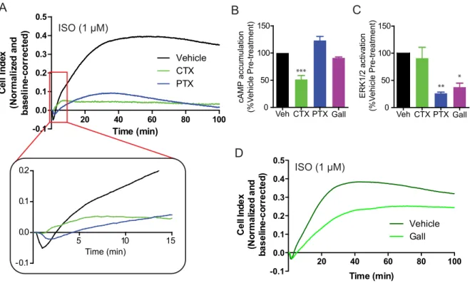
Using the β2-‐adrenergic receptor (β2AR) as a model system, we have demonstrated that changes in cellular impedance reflect the activity of multiple signalling events elicited following ligand stimulation of the receptor. Isoproterenol, the prototypical agonist of the β2AR, was found to elicit a dose-‐dependent impedance response consisting of multiple, discrete features over time, which could be blocked in a competitive manner by the antagonist ICI118,551. Using pathway-‐selective inhibitors, we were able to dissect the contribution of many of the canonical pathways activated by the β2AR, including Gs-‐ and Gi-‐ dependent signalling, as well as cAMP production and ERK1/2 activation. Furthermore, through the pharmacological dissection of this impedance response, we identified a novel
v
Ca2+ mobilization pathway that contributes to the overall cellular response to β2AR stimulation. In a separate study of the mechanism generating this β2AR-‐promoted Ca2+ response, we revealed a Gs-‐dependent transactivation mechanism of the Gq-‐coupled P2Y11 purinergic receptor. Given the ability of impedance measurements to capture this pleiotropic signalling activity, we then reasoned that ligands exhibiting different signalling profiles should generate distinct impedance signatures. In screening a library of functionally selective compounds targeting the β2AR, we obtained a wide variety of impedance signatures. Through the development of a novel computational approach, we were able to cluster these signatures into five distinct compounds classes, which were highly correlated with signalling profiles of the ligands.
In an extension of this approach, we then combined impedance screening with the use of pathway-‐selective inhibitors to determine if this would provide greater resolution in distinguishing among functionally distinct compounds. By assessing if and how a given signalling pathway contributes to a ligand’s impedance signature, we were able to reveal even more texture among ligands targeting the β2AR. Furthermore, this approach was found to be predictive of the signalling profiles of a library of uncharacterized compounds for the β2AR. This work led to the development of a visualization method to express ligand functional selectivity and revealed potentially novel classes of compounds for the receptor. These compound classes were then validated in human cardiomyocytes, confirming that compounds clustering into different classes produced distinct effects on cardiomyocyte contractility.
Altogether, this work demonstrates the ability of cellular impedance to accurately measure functional differences among compounds targeting GPCRs. In providing a representation of the pluridimensionality of GPCR signalling using a single, label-‐free assay, impedance profiling represents an innovative strategy to assess ligand functional selectivity and may be a valuable addition to future drug discovery campaigns.
Key words: G protein-‐coupled receptor (GPCR), β2-‐adrenergic receptor (β2AR), functional selectivity, signalling networks, cellular impedance, cardiomyocytes, drug discovery
vi
vii
TABLE OF CONTENTS
INTRODUCTION ... 1
1. Introduction to G protein-‐coupled receptors ... 1
2. The evolution of receptor theory ... 4
3. The pleiotropic nature of GPCR-‐mediated signalling ... 12
1. Promiscuity of G protein coupling and activation ... 12
2. Non-‐G protein effectors ... 16
i. β-‐arrestin ... 16
ii. G protein-‐coupled receptor kinases ... 19
iii. Second messenger kinases ... 19
iv. Src family kinases ... 20
v. GPRC scaffolding proteins ... 21
vi. Other effectors ... 22
4. Functional selectivity ... 25
1. Introduction to functional selectivity ... 25
2. Biased agonism at G protein-‐dependent signalling pathways ... 26
3. G protein-‐dependent vs. -‐independent signalling bias ... 28
4. Uncoupling of signalling from receptor desensitization ... 29
5. Dimerization ... 32
6. Selective antagonism ... 33
7. Physiological and clinical relevance ... 33
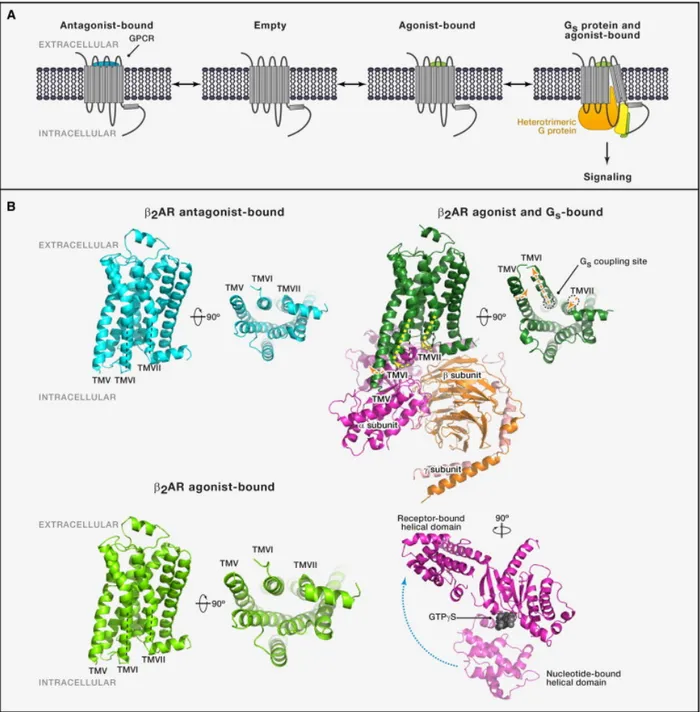
5. Conformational basis of functional selectivity ... 35
1. Spectroscopic studies ... 35
2. Crystal structures ... 38
3. Nuclear magnetic resonance studies ... 43
6. Experimental assessment of functional selectivity ... 45
1. Measurement of discrete signalling outcomes ... 46
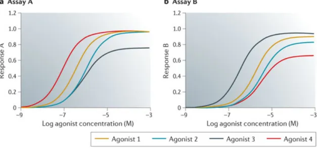
2. Ligand signalling fingerprints ... 47
3. Quantifying ligand bias ... 49
4. Limitations of endpoint assays ... 54
7. The use of cell-‐based, label-‐free techniques to assess functional selectivity ... 56
1. Cellular impedance vs. dynamic mass redistribution ... 56
2. Other applications of label-‐free assays ... 57
3. Advantages of label-‐free techniques to study GPCR signalling ... 57
OBJECTIVES ... 59
RESULTS ... 62
ARTICLE 1 ... 63
Impedance responses reveal β2-‐adrenergic receptor signaling pluridimensionality and allow classification of ligands with distinct signaling profiles ARTICLE 2 ... 110
β2 adrenergic receptors promote a Gs-‐dependent but cAMP-‐independent transactivation of
viii
ARTICLE 3 ... 140
Chemical Systems Biology Approach for the Functional Classification of Ligands Targeting the β2 adrenergic receptor DISCUSSION ... 181
1. A return from reductionism ... 181
2. The use of cellular impedance in drug discovery ... 184
3. The influence of network architecture on GPCR signalling and pharmacology ... 188
4. Receptor theory: Redux ... 194
CONCLUSION ... 200
REFERENCES ... 202
ANNEX I: List of other publications ... xvii
ANNEX II: Review paper ... xviii
ix
List of Tables
Table 1: Heterotrimeric G proteins and their signalling properties. ... 3
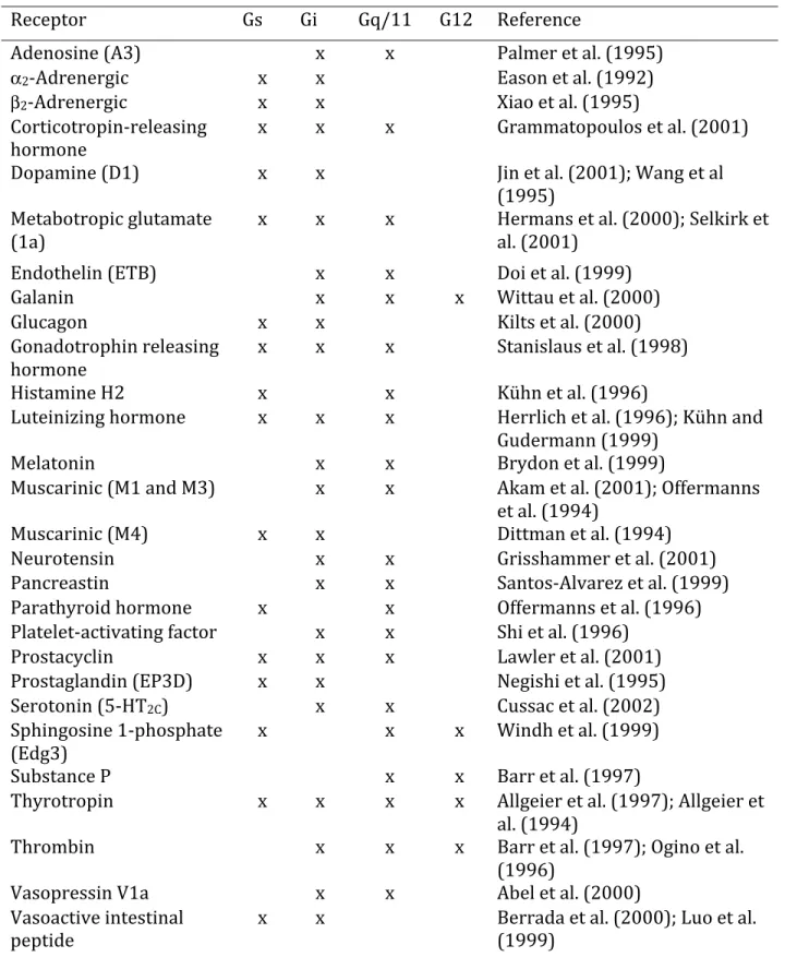
Table 2: Typical examples of receptors showing multiplicity in G protein coupling. ... 15
x
List of Figures
Figure 1: Mechanism of G protein activation. ... 4
Figure 2: Structural differences in the antagonist-‐ and agonist-‐bound β2AR. ... 41
Figure 3: Exploiting functional selectivity for drug discovery. ... 48
Figure 4: Bias plots to visual ligand functional selectivity. ... 51
Figure 5: Calculation of bias factors. ... 53
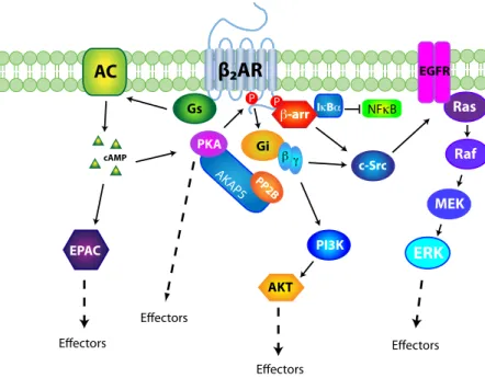
Figure 6: Known components of the β2AR signalling network. ... 60
Figure 7: Integration of cAMP, MAPK and Ca2+ on CRE-‐dependent transcription ... 189
Figure 8: Influence of Akt and ERK1/2 pathways on cell proliferation and differentiation. ... 190
Figure 9: Conformational landscapes of GPCR activation with ligands of different efficacies. ... 197
Figure 10: Divergent conformational pathways in receptor activation. ... 199
xi
Abbreviations
5-‐HT 5-‐hydroxytryptamine
α2AR α2 adrenergic receptor AA arachadonic acid
AKAP A-‐kinase anchoring protein AT1AR angiotensin 1A receptor
β1AR β1 adrenergic receptor
β2AR β2 adrenergic receptor
β-‐arr β-‐arrestin
BRET bioluminscence resonance energy transfer Ca2+ calcium
cAMP cyclic adenosine monophosphate CCKBR type B cholecystokinin receptor CHO Chinese hamster ovary
CRE cAMP response element CTC cubic ternary complex D2R Dopamine D2 receptor
DAMGO [D-‐Ala2,N-‐MePhe4,Gly-‐ol5]-‐enkephalin DMR dynamic mass redistribution
DVG (Asp1, Val5, Gly8)-‐angiotensin II EGFR epidermal growth factor receptor
EPAC exchange nucleotide protein directly activated by cAMP ERG ergotamine
xii FAK focal adhesion kinase
FRET fluorescence resonance energy transfer GABA γ-‐aminobutyric acid
GAP GTPase activating protein GDP guanosine diphosphate GEF guanosine exchange factor GGL G protein gamma-‐like GIP GPCR interacting protein GMP guanosine monophosphate GPCR G protein-‐coupled receptors GRK GPCR kinase
GSK glycogen synthase kinase 3 GTP guanosine triphosphate HDL high density lipoproteins HEK293 human embryonic kidney-‐293 HIV human immunodeficiency virus IGFR insulin-‐like growth factor receptor IKK IκB kinase
Ins-‐1-‐P inositol-‐1-‐phosphate IP inositol phosphate IP3 inositol trisphosphate
IPSC induced pluripotent stem cell JAK Janus kinase
µOR µ-‐opioid receptor
xiii MAPK mitogen activated protein kinase MC4 type 4 melanocortin receptor MD molecular dynamics
mGluR metabotropic glutamate receptor MSH melanocortin stimulating hormone NGF nerve growth factor
NHE3 Na+/H+ exchanger type 3
NHERF1/2 Na+/H+ exchanger regulatory factors 1 and 2 NMDA N-‐methyl-‐D-‐aspartate
NMR nuclear magnetic resonance
NPA R-‐(-‐)-‐10,11-‐dihydroxy-‐N-‐n-‐propylnoraporphine PACAP pituitary adenylyl cyclase-‐activating polypeptide PAF platelet-‐activating factor
PDE phosphodiesterase
PDZ PSD-‐95/Discs-‐large/ZO-‐1 PI3K phosphatidylinositol-‐3-‐kinase PKA protein kinase A
PKC protein kinase C PLC phospholipase C
PP2A protein phosphatase 2A
PSD-‐95 postsynaptic density protein 95 PTB phosphotyrosine binding
PTEN phosphatase and tensin homolog PTH parathyroid hormone
xiv PTH1R type-‐1 parathyroid hormone receptor RAMP receptor activity modifying protein RGS regulators of G protein signalling RhoGEF Rho guanosine exchange factor RKIP Raf kinase inhibitor protein RSK2 p90 ribosomal S6 kinase 2 RTK receptor tyrosine kinase SHP-‐1 Src homology phosphatase 1 SHP-‐2 Src homology phosphatase 2 SI (Sar1, Ile8)-‐angiotensin II SII (Sar1, Ile4, Ile8)-‐angiotensin II
SSNMR solid state nuclear magnetic resonance
STAT signal transducers and activators of transcription TM transmembrane domain
TPR1 tetratricopeptide repeat 1 V2R V2 vasopressin receptor
xv
Acknowledgments
First of all, I would like to graciously thank my supervisor, Dr. Michel Bouvier. Being given the privilege to conduct my doctoral studies in his laboratory has been an honour and pleasure. Dr. Bouvier has been and, I sincerely hope, will continue to be an exceptional mentor. I have learned so much from our discussions and debates, and I have developed both scientifically and personally as a result of his mentorship.
I would also like to thank all the members of the laboratory, past and present. I could not ask for a better group of scientists and friends to pass the long days toiling away in the lab. I will sincerely miss the fruitful discussions, the stimulating debates and the laughs. I wish everyone great success in their future endeavours. In particular, I would like to thank Eric Carpentier, who, in addition to patiently walking me through basic biochemistry techniques (often more than once), also provided indispensible assistance in the writing of this thesis.
And to my friends and family, near and far, thank you all very much for all your love and support. You have made this time all the more pleasant.
And finally, thank you to the agencies who have provided funding for my studies, including the Canadian Institutes of Health Research (CIHR), the Groupe de recherche universitaire sur le médicament (GRUM) and the Faculté des études supérieures de l’Université de Montréal (FES).
xvi
INTRODUCTION
1. Introduction to G protein-‐coupled receptors
G protein-‐coupled receptors (GPCRs) are an extraordinary family of cell surface receptors. With their seven transmembrane tertiary structure, they weave themselves into molecular machines perfectly tuned to transduce extracellular signals, in the form of small molecules, peptides, ions and even photons, into intracellular signalling events that allow a precise and appropriate response to a diverse environmental context. In fact, this capacity of cells to transduce extracellular signals into an intracellular response is the basis for organism multicellularity and underlies a host of fundamental and diverse cellular activities in eukaryotes. As a result, GPCRs are found in eukaryotes ranging from protists and fungi, to plants and animals (Perez, 2005).
In humans, GPCRs represent the largest family of cell surface receptors, with over 900 individual receptor subtypes (Lappano & Maggiolini, 2011). This remarkable abundance exemplifies the broad reactivity of GPCRs to an extraordinary variety of stimuli, ranging from photons and Ca2+ ions, to small molecules such as hormones and neurotransmitters, odorants and tastants, to peptides and even larger proteins. In accordance with such a broad spectrum of stimuli, GPCRs are involved in nearly every facet of human physiology including organism development (Katanaev, 2010), perception of sight (Palczewski, 2012), smell (DeMaria & Ngai, 2010), and taste (Palmer, 2007), modulation of mood and behaviour (Catapano & Manji, 2007), cardiac function (Salazar, Chen, & Rockman, 2007), blood vessel tone (Kauffenstein, Laher, Matrougui, Guérineau, & Henrion, 2012), innate and adaptive immunity (Yang, Chertov, & Oppenheim, 2001; Yona, Lin, & Stacey, 2010) and in the maintenance of various homeostatic mechanisms (Shioda et al., 2008; Tsunematsu & Yamanaka, 2012), among countless other essential functions.
Given their ubiquitous role in human physiology, it is not surprising that GPCRs also represent the largest family of therapeutic targets (Imming, Sinning, & Meyer, 2006; Overington, Al-‐Lazikani, & Hopkins, 2006; Pierce, Premont, & Lefkowitz, 2002). Whether
2
playing a direct role in the pathology of a given disease, or by providing an indirect mechanistic route to therapeutic benefit, GPCRs often represent an effective clinical target for the treatment of pathologies ranging from diabetes (Ahrén, 2009) to asthma (Barnes 2011), cardiovascular disorders such as hypertension (Brinks & Eckhart, 2010) and heart failure (Lymperopoulos, Rengo, & Koch, 2007), neuropsychiatric disorders like schizophrenia (Capuano, Crosby, & Lloyd, 2002) and Parkinson’s disease (Xu et al. 2012), human immunodeficiency virus (HIV) infection (Maeda, Das, Nakata, & Mitsuya, 2012), pain (Pan et al., 2008) and in the treatment of various forms of cancer (Lappano & Maggiolini, 2011).
GPRCs transduce extracellular signals into intracellular effects through the modulation of an important class of cytoplasmic effector proteins – the heterotrimeric G proteins. These effectors consist of three subunits. The Gα subunit, of which there are four main families (see Table 1), act as the primary determinant of the signalling response induced upon activation of the heterotrimeric protein. The β and γ subunits, of which there are at least five and thirteen isoforms, respectively, are generally regarded as a non-‐dissociable complex and can elicit additional signalling events themselves (Table 1). The G protein acts as a “molecular switch” whose activity is dependent on the guanosine nucleotide bound to it. When bound by guanosine triphosphate (GTP), the heterotrimeric G protein is in its active signalling state, capable of interacting with and activating downstream effectors. When the bound GTP is hydrolyzed to guanosine diphosphate (GDP), the G protein returns to its inactive state. By possessing intrinsic GTPase activity, the G protein itself limits the duration of its signalling activity by spontaneously hydrolyzing its bound GTP to GDP. This activity cycle can be influenced by other proteins to either increase the activation of G proteins by stimulating the exchange of GDP for GTP (i.e. guanosine exchange factors, GEFs) or by stimulating the hydrolysis of GTP to GDP (i.e. GTPase activating proteins, GAPs) to return the protein to its inactive state. GPCRs exert their intracellular effect largely through the activation of G proteins. Receptor activation stimulates the exchange of GDP for GTP, thus activating the G protein to elicit downstream signalling events (Figure 1) through either the α-‐ or βγ-‐subunits. GPCR subtypes possess a selectivity for distinct G proteins, which determines, to a large extent, the subsequent signalling events modulated by the
3
receptor and ultimately its role cellular physiology. Consequently, GPCRs were traditionally classified by the family of G proteins to which they preferentially couple; however, the notion of GPCR-‐G protein coupling specificity has been challenged by the observation that many (if not most) receptors can couple to and activate multiple G protein subtypes (See Section 3.1, page 12).
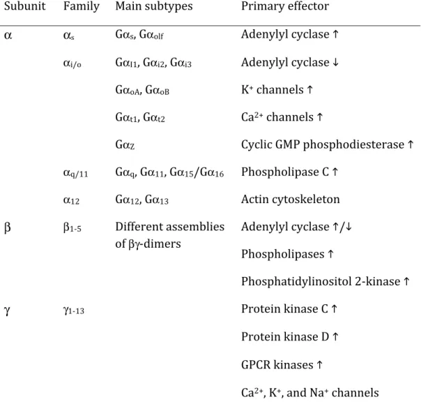
Table 1: Heterotrimeric G proteins and their signalling properties.
Adapted from Hermans (2003).
Subunit Family Main subtypes Primary effector
α αs Gαs, Gαolf Adenylyl cyclase
αi/o GαI1, Gαi2, Gαi3 Adenylyl cyclase GαoA, GαoB K+ channels Gαt1, Gαt2 Ca2+ channels GαZ Cyclic GMP phosphodiesterase αq/11 Gαq, Gα11, Gα15/Gα16 Phospholipase C α12 Gα12, Gα13 Actin cytoskeleton β β1-‐5 Different assemblies of βγ-‐dimers Adenylyl cyclase / Phospholipases Phosphatidylinositol 2-‐kinase γ γ1-‐13 Protein kinase C Protein kinase D GPCR kinases
Ca2+, K+, and Na+ channels
4
Figure 1: Mechanism of G protein activation.
Receptor activation couples to G protein activation by promoting the exchange of GDP for GTP on the α subunit of the heterotrimeric G protein. Once activated, the α-‐ and βγ-‐subunits are capable of interacting independently with and activating downstream effectors to elicit a cellular response. Hydrolysis of the Gα-‐bound GTP to GDP, either through its intrinsic GTPase activity, or with the assistance of additional regulatory GTPase-‐activating proteins (GAPs, e.g. regulators of G protein signalling (RGS) proteins, see Section 3.2.vi, page 22), inactivates the heterotrimeric G protein, terminating its signalling activity. Adapted from Siderovski and Willard (2005)
2. The evolution of receptor theory
GPCRs have long been the cornerstone of pharmacological theory. Many of the key pharmacological concepts that are used to this day to define the properties of drugs were discovered and developed in GPCR model systems.
Much of this early work was performed by a select group of scientists working in the United Kingdom in the first half of the 20th century, including Gaddum, Clark, Schild & Stephenson, who developed the quantitative methods to measure and the theoretical approaches to
5
explain the response of endogenous substances or drugs (collectively referred to as ligands) in isolated tissues (Hill, 2006). Early measurements of concentration-‐response profiles in these isolated systems and the application of the law of mass action to these data (Gaddum, 1937) allowed scientists to quantitatively determine the affinity of a compound, or its propensity to recognize and bind to a given target (Clark, 1937). These seminal studies assumed that the tissue response to the ligand was proportional to the percentage of occupied receptors, often termed the receptor occupancy model. However, evidence began to accumulate indicating that such a model could not account for all of the pharmacology being observed. In 1954, E.J. Ariëns demonstrated that different agonists acting on the same receptor could produce different maximal responses in a tissue and proposed a proportionality term, the “intrinsic activity” of a ligand, to describe these quantitative differences in agonist strength (Ariëns, 1954). In 1956, R.P. Stephenson provided clear evidence of the quantitative disparity between the occupancy of a ligand and the magnitude of its pharmacological effect. Like Ariëns, he introduced a new pharmacological parameter to represent the strength of an agonist to elicit a response. He thus defined efficacy as the capacity of a ligand, once bound, to initiate a reaction in the receptor that culminates in a cellular response (Stephenson, 1956). To further explain this new pharmacological parameter, Stephenson claimed that:
“Different drugs may have varying capacities to initiate a response and consequently occupy different proportions of the receptors when producing equal responses” (Stephenson, 1956)
Thus, contrary to prior assumptions, the propensity of a ligand to bind to a receptor does not solely explain its ability to generate a response through that receptor. In other words, affinity and efficacy are two independent properties of a given ligand-‐receptor pair. Whereas affinity provides information about the strength of the physical interaction between the ligand and receptor, which can be used to predict the selectivity of a drug for its target, it is the efficacy of a ligand that ultimately determines the magnitude and nature of the cellular response that is generated. This work ultimately helped establish a mathematical formalism to describe the response of a drug at a given receptor:
6
𝑅𝑒𝑠𝑝𝑜𝑛𝑠𝑒 = 𝑓 𝐴 × 𝜀[𝑅!] 𝐴 ×𝐾!
(Equation 1; Kenakin, 2004, Stephenson, 1956)
where ε is an empirical proportionality constant representing the intrinsic efficacy of drug A, Rt is the receptor density in the tissue, KA is the affinity constant of the drug for the
receptor. The function f relates the initial stimulus of the system to a tissue response.
These advancements also established the concept of partial agonists, or ligands that, even when occupying all of the available receptors in a system, do not elicit a maximal response as compared to a full agonist. The intrinsic efficacy, ε, of a ligand could vary from zero, as is true for antagonists, to a large positive value, for full agonists, or anywhere in between. The potency of a drug, a property that refers to concentration of the drug required to produce a response of a given magnitude, would therefore be influenced by both the affinity and the efficacy of the drug at a given receptor. The experimental and analytical techniques developed during this time were employed for decades to characterize and develop numerous drugs that remain on the shelves of pharmacies today, including the Nobel Prize winning work by Sir James Black during the 1960s for the development of beta blockers (antagonists of the β adrenergic receptors) for the treatment of angina and other cardiovascular disorders and H2 histamine receptor antagonists to treat stomach ulcers (Black, 1988).
Despite the extraordinary power of such a model, researchers were still confounded with the biochemical nature of the efficacy parameter and its dependence on the tissue in which it is assessed for a given ligand-‐receptor pair. Since the precise steps linking the binding of an agonist to a tissue response could not easily be known or quantified, Black & Leff (1983) sought to derive a more practical or ‘operational’ approach to quantifying efficacy. With the operational model of pharmacological agonism these researchers provided a mathematical formalism for the relationship between the concentration of an agonist and the tissue response it generated in purely biochemical and experimentally accessible terms:
7
𝑅𝑒𝑠𝑝𝑜𝑛𝑠𝑒 = 𝐴 × 𝜏 ×𝐸!"# 𝐴 𝜏 + 1 + 𝐾!
(Equation 2; Black & Leff, 1983)
𝜏 = [𝑅!] 𝐾!
(Equation 3; Black & Leff, 1983)
Key to this new model of receptor theory is the introduction of the τ parameter, which
functions as a ‘transducer ratio’ and is a measure of the efficiency of receptors occupied by agonist A to elicit a response in a given system. τ is inversely proportional to the
concentration of occupied receptors necessary to elicit a half-‐maximal response in the system, KE (Equation 3). Therefore, in systems where receptor activation is very efficiently
coupled to a tissue response (i.e. relatively few receptors need to be activated to produce a response), τ would be large. Only in systems with a very high receptor number and
stimulated with a strong agonist would the tissue response approach the system maximum,
EMAX. This mathematical formalism was extremely powerful for analyzing pharmacological
systems in several ways. First, the definition of the τ parameter provided a quantitative
description of tissue-‐specific differences in the coupling of receptor stimulation to a tissue response. Although the value of τ for a given agonist can vary from tissue to tissue, the rank
order of τ among a collection of agonists should not change. Therefore, τ also provided a
practical means of ranking agonists according to their relative efficacies regardless of the system used for the measurement.
While this conceptual framework remains very effective to explain many aspects of GPCR pharmacology to this day, receptor theory has continued to develop and provide even greater insights into mechanistic basis of drug activity at GPCRs. The operational model was developed, for example, on the assumption of a two-‐state model of receptor activation (see Equation 4). According to this “lock-‐and-‐key” type model, receptors existed in two distinct states or conformations: an inactive (Ri) and an active receptor conformation (Ra).
8
Unliganded receptors would remain in an inactive state, Ri, until bound by agonist, which would induce a conformational change to convert the receptor into an active signalling state Ra to elicit a cellular response. If bound by an antagonist, however, the receptor would remain locked in an inactive conformation, Ri, and thus not produce a cellular response, but would also prevent an endogenous agonist access to activate the receptor.
(Equation 4) However, several studies suggested that receptors could spontaneously assume the Ra conformation and elicit constitutive signalling activity in the absence of bound ligand. Kjelsberg et al. (1992) identified a single amino acid (alanine 293) in the α1B adrenergic receptor that when substituted for any other amino acid conferred an increase in the basal signalling activity of the receptor and agonist affinity. Further evidence of a wild-‐type receptor exhibiting constitutive activity was obtained by Tiberi & Caron (1994) with the observation that increasing the expression level of the dopamine 1A and 1B receptors correlated in a linear manner with cellular adenylyl cyclase activity in the absence of agonist. These data suggested that the receptor exists in a dynamic conformational equilibrium between Ri and Ra. The apparent activation of the receptor by an agonist, therefore, would simply reflect a greater affinity for the active conformation and a shift in the equilibrium towards the Ra state (Equation 5). Furthermore, this equilibrium model also accounted for the observed ability of certain ligands to decrease the signalling activity of a receptor below that of the basal level (Chidiac, Hebert, Valiquette, Dennis, & Bouvier, 1994; Costa & Herz, 1989; Costa, Ogino, Munson, Onaran, & Rodbard, 1992; Samama, Pei, Costa, Cotecchia, & Lefkowitz, 1994). Termed inverse agonists (Chidiac et al., 1994), these ligands would therefore preferentially stabilize the Ri state of the receptor, shifting the equilibrium of the system towards the inactive receptor reducing receptor activity below that of the unliganded state (Equation 5). In addition, a neutral antagonist would be a ligand that binds both Ri and Ra with similar affinity, so as to not significantly shift the equilibrium
R
iR
aantagonist
9
from that of the basal, unliganded state and consequently elicit no detectable signalling activity through the receptor.
(Equation 5)
Another significant development on the operational model occurred upon the discovery that certain proteins interacting directly with the receptor could influence its pharmacology through allosteric effects on the receptor itself. The first proximal effectors found to exhibit this property were the heterotrimeric G proteins. First indications of this phenomenon were the observations that the addition of guanosine nucleotides could change the affinity of an agonist for a receptor (Kent, De Lean, & Lefkowitz, 1980; Lefkowitz, Mullikin, & Caron, 1976; Maguire, Van Arsdale, & Gilman, 1976; Stadel, DeLean, & Lefkowitz, 1980; Williams & Lefkowitz, 1977). Increasing the amount of GTP or a non-‐ hydrolyzable GTP variant (Gpp(NH)p) was found to shift the competition binding curves of agonists to the right, demonstrating a progressively lower affinity of the receptor for the agonist with increasing concentrations of guanosine nucleotide. Interestingly, this effect was only found for agonists, as the binding curves of antagonists were unaffected. De Lean et al. (1980) proposed the ternary complex model to account for the high and low affinity states of agonists, suggesting that the active signalling species of the receptor was a complex between the agonist (A), receptor (R) and some cytoplasmic effector (X) capable of binding GTP:
R
iR
a agonist inverse agonist10
(Equation 6, Adapted from De Lean, Stadel, and Lefkowitz 1980)
This model proposed that the high affinity state of the receptor was the ternary complex ARX and that binding of a guanosine nucleotide to X would both lead to the activation of downstream effectors (E) and destabilize the high affinity complex. The identity of X, of course, was subsequently discovered to be the heterotrimeric G proteins (reviewed by Limbird, 1981), thus conferring the name G protein-‐coupled receptors to this family of membrane proteins.
This early model was subsequently refined to synthesize the above observations with the fact that receptors could spontaneously assume active signalling conformations, and thus form the active RaG protein complex in the absence of ligand. This extended ternary complex (ETC) model (Samama, Cotecchia, Costa, & Lefkowitz, 1993) provided a more complete description of the interactions between agonist (A), receptor (R) and G protein (G):
(Equation 7; Kenakin, 2004) A A A A
ligands confer unique conformations to receptors that
differ from the constitutively active receptor state[34].
In thermodynamic terms, there must be a provision for the inactive receptor to also interact with G proteins; this is allowed in a more complete but more complex model for GPCRs, named the cubic ternary complex (CTC) model
(Box 2)[35]. Recent evidence indicates that antagonists
form GTP-sensitive, non-signaling ternary complexes with
receptors (e.g. opioid peptide receptors[36]and histamine
H2receptors[37]) and that unliganded wild-type receptors
(e.g. pheromone receptors Ste2p and Ste3p [38], and
cannabinoid CB1 receptors[39]) and receptors bound to
inverse agonists {SR141716A (see Chemical names) for
CB1receptors[40], and tiodidine for H2receptors[37]} can
sequester G proteins (in the form of antagonist-bound, non-signaling ternary complexes) from other systems to
reduce constitutive activity. These data suggest that the CTC model applies for some receptor systems.
In the worst-case scenario, recombinant systems can simply show uncharacteristic behavior of receptors or receptors under extreme conditions (i.e. the data take on a ‘Pandora’ aspect whereby the resulting information is misleading and dissimulating). From this standpoint, such data reflect what a receptor can do, and not necessarily what it does under normal physiological circumstances. However, pathological processes change synoptic receptor systems to set-points that might not have been imagined in normal in vitro pharmacological test systems. Therefore, such extremes can be therapeutically relevant.
Beyond linkage models
Linkage models such as the ETC and CTC models are extremely useful for deriving methods to quantify drug
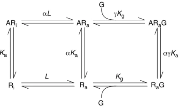
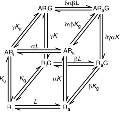
Figure 2. Ternary complex models for G-protein-coupled receptors (GPCRs). (a)
Extended ternary complex model. Receptor states (Ri) and (Ra) coexist according to
the allosteric constant L. G protein (G) enters the system and binds to the activated
receptor state Rato produce the physiological response. Ligand A binds to either
receptor state and also to Rawhen it is bound to the G protein. The propensity of the
system to produce constitutive activity (spontaneous formation of the active state
RaG species) is defined by the allosteric constant L {L¼ [Ra]/[Ri]}. The affinity of
ligands for the receptors is given by Kawhereas the efficacy is described by two
terms, a and g. The term a is the differential affinity of the ligand for Raand the term
gis the differential affinity of the ligand-bound ARafor G proteins. (b) Cubic ternary
complex model. The inactive receptor species Riand ARiare allowed to interact
with G proteins (but not signal) in this variant model. b refers to the differential affinity of the receptor active state (over the inactive state) for the G protein.
TRENDS in Pharmacological Sciences
ARa ARi Ra ARaG RaG Ri Ka αKa γKg αL Kg L G G αγKa ARiG ARaG ARi ARa RiG RaG Ri Ra Ka Kg L αK αL βL δαβL βKg γK δγβKg δγαK γKg
(a) Extended ternary complex model
(b) Cubic ternary complex model
Box 2. Ternary complex models for GPCRs
The extended ternary complex (ETC) model (Figure 2a in the main text) describes a receptor that can exist in two states, active (Ra) and
inactive (Ri), named for their ability to activate G proteins (G). These
conformations coexist according to an allosteric constant unique for the receptor type (denoted L¼ ½Ra#=½Ri#Þ: Ligands have affinity for Ri
denoted by Ka(equilibrium association constant) and a differential
affinity for Raof aKa. Similarly, Rathat is not bound to ligand has an
affinity for G proteins of Kg;ligands can confer a different affinity of
the receptor for G protein denoted gKg.
The ETC model describes response production (elevated concen-trations of Raand ARa) as a fraction of the total receptor species
(denoted by r) as:
r¼ L½G#=KGð1 þ ag½A#=KAÞ
½A#=KAð1 þ aLð1 þ g½G#=KGÞÞ þ Lð1 þ ½G#=KGÞ þ 1 ½Eqn I#
where KAand KGare equilibrium dissociation constants (reciprocals
of association constants). Figure I shows the effects of changing a on the dose–response curves of a system with existing constitutive activity (note the elevated basal activity in the absence of ligand). Formally identical effects are observed with changes in g values. The cubic ternary complex model (Figure 2b in the main text) allows interaction between the Riand G proteins.
Figure I. Response according to the extended ternary complex model for G-protein-coupled receptors (GPCRs). Response (ordinate axis) given as the
concentration of the response-producing species [RaG]þ [ARaG] as a fraction
of the total receptor number according to Eqn I. Curves were calculated for
agonists of fixed value for g (g¼ 5) and varying magnitudes for a in a system
with constitutive receptor activityðL ¼ 0:01Þ:
TRENDS in Pharmacological Sciences
–5 –3 –1 1 3 1.0 0.8 0.6 1.2 0.0 0.4 0.2 Log ([A]/KA) F
raction of maximal response
10 α = 30 3 1 0.3 0.1 0.03 0.01 Chemical names SR141716A: N-(piperidin-1-yl)-5-(4-chlorophenyl)-4-methyl-1H-pyrazole-3-carboxamide HCl
Review TRENDS in Pharmacological Sciences Vol.25 No.4 April 2004 189