Recognition of Hepatitis C Virus RNA by Toll like Receptors 7 and 8: Implications for the Initiation of Innate Immune Response
Reconnaissance de l’ARN du virus de l’hépatite C par les récepteurs de type Toll 7 et 8 Implications dans l’initiation de la réponse immunitaire innée
By Yuwei Zhang
Département de Microbiologie et immunologie Faculté de Médecine
Thesis submitted to the Faculty of Medicine to obtain the degree of Philosophiae Doctor (Ph.D.)
in Immunology
II
Abstract
Hepatitis C virus (HCV), a positive single stranded RNA (ssRNA) virus that replicates in the liver, infects 200 million people worldwide, with approximately 80% of infected individuals ultimately suffering from chronic HCV infection. Antiviral therapies, including interferon and ribavirin, have improved considerably in recent years, but are effective in only about one-half of those treated, and are associated with significant side effects and toxicity.
Innate immune defenses are essential to control viral infection; the innate response is activated through recognition of viral macromolecular motifs known as pathogen-associated molecular patterns (PAMPs) that are recognized by a multitude of Pathogen recognition receptors (PRRs). Although immune activation induced by HCV RNA or proteins has been extensively studied, the detection of HCV by the innate immune system remains poorly understood. Despite activation of the immune response early after HCV infection in vivo, the persistent increase inpatient HCV viral load suggests the failure of the immune response to control HCV infection. A better understanding of the mechanisms of immune activation induced by HCV is crucial for development of effective strategies for HCV treatment.
Here we demonstrate in primary cell models that the HCV genome contained GU-rich RNA sequences that specifically trigger Toll-like receptors (TLR) 7/8, resulting in maturation of plasmacytoid dendritic cells (pDCs) and production of type I interferon (IFN), induction of inflammatory cytokines and chemokines in different antigen
III presenting cells (APCs). Cytokines produced by monocytes and pDCs upon stimulation of HCV-derived ssRNA inhibited HCV production in an IFN-dependent manner, whereas cytokines produced by myeloid DCs (mDCs) and macrophages had no inhibitory effect on virus production, due to a defect in IFN induction by HCV ssRNA in these cells.
TLR7/8 stimulated cytokines also down-regulated the expression of the HCV receptor CD81 on Huh7.5 in an IFN-independent manner that may restrict HCV infection. However, although HCV receptors like CD81 were widely expressed on different subsets of lymphocytes, DCs and monocytes did not respond to HCV particles, and our data indicates that only macrophages sense HCV and produce inflammatory cytokines. The recognition of HCV by macrophages is related to the expression of DC-SIGN on macrophages, and results in TLR7/8 engagement. Similar to a TLR7/8 agonist, HCV stimulation induces inflammatory cytokine production (TNF-α, IL-8, IL-6 and IL-1b) by macrophages but does not stimulate interferon-beta production in macrophages, Congruously, TLR7/8 and HCV RNA mediated cytokine production in macrophages did not inhibit HCV replication.
Our results reveal that HCV RNA has the potential to trigger TLR7/8 in DC populations, and initiate an innate immune response against HCV infection that leads to an IFN-dependent suppression of viral replication. However, HCV is able to escape detection by monocytes and DCs, which produce type I IFN and suppress HCV replication when TLR7/8 is engaged. Macrophages possess the capacity to detect HCV, in part because of surface expression of DC-SIGN. In turn, macrophages produce inflammatory cytokines that nevertheless fail to inhibit HCV replication due to lack of IFN-beta production. Evasion of antiviral defense by HCV may explain the failure of innate immunity in
IV control of HCV infection. Moreover, inflammatory cytokine production by macrophages upon HCV stimulation in vitro suggests that activation of macrophages may contribute to inflammation in HCV infected individuals.
Key words: Hepatitis C virus, dendritic cells, interferon, single-strand RNA, TLR7, TLR8, CD81, DC-SIGN, macrophages
V
Résumé
Le virus de l’hépatite C (VHC) est un virus à ARN simple brin positif (ssARN) qui se replique dans le foie. Deux cents millions de personnes sont infectées par le virus dans le monde et environ 80% d’entre elles progresseront vers un stade chronique de l’infection. Les thérapies anti-virales actuelles comme l’interféron (IFN) ou la ribavirin sont de plus en plus utilisées mais ne sont efficaces que dans la moitié des individus traités et sont souvent accompagnées d’une toxicité ou d’effets secondaires indésirables.
Le système immunitaire inné est essentiel au contrôle des infections virales. Les réponses immunitaires innées sont activées suite à la reconnaissance par les Pathogen Recognition Receptors (PRRs), de motifs macromoléculaires dérivés du virus appelés Pathogen-Associated Molecular Patterns (PAMPs). Bien que l'activation du système immunitaire par l'ARN ou les protéines du VHC ait été largement étudiée, très peu de choses sont actuellement connues concernant la détection du virus par le système immunitaire inné. Et même si l’on peut très rapidement déceler des réponses immunes in vivo après infection par le VHC, l’augmentation progressive et continue de la charge virale met en évidence une incapacité du système immunitaire à contrôler l’infection virale. Une meilleure compréhension des mécanismes d’activation du système immunitaire par le VHC semble, par conséquent, essentielle au développement de stratégies antivirales plus efficaces.
VI Dans le présent travail nous montrons, dans un modèle de cellule primaire, que le génome ARN du VHC contient des séquences riches en GU capables de stimuler spécifiquement les récepteurs de type Toll (TLR) 7 et 8. Cette stimulation a pour conséquence la maturation des cellules dendritiques plasmacytoïdes (pDCs), le production d’interféron de type I (IFN) ainsi que l’induction de chémokines et cytokines inflammatoires par les différentes types de cellules présentatrices d’antigènes (APCs). Les cytokines produites après stimulation de monocytes ou de pDCs par ces séquences ssARN virales, inhibent la production du virus de façon dépendante de l’IFN. En revanche, les cytokines produites après stimulation de cellules dendritiques myéloïdes (mDCs) ou de macrophages par ces mêmes séquences n’ont pas d’effet inhibiteur sur la production virale car les séquences ssARN virales n’induisent pas la production d’IFN par ces cellules.
Les cytokines produites après stimulation des TLR 7/8 ont également pour effet de diminuer, de façon indépendante de l’IFN, l’expression du récepteur au VHC (CD81) sur la lignée cellulaire Huh7.5, ce qui pourrait avoir pour conséquence de restreindre l’infection par le VHC. Quoiqu’il en soit, même si les récepteurs au VHC comme le CD81 sont largement exprimés à la surface de différentes sous populations lymphocytaires, les DCs et les monocytes ne répondent pas aux VHC, Nos résultats indiquent que seuls les macrophages sont capables de reconnaître le VHC et de produire des cytokines inflammatoires en réponse à ce dernier. La reconnaissance du VHC par les macrophages est liée à l’expression membranaire de DC-SIGN et l’engagement des TLR 7/8 qui en résulte. Comme d’autres agonistes du TLR 7/8, le VHC stimule la production de cytokines inflammatoires (TNF-α, IL-8, IL-6 et IL-1b) mais n’induit pas la production d’interféron-beta par les macrophages. De manière attendue, la production de cytokines
VII par des macrophages stimulés par les ligands du TLR 7/8 ou les séquences ssARN virales n’inhibent pas la réplication virale.
Nos résultats mettent en évidence la capacité des séquences ssARN dérivées du VHC à stimuler les TLR 7/8 dans différentes populations de DC et à initier une réponse immunitaire innée qui aboutit à la suppression de la réplication virale de façon dépendante de l’IFN. Quoiqu’il en soit, le VHC est capable d’échapper à sa reconnaissance par les monocytes et les DCs qui ont le potentiel pour produire de l’IFN et inhiber la réplication virale après engagement des TLR 7/8. Les macrophages possèdent quant à eux la capacité de reconnaître le VHC grâce en partie à l’expression de DC-SIGN à leur surface, mais n’inhibent pas la réplication du virus car ils ne produisent pas d’IFN. L’échappement du VHC aux défenses antivirales pourrait ainsi expliquer l’échec du système immunitaire inné à contrôler l’infection par le VHC. De plus, la production de cytokines inflammatoires observée après stimulation in vitro des macrophages par le VHC suggère leur potentielle contribution dans l’inflammation que l’on retrouve chez les individus infectés par le VHC.
Mots clefs: virus de l’hépatite C, cellules dendritiques, interféron, ARN simple brin, TLR7, TLR8, CD81, DC-SIGN, macrophages
VIII
Table of Contents
ABSTRACT ... II RÉSUMÉ ... V TABLE OF CONTENTS ... VIII LIST OF FIGURES ... XI CONTRIBUTION OF AUTHORS ... XII ABBREVIATIONS ... XIV ACKNOWLEDGEMENTS ... XVI
CHAPTER 1 ... 1
INTRODUCTION ... 1
1. Hepatitis C virus- prevalence ... 2
2. Discovery of HCV ... 4
3. HCV virology ... 5
3.1 HCV genome and proteins 6 3.2 Viral life cycle 9 4. Infectious HCV culture system in vitro ... 10
5. HCV treatments and limitations ... 12
6. Acute, chronic infection and viral clearance ... 17
7. Immune response and HCV infection ... 19
7.1 Innate immunity 20 7.2 Adaptive immunity 21 8. TLR7 and TLR8 ... 23
IX
10.HCV detection by antigen-presenting cells ... 27
HYPOTHESIS AND OBJECTIVES ... 30
CHAPTER 2 ... 32
HCV derived ssRNA induce innate immune response against HCV infection via TLR7 and TLR8 ... 32
Abstract ... 34
Results ... 37
1. Identification of ssRNA sequences in HCV genome that stimulate innate immune responses 37 2. Specific recognition of HCV-derived ssRNA by TLR7 and TLR8 38 3. Cytokine production and DC maturation upon ssRNA stimulation 39 4. Effect of HCV-derived ssRNA-induced cytokines on HCV infection 40 5. HCV-derived ssRNA-mediated cytokines down-regulate CD81 expression on huh7.5 41 6. HCV-derived ssRNA pretreatment impairs pDCs capacity to produce IFNα 42 Discussion ... 44
Materials and methods ... 49
Reference ... 73
CHAPTER 3 ... 76
Macrophages sense Hepatitis C virus to initiate innate immunity ... 76
Abstract ... 78
Results ... 83
X 2. Induction of inflammatory cytokines by HCVcc stimulation 84 3. HCVcc as well as TLR7/8 agonist do not trigger interferon pathway in
macrophages 84
4. Antiviral activity and responsiveness to HCVcc 85
5. RNA sensors involved in HCV recognition 86
6. DC-SIGN expression and responsiveness to HCV 87
7. Difference in responsivenss of DC-SIGN expressing cells to HCV 88
Discussion ... 90
Materials and methods ... 94
Reference ... 112
CHAPTER 4 ... 115
DISCUSSION ... 115
1. Summary of findings ... 116
2. Immunogenicity of HCV ... 117
3. TLR7 and innate immunity against HCV infection ... 119
4. Induction of innate immune response and inflammation in HCV infection ... 121
5. Detection of HCV by antigen-presenting cells ... 123
6. Interferon and control of HCV infection ... 126
Perspectives ... 130
XI
LIST OF FIGURES
Chapter 1
Figure 1. Geographic distribution of Hepatitis C prevalence. ... 3
Figure 2 Global distribution of HCV genotypes ... 4
Figure 3. Genome and proteins of HCV virus. ... 8
Figure 4 Hepatitis C virus (HCV) viral cycle and targets for drug development ... 14
Chapter 2 Figure 1 GU-rich sequences in HCV genome induce TNF-α production. ... 56
Figure 2 Specificity of TLR7/8 triggering by HCV derived ssRNA ... 58
Figure 3 Inflammatory cytokines and chemokines production by monocytes, mDCs and pDCs. ... 60
Figure 4 Maturation and type I IFN production of pDC ... 63
Figure 5 HCV-ssRNA mediated cytokines inhibit HCV production in IFN-dependent mannar. ... 66
Figure 6 HCV-ssRNA mediated cytokines inhibit HCV production in IFN-dependent mannar. ... 68
Figure 7 HCV-derived ssRNA down-regulate CD81 expression on huh7.5. ... 70
Figure 8 Impairment of IFN expression in pDC with HCV-derived ssRNA pre-treatment ... 72
Chapter 3 Figure 1 Macrophages sense HCV . ... 98
Figure 2 Dose-dependent activation and Inflammatory cytokines induction by HCVcc stimulation. ... 100
Figure 3 Expression of Interferon and interferon-stimulated genes in macrophage upon HCV stimulation. ... 102
Figure 4 Antiviral activity and responsiveness to HCV. ... 104
Figure 5 Pathway involved in HCV recognition. ... 106
Figure 6 DC-SIGN mediated uptake of HCV. ... 109
XII
CONTRIBUTION OF AUTHORS
Chapter 2 HCV derived ssRNA induce innate immune response against HCV infection via TLR7 and TLR8
Yuwei Zhang, Elias Said, Abdel-Hakeem MS, Zhong He, Francesco A. Procopio,
Rafick-Pierre Sekaly
Yuwei Zhang and Elias Said defined the criteria of GU-rich sequence and selected nine sequences from HCV genome. Yuwei Zhang performed screening of RNA sequences and TNF-α ELISA. Yuwei Zhang, with help of Zhong He, performed the luciferase assay. Elias Said performed DC separation and cytokine/chemokine dectection by Cytometric Bead Array with the help of Yuwei Zhang. Francesco A. Procopio optimized the method for HCV quantification with the help of Abdel-Hakeem MS. Yuwei Zhang and Zhong He performed viral production and infection in vitro. The manuscript and the figures were generated by Yuwei Zhang under the supervision of Rafick-Pierre Sekaly.
XIII Chapter 3 Macrophages sense Hepatitis C virus to initiate innate immunity
Yuwei Zhang, Elias Said, Zhong He, Francesco A. Procopio, Rafick-Pierre Sekaly
Yuwei Zhang performed cell separation and macrophage generation. Yuwei Zhang performed TNF-α ELISA. Zhong He performed cytokine/chemokine dectection by Cytometric Bead assay. Francesco A. Procopio optimized the method for HCV quantification. Yuwei Zhang and Zhong He performed viral production and infection in vitro. Yuwei Zhang performed the measurement of gene expression and viral quantification by real-time PCR with the help of Francesco A. Procopio. Yuwei Zhang performed the phenotypic staining and flow cytometric analysis of innate immune cells and macrophages. The manuscript and the figures were generated by Yuwei Zhang, in collaboration with Elias Said, under the supervision of Rafick-Pierre Sekaly.
XIV
ABBREVIATIONS
APC: Antigen presenting cell
ARFP: Alternate reading frame protein CBA: Cytometric Bead Array
CLDN1: Claudin 1
CTL: Cytotoxic T lymphocyte
DC-SIGN: Dendritic Cell-Specific Intercellular adhesion molecule-3- Grabbing non-integrin dsRNA: Double-strand RNA
Flu: Influenza virus
GM-CSF: Granulocyte-macrophage colony-stimulating factor HCV: Hepatitis C virus
HCVcc: Cell culture-derived HCV HCVpp: HCV pseudotype particles HIV: Human immunodeficiency virus HSC: Hepatic stellate cell
IFN: Interferon IL-4: Interleukin 4
IRES: Internal ribosome entry site IRF-3: Regulatory factor-3
ISGs: Interferon-stimulated genes KC: Kupffer cell
LDLR: Low-density lipoprotein receptor
MDA-5: Melanoma Differentiation-Associated protein 5 mDC: Myeloid dendritic cell
XV MDM: Monocytes-derived macrophage
nAb: Neutralizing antibody NK: Natural killer cell
NLP: Nucleocapsid-like particle NS: Nonstructural protein NTR: Non-translated region
PAMPs: pathogen-associated molecular patterns PBMC: peripheral blood mononuclear cell pDC: Plasmacytoid dendritic cell PRRs: Pathogen recognition receptors RIG-I: Retinoic acid-inducible gene-I RLH: RIG-I-like RNA helicase SRB1: Scavenger receptor class B-1 ssRNA: Single stranded RNA
Stat1: Signal transducer and activator of transcription 1 SV: Sendai virus
SVR: Sustained antiviral response TLR: Trigger Toll-like receptors VLP: Viral-like particles
XVI
ACKNOWLEDGEMENTS
First of all, I would like to express my sincerest appreciation to my supervisor, Dr. Rafick-Pierre Sékaly, who has provided me with the personal and financial resources for the completion of this work. With his expertise, extensive knowledge, patience, and attention, I have been able to learn and grow professionally. The experiences with him have been invaluable in shaping the researcher that I want to be.
I would like to express my gratitude to Dr. Elias Said for his valuable contribution through supervising and the numerous scientific discussions, as well as Dr. Naglaa Shoukry for her support. I also would like to thank PhD candidate Mohamed S. Abdel– Hakeem, Dr. Zhong He and Dr. Mohamed El-Far for their significant help with viral production and technical assistance. I also would like to thank Dr. Francesco Procopio for his help with optimization of real-time PCR. I also thank Dr. Jeff Ahlers, Dr. Yu Shi, Dr. John Hiscott, Dr. Joumana Zeidan and Dr. Petronela Ancuta for their editorial assistance.
The encouragement and love from my family and friends are very important to me, they have support me during my Ph.D. studies. I would not have been able to complete without their financial and emotional support.
CHAPTER 1
2 1. Hepatitis C virus- prevalence
The hepatitis C virus, also known as HCV, is now a widespread public health problem that affects a global audience estimated to be 200 million individuals – equaling 3.3% of the world’s population. Annually, the number of new reported cases of the hepatitis C infection reaches as high as 3 to 4 million (Perz, Armstrong et al. 2006, WHO 2011). The prevalence of HCV is comparatively lowest in Asia Pacific, Tropical Latin America and North America (<1.5%). According to a particular survey, the occurrence of HCV antibodies with respect people donating blood averages less than a meager one percent for the aforementioned region. On the flip side, the rates are incrementally higher as seen in South Asia, Southern Latin America and Europe (1.5%-3%). The occurrence of hepatitis C antibodies in Central and East Asia, North Africa/Middle East were reported around 3.8% (Mohd Hanafiah, Groeger et al. 2013). As far as many African countries were concerned though, alarming rates have been reported, reaching as high as 14.7% in Egypt (Mohamoud, Mumtaz et al. 2013).
HCV infection primarily takes place in the liver and causes hepatitis C (Choo, Weiner et al. 1990). The infection is usually asymptomatic in nature (Alberti, Noventa et al. 2002, Mendez-Sanchez, Ponciano-Rodriguez et al. 2005), however chronic infection can lead to scarring of the liver and subsequently lead to cirrhosis (Freeman, Dore et al. 2001). Progression of liver disease caused by HCV usually takes decades - As high as one-fifth of the people that are infected may go on to develop complications such as liver failure, cirrhosis or even hepatocellular carcinoma (Perz, Armstrong et al. 2006). Barring hepatocellular carcinoma, with respect to patients that develop cirrhosis or liver cancer, a
3 liver transplant may be required. HCV-induced liver inflammation and cirrhosis is considered as one of the major causes for liver transplants (Davis, Albright et al. 2003). Only 10% to 40% of HCV-infected individuals can be cured by spontaneous viral clearance, and 60% to 90% infected individuals develop chronic infection, depending on the genotype of the virus they are contracted (Gerlach, Diepolder et al. 2003, Lehmann, Meyer et al. 2004, Micallef, Kaldor et al. 2006). There is no effective vaccine to prevent the hepatitis C infection. A combination of peg-interferon and ribavirin is the current standard therapy.
Figure 1. Geographic distribution of Hepatitis C prevalence
This image is originally from
4 Figure 2. Global distribution of HCV genotypes
This image is originally from
http://www.who.int/vaccine_research/documents/ViralCancer7.pdf
2. Discovery of HCV
In the mid 1970s, a new hepatitis virus was recognized in blood transfusion recipients, identified as non-A, non-B hepatitis (Feinstone, Kapikian et al. 1975). A few distinctive characteristics aided in separating this new virus from HAV and HBV. Infection in chimpanzees produced chronic disease is characterized by inflammation and hepatocyte structural changes (Popper, Dienstag et al. 1980, Dienes, Popper et al. 1982). Complete identification of the new virus was hindered by difficulties in producing a cellular model that allowed viral replication (Lohmann, Korner et al. 1999). In 1987, Michael Houghton and Daniel Bradley collaborated to create a molecular cloning technique to sequence the viral genome (Choo, Kuo et al. 1989). Once HCV was identified, screening for it in blood transfusions became mandatory starting in 1990, which subsequently decreased the rate of infection (Kimber 1990, Weiner, Kuo et al.
5 1990). The highest transmission rates were initially from unsanitary injection drug use or unsterile needle use such as in unregulated body tattoo art and blood transfusions (Conry-Cantilena, VanRaden et al. 1996). Occupational exposure to HCV is and has been the most difficult type of transmission to control as most infections occur accidentally, even with proper precautions observed (Yazdanpanah, De Carli et al. 2005). Since blood screening started to be applied, there has been a decrease of HCV infection in developed countries. However, transmission of the virus in some areas is still growing despite advanced techniques to identify HCV in blood (Alter 2007, Martins, Narciso-Schiavon et al. 2011). Owing to the expensive nature of testing for the virus, underdeveloped countries are still struggling to control the spread of the virus (van der Poorten, Kenny et al. 2008). Still, continued HCV research has led to improved treatments against the virus.
3. HCV virology
Hepatitis C virus, classified as part of the Flaviviridae family, is predominantly a singularly stranded positive-sense RNA virus. This genome is almost 9.6 kilo-bases and has six different major genotypes, genotypes 1-6 (Okamoto 1995). HCV has a high propensity for genetic variations due to the error-prone nature of the RNA replicase (Bukh, Miller et al. 1995, Pawlotsky 1998). Therefore, HCV-infected individuals with certain genotypes and subtypes respond atypically to treatment (Manns, McHutchison et al. 2001), and the identity of HCV genotypes in infected individuals is important to the specific treatment regimen. Throughout the history of HCV therapy, treatment has been conventionally less effective—up to 30% to 50%—in patients harboring HCV genotype 1
6 compared to up to 80% effective clearance in genotypes 2 and 3 (Zein, Rakela et al. 1996, Neumann, Lam et al. 2000, Lehmann, Meyer et al. 2004). Thus, genotype 1 is associated with the development of chronic infection and is also the most prevalent genotype in the world (Fig 2).
3.1 HCV genome and proteins
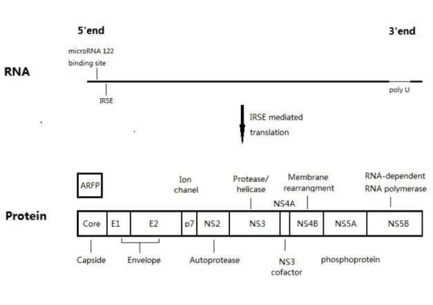
HCV is a relatively small 50 nm enveloped RNA virus (Kaito, Watanabe et al. 1994). The genome, flanked by small 5’ and 3’ non-translated regions (NTRs), has one open reading frame that translates to a number of proteins of roughly 3000 residues (Choo, Richman et al. 1991). The 5’ NTR region is known to contain an internal ribosomal entry site, denoted as IRES, and is considered extremely conserved among the various genotypes and subtypes. It is essential for RNA translation because it binds the 40S ribosome subunit, positioning it near the initiation site (Pestova, Shatsky et al. 1998, Friebe, Lohmann et al. 2001). The 3’ NTR region with its three domains—6variable region, poly U region, and a conserved terminal domain—is highly inconsistent among the genotypes (Friebe and Bartenschlager 2002). Areas in the latter two regions are required for RNA replication.
The virus has ten proteins, three of which are structural proteins: core and envelope 1 and 2 (Takeuchi, Kubo et al. 1990). In between these non-structural as well as structural proteins happens to be a rather small yet integral membrane protein: p7 (Lin, Lindenbach et al. 1994). The nonstructural proteins are NS2 and NS3, NS4 (A and B) and NS5 (A and B). Barring NS2, the remaining nonstructural proteins are deemed to be an
7 essential requirement for replication (Brenndorfer, Karthe et al. 2009, Morikawa, Lange et al. 2011). Extensive research into each of these proteins has revealed specific roles for viral replication and survival. Alternatively, a frame shift may occur in the Core protein-coding region to produce an alternate reading frame protein (ARFP) (Branch, Stump et al. 2005).
The hepatitis C core protein is an RNA binding protein that is highly conserved in nature (Bukh, Purcell et al. 1994, Hitomi, McDonnell et al. 1995). They are also presumed to form the viral capsid. When expressed in several in vitro systems, the hepatitis C core protein is known to participate in the formation of NLPs. (Baumert, Ito et al. 1998, Blanchard, Brand et al. 2002). The two envelope glycoproteins, that are namely E1 and E2, are essential for viral entry and fusion (Bartosch, Dubuisson et al. 2003, Nielsen, Bassendine et al. 2004). E2 plays an important role during early stages of the infection. E2 interaction with either one or multiple receptor complex components is considered to be the root cause behind the initiation of viral attachment (Rosa, Campagnoli et al. 1996, Flint and McKeating 2000). p7 is predominantly a small, 63aa polypeptide, which is understood to be a fundamental membrane protein (Carrere-Kremer, Montpellier-Pala et al. 2002, Sakai, Claire et al. 2003).p7 appears to be essential for HCV infectivity (Sakai, Claire et al. 2003). NS2, along with the amino-terminal of the NS3 protein, along with the NS2-3 protease, splits the NS2 and NS3 (Grakoui, McCourt et al. 1993). NS3 is deemed to be a viral protein exhibiting multiple functionalities. NS4A plays a supporting role during the NS3 protease activity. NS3-4A has also been seen to exhibit added properties while interacting with the pathways of host cells as well as proteins, and resultantly may be considered as a vital factor during the lifecycle and
8 pathogenesis of infection (Bartenschlager, Lohmann et al. 1995, Lin, Thomson et al. 1995).
Figure 3. Genome and proteins of HCV virus
The 9.6 kb positive-strand RNA genome can be schematically depicted at the upper side of the figure. The RNA arrangements at the 5’ and 3’ ends of the hepatitis C genome are important inputs for translation and replication. The IRES-mediated translation produces a poly-protein precursor of almost 3000 amino acids. The host and viral proteases split poly-poly-protein during and after translation to subsequently result in the 10 proteins depicted here. Adjacent to the colored boxes the representation of the known functionality of each protein is depicted. The translational frames shift may result in the production ARFP.
9 3.2 Viral life cycle
HCV infects and replicates in human hepatocytes. Chimpanzees are the only other animal model that will sustain replication (Bukh 2004). The HCV life cycle consists of five distinct steps: entry, protein translation, RNA replication, virion assembly, and virion release. The viral infection starts by binding of the envelope proteins to several host cellular receptors: CD81 (Pileri, Uematsu et al. 1998, Cormier, Tsamis et al. 2004), scavenger receptor class B-1 (SRB1) (Bartosch, Vitelli et al. 2003), glycosaminoglycans (Olenina, Kuzmina et al. 2005), low-density lipoprotein receptor (LDLR) (Monazahian, Bohme et al. 1999), and claudin-1 (Evans, von Hahn et al. 2007). But Albecka A et al showed recently that LDLR is not essential for infectious HCV particle entry (Albecka, Belouzard et al. 2011). Endocytosis of the virion occurs by clathrin mediation and acidification of the endosomal compartment (Blanchard, Belouzard et al. 2006). After entry, the viral and endosomal membrane fuse, allowing for the release of hepatitis C RNA into the cytoplasm. The translation of the viral polyprotein is dependent on the 5’ NTR IRES of the positive single-stranded hepatitis C RNA (Otto and Puglisi 2004). The proteins—core, E1, E2, p7, NS2, NS3, NS4A, NS4B, NS5A, NS5B—are manufactured in the cytoplasm and localized to the endoplasmic reticulum membrane where HCV negative-sense strand RNA production occurs (Liu, Aizaki et al. 2012). In the cytoplasm, while HCV proteins are translated from RNA, the proteins may interact with host factors to ensure viral survival. HCV proteins, in particular NS4B, mediate the formation of membranous webs postulated to be partial ER membranes, where further RNA replication from negative strand to positive strand occurs (Elazar, Liu et al. 2004). Close to the membranous webs, the RNA and structural proteins assemble into viral particles,
10 which bud into the ER or similar membrane compartments to exocytose out of the cell, leaving it intact (Bartenschlager, Penin et al. 2010, Lindenbach and Rice 2013).
4. Infectious HCV culture system in vitro
It has been very difficult to observe a complete infection cycle of HCV in vitro due to the absence of a hepatitis C clone with robust replication and cell line that is permissive for HCV infection. HCV replicon (self-replicating sub-genome with selection transfected into hepatoma cell lines) was able to provide significant insights regarding the functionality of viral RNA and proteins in infection (Lohmann, Korner et al. 1999, Blight, Kolykhalov et al. 2000). Replicon-based systems have been broadly used for the selection of anti-HCV drugs. Pseudo particles decorated with the HCV envelope GPs (HCVpp) allowed the study of steps in viral entry. However, the assembling and the release of infectious hepatitis C were not effectively summarized and restated by these experimental systems. Many attempts have already been made in the direction of developing a proper system for hepatitis C infection and replication in terms of a cell culture, however, as far as reports about the latter were concerned, the results were far from efficient. Fortunately, this situation took a change for the better with the establishment of a new, efficient culture model for hepatitis C infection. The infectious HCV system provides an important tool to study HCV infection, anti-HCV drug selection, and even HCV-related immune response. The first robust infectious clone, JFH1 strain, was isolated from a HCV host. Transfection of in vitro recorded full length JFH1 HCV RNA into Huh7 cells was able to replicate efficiently JFH-1 RNA along with
11 the secreting recombinant viral particles into the culture medium (Wakita, Pietschmann et al. 2005, Zhong, Gastaminza et al. 2005, Bukh and Purcell 2006). Significantly, the secreted viral particles were deemed to be infectious in the case of cultured cells and chimpanzees (Kato, Matsumura et al. 2007). It was also reported that cell culture-adapted mutations in JFH1 yield higher titers of infectious particles (Liu, Xiao et al. 2012). To expand the scope of this novel HCV infection system, several groups have constructed chimeric HCV genomes comprising JFH1-derived replicase proteins and structural proteins from heterologous HCV strains or other virus (Kylefjord, Danielsson et al. 2013, Li, Zhu et al. 2013, Lu, Tao et al. 2013), which might provide a useful tool for virological studies and evaluation of antiviral therapies.
Attempts to propagate HCV in vitro have proven difficult. Developments have led to establishment of several systems that support HCV replication in vitro, including hepatoma cell lines, primary or immortalized hepatocytes et al (Fournier, Sureau et al. 1998, Lohmann, Korner et al. 1999, Kanda, Basu et al. 2006, Gondeau, Briolotti et al. 2013). The rate of infection in the case of cultured cells was comparatively enhanced by using permissive cell lines, t but it is still far from perfect due to evidently low yields of the virus and minimal spread in cell culture (Sainz, Barretto et al. 2012). Expression of essential genes for HCV replication, such as microRNA122, may permit efficient HCV RNA replication and infectious virion production in hepatoma cell lines (Kambara, Fukuhara et al. 2011, Narbus, Israelow et al. 2011). The most permissive cell line for efficient RNA replication in vitro is the human hepatoma cell line Huh-7 and its clonal descendants (Blight, McKeating et al. 2002). Huh7.5, a hepatoma cell derived from HCV-harboring huh7 clones with prolonged treatment with alpha interferon
(IFN-12 alpha) to eliminate self-replicating subgenomic RNA within the cells. Huh7.5 appears highly permissive for HCV infection and replication. A single point mutation was observed in the dsRNA sensor RIG-I and deemed to positively correlate to seemingly increased permissiveness for the replication of HCV RNA (Sumpter, Loo et al. 2005). This observation illustrates that lack of antiviral restriction may substantially increase the permissiveness for HCV. Furthermore, although hepatocarcinoma cell lines have became important new virological tools to study the mechanisms of HCV infection, however, this experimental model remains distantly related to physiological and pathological conditions. It was reported recently that a new ex vivo model using human adult liver slices culture supports efficient replication of primary or culture hepatitis C virus isolates (Lagaye, Shen et al. 2012), this provides a powerful tool for studying the viral life cycle and dynamics of virus spread in native tissue.
5. HCV treatments and limitations
HCV therapy has improved considerably since the discovery of the virus in 1989 (Choo, Kuo et al. 1989). As far as chronic hepatitis C is concerned, the common course of treatment is with a glycoprotein commonly known as IFN-alpha, which is integral for the treatment since it accents the immune response against the virus (Carithers and Emerson 1997). Studies based on a long-term timeframe have suggested that a sustained antiviral response (SVR) shows an overall clearance of virus, and is associated with improved clinical outcomes and subsequent cure of the disease (Bizollon, Ahmed et al. 2003, Veldt, Heathcote et al. 2007, Pearlman and Traub 2011). Furthermore, the starting
13 of IFN monotherapy combined with recommendations of prg-IFN and ribavirin, there has been a noticeable increase in the number of patients achieving SVR (Jacobson, Gonzalez et al. 2005). IFN, although incompletely understood, acts as a direct anti-viral along with acting on the host’s immune system, ribavirin on its own is unable to fully constrain the replication of HCV much expressively but enhances the anti-viral action of IFN (Chung, Gale et al. 2008).
However, the general response to therapy largely depends on the viral genotype and characteristics of the patient. Patients having different hepatitis C genotypes tend to react differently to IFN-alpha (Manns, McHutchison et al. 2001, Fried, Shiffman et al. 2002). This is largely due to the fact that the genotype is one of the strongest prognostic aspects of SVR (Zein, Rakela et al. 1996, Idrees and Riazuddin 2009). Genotype 1 is the viral strain that is most prevalent and most difficult to treat. More SVR was observed in patients suffering from hepatitis C genotype 2 or genotype 3 – more so when compared to HCV-infected hosts having genotype 1. Patients that suffer from HCV genotype 2 or 3 tend to show 75% SVR while people with the HCV genotype 1 have been reported to show 40% SVR (Veldt, Brouwer et al. 2003, Jeffers, Cassidy et al. 2004).
However, there are certain percentages that are not easy to treat. These may include people who do not respond to treatment when exposed to IFN-based therapies in the past, hosts that are suffering from severe liver fibrosis or cirrhosis, and individuals co-infected with HIV (Bourliere, Ouzan et al. , Poynard, Moussali et al. 1999, Jackson 2009, Lerias de Almeida, Alves de Mattos et al. 2010). There are however, no approved options for treatment in the case of people failing to respond to prior treatments. Conducted studies so far indicate that retreatment with extending the treatment duration can improve
14 SVR rates to 20% in non-responders (Goncales, Moma et al. 2010). Patients with HIV co-infection usually exhibit a delayed response, studies conducted on patients that are also infected with the HIV virus have exhibited SVR rates ranging from 17% to 62% (Carrat, Bani-Sadr et al. 2004, Torriani, Rodriguez-Torres et al. 2004). This could be down to a combination of two primary reasons – because of an increased viral load in the co-infected cases and the deficiency in the host’s immune system.
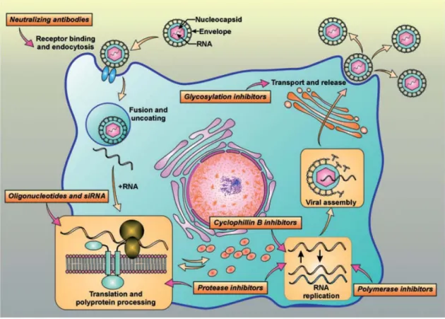
Figure 4 Hepatitis C virus (HCV) viral cycle and targets for drug development The HCV lifecycle starts with virion attachment to its specific receptor. The HCV RNA genome serves as a template for viral replication and as a viral messenger RNA for viral production. It is translated into a polyprotein that is cleaved by proteases. Then, viral assembly occurs. Potentially, each step of the viral cycle is a target for drug development.
15 Current interferon based therapies, while already marred with inadequate response rates, also have numerous side effects associated with them (McHutchison, Gordon et al. 1998, Manns, McHutchison et al. 2001). As a result, there has been growing focus on coming up with new interferon-free approaches that target various steps of the viral lifecycle as either replacements or further improvements for current strategies. This has resulted in developing several agents associated with targeting specific viral components (Stedman 2012). There have been major advancements during last several years with large numbers of ongoing trials with various direct-acting antivirals showing high potency, favorable tolerability profile, higher barrier to resistance, shortened treatment duration, all oral regimen, pan-genotypic (Asselah and Marcellin 2013).Presently there is increased emphasis in the development of more encouraging new agents such as polymerase inhibitors and protease. Moreover, there have been numerous compound inhibitors that have been designed successfully of the NS3 protease. They are subsequently an integral course of action spearheading pre-clinical and clinical development. BILN 2061 played a vital part in understanding the importance of these particular class of compounds, providing at least a decrease of 2 or 3 log10 in hepatitis C load within a timeframe of 48 hours (Lamarre, Anderson et al. 2003). Additionally, several NS5B polymerase compound inhibitors are also undergoing clinical development based on their encouraging results (Gane, Roberts et al. 2010, Le Pogam, Yan et al. 2012). In 2011, The U.S. Food and Drug Administration (FDA) approved Victrelis (boceprevir) and Incivek (telaprevir), known as inhibitor of HCV protease, for the treatment of hepatitis C. In late of 2013, FDA approved Olysio (simeprevir), a new therapy to treat chronic HCV infection. Most recently, a phase 2 clinical trial gave
16 encouraging result, treatment with Sofosbuvir and ledipasvir fixed-dose combination achieved over 95% SVR12 (sustained virological response 12 weeks after treatment) in patients with genotype 1 HCV infection, similar SVR12 was achieved even in those who previously had virological failure after receiving a protease inhibitor treatment (Lawitz, Poordad et al. 2013). The observation that hepatitis C virus (HCV) could only replicate in miR-122-positive hepatocytes led to the discovery that miR-122 is essential for HCV replication, and miR-122 is now one of the crucial host factors for anti-HCV therapy (Henke, Goergen et al. 2008, Lanford, Hildebrandt-Eriksen et al. 2013). Currently, the most advanced miR-122 targeting therapy is SPC3649 (miravirsen), a locked nucleic acid-modified oligonucleotide antagonizing miR-122, which is currently in phase 2 clinical trials for treatment of HCV infection (Janssen, Reesink et al. 2013).
Various factors ranging from treatment related, host characteristics, viral related and disease related, may cause resistance to IFN therapy (Pawlotsky 2003, Chen, Borozan et al. 2005, Puig-Basagoiti, Forns et al. 2005). In addition to that, the apparent generation subsequent selection of resistant variants may lead to the virus escaping the anti-viral pressure that is exerted by HCV-targeting drugs. Verily, mutations pertaining to the polymerase and protease enzymes have been identified already (Tomei, Altamura et al. 2003, Lin, Lin et al. 2004, Lu, Pilot-Matias et al. 2004, De Francesco and Migliaccio 2005, Lin, Gates et al. 2005, Le Pogam, Jiang et al. 2006, Le Pogam, Kang et al. 2006, Tong, Chase et al. 2006, Koev, Dekhtyar et al. 2007). Mutations of drug-targeted viral protein occur rapidly, causing resistance to treatment and leading to treatment failure (Rong, Dahari et al. 2010, Pawlotsky 2011). Various mutations are known to exhibit either an increased or decreased level of resistance along with the changing of the overall
17 prevalence of individual mutations through the passage of time during the treatment stage. In vitro data further suggests a seemingly lowered probability entailing the cross resistance between a certain number of the diverse anti-HCV drugs (Tomei, Altamura et al. 2003, Lin, Lin et al. 2004, Lu, Pilot-Matias et al. 2004, De Francesco and Migliaccio 2005, Lin, Gates et al. 2005, Le Pogam, Jiang et al. 2006, Le Pogam, Kang et al. 2006, Tong, Chase et al. 2006, Koev, Dekhtyar et al. 2007). Adding to that, molecular biology seems to suggest no cross-resistance between protease and polymerase inhibitors (De Francesco and Migliaccio 2005). Based on those studies, it is advisable to use agents that show a low probability of cross-resistance to supplement the therapy, such as different inhibitors or an inhibitor plus pegIFN-alpha. Indeed, administrating telaprevir together with pegIFN-α2a or further combined with ribavirin seemed to prevent viral rebound (Sarrazin, Rouzier et al. 2007). Simeprevir has also shown to increase the rate of sustained virologic response among treatment-experienced patients with HCV genotype-1 infection (Zeuzem, Berg et al. 2013)
6. Acute, chronic infection and viral clearance
Acute hepatitis C infection usually occurs during the first six-month time-bracket following the onset of viremia. It is usually asymptomatic since merely a minority of patients (10%–15%) is known to report symptoms (Ozaras and Tahan 2009). The symptoms of acute hepatitis C infection include a noticeable decrease in appetite, increased fatigue, consistent abdominal pain, jaundice, repetitive itching, and symptoms similar to a case of flu. When the time these symptoms come forth, usually they are
18 related to a comparatively high rate of spontaneous clearance (Gerlach, Diepolder et al. 2003). Spontaneous viral clearance rates can be considerably varied —between 10% - 60% of HCV infected individuals (Lehmann, Meyer et al. 2004, Micallef, Kaldor et al. 2006, Santantonio, Medda et al. 2006). HCV is usually detectable in the blood by PCR within a timeframe ranging from one - three weeks after contracting the infection. Serum viral load tends to increase considerably during the early stages of infection without any clear-cut evidence of liver damage. The responses with respect to the cellular immune are usually evident first among circulating T cells 4 to 8 weeks after the beginning of infection (Thimme, Oldach et al. 2001, Woollard, Grakoui et al. 2003, Cox, Mosbruger et al. 2005). The start of cellular immunity is consistent with a spike in serum transaminases which may subsequently suggest a transient immune-associated liver injury (Thimme, Oldach et al. 2001).
Although spontaneous viral clearance occurs in a minority of patients during acute infection stage, recurring infections are quite common, and most patients along the way are seen to develop chronic hepatitis C (Micallef, Kaldor et al. 2006). Chronic HCV occurs when the infection bearing the hepatitis C virus persists for more than a period of six months. Clinically though, it is often asymptomatic in nature and is mostly discovered by accident due to elevated liver enzymes (Hoofnagle 2002). The natural course of chronic hepatitis C could be varied from person to person. While documented cases of hepatitis C usually suggest almost most people suffering from an inflammation on liver biopsy, the progression rate entailing liver fibrosis is considerably variable among patients. Within the untreated population, roughly 33.3% progress to liver cirrhosis within a period of 20 years while an additional one-third of the remaining portion have
19 been known to progress to cirrhosis within a period of 30 years (Poynard, Ratziu et al. 2000, Poynard, Ratziu et al. 2001, Ghany, Kleiner et al. 2003). Usually, factors adjudged to have a bearing on the rate of the hepatitis C disease’s progress include classifications like age (the higher the age, the increased chances of rapid progression) (Poynard, Bedossa et al. 1997, Poynard, Ratziu et al. 2001), gender (men have been known to experience more rapid progression of the disease than women) (Wiese, Berr et al. 2000, Rodriguez-Torres, Rios-Bedoya et al. 2006), alcohol consumption (directly proportional) (Wiley, McCarthy et al. 1998), HIV co-infection (direct relation) (Rotman and Liang 2009, Konerman, Mehta et al. 2014), and fatty liver (positive correlation) (Younossi, McCullough et al. 2004, Younossi, Afendy et al. 2009, Younossi and McCullough 2009, Bhala, Angulo et al. 2011).
7. Immune response and HCV infection
Immune activation is associated with HCV infection. An immune response induced by HCV infection is characterized as rapid innate immune response and delayed adaptive immune response, regardless of the outcome of the disease progression (Shoukry, Grakoui et al. 2003, Dustin and Rice 2007). Innate immune activation was observed at the early stage after HCV infection in chimpanzees (Bigger, Guerra et al. 2004). Inflammatory cytokines also have been shown to be up-regulated in chronically HCV-infected individuals (Polyak, Khabar et al. 2001). Studies estimate that in the case of an infected chimpanzee at the peak of viremia, ten percent of the hepatocytes support
20 HCV replication at max (Bigger, Brasky et al. 2001). This goes on to suggest that an anti-viral state is induced quite early in the liver during HCV infection.
7.1 Innate immunity
HCV is usually recognized by pathogen recognition receptors (PRRs) which further goes on to initiate an innate immune response against HCV infection. Engagement of those receptors may activate and subsequently translocate latent IRF-3, further going on to activate the IFN-β promoter (Saito, Owen et al. 2008). Intrahepatic interferon signaling was observed and associated with HCV viremia, regardless of the outcome of the disease progression (Su, Pezacki et al. 2002, Thimme, Bukh et al. 2002). The cellular source of interferon is not certain however.
Several different cell types within the liver may take part in innate immune surveillance on HCV infection, including hepatocytes, Kupffer cells (KCs) that serve as resident liver macrophages, DCs and natural killer cells (NKs) (Crispe 2009). The first immune response to HCV infection was thought to be interferon induction in infected hepatocytes (Bigger, Guerra et al. 2004). Hypothetically, infected hepatocytes produce the first interferon, which acts on surrounding cells and amplifies interferon signals. Several studies showed that HCV infection induced IFN signaling in immortalized human hepatocytes (Kanda, Steele et al. 2007) or hepatoma cell lines (Lau, Fish et al. 2008). There is no direct evidence of interferon induction in hepatocytes of HCV patients in
vivo, but intrahepatic interferon-induced genes were detected in infected chimpanzees
(Lau, Fish et al. 2008). Type I IFNs production - as a reaction to hepatitis C infection - may lead to an antiviral state with respect to the surrounding cells. Plasmacytoid DCs
21 (pDCs) were reported to be activated by HCV-infected hepatocytes through cell-cell contact, resulting in producing type I IFN in vitro (Takahashi, Asabe et al. 2010). However, in vivo study has not supported pDCs as the source of intrahepatic interferon during HCV infection (Lau, Fish et al. 2008).
Kupffer cells form the highest percentage of tissue-resident macrophages in the liver (Laskin, Weinberger et al. 2001). Kupffer cells are responsible for the recruitment of CTLs and APCs to eliminate infected cells in the liver (Uwatoku, Suematsu et al. 2001), and they also carry out an important function in modulating inflammation in liver fibrosis development (Liu, Tao et al. 2010). NK cells are found to be in high quantity in the liver and are also essential early responders during HCV infection. NK cells facilitate lysis of infected cells and produce IFN-γ that control viral infection (Kokordelis, Kramer et al. 2013). NK cells may also contribute to maturation of DCs and bridge the gap between innate and adaptive immunity. The importance of NK cells in the resolution of HCV infection is revealed by the observation of genetic polymorphisms that affect the threshold of NK cell activation and influence the outcome of HCV infection (Khakoo, Thio et al. 2004).
7.2 Adaptive immunity
Controlling acute HCV infection is related to strong, broad directed, and most importantly a sustainable activation of hepatitis C-specific T cells (Thimme, Oldach et al. 2001, Thimme, Bukh et al. 2002, Flynn, Dore et al. 2013). The activation of T cell can be transiently observed at least, as hepatitis C is able to establish infection persistently (Ulsenheimer, Lucas et al. 2006). However, in the case of majority of the patients,
cell-22 mediated immunity is unable to eliminate hepatitis C infection completely (Guobuzaite, Chokshi et al. 2008). Even in the case of humans and chimpanzees, the chances of contracting reinfections through homologous and heterologous hepatitis C strains remain a plausible outcome. Protective immunity on the other hand can reduce the overall duration and extent of viremia in the case of repeated infection (Major, Mihalik et al. 2002, Mehta, Cox et al. 2002, Lanford, Guerra et al. 2004), but completely sterilizing immunity has still evaded proof.
Spontaneous resolution of hepatitis C is commonly linked with a strong and sustained T cell response that targets multiple hepatitis C epitopes at once and with intrahepatic production of IFN-γ (Cooper, Erickson et al. 1999, Lechner, Wong et al. 2000, Thimme, Oldach et al. 2001). HCV-specific CD4+ T cell activation is relatively easy to detect during the early stage of acute infection than in chronic infection (Lechner, Wong et al. 2000). Epitope-specific T cells can be identified when stained with MHC class I or class II tetramer peptide complexes. Tetramer-staining T cells may not be active in a functional capacity during acute or chronic infection. Indeed, studies conducted have shown that a significant percentage of hepatitis C-specific T cells have a “stunned” phenotype, even in the case of those hosts that go on to resolve infection (Lechner, Wong et al. 2000, Thimme, Oldach et al. 2001). Resolution of acute infection is associated with T cell recovery of an activated phenotype and the ability to produce IFN-γ. The broad patterns of CD4+ and CD8+ T cell specificities in individuals with resolved hepatitis C infection varies substantially with the rather limited amount of hepatitis C epitopes that are targeted by T cells with respect to people having chronic infection (Day, Lauer et al. 2002) (Lauer, Ouchi et al. 2002).
23 The role of humoral immunity to HCV is yet to be comprehended accurately. Seroconversion is seen to be a comparatively delayed phenomenon in the case primary hepatitis C infection (Netski, Mosbruger et al. 2005). The response of antibodies are not required for clearance of acute infection (Cooper, Erickson et al. 1999, Thimme, Bukh et al. 2002, Logvinoff, Major et al. 2004) and likely to diminish after spontaneous recovery (Takaki, Wiese et al. 2000). Studies suggest that broadly reactive neutralizing antibodies (nAbs) that target the viral envelope can be found in the serum of persistently infected hosts (Bartosch, Bukh et al. 2003, Logvinoff, Major et al. 2004, Meunier, Engle et al. 2005). However, the overall development of nAbs is substantially delayed in the case of acute infection (Logvinoff, Major et al. 2004, Netski, Mosbruger et al. 2005). In the event that patients remain persistently infected in spite of the presence of high titers of nAbs may subsequently indicates that nAbs do not facilitate sterilizing immunity, possibly because of continued viral mutation and antibody-dependent selection.
8. TLR7 and TLR8
Receptor-mediated detection of pathogen-derived nucleic acids helps to protect genomic nucleic acid in the event of foreign genetic material invasion. A number of pattern recognition receptors (PRRs) have been known to participate in the recognition of nucleic acid (Barbalat, Ewald et al. 2011). Generally speaking, PRRs can be classified into numerous families. The Toll-like receptor (TLR) family is known to consist of ten members. Moreover, viral RNA bearing certain traits can also be detected by members of the RIG-I-like RNA helicase (RLH) family, such as RIG-I and MDA-5. TLRs along with
24 the two RLH members namely RIG-I and MDA-5 can be differentiated with respect to their cellular localization, ligand specificity along with downstream signaling pathways (Thompson and Locarnini 2007, Thompson, Kaminski et al. 2011, O'Neill, Golenbock et al. 2013). Thus it suggests that host cells can employ several, non-redundant defense mechanisms in the detection of invading pathogens. While RIG-I and MDA-5 can be classified as cytosolic receptors (Barral, Sarkar et al. 2009), the four members of the TLR family (TLR3, TLR7, TLR8, and TLR9) involved in viral nucleic acid recognition are all present in the endosomal membrane (Lee and Barton 2014). TLR3 and MDA-5 recognize long double-stranded RNA (Liu, Botos et al. 2008, Wu, Peisley et al. 2013). Either single- or double-stranded RNA belonging to the triphosphate group at the 5’ end was observed to be the ligand for RIG-I (Hornung, Ellegast et al. 2006), and also the ligand for TLR9 is DNA-containing CpG motifs (Rutz, Metzger et al. 2004). TLR7 and TLR8 detect single-strand RNA with certain motifs (Heil, Hemmi et al. 2004).
Sequence comparisons across vertebrate TLRs go on to reflect that TLR7 and TLR8 are the most closely related of the nucleic acid–sensing TLRs (Roach, Glusman et al. 2005, Mikami, Miyashita et al. 2012). In humans, remiquimod in known to stimulate TLR7 and TLR8, yet the related imidazoquinoline, imiquimod, predominantly activates TLR7, indicating considerable differences in ligand specificity and functions between the respective receptors (Gorden, Gorski et al. 2005, Larange, Antonios et al. 2009). As far as humans are concerned, TLR7 and TLR8 are able to aptly recognize distinct sequence motifs in ssRNA (Heil, Hemmi et al. 2004, Hornung, Guenthner-Biller et al. 2005, Forsbach, Nemorin et al. 2008). Expression of TLR7 and TLR8 is mainly limited to immune cell subsets. Moreover, in the human system, the expression of TLR7 is
25 restricted to PDCs and B cells. TLR8 is predominantly expressed in cells belonging to the myeloid lineage, such as monocytes, myeloid dendritic cells, and macrophages (Krug, Towarowski et al. 2001, Hornung, Rothenfusser et al. 2002). This differential expression is most likely to explain the distinct cytokine profiles influenced by TLR7 and TLR8 ligands in human cells (Gorden, Gorski et al. 2005). Whereas TLR7 and TLR8 are regulated by cytokines and can be induced upon cell activation, recent reports have indicated that NK cells and T cells also express these TLRs in some instances (Hart, Athie-Morales et al. 2005, Song, Zhuang et al. 2009).
When receptor/ligand interacts, TLR7 and TLR8 recruit the universal TLR adaptor protein MyD88 through its TIR domain (Hemmi, Kaisho et al. 2002, O'Neill and Bowie 2007). Instead, MyD88 recruits IRAK1 and IRAK4. IRAK4 then goes on to activate IRAK-1 by phosphorylation (Uematsu, Sato et al. 2005, Kim, Staschke et al. 2007). Both IRAK-1 and IRAK4 leave the MyD88-TLR complex and link up temporarily with TRAF6, which leads to its ubiquitination (Kawai, Sato et al. 2004). After ubiquitination, TRAF6 is able to form a complex with TAB2/TAB3/TAK1, inducing TAK1 activation (Besse, Lamothe et al. 2007). TAK1 then links up to the IKK complex which leads to the phosphorylation of IκB and the consequent nuclear localization of NF-κB (Adhikari, Xu et al. 2007). Activation of NF-NF-κB induces the production of pro-inflammatory cytokines. Moreover, TLR7/8 can bind with MyD88 and activate IRAK1 and TRAF6. TRAF6mediates IRF-5 ubiquitination, which is important for IRF-5 nuclear translocation and interferon induction (Schoenemeyer, Barnes et al. 2005, Balkhi, Fitzgerald et al. 2008). Engagement of TLR7 or TLR8 may also activate IRF7 through activation of MyD88, BTK, and TRAF6 (Konno, Yamamoto et al. 2009). Formation of
26 the MyD88-TRAF6-IRF7 complex induces phosphorylation and IRF7 activation. Activation of IRF5 and IRF7 results in induction of type I interferon (Lazear, Lancaster et al. 2013).
9. Role of TLR7 in immune response against HCV infection
TLR7, which induces a strong type I interferon in pDCs and hepatocytes when it is engaged, has been expected to play an instrumental immunological role in HCV infection (Gibson, Lindh et al. 2002, Sagan and Sarnow 2010, Takahashi, Asabe et al. 2010). Indeed, TLR7 agonists have been shown remarkable antiviral activities by in vitro and in vivo studies. It was reported by several groups that TLR7 engagement initiated a strong innate immune response against HCV infection. TLR7 agonists have induced an antiviral response in hepatoma cell lines through both interferon-dependent and interferon-independent mechanisms (Lee, Wu et al. 2006). The potential of TLR7 agonist for hepatitis C therapy has been estimated (Lee, Wu et al. 2006, Thomas, Laxton et al. 2007), and the efficiency and safety of those compounds are under evaluation in clinical trials. The administration of TLR7 agonist results in significant interferon induction and suppression of viremia (up to 3 log10) (Horsmans, Berg et al. 2005, Bergmann, de Bruijne
et al. 2011, Fidock, Souberbielle et al. 2011). Recently, Erilka et al. have proved the involvement of TLR7 in induction of interferon by HCV and in antiviral response against HCV infection. Knockdown of TLR7 in hepatoma cell lines abolished the induction of interferon-β. Recently Erilka et al. have proved the involvement of TLR7 in induction of interferon by HCV in cell lines that was highly permissive to HCV (Eksioglu, Zhu et al.
27 2011). However, the role of TLR7/8 in detection of HCV RNA by APCs remains unknown.
The potential implication of TLR7 in the innate immune response against HCV was postulated, nevertheless little is known about it and it is not fully demonstrated. In fact, pDCs have been shown to be able to be stimulated by infected Huh-7 cells in cell-culture by recognizing the viral RNA likely via a TLR7-dependent pathway (Python, Gerber et al. , Takahashi, Asabe et al. 2010). Moreover, the presence of a GU-rich fragment in HCV genome was shown to be detected by TLR7 (Zhang, Guo et al. 2009). Furthermore, the single nucleotide polymorphisms (SNPs) in TLR7 and TLR8 were associated with the magnitude of inflammation and fibrosis during HCV infection, with the response to IFN-α-based therapy and with the susceptibility to the infection with HCV (Schott, Witt et al. 2008, Wang, Eng et al. 2011). However, the role of TLR7 in HCV detection in APCs and the mechanisms leading to this detection are not known. Additionally, the role of TLR8 in the innate immune response against HCV is not known.
10. HCV detection by antigen-presenting cells
Antigen-presenting cells (APCs) are crucial for initiation of antiviral immunity. APCs sense viral components via PRRs to induce an innate immune response, they also capture viral antigens and present to naïve T cells to elicit an adaptive immune response. HCV-specific T cells were detected in HCV-infected chimpanzees and humans, regardless of the outcome of the disease progression, suggesting the capability of APCs for HCV uptake. However, although the immunogenicity of HCV components (RNA or proteins) have been shown, DCs appear “silent” when they encounter HCV. Many efforts
28 have been made to prove the immunogenicity of HCV particles, but the only common element in these studies is the unresponsiveness of DCs to HCV stimulation. Neither DC maturation nor induction of cytokines was observed (Shiina and Rehermann 2008, Liang, Russell et al. 2009, Zhang, Guo et al. 2009, Takahashi, Asabe et al. 2010). The mechanisms of DC tolerance to HCV are mysterious, and the causes that endow HCV with an “undetectable” character remain unknown. The HCV uptake capability of DCs still remains controversial. The internalization of HCV-like particles by DCs (Ludwig, Lekkerkerker et al. 2004, Barth, Ulsenheimer et al. 2005) and the absence of HCV in DCs from HCV infected-patients (Rollier, Drexhage et al. 2003, Longman, Talal et al. 2004) have both been reported. The presence of the HCV genome in blood cells, which includes DC, has been depicted in numerous studies through reverse transcription (RT)-PCR. However, the presence of viral RNA does not explain whether HCV enters cells or merely sticks to their surface. Recently, it has been shown that PBMCs are certainly not susceptible to HCV (Cormier, Tsamis et al. 2004), HCV in PBMCs from patients are mainly carried on the surface of cells (Natarajan, Kottilil et al. 2010). Freshly isolated plasmacytoisd DCs (pDCs) and myeloid DCs (mDCs) are not susceptible to pseudotype HCV, however, incubation with GM-CSF makes mDCs highly susceptible to pseudotype HCV (Kaimori, Kanto et al. 2004). In contrast, Monocyte-derived DCs (mDDCs) appear to have great capability for HCV uptake, which depends on DC-SIGN that is induced by GM-CSF/IL-4 (Ludwig, Lekkerkerker et al. 2004, Barth, Ulsenheimer et al. 2005).
The trail by which DCs are able to internalize antigens for presentation of T cells is receptor-mediated endocytosis, in which C-type lectins act in an important capacity. An increasing number of C-type lectins with specificity for mannosylated antigens have been
29 discovered to be expressed by DCs such as Langerin (Valladeau, Ravel et al. 2000), mannose receptor (Sallusto, Cella et al. 1995), DEC-205 (Kato, Neil et al. 1998), and DC-SIGN (Engering, Geijtenbeek et al. 2002). Studies suggest that DC-SIGN is understood to play an active in infection of DCs by the dengue virus (Navarro-Sanchez, Altmeyer et al. 2003, Tassaneetrithep, Burgess et al. 2003), another member of the
Flaviviridae family. Recently, studies depicting recombinant HCV envelope glycoprotein
2 (E2) and HCV pseudotype particles (HCVpp’s) have indicated that DC-SIGN and its liver-expressed homologue L-SIGN are vital receptors for HCV envelope glycoproteins E1 and E2 (Pohlmann, Zhang et al. 2003, Lozach, Amara et al. 2004, Ludwig, Lekkerkerker et al. 2004). Both HCV E1 and E2 are able to bind the same binding site on DC-SIGN as HIV and mycobacteria. Remarkably, internalized HCV virus-like particles were targeted towards non-lysosomal compartments within immature DCs in a not so dissimilar manner to that established for HIV-1 (Ludwig, Lekkerkerker et al. 2004), suggesting that HCV may target DC-SIGN to escape detection and facilitate viral dissemination.
30
HYPOTHESIS AND OBJECTIVES
Hypothesis:
Immune response induced by HCV infection is characterized as rapid innate immune response and delayed adaptive immune response (Dustin and Rice 2007), regardless of the outcome of the disease progression. Innate immune activation was observed at an early stage after HCV infection in chimpanzees (Bigger, Brasky et al. 2001). Antigen presenting cells (APCs) are deemed to be responsible for HCV detection, however, the mechanisms of immune activation induced by HCV infection has been difficult to clarify. It has been shown that HCV RNA is capable to induce anti-viral response (Saito, Owen et al. 2008, Zhang, Guo et al. 2009), nevertheless, the detection of HCV by APCs still remains elusive. Furthermore, the single nucleotide polymorphisms (SNPs) in TLR7 and TLR8 were associated with the magnitude of inflammation and fibrosis during HCV infection, with the response to interferon based therapy and with the susceptibility to the infection with HCV (Schott, Witt et al. 2008, Wang, Eng et al. 2011). However, the role of TLR7 and TLR8 in HCV detection by APCs and the mechanisms leading to this detection are still not known. We speculate that APCs has a unique way to detect HCV and TLR7/8 is involved in this process.
31 Objectives
1. Determine the capacity of HCV RNA to induce immune response. Is HCV RNA able to induce inflammatory cytokine? Dose HCV RNA contains GU-rich motifs which may be recognized by TLR7/8? Are those GU-rich sequences able to induce inflammatory cytokine and DC maturation?
2. Determine the role of TLR7 and TLR8 in HCV RNA detection. Does HCV RNA specifically trigger TLR7/8? Are those GU-rich sequence selected from HCV genome able to induce cytokines in similar profile as validated TLR7/8 agonist? 3. Determine the immune response induced by HCV RNA in different antigen
presenting cells (APCs) and the anti-viral activity of such immune response. Does HCV RNA induce type I interferon? Do cytokines produced by APCs upon HCV RNA stimulation inhibit HCV replication?
4. Determine the role of different APCs in HCV detection and anti-HCV immune response. Are APCs able to respond to HCV particles? Is there any preference of APCs in HCV detection? Does HCV induce type I interferon in APCs? Is there any difference between the immune response induced by HCV RNA and particle? What are the mechanisms involved in HCV detection by APCs?
32
CHAPTER 2
HCV derived ssRNA induce innate immune response
against HCV infection via TLR7 and TLR8
33
HCV derived ssRNA induce innate immune response against
HCV infection via TLR7 and TLR8
Yuwei zhang1-4, Elias Said1-3, Abdel-Hakeem MS1, Zhong He1-4, Francesco A. Procopio1-4, Rafick-Pierre Sekaly1-6
1Centre de recherche du centre Hospitalier de l’Université de Montréal (CRCHUM), Hôpital
Saint-Luc, Québec H2X 1P1, Canada
2Laboratoire d’Immunologie, Département de Microbiologie et d’Immunologie, Université de
Montréal, Montréal, Québec H3T 1J4, Canada
3Institut national de la Santé et de la Recherche médicale U743, Centre de Recherche, CHUM,
Université de Montréal, Montréal, Québec H2X 1P1, Canada
4Vaccine and Gene Therapy Institute-Florida (VGTI-Fl), 11350 SW Village Pkwy, third floor.
Port St. Lucie, Florida. USA, 34987.
5Departement of Microbiology and Immunology, McGill University, Montréal, Québec H3A
2B4, Canada
6Corresponding authors
Correspondence should be addressed to Rafick-Pierre Sekaly
COMPETING INTERESTS STATEMENT
34
Abstract
The hepatitis C virus (HCV) is a positive single-stranded RNA (ssRNA) virus that replicates in the liver and has infected 200 million people worldwide. Recognition of HCV-associated molecular patterns is crucial for initiation of innate immune response against HCV infection (Schoggins and Rice 2013). As a ssRNA virus, HCV RNA might be detected by Toll-like receptor 7 or 8, an endosomal/lysosomal RNA sensor, resulting in induction of type I interferon that limits infection. Although antiviral activities of the TLR7 agonist have been reported (Horsmans, Berg et al. 2005, Bergmann, de Bruijne et al. 2011), the role of TLRs in recognizing HCV and the induction of an immune response against HCV have not been well defined. In this study, we demonstrate that the HCV genome contains GU-rich sequences that can specifically trigger TLR7/8. The engagement of TLR7/8 by HCV-derived ssRNA results in the maturation of pDC and the production of type I interferon, and also leads to induction of inflammatory cytokines and chemokines in different APCs. Cytokines produced by monocytes and pDCs upon stimulation of HCV-derived ssRNA inhibit HCV production in an IFN-dependent manner, whereas HCV-ssRNA and cytokines produced by activated mDCs do not affect viral production due to lack of interferon signaling. HCV ssRNA-mediated cytokines also down-regulate CD81 expression on huh7.5 in an IFN-independent manner, which might inhibit HCV infection. Our results suggest that HCV RNA has the potential to trigger TLR7/8, which subsequently initiates an innate immune response against HCV infection.
35
Introduction
The hepatitis C virus (HCV) is a worldwide public health problem affecting an estimated 200 million individuals (Lauer and Walker 2001). There are approximately 3 to 4 million new cases of HCV infection each year (WHO 2011). No effective anti-HCV vaccine currently exists. Only 20% of HCV-infected individuals can be cured by spontaneous viral clearance. Approximately 80% of infected individuals develop chronic infection (Manns, Foster et al. 2007). The progression of liver disease usually takes decades. Up to 20% of those infected may develop complications including cirrhosis, liver failure, or hepatocellular carcinoma (Afdhal 2004). Although antiviral therapies have improved considerably in recent years, they are associated with significant toxicity and are effective in only about one-half of those treated (Manns, Foster et al. 2007). Novel anti-HCV drugs targeting HCV proteins that are essential for HCV replication are under development. These drugs have achieved great effectiveness in HCV treatment (Asselah and Marcellin 2013). However, mutations of target viral proteins occur rapidly, causing resistance and failure of treatment (Rong, Dahari et al. 2010, Pawlotsky 2011). Thus, a better understanding of immunity against HCV is required to develop a solution for hepatitis C.
Innate immune defenses are essential to control viral infection through recognition of viral macromolecular motifs known as pathogen-associated molecular patterns (PAMPs). Pathogen recognition receptors (PRRs) recognize PAMPs and initiate innate immune responses (Gordon 2002, Janeway and Medzhitov 2002, Arpaia and Barton 2011). RNA viruses have the potential to be recognized by RNA sensors of immune cells that initiate immune responses against viral infection, i.e., influenza virus