ORIGINAL PAPER
Seasonal plasticity of auditory saccular sensitivity in “sneaker”
type II male plainfin midshipman fish, Porichthys notatus
Ashwin A. Bhandiwad1 · Elizabeth A. Whitchurch1 · Orphal Colleye1,2 ·
David G. Zeddies3 · Joseph A. Sisneros1,4,5
Received: 14 September 2016 / Revised: 16 February 2017 / Accepted: 17 February 2017 © Springer-Verlag Berlin Heidelberg 2017
auditory sensitivity may potentially facilitate eavesdrop-ping by sneaker males and their assessment of vocal type I males for the selection of cuckoldry sites during the breed-ing season.
Keywords Hearing · Particle acceleration · Teleost · Saccule · Hair cells
Introduction
Many teleost fishes use acoustic signals for social com-munication, and in many species, the use of these social acoustic signals is essential for successful courtship and reproduction (Bass and McKibben 2003; Ladich 2004; Fine and Parmentier 2015). Recent studies have shown that many teleosts possess both behavioral and neural adapta-tions for the production and perception of acoustic signals that are necessary for reproduction and survival (Bass and McKibben 2003; Bass and Ladich 2008; Kelley and Bass
2010). One vocal teleost that is a good species for inves-tigating neural mechanisms of vocal production and sig-nal perception is the plainfin midshipman fish, Porichthys
notatus (Bass et al. 1999; Sisneros 2009a; Forlano et al.
2015). This nocturnally active marine teleost produces a relatively simple repertoire of acoustic signals for intraspe-cific social communication that includes “grunts”, “growls” and “hums” (Bass et al. 1999; Sisneros 2009a). While all three adult sexual phenotypes (female and two male sex-ual phenotypes: types I and II) are known to produce the short-duration, broadband agonistic “grunts”, only type I “singing” males build nests and produce the long-duration, broadband agonistic “growls” and multiharmonic adver-tisement calls or “hums” during the summer breeding sea-son (Bass and McKibben 2003; Brantley and Bass 1994). Abstract Adult female and nesting (type I) male
mid-shipman fish (Porichthys notatus) exhibit an adaptive form of auditory plasticity for the enhanced detection of social acoustic signals. Whether this adaptive plasticity also occurs in “sneaker” type II males is unknown. Here, we characterize auditory-evoked potentials recorded from hair cells in the saccule of reproductive and non-reproductive “sneaker” type II male midshipman to determine whether this sexual phenotype exhibits seasonal, reproductive state-dependent changes in auditory sensitivity and frequency response to behaviorally relevant auditory stimuli. Sac-cular potentials were recorded from the middle and cau-dal region of the saccule while sound was presented via an underwater speaker. Our results indicate saccular hair cells from reproductive type II males had thresholds based on measures of sound pressure and acceleration (re. 1 µPa and 1 ms−2, respectively) that were ~8–21 dB lower than non-reproductive type II males across a broad range of frequencies, which include the dominant higher frequen-cies in type I male vocalizations. This increase in type II A. A. Bhandiwad and E. A. Whitchurch contributed equally.
* Joseph A. Sisneros sisneros@uw.edu
1 Department of Psychology, University of Washington, Seattle, WA 98195-1525, USA
2 Laboratoire de Morphologie Fonctionnelle et Evolutive, Université de Liège, Institut de Chimie, Bât. B6c, Quartier Agora, 4000 Liège, Belgium
3 JASCO Applied Sciences, Silver Springs, MD 20902, USA 4 Department of Biology, University of Washington, Seattle,
WA 98195-1800, USA
5 Virginia Merrill Bloedel Hearing Research Center, University of Washington, Seattle, WA 98195-7923, USA
Previous studies have shown that both the female and type I male auditory systems are seasonally adapted to detect the broadband agonistic calls and the dominant harmonic components of the advertisement calls produced by type I males during the breeding season (Sisneros and Bass 2003; Rohmann and Bass 2011). These adaptations are poten-tially important for conspecific detection and localization, mate choice decisions and intraspecific male competition (Sisneros 2009a, b).
In contrast to “singing” type I males, “sneaker” type II males employ an alternative reproductive strategy that does not require building a nest or producing an advertise-ment call to attract mates. Instead, type II males, which have neuroendocrine traits similar to that of females, engage in satellite or sneak-spawning to steal fertilizations from type I males that are actively courting females in the nest (Brantley and Bass 1994). While females and type I males are known to be better suited to detect midshipman vocalizations during the breeding season (Sisneros and Bass 2003; Sisneros 2009b; Rohmann and Bass 2011), the auditory sensitivity of sneaker type II males has yet to be characterized. There is no a priori reason to expect that sea-sonal plasticity of midshipman auditory sensitivity would be limited to females and type I males.
The sensitivity of auditory systems in fishes can be measured in terms of acoustic particle motion and pressure. All fishes are likely sensitive to the particle motion compo-nent of sound via their otolithic end organs which essen-tially function as inertial accelerometers (de Vries 1950; Fay 1984). This common mode of hearing in fishes enables them to detect particle motion, the directional vector com-ponent of sound, as opposed to sound pressure, which is a scalar quantity containing no directional information. Some more recently derived fishes such as Otophysan fishes (e.g., goldfish, carp, zebrafish, etc.) have evolved pressure sensi-tivity as a result of having a specific connection between the inner ear and an air bubble (e.g., the swim bladder) (Fay and Popper 1980; Blaxter 1981; Schellart and Popper
1992). Recently Popper and Fay (2011) proposed that all future fish hearing studies should independently character-ize particle motion and sound pressure sensitivities of fish regardless of any hearing specializations that fish may pos-sess (e.g., accessory hearing structures such as Weberian ossicles that connect the swim bladder to the inner ear). Previous auditory physiology studies of the plainfin mid-shipman fish have primarily reported auditory responses with regard only to sound pressure (McKibben and Bass
1999, 2001; Sisneros et al. 2004; Sisneros and Bass 2003,
2005; Sisneros 2007, 2009b; Alderks and Sisneros 2011; Rohmann and Bass 2011; but for the exception see Weeg et al. 2002). Thus, better characterization of particle motion sensitivity in this species and across the different sexual phenotypes is warranted.
The primary goal of this study was to characterize audi-tory-evoked potentials from the saccule in reproductive and non-reproductive type II male midshipman to determine whether this sexual phenotype exhibits seasonal differences in auditory threshold and frequency response of the sac-cular hair cells to behaviorally relevant auditory stimuli. A secondary goal was to provide a description of the acous-tic paracous-ticle motion sensitivity of the midshipman auditory system that could be used for future comparisons across midshipman sexual phenotypes and with other species. We hypothesized that the auditory saccular sensitivity of type II males during the breeding season would be adapted to detect the dominant frequency content of the type I male vocalizations, which would be beneficial for type II males in their selection of type I male cuckoldry sites for sneak or satellite spawning. We also compare our findings to that of the saccular potentials reported for females (Sis-neros 2009b) and type I males (Rohmann and Bass 2011) and interpret our results as they relate to possible auditory adaptations for acoustic communication during the repro-ductive season.
Materials and methods
Experimental animalsSummer reproductive adult type II male midshipman fish (Porichthys notatus) were hand-collected from exposed intertidal nests at low tide in Tomales Bay, CA, USA, dur-ing the reproductive season (May–July 2010). Winter non-reproductive adult type II males were collected by otter trawl (RV Kittiwake, Bio-Marine Enterprises) at depths from 70 to 100 m in the Puget Sound near Edmonds, WA (47.82N, 122.38W) and in Shilshole Bay near Ballard, WA (47.68N, 122.41W) during the winter months (Decem-ber–February) of 2007, 2010 and 2016. Note that the lim-ited number of non-reproductive type II males used in this study was sampled over multiple years due to their scarcity during the non-reproductive winter (personal observations, Sisneros). Based on our experience, the collection of type II males during winter trawls in the waters of Puget Sound, WA, is often a rare and serendipitous event.
Type II males were distinguishable from type I males based on their standard body length (SL), gonadosomatic index (GSI) and visual inspection of their genital papilla and their swim bladder sonic muscles. The reproductive type II males used in this study were noted to have pale and underdeveloped sonic muscles instead of the typical large red sonic muscles observed in summer reproductive type I males. In addition, the SL and GSI ranges of type II males reported here were well within the ranges reported for type II males in a previous study (Grober et al. 1994) that were
collected at the same sites in Tomales Bay, CA. GSI was calculated as 100 x gonad mass/(body mass – gonad mass), based on Tomkins and Simmons (2002). Soon after summer field collection (<4–5 days), type II males were transported to the University of Washington in Seattle, WA, where they were housed in aquaria at 13–15 °C and kept under reverse-light cycle (reverse-light:dark—14:10 h). The experiments per-formed with summer reproductive type II males were con-ducted during the dark phase of the light/dark cycle since these fish are known to be nocturnally active. Animals were hand-fed defrosted shrimp and/or live feeder guppies two to three times a week. Auditory physiology recordings and measurements were made within 26 of collection for most animals except for two non-reproductive type II males that were recorded approximately a few months after collec-tion while being maintained under a short-day winter pho-toperiod (8 h light, 16 h dark). A simple linear regression between gonadosomatic index (GSI) and days in captivity was performed to test for time-dependent changes in breed-ing state, with the regression coefficient (r2) reported. Experimental procedures
The procedures for exposing and recording from the inner ear saccule followed those used in previous studies (Sis-neros 2007, 2009b; Alderks and Sisneros 2011). Briefly, animals were initially anesthetized in a 0.025% ethyl-p-am-inobenzoate/saltwater bath until the fish’s opercula ceased movement for at least 5 min. The animals were then immo-bilized with an intramuscular injection of pancuronium bromide (approx. 0.5 µg g−1 body mass). Skin, muscle and bone were removed just dorsal to the otic capsules, and a 2.5-cm hydrophobic dam was built around the craniotomy to enable submersion without exposing the brain and inner ear to salt water. The cranial cavity was then filled with Teleost Ringer’s solution to prevent drying. A subcutane-ous injection of 0.25% bupivacaine (approx. 1 µg g−1 body weight) was administered around the surgical site as an analgesic. During the experiment, the animals were sub-merged underwater such that the saccules were approxi-mately 3 cm below the water surface while fresh saltwater was circulated over the gills. The craniotomy was periodi-cally checked visually to verify stable blood flow in the brain and inner ear organs.
Animals were suspended by a custom-built acrylic stereotaxic head-holder that was positioned 10 cm above an underwater speaker (UW-30, Telex Communications, Burnsville, MN, USA) in the center of a 40-cm-diameter Nalgene recording tank. The bottom of the tank contained an approximately 4.5 cm layer of small rock substrate in which the speaker was embedded so that only the top 2 cm of the speaker projected upwards into the water column. The water was initially chilled to 12 °C and maintained
between 14 and 15 °C for the duration of the recording ses-sion. The distance from the water surface to the surface of the speaker was 13 cm. The tank was positioned on an inflated pneumatic table, inside an acoustic isolation cham-ber (Industrial Acoustics, New York, NY, USA), and all recording equipment were housed outside this chamber.
Saccular potentials were recorded using glass electrodes (1.0–4.0 MΩ) filled with a 3 M KCl solution. The elec-trodes were visually positioned in the space between the otolith and sensory maculae in the middle/caudal region of the saccule. Both left and right saccules were used in this study. Recording fidelity was assessed by comparing the magnitude of the saccular potentials recorded during the blank (no stimulus presented) and auditory stimulus test trials. The auditory stimuli were presented were of equal amplitude (with ±1–2 dB) at a given sound level across all frequencies tested. Electrode signals were band-pass-fil-tered (130–3000 Hz, Stanford Research Systems SR 650), amplified by 100× (Getting 5A), and then sent to a lock-in amplifier (Stanford Research Systems SR 830) and digi-tized at 20 kHz (Micro1401 mkII, Cambridge Electronic Design). Data acquisition and stimulus timing were con-trolled by a custom MATLAB (MathWorks Inc., Natick, MA) script. The output of the lock-in amplifier reflects the relative amplitude of the inner ear saccule’s hair cell response to a tonal stimulus. Because the opposing hair cell orientations within the saccular sensory maculae result in a double-frequency response, the saccular potential is defined here as the amplitude the hair cell response at the second harmonic of the stimulus (Cohen and Winn 1967; Sisneros 2007).
Acoustic stimulus generation and calibration
Single tones were presented at 75–85 Hz in 10 Hz steps and 105–385 Hz in 20 Hz steps. Stimuli were generated by the lock-in amplifier, amplified by an audio amplifier, and then played through the underwater speaker. Stimuli were 750 ms in length and separated by a 500-ms interval. Lock-in measurements were sampled during the last 20 ms of the stimulus. This data collection procedure and use of the lock-in amplifier has been shown to be very sensitive and effective in measuring the saccular potentials at the reported frequencies (Sisneros 2007, 2009b; Rohmann and Bass 2011; Vasconcelos et al. 2011). Each recording ses-sion began with control trials (no auditory stimulus) fol-lowed by stimulus test trials in which auditory frequency stimuli at a given sound level were presented in a pseudor-andom order.
Calibrations were performed before every experiment, after the tank water was initially cooled, but before the fish was placed in the tank. During calibration of the auditory stimuli, the hydrophone and accelerometer were centered
10.5 cm above the speaker (2.5 cm below water surface) at the position of the fish’s ear. Stimuli were equalized in SPL using an iterative MATLAB script that measured power spectral density for each frequency from recordings taken through a mini-hydrophone (model 8103, Bruel and Kjaer). At each iteration, the voltage signal sent to the speaker was scaled until the measured SPL output at each frequency was within 0.5 dB of the desired amplitude (130 dB re.: 1 µPa). This SPL level was chosen for calibration because 130 dB SPL is well above the noise floor for our measure-ment devices, and it is consistent with sound levels for type I male midshipman calls within and near their nests in the field and therefore within the range of stimulus amplitudes tested here (Bass and Clark 2003). The frequency stimuli used in this study were based on that used in previous stud-ies (Sisneros and Bass 2003; Sisneros 2007, 2009b; Rohm-ann and Bass 2011). The intentional of this study was to compare the saccular sensitivity of type II male P. notatus across season (non-reproductive vs. reproductive state) and compare the saccular sensitivity with that of females (Sis-neros 2009b) and type I males (Rohmann and Bass 2011) under similar experimental conditions. The data presented
in sound pressure and particle acceleration to describe auditory sensitivities should not be considered in terms of absolute valve given the constrains of our experimental tank setup, but instead should be used as a means to make quantifiable comparisons of the relative seasonal, reproduc-tive state-dependent differences in auditory saccular sensi-tivity across the three sexual phenotypes (females, and type I and II males).
Consistent with previous studies, acceleration meas-urements were collected in three dimensions relative to fish orientation inside the tank: x (anterior/posterior), y (left/right) and z (dorsal/ventral) (Casper and Mann 2006; Horodysky et al. 2008; Wysocki et al. 2009) using a cus-tom-modified three-dimensional underwater accelerom-eter (PCB model VW356A12, sensitivity = 100 mV g−1 (10.2 mV ms−2) PCB Piezotronics, Depew, NY) that was encased in syntactic foam and epoxy to make it neutrally buoyant. We characterized the acoustic impedance (ratio of sound pressure to particle velocity) of our experimen-tal tank conditions as suggested by Popper and Fay (2011), using the calibration measurements at different three SPLs: 118, 130, and 145 dB (re.: 1 µPa). Figure 1a shows the
Fig. 1 Acoustic characteriza-tion of the tank used in this study. a Acoustic impedance, calculated as the ratio of sound pressure level to particle motion level, measured in the frequency ranges tested at three sound pressure levels (118, 130 and 145 dB re. 1 µPa). Note that the three sound pressure levels tested are parallel and show no major resonances at any of the tested frequencies. b Amplitude profiles of three sound pressure levels (118, 130 and 145 dB re. 1 µPa) are relatively constant and frequency-invariant. c Particle acceleration levels measured at 130 dB re. 1 µPa at each of the frequencies used in this study in the three Cartesian axes [x, y, z], represented by pink, blue and red, respectively. The majority of the energy is contained in the dorsoventral (“z”) axis (red bars)
ratio of sound pressure level to particle velocity for each frequency recorded in our experimental tank setup. The calibrated sound pressure levels were relatively constant across the test frequencies regardless of amplitude (e.g., see Fig. 1b which shows stimulus calibrations at 118, 130 and 145 dB re. 1 µPa). For any given sound level, parti-cle acceleration was relatively constant and greatest in the
z dimension relative to the dimensions (x and y) orthogonal
to speaker motion (Fig. 1c) and particle acceleration in all three dimensions scaled linearly across the SPLs tested. Data analyses
The acoustic stimuli and the evoked saccular potentials were measured simultaneously and sampled at 20 kHz. During the recordings, the hydrophone and accelerometer were placed approximately 13 cm from the center of the tank (where the fish was positioned) at a depth of 10 cm.
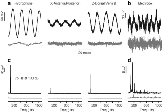
Representative examples of a control (no auditory stimu-lus) and test trial (75 Hz at 130 dB) are shown in Fig. 2. The middle 700 ms of the recorded signals was used to cal-culate the relative power of each frequency using the fast Fourier transform (“FFT,” MATLAB version 2007). Note that any acoustic reflections of the stimulus from the tank walls and water surface did not alter the sound pressure waveform of the acoustic signal, even at the lowest frequen-cies tested (e.g., 75 Hz) as shown in Fig. 2. There was no significant background noise measured by the hydrophone or accelerometer during the control trials (note gray traces in Fig. 2). Saccular potentials contained only relative small peaks of noise at the harmonics of 60 Hz during the record-ings of the controls. During test trials, the saccular poten-tials contained significant energy at the second harmonic of the stimulus frequencies (Fig. 2d). Only the second harmonic component was used by the lock-in amplifier to determine the saccular potential.
Fig. 2 Representative examples of the acoustic stimulus and the evoked saccular potentials recorded from type II male P. notatus that were simultaneously measured during physiology experiments. a During the experiments, the fish was positioned in the center of the tank and the hydrophone and/or accelerometer was positioned at a depth of 10 cm and placed horizontally approximately two-third of the distance from the center to the rim of the experimental tank [black trace shows recorded signal during stimulus presentation while gray trace shows the absence of signal during control trial (no audi-tory stimulus); note only two of the three recorded accelerometer axes are shown here—X (anterior/posterior) and Y (dorsal/ventral)]. b During stimulus presentation, the saccular potentials were evoked during the duration of stimulation [black trace shows the recorded evoked saccular potential from the recording electrode during a stim-ulus presentation of 75 Hz at 130 dB re: 1 µPa, while the gray trace
shows only background noise during a control (no auditory stimulus) trial]. c Power spectrum of the auditory stimulus as measured by the hydrophone and accelerometer shown in a. The corresponding power spectra are shown below the measurement for the hydrophone and the recorded X and Z accelerometer axis [black trace is the power spectra for the stimulus frequency of 75 Hz at 130 dB re: 1 µPa, while the gray trace shows power spectra for control trial (no auditory stimu-lus)]. d Power spectrum of the recorded evoked saccular potential shown in b black trace is the power spectra of the saccular potentials during auditory stimulation while gray trace shows power spectra of saccular potentials during control (no auditory stimulus) trial. Note that the greatest energy in the power spectra of the evoked saccular potentials occurs at the 2nd harmonic of the stimulus frequency (the expected response from opposite oriented hair cells in the saccule)
The threshold of the evoked saccular response was determined using: (1) the average potential recorded in the absence of a stimulus plus two standard deviations and (2) a continuous estimate of the saccular responses (yest) based on the recorded evoked responses near-threshold levels, which was generated by fitting the data with an exponen-tial function (see Fig. 3, thick fitted line using the equation below). Auditory thresholds were determined using expo-nential rate-level functions that were fitted to the records of the evoked potentials and then compared to the saccular potential recorded in the absence of an auditory stimulus.
Figure 3 shows two illustrated examples of threshold deter-mination using the rate-level function near threshold at the minimum and maximum frequencies tested. Auditory threshold was defined as the minimum stimulus level above which the estimated saccular response (yest) exceeded 2 standard deviations above the recorded evoked potential in the absence of a stimulus (Fig. 3 horizontal line). Expo-nentials were fit using data points from near-threshold trials (10–15 dB above and below threshold) using the following equation:
where x is the stimulus amplitude, yest is the estimated sac-cular response, and b1 and b2 are the fitted variables defin-ing the curve. Particle motion thresholds were derived from sound pressure thresholds using best-fit linear coeffi-cients derived from calibrations. Because there was a lin-ear relationship between pressure and particle motion at all frequencies tested, best-fit linear transformations were derived for each frequency using pressure calibration meas-urements and the equivalent particle motion measmeas-urements in the x, y and z axes. After deriving thresholds in the x,
y and z dimensions, particle motion threshold reported
as the combined magnitude vector, calculated as 20 log (√(x2 + y2 + z2)) similar to Wysocki et al. (2009) and Vas-concelos et al. (2011).
The overall effect of reproductive state on the auditory saccular thresholds based on sound pressure and particle acceleration in non-reproductive and reproductive type II male midshipman was analyzed using a repeated measures ANOVA with thresholds for each of the 17 frequencies tested (75–385 Hz) as the repeats (i.e., within-subject fac-tors) and reproductive state of the animal as the between-subject factor followed by the HSD test with unequal N for post hoc planned comparisons of thresholds at each stimu-lus frequency. For all statistical tests, α was set at 0.05. Sta-tistical analyses were performed using Statistica for Win-dows (StatSoft).
Results
Evoked saccular potentials were recorded from a total of 32 adult type II male midshipman: 5 non-reproduc-tive type II males with a size range of 10.2–16.0 cm SL [mean SL = 13.5 ± 1.8 cm SD, mean body mass (BM) = 35.2 ± 17.1 g SD, and mean GSI = 4.4 ± 3.1 SD] and 32 reproductive type II males with a size range of 8.0–10.5 cm SL [mean SL = 9.5 ± 0.7 cm SD, mean body mass (BM) = 10.4 ± 2.3 g SD, and mean GSI = 13.5 ± 4.5 SD]. The evoked saccular potentials were recorded from all type II males within 26 days of field collection, with
yest= ex∗b1
1 + ex∗b2
,
Fig. 3 Illustrated examples of how the evoked saccular potential thresholds were determined at the tested frequencies. Figure shows examples at the lowest frequency tested (i.e., 75 Hz) (a) and the high-est frequency thigh-ested (i.e., 385 Hz) (b) for reproductive (open circles) and non-reproductive fish (filled circles). Each data set was fitted with a best-fit model curve (black lines) and thresholds were interpolated from the best-fit curve (see detailed description in “Materials and methods”). Auditory threshold level was calculated as two standard deviations above baseline and is represented in figure as the horizon-tal red line
the exception of two non-reproductive type II males that were recorded approximately 5–7 months after collection; however, both of these males were maintained on a short-day “winter” photoperiod. No relationship was observed between GSI and the number of days that the animal were kept in captivity within 26 days of collection (r2 = 0.047,
p > 0.2).
Auditory thresholds based on sound pressure and par-ticle acceleration were determined for whole populations of hair cells in the saccule of non-reproductive and repro-ductive type II males. Threshold tuning curves were con-structed using an exponential rate-level function that was fitted to the records of evoked saccular potentials and com-pared to the evoked potentials recorded in the absence of a stimulus (background noise measurement) (Fig. 3). The auditory threshold at each stimulus frequency was desig-nated as the lowest stimulus level that evoked a saccular potential greater than 2 SD above the background noise measurement (Fig. 3, red line). The auditory thresholds for the recorded saccular hair cells based on sound pressure and particle acceleration are summarized in Fig. 4.
The threshold tuning curves based on sound pressure generally consisted of tuning profiles with lowest thresh-olds at 75 and 85 Hz that gradually increased to highest thresholds at frequencies ≥205 Hz (Fig. 4a). Significant pressure threshold differences between non-reproductive and reproductive type II males were observed at every tested frequency except 75 Hz (repeated measures ANOVA: between-subject factor reproductive state, HSD test with unequal n: F(17,21) = 4.94, p < 0.001). The auditory thresh-olds of the saccular hair cells from reproductive male type II were ~8–20 dB (re. 1 µPa) lower than non-reproductive type II males at frequencies from 85 to 385 Hz (Fig. 4a).
The threshold tuning curves based on particle accelera-tion were similar in shape to the pressure threshold pro-files with lowest thresholds at 75 and 85 Hz that gradually increased to highest thresholds at frequencies ≥185 Hz (Fig. 4b). Significant threshold differences based on accel-eration between non-reproductive and reproductive type II males were observed at every frequency except 75 Hz (repeated measures ANOVA: between-subject factor repro-ductive state, HSD test with unequal n: F(17,21) = 4.95,
p < 0.001). The auditory acceleration thresholds of the
saccular hair cells from reproductive male type II were ~8–20 dB re. 1 m s−2 lower than non-reproductive type II males at frequencies from 85 to 385 Hz (Fig. 4b).
Discussion
The goal of this study was to characterize the evoked sac-cular potentials of type II male midshipman to determine whether auditory threshold and frequency response of
saccular hair cells change seasonally to behaviorally rel-evant auditory stimuli. Our results indicate that the summer reproductive type II males were more sensitive than winter
Fig. 4 Seasonal changes in saccular sensitivity-based sound pres-sure and particle acceleration meapres-surements in the plainfin midship-man fish (Porichthys notatus). a Auditory threshold tuning curves for non-reproductive and reproductive type II males based on saccular potential recordings and measurements of sound pressure. All data are plotted as mean ± 95% confidence limit. The number of animals and records are indicated in parentheses. b Auditory threshold tuning curves for non-reproductive and reproductive type II males based on saccular potential recordings and measurements of acoustic particle acceleration. All data are plotted as mean ± 95% confidence limit. The number of animals and records are indicated in parentheses
non-reproductive type II males across a range of frequen-cies tested (85–385 Hz). Our data support the hypothesis that the saccular sensitivity of reproductive type II males is similar in sensitivity to the other reproductive sexual phe-notypes (females and type I males) and better adapted than that of non-reproductive type II males to detect the domi-nant frequency content of the type I male vocalizations. In this discussion, we also interpret our results as they relate to the reception of social acoustic signals based on sound pressure and particle motion and how the changes in audi-tory sensitivity of type II males may facilitate the localiza-tion and assessment of midshipman social acoustic signals during the breeding season.
Appropriate stimuli
There has been much discussion regarding the appropri-ate sound stimuli to measure in fish hearing studies (Pop-per and Fay 1993, 2011; Fay and Popper 2012). Sound can be measured in terms of both acoustic particle motion and pressure. Fish have evolved at least two modes of hearing: an inertial (particle motion) mode and a pressure-mediated mode. All teleost fishes are thought to be capable of detect-ing a particle motion vector, such as acoustic particle accel-eration, via their inner otolithic end organs which essen-tially act as acceleration detectors that respond directly to displacement of the fish by particle motion (De Vries 1950; Hawkins 1993; Sisneros and Rogers 2016). In contrast, the pressure-mediated mode of fish hearing requires the use of specialized morphological adaptations that enable fish to transduce the pressure-induced vibrations of the swim bladder to the otolithic end organs to detect sound pressure. Otophysan fish, such as the goldfish (Carassius auratus) and zebrafish (Danio rerio), possess skeletal adaptations known as Weberian ossicles that link the swim bladder to the inner ear for enhanced pressure detection while other fishes such the Hawaiian squirrelfish (Myripristis kuntee) and the West African ladyfish (Elops lacerta) have no spe-cialized connections between the swim bladder and inner ear, but do exhibit increased pressure sensitivity simply due to the close proximity of these gas-filled structures to the inner ear (Greenwood 1970; Coombs and Popper
1979; Braun and Grande 2008; Tricas and Boyle 2015). Additional studies have shown that natural or artificial gas-filled bladders without direct apposition to the inner ear can also significantly enhance hearing sensitivity in fishes (Chapman and Sand 1974; Jerko et al. 1989). Given that the swim bladder in female and type II male midshipman extends anteriorly via “horn-like” extensions of the swim bladder to caudal regions of the otic capsule (Whitchurch et al. 2012), and that this caudal region of otic capsule has been proposed to be an “acoustic channel” to the saccule through the neurocranium (Edds-Walton et al. 2015), the
midshipman is very likely sensitive to both sound pres-sure and particle motion. Thus, in the current study we characterized the midshipman auditory thresholds in terms of both sound pressure and particle motion. As suggested by Popper and Fay (2011), we also report the impedance (ratio of pressure to particle velocity) of our test tank (see Fig. 1a) so that the acoustic impedance of the tank condi-tions in this study can be used in comparison with that of future fish hearing studies.
Reproductive state-dependent changes in auditory saccular sensitivity
We observed dramatic reproductive state-dependent dif-ferences in the sound pressure and particle acceleration thresholds of the saccule in type II males. The relative differences in sensitivity between reproductive and non-reproductive type II males for both pressure and accelera-tion (re. 1 µPa and 1 ms−2, respectively) varied between 8 and 21 dB at frequencies from 75 to 385 Hz (Fig. 4). Fur-thermore, the saccular thresholds of reproductive type II males were at least 13 dB lower (an enhanced sensitivity equivalent to 4.5 times greater) than non-reproductive type II males at frequencies >185 Hz, which corresponds to the higher frequency components with the majority of energy in type I male vocalizations (Fig. 5). Nesting type I males produce three types of acoustic signals that include ago-nistic grunts and growls and advertisement calls known as hums (Bass et al. 1999; Forlano et al. 2015). Reproductive type I males often produce trains of broadband, short-dura-tion (50–200 ms) grunts (Fig. 5a) to fend or ward off poten-tial nest intruders (Brantley and Bass 1994). Grunts trains produced by type I males can have a repetition rate of about 2.5 Hz with grunt intervals of approximately 400 ms (Bass et al. 1999). Growls are relatively long-duration (>1 s), multiharmonic signals (Fig. 5b) with fundamental frequen-cies that gradually change over the duration of the call from 116 to 59 Hz (Bass et al. 1999). In comparison, the hum is a long-duration, multiharmonic signal with a fundamen-tal frequency that can range from 80 to 102 Hz (personal observation, JAS; also see Bass et al. 1999). In addition to the fundamental frequency, the hum also contains a number of prominent harmonics that range up to 400 Hz (Fig. 5c) with additional lower amplitude harmonics ranging up to 800 Hz and beyond (Bass et al. 1999). In the context of signal recognition and preference, type II males have been observed to exhibit positive phonotaxis to the play-back of simulated hums (McKibben and Bass 1998). Our results suggest that the saccular sensitivity of reproductive type II males is well suited to encode frequencies ≥165 Hz which include the dominant higher frequency components in type I male vocalizations including hums (Fig. 5). This enhanced saccular sensitivity may be adaptive and function
to increase the probability of signal detection by sneaker males in shallow water environments where the propaga-tion of acoustic signals is greatly affected by substrate com-position and water depth (Rogers and Cox 1988; Bass and Clark 2003; Maruska and Sisneros 2015). The relative high frequencies (approximately those greater than 180 Hz) con-tained with type I vocalizations will propagate further than the calls’ fundamental frequencies in shallow water due to the inverse relationship between water depth and the cutoff frequency of sound transmission (Fine and Lenhardt 1983; Rogers and Cox 1988; Bass and Clark 2003). In addition, the substrate composition (e.g., the rocky substrate of the intertidal zone where midshipman breed) can greatly influ-ence the speed of sound in the bottom substrate and affect the cutoff frequency of signal propagation which can atten-uate the transmission of low-frequency acoustic signals (e.g., the fundamental frequencies of type I male vocaliza-tions) (Rogers and Cox 1988). Thus, the enhanced sensi-tivity of type II males to the dominant higher frequencies within type I male vocalizations may potentially facilitate eavesdropping by sneaker males for the assessment of type I male vocalizations and the selection of cuckoldry sites during the breeding season.
The observed seasonal, reproductive state-dependent changes in auditory saccular sensitivity based on sound pressure for type II males were remarkably similar to that reported for female and type I male midshipman. Previ-ously, seasonal and reproductive state-dependent changes in auditory saccular sensitivity based on sound pressure have been reported for both females and type I males across the same range of frequencies from 75 to 385 Hz (Sisneros
2009b; Rohmann and Bass 2011). Results from this study revealed that the difference in saccular sensitivity based on sound pressure between reproductive and non-reproduc-tive type II males ranged from 9 to 21 dB at 75 to 385 Hz, respectively (Fig. 4), which is similar to the reproductive
state-dependent differences observed for females and type I males (Fig. 6). Although the sound pressure tuning curves were similar for all three sexual phenotypes, there appear to be slight seasonal differences in the mean threshold profiles between the reproductive and non-reproductive sexual phe-notypes. Here, we show that reproductive state-dependent
Fig. 5 Comparison between the vocal characteristics of type I male vocalizations and the change in auditory saccular tuning of type II males based on seasonal reproductive state. a Sensitivity increase in auditory saccular thresholds (left y axis) of reproductive type II males compared to non-reproductive type II males at each frequency tested and the power spectrum of a type I male grunt (right y axis, in rela-tive dB values), inset: the temporal waveform of a type I male grunt recorded at 13.5 °C in a laboratory setting (scale bar 20 ms). b Sen-sitivity increase in auditory saccular thresholds (left y axis) of repro-ductive type II males compared to non-reprorepro-ductive type II males at each frequency tested and the power spectrum of a type I male growl (right y axis, in relative dB values), inset: the temporal waveform of a type I male growl recorded at 13.5 °C (scale bar 20 ms). c Sensitivity increase in auditory saccular thresholds (left y axis) of reproductive type II males compared to non-reproductive type II males at each fre-quency tested and the power spectrum of a nesting type I male adver-tisement call or “hum” (right y axis, in relative dB values), inset: the temporal waveform of a type I male hum recorded at 13.5 °C at Seal Rock, WA, in a nesting site (scale bar 20 ms)
changes in type II male saccular sensitivity were frequency dependent with seasonal threshold differences greatest at frequencies ≥125 Hz (~12–20 dB) and lowest threshold differences at frequencies <125 Hz (~8–9 dB). Similarly, type I males also exhibited reproductive state-depend-ent changes in saccular sensitivity that were frequency dependent with greatest threshold differences at frequen-cies >145 Hz (~12–15 dB) and lowest threshold differences at frequencies ≤145 Hz (~4–9 dB; see Rohmann and Bass
2011). In contrast, seasonal differences in female saccular sensitivity were not frequency dependent but were instead relatively constant (~8–13 dB) across the same range of tested frequencies from 75 to 385 Hz (Sisneros 2009b). Consistent across phenotypes is the observation that repro-ductive midshipman showed significantly lower saccular thresholds than non-reproductive midshipman (Fig. 6). In sum, the results from this study now confirm that seasonal, reproductive state-dependent changes in auditory saccular sensitivity are a species-typical trait found in all midship-man sexual phenotypes.
Although no previous study has examined seasonal differences in acoustic acceleration sensitivity of the sac-cule in type II males, our results suggest that the saccular acceleration sensitivity of non-reproductive type II males is at least similar to that of non-reproductive type I males
(Weeg et al. 2002). Estimates for particle motion sensitiv-ity in non-reproductive type I male saccular afferents range from −15 to 26 dB (re. 1 nm), which corresponds to a dis-placement of 0.8–20 nm, whereas our estimate for parti-cle acceleration threshold at the most sensitive frequency tested (85 Hz) is −45 dB ± 7 dB (re. 1 m s−2). Numerical integration of the mean acceleration threshold value at 85 Hz provides a rough estimate of a displacement thresh-old of 17 nm, which is within the displacement sensitiv-ity range reported for type I males by Weeg et al. (2002). Furthermore, the study by Weeg et al. (2002) demonstrated that type I male saccular afferents responded best at 50 and 100 Hz. Although we did not test the same frequen-cies, we do show that non-reproductive type II males share a similar particle acceleration sensitivity to that of type I males with lowest acceleration thresholds reported at fre-quencies <100 Hz. However, it should be noted that the published results on type I males (Weeg et al. 2002) were obtained using a shaker table system which primarily only delivers particle acceleration stimuli, whereas in our study we used a speaker, which delivers both particle motion and pressure stimuli. Further tests will need to be conducted using a shaker system to more accurately compare the par-ticle acceleration sensitivities for the three midshipman sexual phenotypes.
Potential mechanisms for the plasticity of saccular sensitivity
The underlying mechanism(s) responsible for the seasonal reproductive-dependent changes in saccular sensitivity of type II males is likely similar to that proposed for females and type I males. Previous studies by Rohmann and Bass (2011) and Rohmann et al. (2013) showed that the seasonal variation in the frequency sensitivity of saccular hair cells in type I males could be explained by seasonal changes in the abundance of calcium-activated potassium BK chan-nels, which are known to be responsible for a large con-ductance, outward current that produces electrical receptor oscillations along the hair cell epithelium (Lewis and Huds-peth 1983; Roberts et al. 1988; Fettiplace and Fuchs 1999). The electrical resonance that arises from the ion-channel current kinetics of hair cells is thought to be the major con-tributing factor that influences low-frequency (<1 kHz) hair cell tuning in reptiles and birds (Fettiplace and Fuchs
1999) and in fish (Steinacker and Romero 1991, 1992). Rohmann et al. (2013) showed that by using the BK chan-nel antagonist iberiotoxin to reduce BK chanchan-nel availability they could effectively induce a saccular neurophysiological phenotype similar to that found in non-reproductive type I males. Thus, the BK channels of hair cells are thought to be a primary mechanism that can account for the seasonal changes in the frequency selectivity of hair cells in males
Fig. 6 Frequency sensitivity of saccular hair cells in the plainfin midshipman fish (Porichthys notatus). Shown here is a plot of the auditory saccular tuning curves based on evoked potential record-ings in all three adult sexual phenotypes (females, type I and type II males) in reproductive and non-reproductive conditions. All data are plotted as mean ± 95% confidence limit. The number of animals and records are indicated in parentheses. Auditory saccular tuning data for females is adapted from Sisneros (2009b), and from Rohmann and Bass (2011) for type I males
and females. Another potential mechanism responsible for seasonal changes in saccular sensitivity is the reproductive state-dependent increases in saccular hair cell density and decreases in hair cell death observed in females by Cof-fin et al. (2012). Such seasonal increases in saccular hair cell density were independent of body size (age) and not observed in the other inner ear end organs, the lagena and utricle. In addition, Coffin et al. (2012) also observed in reproductive females that the increase in saccular hair cell density was concurrent with increases in overall magnitude of the evoked saccular potentials and decreases in auditory saccular thresholds (.i.e., increased auditory sensitivity). The above-mentioned mechanisms potentially responsible for the observed seasonal changes in saccular sensitivity have been proposed to be induced by estrogens and andro-gens acting via long term signaling pathways to upregulate BK channels and modulate hair cell proliferation and death to ultimately enhance the hearing sensitivity in this species (Coffin et al. 2012; Forlano et al. 2015, 2016). However, this steroid-mediated mechanism hypothesis remains to be tested.
In summary, our results indicate that the seasonal plas-ticity of auditory sensitivity previously reported for females and type I males also occurs in type II male midshipman. This form of reproductive state-dependent plasticity of auditory sensitivity provides an adaptive mechanism that enhances hearing sensitivity in type II males during the reproductive season. Such an increase in auditory sensitiv-ity should increase the probabilsensitiv-ity of conspecific detection by type II sneaker males and facilitate eavesdropping of type I male vocalizations for the assessment of type I male rivals and the selection of cuckoldry sites during the breed-ing season.
Acknowledgements The authors would like to thank the members of the Sisneros Lab and Captain Charlie Eaton and his crew of the R/V Kittiwake for field assistance in animal collections. This work was supported by an NIH Auditory Neuroscience Training Grant 2T32DC005361-11 (to A. A. B. and E. A. W.) and a University of Washington Bridge Fund (to J. A. S.). All experimental procedures were approved by the University of Washington Institutional Ani-mal Care and Use Committee and followed the National Institutes of Health guidelines for the care and use of animals.
References
Alderks PW, Sisneros JA (2011) Ontogeny of auditory saccular sen-sitivity in the plainfin midshipman fish, Porichthys notatus. J Comp Physiol A 197(4):387–398
Bass AH, Clark CW (2003) The physical acoustics of underwater sound communication. In: Simmons AM, Popper AN, Fay RR (eds) Acoustic communication. Springer, New York, pp 15–64 Bass AH, Ladich F (2008) Vocal–acoustic communication: from
neu-rons to behavior. In: Webb JF, Popper AN, Fay RR (eds) Fish bioacoustics. Springer, New York, pp 253–278
Bass AH, McKibben JR (2003) Neural mechanisms and behaviors for acoustic communication in teleost fish. Progr Neurobiol 69(1): 1–26.
Bass AH, Bodnar DA, Marchaterre MA (1999) Complementary explanations for existing phenotypes in an acoustic communica-tion system. In: Hauser MD, Konishi M (eds) The design of ani-mal communication. MIT, Cambridge, pp 493–514
Blaxter JHS (1981) The swim bladder and hearing. In: Tavolga WN, Popper AN, Fay RR (eds) Hearing and sound communication in fishes. Springer, Berlin, pp 61–72
Brantley RK, Bass AH (1994) Alternative male spawning tactics and acoustic signals in the plainfin midshipman fish Porichthys nota-tus Girard (Teleostei, Batrachoididae). Ethology 96(3):213–232 Braun CB, Grande T (2008) Evolution of peripheral mechanisms for
the enhancement of sound reception. In: Webb JF, Popper AN, Fay RR (eds) Fish bioacoustics, Springer, New York, pp 99–144 Casper BM, Mann DA (2006) Evoked potential audiograms of the
nurse shark (Ginglymostoma cirratum) and the yellow stingray (Urobatis jamaicensis). Environ Biol Fish 76(1):101–108 Chapman CJ, Sand O (1974) Field studies of hearing in two
spe-cies of flatfish Pleuronectes platessa (L.) and Limanda limanda (L.) (Family Pleuronectidae). Comp Biochem Physiol A 47(1):371–385
Coffin AB, Mohr RA, Sisneros JA (2012) Saccular-specific hair cell addition correlates with reproductive state-dependent changes in the auditory saccular sensitivity of a vocal fish. J Neurosci 32(4):1366–1376
Cohen MJ, Winn HE (1967) Electrophysiological observations on hearing and sound production in the fish, Porichthys notatus. J Exp Zool 165(3):355–369
Coombs S, Popper AN (1979) Hearing differences among Hawaiian squirrelfish (family Holocentridae) related to differences in the peripheral auditory system. J Comp Physiol 132(3):203–207 De Vries HL (1950) The mechanics of the labyrinth otoliths. Acta
Otolaryngol 38(3):262–273
Edds-Walton PL, Arruda J, Fay RR, Ketten DR (2015) Computerized tomography of the otic capsule and otoliths in the oyster toad-fish, Opsanus tau. J Morphol 276(2):228–240
Fay RR (1984) The goldfish ear codes the axis of acoustic particle motion in three dimensions. Science 225(4665):951–954 Fay RR, Popper AN (1980) Structure and function in teleost auditory
systems. In: Popper AN, Fay RR (eds) Comparative studies of hearing in vertebrates. Springer, Berlin, pp 3–42
Fay RR, Popper AN (2012) Fish hearing: new perspectives from two ‘senior’ bioacousticians. Brain Behav Evol 79(4):215–217 Fettiplace R, Fuchs PA (1999) Mechanisms of hair cell tuning. Annu
Rev Physiol 61:809–834
Fine ML, Lenhardt ML (1983) Shallow-water propagation of the toadfish mating call. Comp Biochem Physiol A 76:225–231 Fine ML, Parmentier E (2015) Mechanisms of fish sound production.
In: Ladich F (ed) Sound communication in fishes. Animal sig-nals and communication, vol 4. Springer, Berlin, pp 77–126 Forlano PM, Sisneros JA, Rohmann KN, Bass AH (2015)
Neuroen-docrine control of seasonal plasticity in the auditory and vocal systems of fish. Front Neuroendocrinol 37:129–145.
Forlano PM, Maruska KP, Sisneros JA, Bass AH (2016) Hormone-dependent plasticity of auditory systems in fishes. In: Bass AH, Sisneros JA, Popper AN, Fay RR (eds) Hearing and hormones. Springer International, New York, pp 15–51
Greenwood PH (1970) Skull and swimbladder connections in the fishes of the family Megalopidae. Bull British Mus Nat Hist (Zool) 14:121–135
Grober MS, Fox SH, Laughlin C, Bass AH (1994) GnRH cell size and number in a teleost fish with two male reproductive morphs: sexual maturation, final sexual status and body size allometry. Brain Behav Evol 43(2):61–78
Hawkins AD (1993) Underwater sound and fish behavior. In: Pitcher TJ (ed) The behaviour of teleost fishes. Chapman and Hall, Lon-don, pp 114–151
Horodysky AZ, Brill RW, Fine ML, Musick JA, Latour RJ (2008) Acoustic pressure and particle motion thresholds in six sciaenid fishes. J Exp Biol 211(9):1504–1511
Jerko H, Turunen-Rise I, Enger PS, Sand O (1989) Hearing in the eel (Anguilla anguilla). J Comp Phys A 165(4):455–459
Kelley DB, Bass AH (2010) Neurobiology of vocal communication: mechanisms for sensorimotor integration and vocal patterning. Curr Opin Neurobiol 20(6):748–753
Ladich F (2004) Sound production and acoustic communication. In: von der Emde G, Mogdans J, Kapoor BG (eds) The senses of fishes: adaptations for the reception of natural stimuli. Narosa Publishing House Pvt Ltd, New Delhi, pp 210–230
Lewis RS, Hudspeth AJ (1983) Voltage-and ion-dependent conduct-ances in solitary vertebrate hair cells. Nature 304:538–541 Maruska KP, Sisneros JA (2015) Sex steroid-dependent
modula-tion of acoustic communicamodula-tion systems in fishes. In: Ladich F (ed) Sound communication in fishes. Springer, New York, pp 207–233
McKibben JR, Bass AH (1998) Behavioral assessment of acoustic parameters relevant to signal recognition and preference in a vocal fish. J Acoust Soc Am 104(6):3520–3533
McKibben JR, Bass AH (1999) Peripheral encoding of behaviorally relevant acoustic signals in a vocal fish: single tones. J Comp Physiol A 184(6):563–576
McKibben JR, Bass AH (2001) Peripheral encoding of behaviorally relevant acoustic signals in a vocal fish: harmonic and beat stim-uli. J Comp Physiol A 187(4):271–285
Popper AN, Fay RR (1993) Sound detection and processing by fish: critical review and major research questions. Brain Beh Evol 41:14–25
Popper AN, Fay RR (2011) Rethinking sound detection by fishes. Hear Res 273(1):25–36
Roberts WM, Howard J, Hudspeth AJ (1988) Hair cells: transduc-tion, tuning, and transmission in the inner ear. Ann Rev Cell Biol 4(1):63–92
Rogers PH, Cox M (1988) Underwater sound as a biological stimulus. In: Atema J, Fay RR, Popper AN, Tavolga WN, Sensory biology of aquatic animals. Springer, New York, pp 131–149
Rohmann KN, Bass AH (2011) Seasonal plasticity of auditory hair cell frequency sensitivity correlates with plasma steroid levels in vocal fish. J Exp Biol 214(11):1931–1942
Rohmann KN, Fergus DJ, Bass AH (2013) Plasticity in ion channel expression underlies variation in hearing during reproductive cycles. Curr Biol 23(8):678–683
Schellart NAM, Popper AN (1992) Functional aspects of the evolu-tion of the auditory system of actinopterygian fish. In: Webster
DB, Fay RR, Popper AN (eds) The evolutionary biology of hear-ing. Springer, New York, pp 295–322
Sisneros JA (2007) Saccular potentials of the vocal plainfin midship-man fish, Porichthys notatus. J Comp Physiol A 193:413–424 Sisneros JA (2009a) Adaptive hearing in the vocal plainfin
midship-man fish: getting in tune for the breeding season and implications for acoustic communication. Integr Zool 4:33–42
Sisneros JA (2009b) Seasonal plasticity of auditory saccular sensitiv-ity in the vocal plainfin midshipman fish, Porichthys notatus. J Neurophysiol 102(2):1121–1131
Sisneros JA, Bass AH (2003) Seasonal plasticity of peripheral audi-tory frequency sensitivity. J Neurosci 23:1049–1058
Sisneros JA, Bass AH (2005) Ontogenetic changes in the response properties of individual, primary auditory afferents in the vocal plainfin midshipman fish Porichthys notatus Girard. J Exp Biol 208(16):3121–3131
Sisneros JA, Rogers PH (2016) Directional Hearing and Sound Source Localization in Fishes. In: Sisneros JA (ed) Fish hearing and bioacoustics. Springer International, New York, pp 121–155 Sisneros JA, Forlano PM, Knapp R, Bass AH (2004) Seasonal vari-ation of steroid hormone levels in an intertidal-nesting fish, the vocal plainfin midshipman. Gen Comp Endocrinol 136:101–116 Steinacker A, Romero A (1991) Characterization of voltage-gated and
calcium-activated potassium currents in toadfish saccular hair cells. Brain Res 556(1):22–32
Steinacker A, Romero A (1992) Voltage-gated potassium cur-rent and resonance in the toadfish saccular hair cell. Brain Res 574:229–236
Tomkins JL, Simmons LW (2002). Measuring relative investment: a case study of testes investment in species with alternative male reproductive tactics. Anim Behav 63(5):1009–1016
Tricas TC, Boyle KS (2015) Sound pressure enhances the hearing sensitivity of Chaetodon butterfly fishes on noisy coral reefs. J Exp Biol 218:1585–1595
Vasconcelos RO, Sisneros JA, Amorim MCP, Fonseca PJ (2011) Auditory saccular sensitivity of the vocal Lusitanian toadfish: low frequency tuning allows acoustic communication throughout the year. J Comp Physiol A 197(9):903–913
Weeg M, Fay RR, Bass AH (2002) Directionality and frequency tun-ing of primary saccular afferents of a vocal fish, the plainfin mid-shipman (Porichthys notatus). J Comp Physiol A 188(8):631–641 Whitchurch EA, Ketten DR, Forlano PM, Fay RR, Sisneros JA (2012) Swim bladder sexual dimorphism in the plainfin midshipman fish: Implications for acoustic communication in this species. Program No. 296.11/CCC73. 2012 Neuroscience Meeting Plan-ner. Society for Neuroscience, New Orleans (online)
Wysocki LE, Codarin A, Ladich F, Picciulin M (2009) Sound pressure and particle acceleration audiograms in three marine fish species from the Adriatic Sea. J Acoust Soc Am 126(4):2100–2107